ORIGINAL ARTICLE
Procalcitonin and C-reactive protein in pericardial fluid
for postmortem diagnosis of sepsis
Bettina Schrag&Katia Iglesias&Patrice Mangin&
Cristian Palmiere
Received: 21 December 2011 / Accepted: 13 March 2012 / Published online: 29 March 2012 # Springer-Verlag 2012
Abstract The aim of this study was to investigate the pres-ence and concentrations of procalcitonin and C-reactive pro-tein in pericardial fluid and compare these levels to those found in the postmortem serum obtained from the femoral blood. Two groups were formed, a sepsis-related fatalities group and a control group. Postmortem native CT scans, autopsies, histology, neuropathology and toxicology as well as other postmortem biochemistry investigations were per-formed in all cases. Pericardial fluid procalcitonin levels were significantly different between the cases of sepsis-related fatalities and those of the control group. Postmortem serum procalcitonin levels below the detection limit were also reflected in undetectable pericardial fluid levels. Similarly, a large increase in postmortem serum procalcitonin levels was reflected in a large increase of procalcitonin pericardial fluid levels. Based on these findings, pericardial fluid could be an alternative to postmortem serum for the determination of procalcitonin levels in cases where postmortem serum is not
available and measurements of procalcitonin are required to circumstantiate the pathogenesis of death.
Keywords Procalcitonin . C-reactive protein . Sepsis . Inflammation . Postmortem biochemistry
Introduction
In clinical practice, procalcitonin and C-reactive protein (CRP) levels have been investigated, either individually or in conjunction with other cytokines, to distinguish sepsis from severe, systemic, non-infectious inflammations [1–8]. Procalcitonin has been indicated as the best marker of infection in critically ill patients admitted to intensive care units, whereas measurements of CRP and other cytokines, such as interleukin-6, have been shown to be non-optimal in discriminating patients with infections from those with non-infectious diseases or systemic inflammatory response syndrome [2].
In clinical practice, sepsis can be difficult to distinguish from other non-infectious, systemic, inflammatory responses, espe-cially in critically ill patients admitted to the hospital with clinical signs of acute inflammation. Biochemical investiga-tions may therefore provide useful information in distinguish-ing between systemic inflammatory response syndrome and various forms of sepsis [2].
Postmortem diagnosis of sepsis is even more difficult to achieve, mainly because of the lack of medical records at the moment of the autopsy as well as the absence of specific macroscopic and microscopic findings (myocardial ischemia, pulmonary edema, hypoxic liver damage, mesenteric ische-mia, gastrointestinal haemorrhages, spleen infarction, kidney ischemia and brain edema), which may be of infectious or non-infectious origin [3,9,10]. Thus, the postmortem usefulness of
Bettina Schrag and Cristian Palmiere equally contributed as first authors of this work.
B. Schrag
:
P. Mangin:
C. Palmiere (*)University Centre of Legal Medicine, Lausanne-Geneva, Rue du Bugnon 21,
1011 Lausanne, Switzerland e-mail: [email protected] B. Schrag
:
P. Mangin:
C. PalmiereUniversity Centre of Legal Medicine, Lausanne-Geneva, Rue Michel-Servet 1,
1211 Genève 4, Switzerland K. Iglesias
Institute of Social and Preventive Medicine, Lausanne University Hospital,
Rue du Bugnon 17, 1004 Lausanne, Switzerland
procalcitonin and CRP has been studied to discriminate between sepsis-related fatalities and systemic inflammatory response syndrome [9–17]. Procalcitonin concentrations have been principally measured in the postmortem serum obtained from femoral blood, vitreous humor and cerebrospinal fluid [9–11,17]. CRP levels have been tested in the postmortem serum from femoral blood [12–16], liver [14], vitreous humor and cerebrospinal fluid [17].
The results of these investigations confirmed the useful-ness of postmortem procalcitonin measurements in differen-tiating sepsis-related fatalities from non-septic underlying causes of death [9, 11]. Moreover, postmortem investiga-tions have demonstrated that CRP levels can be used in forensic diagnosis in a similar manner to their use in clinical practice [10,12–16].
Pericardial fluid has been used in forensic practice for several applications to measure different markers, including glucose, ketone bodies, calcium, sodium, potassium, magne-sium, urea nitrogen, creatinine, uric acid, markers of myocar-dial ischemia and cardiac function, insulin, C-peptide and serotonin [18–32]. Pericardial fluid levels have been found to be as reliable as blood levels for some of these parameters, thus suggesting that this substrate could be used for postmor-tem analyses when postmorpostmor-tem biochemical investigations are required to circumstantiate the pathogenesis of death and sufficient amounts of blood are unavailable.
The aim of this study was to investigate both the presence and concentrations of procalcitonin and CRP in the pericar-dial fluid of a group of sepsis-related fatalities and a control group in order to compare pericardial fluid levels of these two markers to those found in postmortem femoral blood serum to detect sepsis-related fatalities. Postmortem native CT scans, autopsies, histology, neuropathology and toxicol-ogy as well as other postmortem biochemical investigations were also performed in all cases.
Materials and methods
Subjects
Two groups of study were formed, a sepsis-related fatalities group and a control group. The sepsis-related fatalities group consisted of 12 forensic autopsy cases (eight males, including one 2-month-old infant and four females). The mean age was 40.5 years. All subjects were admitted to the intensive care unit of the local university hospital. Ten cases had documented clinical diagnoses of sepsis in vivo. The period of the septic condition was between 24 h and 4 days. Complete forensic autopsies were performed within a 24-h postmortem period on all the cases of this group. Postmortem microbiological investigations were carried out on cardiac blood revealing the presence of Pseudomonas
aeruginosa (two cases), Klebsiella pneumoniae (four cases), Escherichia coli (two cases) and Streptococcus pneumoniae (one case). In two cases, microbiology revealed the presence of multiple Gram-positive and Gram-negative bacteria. In one case, the postmortem microbiological investigation did not reveal clearly identifiable pathogens. The cause of death was attributed to multiple organ failure in all cases.
The control group consisted of 28 forensic autopsy cases (20 males and 8 females, no children among them) with a mean age of 46 years. As with the group above, the postmortem period was within 24 h. In three cases, the cause of death was not determined when the bodies were admitted to the medico-legal center. For 10 cases, the cause of death was attributed to severe chest and/or abdominal trauma following traffic accidents with survival time up to 2 h. The cause of death was attributed to head injury in another 10 cases, with or without chest and abdominal trauma, following high falls with a survival time up to 6 h. In the remaining five cases, the cause of death was attributed to carbon monoxide intoxication in bodies presenting extensive burns of the skin, fat tissue and skeletal muscles. No individuals in this group had any medical history of infectious diseases or septic conditions prior to their death. Macroscopic and microscopic examination did not reveal any findings that suggested the existence of an underlying infectious disease. Microbiological investigations were not performed.
Samples collection
Postmortem serum samples
Using a sterile needle and a syringe, postmortem blood sam-ples were collected by aspiration of the femoral vein during the autopsy. All blood samples were centrifuged immediately post-collection at 3,000×g for 15 min. After centrifugation, the separated supernatant (postmortem serum) was collected, stored in tubes without preservatives and frozen at−20 °C. Pericardial fluid samples
Undiluted samples of pericardial fluid were collected imme-diately after an incision in the pericardium during the au-topsy. All the samples were immediately centrifuged at 3,000×g for 15 min. After centrifugation, the separated supernatant was collected, stored in tubes without preserva-tives and frozen at−20 °C.
Analyses
Determination of procalcitonin concentration
KRYPTOR® (B·R·A·H·M·S, Henningsdorf, Germany) was used to measure the procalcitonin concentration. KRYPTOR® uses time-resolved amplified cryptate emission technology,
which is based on a non-radiative transfer of energy. This transfer occurs between two fluorescent tracers: europium cryptate (donor) and XL665 (acceptor). The signal measured during the formation of the antigen–antibody complex is accompanied by amplification. KRYPTOR® assays are homogeneous and do not require separation or washing. The molecules of procalcitonin present in the assay samples are sandwiched between the antibodies. The intensity of the signal is proportional to the amount of procalcitonin present. The shape of the standard curve is identical to that obtained using immunometric methods. The results are expressed in micrograms per liter.
Determination of C-reactive protein concentration
SYNCHRON® C-reactive protein reagent (Beckman Coulter, Inc., Brea, USA) was used to measure the CRP concentrations using the turbidimetric method specified in the supplier's instructions. In this reaction, the CRP combines with specific antibodies to form insoluble antigen–antibody complexes. The SYNCHRON® system automatically proportions the appro-priate volumes of the sample and the reagent into a cuvette at the ratio of 1 part sample to 26 parts reagent. The system monitors the change in absorbance at 340 nm. This change in absorbance is proportional to the concentration of CRP in the sample and is used by the system to calculate and express the CRP concentration, based upon a multi-point, non-linear cal-ibration curve. The results are expressed in milligrams per liter.
Statistical method
A sensitivity analysis was carried out in order to examine whether or not postmortem serum procalcitonin and CRP
concentrations correlated to pericardial fluid values. Accord-ing to the laboratory references, procalcitonin values were dichotomized into“non-septic values” (concentrations lower than 2μg/l) and “septic values” (concentrations greater than 2μg/l) and CRP values into “normal values” (concentrations lower than 10 mg/l) and “increased values” (concentrations greater than 10 mg/l). Pearson's correlation coefficient was used to assess the correlation between postmortem serum and pericardial fluid levels for procalcitonin and CRP.
Results
Results are reported in Table 1. The analysis of both pro-calcitonin and CRP markers in the septic group revealed that the diagnostic performance of the former was better than the latter, with a sensitivity of 91.67 %.
Increased postmortem serum procalcitonin levels were noted in all cases of sepsis. Such increases were furthermore accompanied by increases in CRP levels in 11 out of 12 cases. The only CRP-negative case in the sepsis group was the 2-month-old infant (case 1, Table1). Increased postmor-tem serum CRP levels without parallel postmorpostmor-tem serum procalcitonin increases were detected in six of the control cases presenting head injuries, with or without chest and abdominal trauma, having a survival time up to 6 h.
Considering that normal and pathological procalcitonin blood levels in living people show similar ranges in both postmortem serum and pericardial fluid (concentrations greater than 2 μg/l for septic states), postmortem serum procalcitonin levels below the detection limit (<0.06 μg/l) in 25 control cases were reflected in undetectable pericardial fluid levels in the same 25 control cases.
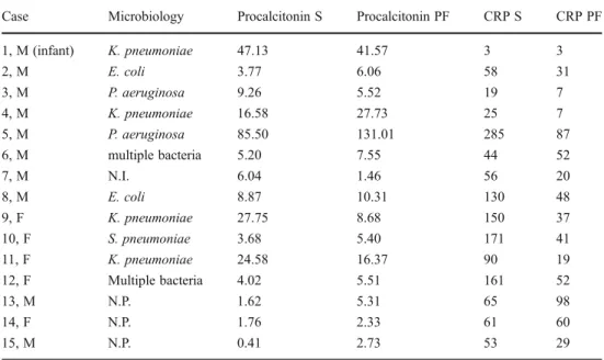
Table 1 Septic cases included in the study
Cases 13, 14 and 15 are control cases showing increased procal-citonin levels in both postmor-tem serum and pericardial fluid. Explanations indicated in text. Procalcitonin results are expressed in micrograms per liter. CRP results are expressed in milligrams per
liter
S postmortem serum from femoral blood, PF pericardial fluid, M male, F female, N.I. not clearly identifiable pathogens, N.P. not performed
Case Microbiology Procalcitonin S Procalcitonin PF CRP S CRP PF
1, M (infant) K. pneumoniae 47.13 41.57 3 3 2, M E. coli 3.77 6.06 58 31 3, M P. aeruginosa 9.26 5.52 19 7 4, M K. pneumoniae 16.58 27.73 25 7 5, M P. aeruginosa 85.50 131.01 285 87 6, M multiple bacteria 5.20 7.55 44 52 7, M N.I. 6.04 1.46 56 20 8, M E. coli 8.87 10.31 130 48 9, F K. pneumoniae 27.75 8.68 150 37 10, F S. pneumoniae 3.68 5.40 171 41 11, F K. pneumoniae 24.58 16.37 90 19 12, F Multiple bacteria 4.02 5.51 161 52 13, M N.P. 1.62 5.31 65 98 14, F N.P. 1.76 2.33 61 60 15, M N.P. 0.41 2.73 53 29
Measurements of procalcitonin in the pericardial fluid appear to be a possible alternative to postmortem serum, with a sensitivity of 90 % and a specificity of 89.29 %. Pearson's correlation coefficient was 0.947 and 0.751 for procalcitonin and CRP, respectively. The analysis of pro-calcitonin in pericardial fluid revealed one false-negative in the group of sepsis-related fatalities (case 7, Table 1) and three false-positive cases in the control group (cases 13–15, Table1).
The pericardial fluid procalcitonin concentration of the only false-negative case in the sepsis group was consistent with what would be expected in a generalised bacterial infec-tion (1.46μg/l), whereas the three false-positive cases in the control group had procalcitonin pericardial fluid levels con-sistent with a state of sepsis (5.31, 2.33 and 2.73μg/l) though postmortem serum levels did not confirm this diagnosis. However, in two cases, procalcitonin postmortem serum con-centrations were consistent with generalised bacterial infec-tions (1.76 and 1.62μg/l).
Measurements of CRP in the pericardial fluid revealed two false-negatives in the sepsis group (cases 3 and 4, Table1), two false-positives in the control group and one false-positive in the control group, with 80.00 % sensitivity and 95.00 % specificity. The pericardial fluid CRP concen-trations in the false-positive and false-negative cases were more difficult to compare to corresponding postmortem serum concentrations.
Discussion
The aim of this study was to investigate the usefulness of measuring procalcitonin and CRP concentration in pericar-dial fluid for the postmortem diagnosis of sepsis. Concen-trations of both these markers were compared in postmortem serum and pericardial fluid in a group of sepsis-related fatalities as well as a control group.
The results of our study indicate that pericardial fluid could be considered an alternative to postmortem serum in cases in which procalcitonin measurement is required and postmortem serum is unavailable. Indeed, postmortem serum procalcitonin levels below the detection level in control cases were also undetectable in the pericardial fluid. Similarly, a marked increase of procalcitonin levels in the postmortem serum of septic cases was reflected in increased pericardial fluid levels.
Procalcitonin is not the only biochemical marker which has been associated, in clinical and medico-legal investiga-tions, with various sepsis-related outcomes. CRP and other cytokines, such as interleukin-6 and interleukin 8, have also been postulated as being potentially useful in the diagnosis of bacterial sepsis. However, more recent studies have somewhat put into question the diagnostic functionality of
CRP in distinguishing sepsis from non-infectious systemic inflammatory response syndrome. These studies have sug-gested that increased CRP levels be considered as a non-specific indicator of an underlying inflammatory process rather than a specific marker of sepsis.
The results of the study herein presented are consistent with the findings above and confirm that CRP is not as discriminating as procalcitonin in the detection of bacterial sepsis. This leads to the conclusion that the diagnostic potential of procalcitonin in detecting sepsis-related out-comes should be opted for over CRP when cases of sudden, unexpected deaths appear in situations where sepsis can be listed as a possible cause of or significant contributing condition to death.
The postmortem diagnosis of sepsis-related fatalities remains a difficult challenge for forensic pathologists due to the absence of typical macroscopic and microscopic morphological findings as well as insufficient, inexistent or unavailable medical records upon autopsy. Postmortem biochemical markers can provide useful information in or-der to support and enforce morphological findings and are therefore necessary to confirm or exclude this diagnosis.
The conversion of clinical analyses and reference values to the postmortem field can be relatively arduous for several reasons. Among these factors are biochemical marker sta-bility in the biological fluids after death, postmortem changes, postmortem haemolysis and often the unavailabil-ity of qualitatively acceptable biological samples.
Thus, it is extremely important to investigate the relation-ship between clinical and postmortem biochemical analyses to test result reliability as well as the applicability of clinical reference values in the postmortem period. It is, however, also important to dispose of alternative biological fluids to analyse for postmortem biochemical investigations. This is especially relevant in forensic practice, where it is not infre-quent that either peripheral or cardiac blood is unavailable for analyses and postmortem biochemistry could provide important information.
Based on these assumptions, we have decided to test the usefulness of pericardial fluid as a biological substrate for postmortem biochemical investigations in order to diagnose sepsis-related fatalities. Pericardial fluid is relatively easy to obtain during autopsy and small amounts can be collected even when a body presents advanced postmortem changes. The composition of normal pericardial fluid has been in-vestigated by several authors [33–37]. In 2005, Ben-Horin et al. [37] proposed a detailed analysis of the normal pericardial fluid obtained from 30 consecutive patients who had been admitted to the hospital for elective open heart surgery due to coronary or valvular disease. Biochemical analyses showed that the concentrations of small molecules, such as urea nitro-gen, creatinine, uric acid, electrolytes and glucose, corre-sponded to an ultrafiltrate of plasma. In contrast, lactate
dehydrogenase (LDH) and protein levels were higher than expected for large molecules in a plasma ultrafiltrate and were usually above the exudate cut-off level used by Light's criteria [38] for the pleural fluid.
Physiological pericardial fluid is believed to be a transu-date generated by the net result of the hydrostatic pressure and the osmotic gradient, like other serosal fluids, such as the pleural and peritoneal fluids. The results of the study performed by Ben-Horin et al. [37] confirmed the transuda-tive nature of the pericardial fluid in relation to the small molecules and the exudative range, as defined for pleural effusions [38], for LDH and protein content. According to the authors, the explanation of the high LDH and protein levels in the physiologic pericardial fluid is uncertain but can be the consequence of a preferential leak from the adjacent perimyocardial tissue.
In medico-legal literature, total ketone body and 3-β-hydroxybutyrate concentrations, as well as insulin, C-peptide, electrolytes, renal function markers, serotonin and markers of cardiac function have been studied in the peri-cardial fluid by several authors [18–32]. Positive correla-tions between blood and pericardial fluid ketone body levels have been shown by Pounder [18]. Our own experience confirmed the conclusions of Pounder, as well as increased insulin concentration in the pericardial fluid of a case of insulin overdose [19].
Zhu et al. [21,22] studied the correlations between urea nitrogen, creatinine and uric acid levels in the postmortem serum and pericardial fluid. They emphasised the usefulness of the comparison between blood and pericardial fluid val-ues to confirm the existence of persistent metabolic disor-ders preceding the death and obtain information pertaining to survival time. Furthermore, measurements of cardiac biomarkers [24–31] in pericardial fluid have been indicated as useful in the investigation of myocardial damage severity. The study herein presented is the first comparison between procalcitonin and CRP postmortem serum and pericardial fluid levels. The results of our investigations suggest that procalcitonin can be reliably measured in pericardial fluid for forensic purposes when postmortem serum is unavailable and biochemical investigations are required to confirm or exclude the diagnosis of sepsis-related fatalities. Conversely, even if further studies and investigations are required to confirm these findings, the data seem to indicate that more caution is necessary when comparing postmortem serum to pericardial fluid CRP levels.
Acknowledgments The authors are grateful to the anonymous reviewers whose constructive and useful comments improved the quality of this article.
Conflict of interest The authors have no conflict of interest to declare.
References
1. Balci C, Sungurtekin H, Gürses E, Sungurtekin U, Kaptanoglu B (2003) Usefulness of procalcitonin for diagnosis of sepsis in the intensive care unit. Crit Care 7(1):85–90
2. Harbarth S, Holeckova K, Froidevaux C, Pittet D, Ricou B, Grau GE, Vadas L, Pugin J, Network GS (2001) Diagnostic value of procalcitonin, interleukin-6, and interleukin-8 in critically ill patients admitted with suspected sepsis. Am J Respir Crit Care Med 164(3):396–402
3. Torgersen C, Moser P, Jochberger S, Hasibeder WR, Dünser MW (2009) Macroscopic postmortem findings in 235 surgical intensive care patients with sepsis. Anesth Analg 108(6):1841–1847 4. Becker KL, Snider R, Nyles ES (2008) Procalcitonin assay in
systemic inflammation, infection, and sepsis: clinical utility and limitation. Crit Care Med 36(3):941–952
5. Becker KL, Snider R, Nyles ES (2010) Procalcitonin in sepsis and systemic inflammation: a harmful biomarker and a therapeutic target. Br J Pharmacol 159(2):253–264
6. Billeter A, Turina M, Seifert B, Mica L, Stocker R, Keel M (2009) Early serum procalcitonin, interleukin-6 and 24-hour lactate clear-ance: useful indicators of septic infections in severely traumatized patients. World Surg 33(3):558–566
7. Fioretto JR, Martin JG, Kurokawa CS, Carpi MF, Bonatto RC, de Moraes MA, Ricchetti SM (2010) Comparison between procalci-tonin and C-reactive protein for early diagnosis of children with sepsis or septic shock. Inflamm Res 59(8):581–586
8. McGrane S, Girard TD, Thompson JL, Shintani AK, Woodworth A, Wesley Ely E, Pandharipande PP (2011) Procalcitonin and C-reactive protein levels at admission as predictors of duration of acute brain dysfunction in critically ill patients. Crit Care 15(2): R78
9. Tsokos M, Reichelt U, Nierhaus A, Püschel K (2001) Serum procalcitonin (PCT): a valuable biochemical parameter for the postmortem diagnosis of sepsis. Int J Legal Med 114(4–5):237– 243
10. Ramsthaler F, Kettner M, Mall G, Bratzke H (2008) The use of rapid diagnostic test of procalcitonine serum levels for the post-mortem diagnosis of sepsis. Forensic Sci Int 178(2–3):139–145 11. Tsokos M, Reichelt U, Jung R, Nierhaus A, Püschel K (2001)
Interleukin-6 and C-reactive protein serum levels in sepsis-related fatalities during the early postmortem period. Forensic Sci Int 119 (1):47–56
12. Uhlin-Hansen L (2001) C-reactive protein (CRP), a comparison of pre- and postmortem blood levels. Forensic Sci Int 124(1):32–35 13. Fujita MQ, Zhu BL, Ishida K, Quan L, Oritani S, Maeda H (2002)
Serum C-reactive protein levels in postmortem blood—an analysis with special reference to the cause of death and survival time. Forensic Sci Int 130(2–3):160–166
14. Astrup BS, Thomsen JL (2007) The routine use of C-reactive protein in forensic investigations. Forensic Sci Int 172(1):49–55 15. Maeda H, Zhu BL, Bessho Y, Ishikawa T, Quan L, Michiue T,
Zhao D, Li DR, Komatsu A (2008) Postmortem serum nitrogen compounds and C-reactive protein levels with special regard to investigation of fatal hyperthermia. Forensic Sci Med Pathol 4 (3):175–180
16. Ishikawa T, Hamel M, Zhu BL, Li DR, Zhao D, Michiue T, Maeda H (2008) Comparative evaluation of postmortem serum concen-trations of neopterin and C-reactive protein. Forensic Sci Int 179 (2–3):135–143
17. Schrag B, Roux-Lombard P, Schneiter D, Vaucher P, Mangin P, Palmiere C (2011) Evaluation of C-reactive protein, procalcitonin, tumor necrosis factor alpha, interleukin-6 and interleukin-8 as diagnostic parameters in sepsis-related fatalities. Int J Legal Med. doi:10.1007/s00414-011-0596-z
18. Pounder DJ, Stevenson RJ, Taylor KK (1998) Alcoholic ketoaci-dosis at autopsy. J Forensic Sci 43(4):812–816
19. Palmiere C, Mangin P (2011) Postmortem chemistry update: part I. Int J Legal Med. doi:10.1007/s00414-011-0625-y
20. Arroyo A, Valero J, Marrón T, Vidal C, Hontecillas B, Bernal J (1998) Pericardial fluid postmortem: comparative study of natural and violent deaths. Am J Forensic Med Pathol 19(3):266–268 21. Zhu BL, Ishikawa T, Michiue T, Li DR, Zhao D, Quan L, Maeda H
(2005) Evaluation of postmortem urea nitrogen, creatinine and uric acid levels in pericardial fluid in forensic autopsy. Leg Med (Tokyo) 7(5):287–292
22. Zhu BL, Ishikawa T, Michiue T, Tanaka S, Zhao D, Li DR, Quan L, Oritani S, Maeda H (2007) Differences in postmortem urea nitrogen, creatinine and uric acid levels between blood and peri-cardial fluid in acute death. Leg Med (Tokyo) 9(3):115–122 23. Li DR, Quan L, Zhu BL, Ishikawa T, Michiue T, Zhao D, Yoshida
C, Chen JH, Wang Q, Komatsu A, Azuma Y, Maeda H (2009) Evaluation of postmortem calcium and magnesium levels in the pericardial fluid with regard to the cause of death in medicolegal autopsy. Forensic Sci Int 11(Suppl 1):S276–S278
24. Zhu BL, Ishikawa T, Michiue T, Li DR, Zhao D, Tanaka S, Kamikodai Y, Tsuda K, Okazaki S, Maeda H (2007) Postmortem pericardial natriuretic peptides as markers of cardiac function in medico-legal autopsies. Int J Legal Med 121(1):28–35
25. Michaud K, Augsburger M, Donzé N, Sabatasso S, Faouzi M, Bollmann M, Mangin P (2008) Evaluation of postmortem mea-surement of NT-proBNP as a marker for cardiac function. Int J Legal Med 122(5):415–420
26. Osuna E, Pérez-Cárceles MD, Alvarez MV, Noguera J, Luna A (1998) Cardiac troponin I (cTn I) and the postmortem diagnosis of myocardial infarction. Int J Legal Med 111(4):173–176
27. Zhu BL, Ishikawa T, Michiue T, Li DR, Zhao D, Oritani S, Kamikodai Y, Tsuda K, Okazaki S, Maeda H (2006) Postmortem cardiac troponin T levels in the blood and pericardial fluid. Part 1. Analysis with special regard to traumatic causes of death. Leg Med (Tokyo) 8(2):86–93
28. Zhu BL, Ishikawa T, Michiue T, Li DR, Zhao D, Kamikodai Y, Tsuda K, Okazaki S, Maeda H (2006) Postmortem cardiac tropo-nin T levels in the blood and pericardial fluid. Part 2: analysis for application in the diagnosis of sudden cardiac death with regard to pathology. Leg Med (Tokyo) 8(2):94–101
29. Zhu BL, Ishikawa T, Michiue T, Li DR, Zhao D, Bessho Y, Kamikodai Y, Tsuda K, Okazaki S, Maeda H (2007) Postmortem cardiac troponin I and creatine kinase MB levels in the blood and pericardial fluid as markers of myocardial damage in medicolegal autopsy. Leg Med (Tokyo) 9(5):241–250
30. Wang Q, Michiue T, Ishikawa T, Zhu BL, Maeda H (2011) Com-bined analyses of creatine kinase MB, cardiac troponin I and myoglobin in pericardial and cerebrospinal fluids to investigate myocardial and skeletal muscle injury in medicolegal autopsy cases. Leg Med (Tokyo) 13(5):226–232
31. Sabatasso S, Vaucher P, Augsburger M, Donzé N, Mangin P, Michaud K (2011) Senditivity and specificity of NT-proBNP to detect heart failure at post mortem examination. Int J Legal Med 125(6):849–856 32. Quan L, Ishikawa T, Hara J, Michiue T, Chen JH, Wang Q, Zhu BL, Maeda H (2011) Postmortem serotonin levels in cerebrospinal and pericardial fluids with regard to the cause of death in medico-legal autopsy. Leg Med (Tokyo) 13(2):75–78
33. Hutchin P, Nino HV, Suberman R (1971) Electrolyte and acid–base composition of pericardial fluid in man. Arch Surg 102(1):28–30 34. Eisalo A, Konttinen A (1972) Composition of pericardial fluid in
cholesterol pericarditis. Acta Med Scand 191(1–2):125–128 35. Gibson AT, Segal MB (1976) Observations on the composition of
the pericardial fluid. J Physiol 256(1):32P–33P
36. Gibson AT, Segal MB (1978) A study of the composition of pericardial fluid, with special reference to the probable mechanism of fluid formation. J Physiol 277:367–377
37. Ben-Horin S, Shinfeld A, Kachel E, Chetrit A, Livneh A (2005) The composition of normal pericardial fluid and its implications for diagnosing pericardial effusions. Am J Med 118(6):636–640 38. Light RW (2002) Clinical practice: pleural effusions. N Engl J Med