HAL Id: inserm-02545538
https://www.hal.inserm.fr/inserm-02545538
Submitted on 17 Apr 2020
HAL is a multi-disciplinary open access
archive for the deposit and dissemination of sci-entific research documents, whether they are pub-lished or not. The documents may come from teaching and research institutions in France or abroad, or from public or private research centers.
L’archive ouverte pluridisciplinaire HAL, est destinée au dépôt et à la diffusion de documents scientifiques de niveau recherche, publiés ou non, émanant des établissements d’enseignement et de recherche français ou étrangers, des laboratoires publics ou privés.
Prospects for Diminishing the Impact of Nonamyloid
Small-Vessel Diseases of the Brain
Anne Joutel
To cite this version:
Anne Joutel. Prospects for Diminishing the Impact of Nonamyloid Small-Vessel Diseases of the Brain. Annual Review of Pharmacology and Toxicology, Annual Reviews, 2020, 60 (1), pp.437-456. �10.1146/annurev-pharmtox-010818-021712�. �inserm-02545538�
1
Prospects for diminishing the impact of non-amyloid small vessel
diseases of the brain
Author
Anne Joutel
Institute of Psychiatry and Neurosciences of Paris, INSERM UMR1266, Paris Descartes University, DHU NeuroVasc, Sorbonne Paris Cité, Paris, France
Department of Pharmacology, College of Medicine, University of Vermont, Burlington, VT, USA
Email: anne.joutel@inserm.fr Tel: +33 1 40 78 92 96
Word counts: 6,232 words 1 table
2
ABSTRACT
Small vessel diseases (SVDs) of the brain are involved in about one-fourth of ischemic strokes and a vast majority of intracerebral hemorrhage, and are responsible for nearly half of dementia cases in the elderly. SVDs are a heavy burden for society, a burden that is expected to increase further in the absence of significant therapeutic advances, given the aging population. Here, we provide a critical appraisal of currently available therapeutic approaches for non-amyloid sporadic SVDs that are largely based on targeting modifiable risk factors. We review what we know about the pathogenic mechanisms of vascular risk factor related-SVDs and CADASIL, the most frequent hereditary SVD, and elaborate on two mechanism-based therapeutic approaches worth exploring in sporadic SVD and CADASIL. We conclude by discussing opportunities and challenges that need to be tackled if efforts to achieve significant therapeutic advances for these diseases are to be successful.
Keywords
3
INTRODUCTION
Small vessel diseases (SVDs) of the brain encompass a heterogeneous group of disorders caused by structural or functional alterations of small intracranial arteries or arterioles, capillaries, and sometimes small veins. SVDs are involved in about 25% of ischemic strokes and more than 90% of cases of intracerebral hemorrhage (ICH)—the most devastating type of stroke; they are also responsible for up to 45% of dementia cases in the elderly (1). Clinically, SVDs can present with sudden-onset stroke symptoms or progressive manifestations, such as cognitive difficulties or physical disability, and apart from rare acute inflammatory forms, they progress slowly over several years. SVDs are among the most prevalent disorders that impact brain health at the population level, and contribute to increased mortality, imposing a huge cost on society.
Since small vessels of the brain are difficult to image in vivo, the diagnosis of SVD relies on the imaging of parenchymal alterations thought to arise as a consequence of the small vessel pathology. As revealed by conventional brain magnetic resonance imaging (MRI), the main features of SVDs are small subcortical infarcts, lacunes (fluid-filled cavities 3 to 15 mm in diameter that are sequelae of previous small subcortical infarcts or hemorrhages), white matter hyperintensities (WMHs), microbleeds, enlarged perivascular spaces (fluid-filled spaces that follow the course of penetrating vessels) and brain atrophy. All of these individual imaging features are highly inter-related (2). Importantly, neuroimaging studies have shown that these MRI features often exist long before symptoms occur, indicating that SVDs progress silently for many years before becoming clinically evident (1).
SVDs can be subdivided into two main types: cerebral amyloid angiopathy (CAA), which is especially prevalent in the elderly, and non-amyloid SVDs, a larger group of pathologies, also called arteriolosclerosis, that are commonly related to vascular risk factors. CAAs, characterized by progressive accumulation of amyloid ß peptide within the vessel wall and loss of mural cells, classically affect small arteries and arterioles (type 2) or capillaries (type 1) in the cerebral cortex and overlying meninges (3). CAAs manifest primarily as numerous cerebral microbleeds and recurrent lobar ICHs that are sometimes associated with cortical microinfarcts and diffuse WMHs (4). Non-amyloid (or vascular risk-factor–related) SVDs are characterized by progressive degeneration of smooth muscle cells (SMCs), accumulation of extracellular matrix proteins, and sometimes infiltration of plasma proteins or lipids (5). In contrast to CAAs, non-amyloid SVDs predominantly affect small penetrating vessels within the white matter and deep parts of the brain, and classically manifest as lacunar infarcts, microbleeds or ICHs in
4
these deep brain regions and diffuse WMHs (1). In addition to sporadic SVDs, monogenic forms that are largely indistinguishable from sporadic forms have been characterized in recent years, offering excellent opportunities for mechanistic studies using genetic models.
This review focuses on therapeutic options and opportunities in non-amyloid SVDs. We first examine risk factors of SVDs, and then summarize what is currently known about the pathogenic mechanisms of vascular risk-factor-related SVDs and CADASIL (Cerebral Autosomal Dominant Arteriopathy with Sub-cortical Infarcts and Leukoencephalopathy), the most frequent hereditary SVDs. Next, we review existing preventive and therapeutic treatments, which are largely based on targeting vascular risk factors, followed by a description of some mechanism-based therapeutic strategies. Finally, we discuss new avenues for better monitoring prognosis and treatments effects. We conclude with lessons learned and future directions.
RISK FACTORS Age
The overwhelmingly dominant risk factor for all-cause SVD is increasing age. Notably, WMHs, small infarcts or lacunes, and microbleeds without attributable acute neurological symptoms are highly prevalent in elderly people and their prevalence significantly increases with age, particularly beyond age 65 (6).
Modifiable risk factors
Hypertension is considered a leading risk factor for SVD (1, 7), especially for SVD-related ICHs (8), yet it remains unclear whether systolic or diastolic blood pressure (BP) is more important. Higher BP is associated with the total brain MRI burden of SVD (9) and with each of the main MRI features of SVD, namely small subcortical infarcts or lacunes (10, 11), severe WMHs (12), and microbleeds (13). Importantly, there is a positive linear relationship between BP and the severity of WMHs (14). A recent study suggested that cross-sectional or prospective studies based on baseline BP likely underestimate the importance of hypertension as a risk factor. Indeed, in a large population-based study, long-term premorbid BP was found to correlate more strongly with SVD burden than baseline BP or history of hypertension in a subset of patients with ischemic stroke, especially at younger ages, suggesting a latency effect of BP on SVD (15). Finally, in addition to elevated BP, there is growing interest in BP variability
5
(i.e., wide fluctuations in a person’s BP across consecutive measures), which may also have a prognostic value (16).
Other significant risk factors include type 2 diabetes mellitus, which doubles the risk of lacunar infarcts and is associated with lower cognitive performance (17); smoking (10); and increased triglyceride levels (18). Studies linking LDL or HDL cholesterol with SVD have yielded inconsistent results, possibly owing in part to gender effects. However, it is worth noting that low levels of total or LDL-cholesterol seem to be risk factors for ICH (19). Several studies, including a recent Mendelian randomization analysis, suggest that elevated total blood level of homocysteine is a risk factor for SVD, but not other major ischemic stroke subtypes (20, 21). Nevertheless, the proportion of the burden of SVD explained by all common vascular risk factors combined is likely lower than that for large artery stroke (22).
On the other hand, higher education, in a certain sense, can be considered a protective factor (23).
Genetic factors
To date, highly penetrant mutations in six distinct genes—NOTCH3, COL4A1, COL4A2, HTRA1, TREX1 and FOXF2—have been linked to familial SVDs (24). Among these genetic forms of SVD, CADASIL, caused by dominant mutations in the NOTCH3 receptor, shares a number of clinical and pathological features with sporadic SVDs and has emerged as the most common hereditary SVD (25–27). CADASIL-associated NOTCH3 mutations are highly distinctive pathogenic variants that lead to an odd number of cysteine residues within one of the 34 EGF (epidermal growth factor) repeats that constitute the NOTCH3 extracellular domain (Notch3ECD) (28). Remarkably, cysteine-altering missense mutations have been recently
reported to occur at a surprisingly high frequency (>1:1000) in the general population, suggesting that CADASIL is much more prevalent than previously recognized (29). Moreover, some mutations, especially those located in EGF repeats 7 to 34, may be associated with incomplete penetrance and a milder phenotype (29).
Genome-wide association studies have identified few loci that reach genome-wide significance for association with SVD compared with large artery or cardioembolic strokes (30). Importantly, significant associations have been found between common genetic variants in NOTCH3, COL4A1, COL4A2 and HTRA1 genes and lacunar strokes, ICHs and/or WHMs, suggesting a continuum between genes underlying Mendelian SVDs and those that contribute to common, multifactorial forms of SVD (30–33).
6
CURRENT KNOWLEDGE OF PATHOGENIC MECHANISMS Overview of brain vessels peculiarities
Small vessels of the brain have unique functional properties that ensure that the brain, which consumes considerable amounts of energy but has little capacity for energy storage, normally receives an adequate supply of blood to meet the ever-changing demands of active neurons (functional hyperemia) and cope with daily BP fluctuations (cerebral blood flow autoregulation) (34). Capillaries account for the vast majority of cerebral vessel length, and capillary endothelial cells have recently been shown to play a central role in sensing neural activity and translating it into dynamic changes in cerebral blood flow (CBF) (35). Myogenic tone, which reflects an intrinsic contractile response of arterial SMCs to increases in intravascular pressure, plays a key role in CBF autoregulation and also provides the vasodilatory reserve necessary for parenchymal arterioles to locally dilate and increase blood delivery in response to neuronal activity (36).
Capillaries also constitute a major site of the blood brain barrier (BBB). The BBB limits entry of potentially neurotoxic plasma components and blood cells into the brain and controls chemical composition of the neuronal milieu by regulating the transport of molecules into and out of the brain. The core components of the BBB comprise endothelial cells, pericytes and astrocytic endfeet. Brain endothelial cells differ from those in the periphery in many respects; in particular, they are sealed by tight junctions and adherens junctions, have a very low rate of transcytosis, and express a specific repertoire of transporters. Brain endothelial cells are surrounded over one-third of their surface by pericytes, and both endothelial cells and pericytes are enwrapped by interconnected astrocytic endfeet. Pericytes play a key role in maintaining the integrity of the BBB by regulating astrocyte function and brain-specific gene expression in endothelial cells (37).
Penetrating brain vessels are surrounded by perivascular spaces (Virchow-Robin spaces), delimited by vascular and glial basement membranes, that have been implicated as a clearance pathway for the removal of metabolic wastes and potentially harmful molecules. The functional features of this clearance system, termed the “glymphatic pathway”, consist of the flow of cerebrospinal fluid (CSF) into the brain via peri-arterial spaces, then across astrocytic endfeet to the interstitium; there, CSF mixes with the interstitial fluid before exiting via peri-venous spaces and ultimately along the lymphatic network associated with the meninges, cranial nerves, and large vessels exiting the skull (38).
7
Potential mechanisms of brain lesions in SVD
Occlusion of small vessels
In humans, there is no direct proof that lacunar infarcts are caused by a thromboembolic mechanism. However, it is worth noting that, because a lacunar infarct does not lead immediately to death, neuropathological studies of the acute stage of this subtype of stroke are not available. Yet, in rodents, occlusion of an individual penetrating arteriole or venule can produce a small ischemic lesion, whose size would be consistent with a lacunar infarct in humans (39).
Reduced cerebral blood flow
In humans, CBF can be monitored using arterial spin labeling (ASL) or blood oxygen level-dependent (BOLD) functional MRI (40). Cerebral hypoperfusion, particularly in the white matter, has been documented in sporadic SVDs and CADASIL, although it is not yet clear whether a reduction in CBF is a causative factor or a secondary response to the reduced metabolic demand of the injured brain (41). Furthermore, functional MRI studies have provided convincing evidence that cerebrovascular reactivity—the CBF increase in response to neuronal activation or a vasodilatory stimulus, such as inhaled carbon dioxide (CO2)—is an early
alteration in patients with sporadic SVD or CADASIL (40, 42).
Experimental studies have shown that aging and hypertension have profound impacts on vessel wall structure and function (43, 44). Chronic hypertension induces SMC hypertrophy or hyperplasia, rearrangement of SMCs and remodeling of the extracellular matrix, leading to vessel wall hypertrophy, lumen narrowing, and increased stiffness (45). These changes are of particular importance, because although they can protect the downstream circulation from elevated BP, they also decrease microvascular pressure and vasodilatory capacity, resulting in hypoperfusion. Moreover, in aged or hypertensive rodents, endothelium-dependent dilation and functional hyperemia are attenuated, and the lower limit of CBF autoregulation is shifted to higher BP; thus, the risk of ischemia is increased during hypotension (Toth et al. 2017). Interestingly, cerebral arterioles of hyperhomocysteinemic mice exhibit similar vessel wall hypertrophy and reduced endothelium-dependent dilation (46, 47). Notably, studies in rodents suggest that chronic hypoperfusion can lead to white matter injury, lacunar strokes, hemorrhages, brain atrophy, and memory impairments (48). Endothelial dysfunction takes center stage in cerebrovascular dysfunction related to aging, hypertension or
8
hyperhomocysteinemia, and a reduction in NO signaling due to altered endothelial NO synthase (eNOS) or NO bioavailability is a key driver of these pathologies. Major mechanisms that contribute to compromised NO signaling include NADPH oxidase (NOX)-derived reactive oxygen species (ROS), which are predominantly produced by brain perivascular macrophages, the renin angiotensin aldosterone system, and Rho kinase (49–51).
Remarkably, despite having normal BP, CADASIL mice share a number of neurovascular deficits with hypertensive and aged mice, including an early reduction in cerebral artery lumen diameter, attenuated functional hyperemia and a shift of the lower limit of CBF autoregulation to higher BP. However, unlike the case in hypertensive mice, myogenic responses of cerebral arteries in CADASIL mice are decreased and responses to SMC-vasodilators are attenuated, indicating a more global cerebrovascular dysfunction (52, 53). SMCs and pericytes have been identified as the primary target cells in CADASIL. Indeed, the Notch3 receptor is predominantly expressed in vascular SMCs and pericytes. Moreover, CADASIL-associated NOTCH3 mutations result in the early accumulation of Notch3ECD in and around vascular
SMCs and pericytes (54, 55). Experimental studies have shown that the reduction in lumen diameter in cerebral arteries of CADASIL arises from an increase in Notch3 activity in SMCs (52), whereas cerebrovascular dysfunction results from an elevated level of TIMP3 (tissue inhibitor of metalloproteinases-3), a protein tightly bound to the extracellular matrix of brain arteries that forms complexes with extracellularly deposited Notch3ECD (53, 56). Further studies
have shown that the resulting increase in TIMP3 level and activity inhibits the metalloproteinase, ADAM17, decreasing ADAM17-dependent shedding of the epidermal growth factor receptor (EGFR) ligand, HB-EGF, thereby diminishing EGFR-mediated endocytosis of voltage-gated potassium (KV) channels. The resulting increase in KV channel
activity impairs myogenic responses and CBF autoregulation (57, 58). Dysfunction of the blood brain barrier
The rate and spatial extent of subtle BBB leakage can be assessed in humans by dynamic contrast-enhanced MRI using a gadolinium-based contrast agent combined with pharmacokinetics modeling (40). Current data suggest global BBB dysfunction in both normal and damaged tissue in SVD patients; however, longitudinal studies are required to better understand the contribution of BBB leakage to brain damage, since infarcts, including microinfarcts that are undetectable by conventional MRI, can lead to secondary BBB leakage (41).
9
Increased BBB permeability has been reported in several animal models of hypertension (44), as well as some experimental models of chronic hypoperfusion (48). The two major mechanisms by which the BBB may become more permeable are dysfunction of endothelial cells (34) and pericyte loss. Pericyte coverage decreases with age in rodents, monkeys, and humans (59). Pericyte loss is associated with early leakage of the BBB, compromised functional hyperemia, cerebral hypoperfusion and hypoxia, which ultimately lead to severe white matter damage, neuronal loss and cognitive deficits through a mechanism involving toxicity of blood-derived fibrinogen towards oligodendrocytes and neurons (60, 61). Another potential mechanism of BBB dysfunction is age-related upregulation of brain endothelial cell-derived acid sphingomyelinase, a key sphingolipid-metabolizing enzyme that has recently been shown to increase caveolae-mediated transcytosis (62).
Altered glymphatic clearance
In humans, it has been speculated that widening of perivascular spaces may reflect dysfunction of the glymphatic system, which could result in the accumulation of products that are potentially toxic for the brain (63).
CSF flow in the perivascular space is driven by arterial pulsations (64), and astrocytic endfoot-specific aquaporin 4 (AQP4) plays a critical role in this glymphatic transport (65). In rodents, both aging and hypertension impair glymphatic function by affecting arterial pulsatility or perivascular AQP4 polarization (64, 66).
CURRENT THERAPEUTIC APPROACHES Acute management
Ischemic lacunar stroke. Intravenous thrombolysis, often combined with endovascular thrombectomy in current treatment regimens, is a well-established beneficial treatment in stroke patients with large vessel occlusion (67). However, the effectiveness of intravenous thrombolysis for SVD-related lacunar infarcts has been debated because there is no clear evidence of a thromboembolic mechanism and because this stroke subtype is considered more benign with frequent favorable recovery. Overall, studies strongly support the idea that intravenous thrombolysis is also beneficial against SVDs to a similar degree as other stroke subtypes (68, 69). Nevertheless, these and other studies have raised the concern of a higher risk
10
of symptomatic ICH, especially in patients with moderate to severe WMHs or multiple microbleeds (68, 70).
Intracerebral hemorrhage. Patients with ICH, which has a high rate of mortality and long-term disability, strongly benefit from early active management. Because hypertension is common early after ICH and is thought to be associated with hematoma growth, lowering systolic BP to within the range of 130 to 140 mm Hg is recommended, although randomized control studies (RCTs) have not yet demonstrated a clear benefit on clinical outcomes (71). A recent meta-analysis suggests that minimally invasive surgery, including endoscopy surgery, or stereotactic thrombolysis or aspiration, to decompress the hematoma and minimize brain injury decreases the rate of moderate to severe functional impairment and death at long-term follow-up (72). Additional promising interventions, including antifibrinolytic and neurosurgery, among others, are under investigation (71).
Preventative treatments
General vascular risk management is relevant to SVD prevention. Below, we summarize available data on the impact of antihypertensive medication (Table 1), as well as interventions aimed at modulating hemostasis, or lowering lipid or blood homocysteine levels.
Blood pressure lowering
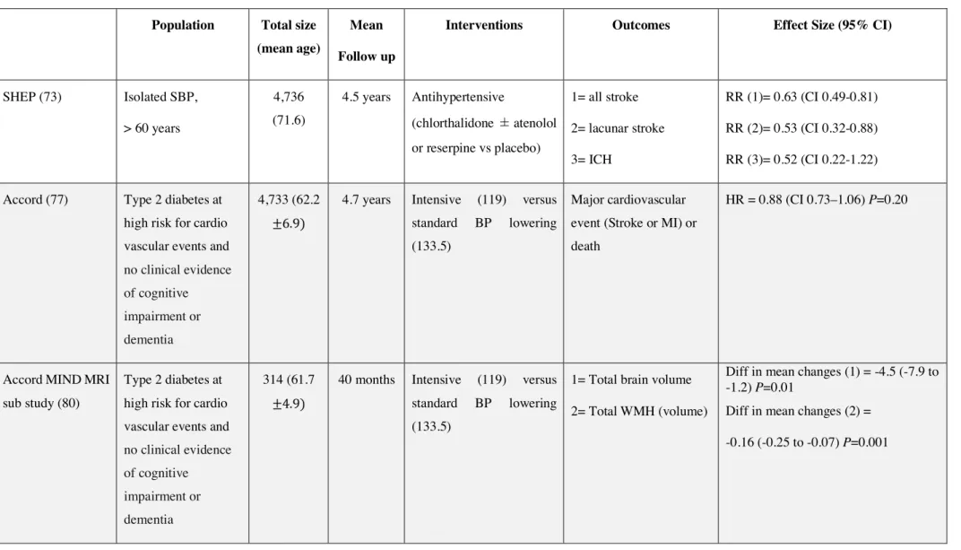
In primary prevention trials, the Systolic Hypertension in the Elderly Program (SHEP), a RCT conducted in elderly individuals with isolated systolic BP greater than 170 mmHg, showed that decreasing systolic BP to below 160 mmHg, and by more than 20 mmHg, significantly reduces the incidence of all strokes, including SVD-related strokes, by one-third. It was found that the incidence of stroke decreased by 1% for every mmHg decrease in systolic BP. The reduction in risk was especially marked for ICH (73).
In secondary prevention trials, the Perindopril PRotection aGainst Recurrent Stroke Study (PROGRESS), a RCT conducted in individuals with previous stroke, showed that lowering BP significantly reduced the risk of recurrent stroke by more than 25%. Notably, however, the effect was lower in lacunar (SVD-related) stroke compared with large-artery stroke. Remarkably, the magnitude of the reduction in stroke risk was similar in both hypertensive and non-hypertensive individuals. These data suggest that BP reduction per se confers the protective effects and not specific properties of a particular antihypertensive agent (74). Moreover, a subsidiary analysis of PROGRESS data indicated that lowering BP significantly decreases the
11
total volume of incident white matter lesions, producing the greatest beneficial effects in patients with severe WMHs at baseline (75). Remarkably, lowering BP has a much stronger effect on ICH than ischemic stroke. For example, the risk of ICH was halved in the PROGRESS study (74).
The optimal target systolic BP in SVD is still unclear. The Systolic Blood Pressure Intervention Trial (SPRINT) study was terminated early because intensive lowering of BP (to <120 mmHg) had a significant benefit for cardiovascular morbidity and mortality, although the rate of all strokes (a secondary outcome) was not significantly changed (76). In the ACCORD study, which enrolled patients with type 2 diabetes at high risk for cardiovascular events, intensive systolic BP reduction (to <120 mmHg) was associated with a significant reduction in the annual rate of all strokes (77). Moreover, a recent network meta-analysis that included 17 RCTs enrolling 55,163 individuals suggested that the lower the systolic BP target (down to 120 mmHg), the greater the reduction in the incidence of all strokes (78). However, in the Secondary Prevention of Small Subcortical Strokes (SPS3) trial, a unique study that tested an intensive (to <130 mmHg) versus standard systolic BP-lowering strategy and enrolled only patients with lacunar infarction, there was a non-significant 20% reduction in stroke in the intensive target group. Nevertheless, the rate of ICH was significantly reduced by two-thirds (79). On the other hand, intensive systolic BP reduction (to <120 mmHg) in the ACCORD-MIND MRI sub-study was associated with a significant reduction in the total volume of WMHs (80). The progression of WMHs was also significantly reduced in the subset of participants in the SPRINT Memory and Cognition IN Decreased Hypertension (SPRINT MIND) that participated in the brain MRI sub-study (81). To date, antihypertensives are used in patients with SVD like in those with other subtype of strokes.
There are controversies about the risks and benefits associated with intensive BP lowering. Serious adverse events, including hypotension, electrolyte abnormalities and kidney failure, have been reported with reductions in systolic BP below 120 mmHg (76, 78). Notably, a post hoc analysis of the SPS3 study showed that intensive lowering of systolic BP in patients with a prior lacunar stroke increased the likelihood of a decline in kidney function, especially during the first year of treatment, independent of the antihypertensive class used (82). In keeping with these concerns, a large meta-analysis has suggested <130 mmHg as a target BP that optimally balances efficacy and safety (78). On the other hand, the SPRINT MIND study showed that intensive BP lowering did not increase the risk of developing dementia, and even significantly reduced the risk of mild cognitive impairment (83). There was also no significant difference in
12
resting CBF among patients with lacunar infarcts and extensive WMHs between those subjected to intensive BP lowering (to <125 mmHg) and those on a standard BP-lowering regimen (to 130–140 mmHg) over a 3-month follow-up period (84).
Some caution should be exercised in interpreting these RCTs. The vast majority of secondary prevention studies, except for SPS3, did not focus specifically on patients with SVD. Another limitation is that these trials did not take into account the duration of premorbid hypertension, which is suspected to be more important than BP values at study entry. Also, the use of clinical stroke event or volume of WMHs as primary or secondary outcomes is unlikely to reflect the wide range of neurological deficits exhibited by patients with SVD (see below). Hence, inadequate or insensitive endpoints may have contributed to negative reported outcomes. Moreover, the average follow-up was ~3–4 years, which is relatively short compared with the long disease duration and chronic nature of SVD. Finally, there is currently no available information on differential effects of distinct classes of antihypertensive treatments.
Antiplatelet agents
RCTs have shown that there is no significant benefit of oral anticoagulants for secondary stroke prevention in SVD patients; in fact, these agents are associated with a higher risk of hemorrhage (85). In contrast, several antiplatelet drugs, including aspirin alone or in combination dipyridamole, clopidogrel or ticlopidine, have been approved by the Food and Drug Administration (FDA) for secondary stroke prevention. All RCTs on the subject have concluded that prolonged antiplatelet therapy reduces the risk of stroke recurrence by ~20% among patients with a previous transient ischemic attack or ischemic stroke, although it should be noted that SVD patients were not distinguished from patients with other subtypes of stroke in these trials (86). Overall, the efficacy of each drug is comparable (85). In contrast, there is no benefit of antiplatelet therapy for primary prevention.
The SPS3 investigators assessed the benefit of dual antiplatelet therapy (aspirin and clopidogrel vs. aspirin alone) in patients with a recent SVD-related stroke. The results of this assessment showed no significant change in recurrent ischemic stroke, but revealed a significant increase in the risk of major bleeding or death (87). The safety and efficacy of emerging antiplatelet agents that are associated with fewer bleeding complications, like the phosphodiesterase 3 inhibitor cilostazol, warrant further investigation.
The benefit-to-risk ratio of restarting antiplatelet agents poses a therapeutic dilemma for patients already taking antiplatelets following an ICH. Available observational studies favor
13
restarting antiplatelet therapy, because the risk of ischemic event for most patients is higher than the risk of another intracerebral hemorrhage (71).
Statins
HMG-CoA reductase inhibitors (statins) decrease LDL-cholesterol and, to a lesser extent, triglycerides. Through their inhibitory effect on the synthesis of isoprenoid intermediates, which are important for RhoA-ROCK/Rac/Cdc42 activity, statins exert pleiotropic effects on the vasculature, including inhibition of NOX and production of ROS, as well as activation of eNOS (88). Several RCTs have established that statins reduce the risk of all strokes in both primary and secondary prevention applications, effects that are closely associated with the reduction in LDL-cholesterol (89–91). A post hoc analysis of the SPARCL (Stroke Prevention by Aggressive Reduction in Cholesterol Levels) trial, in which 30% of subjects were patients with SVD-related strokes, showed similar beneficial trends against large artery and small-vessel strokes following treatment with the statin, Atorvastatin, although the effects did not reach statistical significance in either group (92). Although a lower level of LDL-cholesterol is associated with a higher risk of ICH, there is no evidence from several large meta-analyses that statin treatment is associated with ICH, possibly because the risk, if there is one, is small and compensated by the cardiovascular benefits (93).
B vitamins
The vitamins B6, B9 (folic acid), and B12 play important roles in the metabolism of homocysteine, and deficiencies of these vitamins can result in increased levels of total homocysteine, which has been associated with SVD (see above). The VITAmins TO Prevent Stroke (VITATOPS) study, a large RCT involving 8164 subjects with recent stroke, showed a marginally significant reduction in the incidence of major vascular events (stroke, myocardial infarction, death) in the subgroup of 2762 patients with SVD, but no effect in other stroke subtypes. However, a small MRI sub-analysis of SVD patients showed no evidence for a reduction in the incidence of lacunar infarcts or WMHs (94).
THERAPEUTIC AVENUES
Although we still do not have a firm grasp on the pathogenesis of SVD, some mechanisms have begun to emerge. Below, we elaborate on two mechanism-based therapeutic strategies worth exploring in sporadic SVD and CADASIL, respectively.
14
Targeting endothelial dysfunction
Several lines of evidence support a contribution of endothelial dysfunction to vascular risk-factor-related SVDs. An extensive body of evidence has established that a reduction in endothelial NO signaling is one important mechanism involved in mediating the deleterious neurovascular effects of chronic hypertension or aging. Here, we use the term “endothelial dysfunction” to refer to abnormalities in the production or bioavailability of endothelial-derived NO and resultant deleterious changes.
In vessels, NO is produced from L-arginine by eNOS. Most of the actions of eNOS are mediated through activation by NO of its intracellular receptor, soluble guanylyl cyclase (sGC), which is expressed in SMCs and platelets. Activation of sGC results in the synthesis of cyclic guanosine-3’,5’ monophosphate (cGMP), which acts as a second messenger to modulate SMC relaxation and proliferation and platelet reactivity, and inhibit vascular fibrosis. cGMP effects are terminated by the action of phosphodiesterase 5 (PDE5) (95).
NO-based therapeutics could be used to compensate for reduced NO bioactivity. However, their long-term use is limited by their adverse effects, mainly headaches, postural hypotension, and reflex tachycardia (96). Moreover, NO tolerance or resistance, and the potential scavenging of NO by superoxide, decreases the effectiveness of such therapeutics. Promising new agents include nitroxyl (HNO) donors, which are not scavenged by superoxide, do not induce tolerance, and moreover have the ability to inhibit NOX (97).
Among other potential NO pathway-targeting agents are sCG and cGMP modulators. sCG exists in both NO-sensitive and -insensitive forms; the latter, which is the predominant form under oxidative stress conditions, is the target of sGC activators. Several sGC activators are currently undergoing Phase II or III trials in subjects with chronic heart failure, a condition associated with oxidative stress and reduced NO bioavailability. PDE5A inhibitors, the most popular of which is sildenafil, have the potential to increase cGMP; however, their efficacy may be limited in the context of reduced NO signaling and decreased cGMP production (98). NOX, especially NOX2, is another logical target. Oxidative stress, defined as an imbalance between the production and removal of ROS, is a key contributor to compromised NO signaling, and NOX has been pinpointed as the main source of ROS. Remarkably, cerebral arteries have greater levels of NOX activity than many peripheral arteries, which may explain the higher susceptibility of cerebral vessels to oxidative stress (99). NOX is a family of multi-subunit enzyme complexes comprising seven distinct isoforms, of which NOX2 plays a major
15
role in cerebrovascular dysfunction associated with hypertension or aging (43). Classic NOX inhibitors, such as apocynin, the peptide gp91ds-tat and diphenyleneiodonium, have proven to be invaluable tools for experimental studies. However, their poor isoform selectivity and off-target effects are obvious limitations to their clinical utility. The development of novel, orally bioavailability compounds with better safety profiles is an important initial step, but additional efforts are needed to develop effective and selective NOX2 inhibitors. However, there is a narrow therapeutic window for NOX2 inhibitors, especially for long-term treatment, because of the physiological role of NOX2 in phagocytic cells, where it is predominantly expressed. Notably, phagocyte-derived ROS are important in killing invading pathogens and regulating autoimmunity and immune-mediated inflammatory diseases; moreover, functional deficiencies in NOX2 are associated with a growing number of diseases in humans, including X-linked chronic granulomatous disease, which is caused by a NOX2 mutation (100).
Angiotensin II is a major stimulus for ROS production. Angiotensin-converting enzyme (ACE) inhibitors or blockers of angiotensin II receptor type I (AT1R) protect against deleterious effects of angiotensin II by reducing the production of ROS (101). Although previous RCTs investigating BP-lowering agents have not highlighted superior properties of ACE inhibitors or AT1R blockers in reducing the risk of stroke, this strategy warrants further study in patients with SVD. Other therapeutic targets are emerging from experimental studies. These include, but are not limited to, Rho kinase (ROCK), which exerts inhibitory effects on eNOS and increases production of angiotensin II-induced ROS. Experimental studies have shown that specific inhibition of the ROCK2 isoform protects against compromised NO-mediated dilation of cerebral arteries and arterioles during aging (102).
Targeting NOTCH3 in CADASIL
Notch3ECD accumulation, a consequence of cysteine-altering, CADASIL-associated NOTCH3
mutations and one of the earliest manifestations in CADASIL, induces pathogenic changes in the brain microvascular extracellular matrix (Monet-Leprêtre et al. 2013; Capone, Cognat, et al. 2016).
One option for preventing the detrimental effects of the unpaired cysteine residue is to eliminate the mutant EGF repeat without compromising NOTCH3 activity. Proof of concept of this approach has recently been obtained in vitro using antisense oligonucleotide (AON)-mediated exon skipping to “hide” targeted exons from the splicing machinery, resulting in their exclusion from the mature mRNA (103). In this approach, targeted exons were selected in such a way that
16
the resulting exon-skipped NOTCH3 protein, which contained a chimeric EGF repeat with six correctly spaced cysteine residues, retained almost normal expression and the ability to be activated by its ligand, Jagged-1. Notably, an in silico analysis further indicated that a large majority of CADASIL mutations are eligible for cysteine correction (104). The next challenge will be to establish the preclinical efficacy of this approach. Although the proportion of mutant NOTCH3 that must be eliminated to prevent all pathogenic effects remains an unresolved issue, AON-mediated exon skipping is a promising therapeutic approach. Indeed, one such AON has received approval from the Food and Drug Administration for the treatment of selected patients with Duchenne Muscular Dystrophy (105).
An alternate strategy consists of neutralizing the toxicity of Notch3ECD through passive
immunization against Notch3ECD. Proof of concept of this approach has been established in a
preclinical mouse model of CADASIL. In this approach, a peripherally injected monoclonal antibody against Notch3ECD was found to bind all Notch3ECD deposits in the brain vasculature
of mutant mice, yielding almost complete target engagement. Chronic administration of this antibody markedly protected mutant mice against cerebrovascular dysfunction, normalizing functional hyperemia and vasodilatory responses as well as rescuing defects in myogenic tone and reduced lumen diameter. Intriguingly, this antibody was effective despite failing to attenuate Notch3ECD deposition; thus, the next step before embarking on future clinical trials
will be to clarify its mechanism of action (106).
Some uncommon mutations (present in ~5% of families) located in or around the ligand-binding domain of Notch3 unambiguously abrogate Notch3 signaling (107). It was recently shown that an agonistic Notch3 antibody that targets the heterodimerization domain of NOTCH3 and induces ligand-independent activation of the receptor partially restores the activity of a mutant NOTCH3 receptor containing a CADASIL mutation in the ligand-binding domain (108). However, whether such an approach would be beneficial in CADASIL is questionable, given that genetic studies in both humans and mice argue against a loss-of-function mechanism in the pathological consequences of NOTCH3 mutations (109, 110).
PROSPECTS FOR BETTER MONITORING OF TREATMENT EFFECTS
Evaluating treatment in patients with SVD represents a major challenge, because neurological deficits include not only acute events (lacunar infarcts) but also chronic manifestations (cognitive decline, disability, apathy and mood disorders). Yet, the vast majority of published
17
RCTs have used reduction in stroke events as a primary or secondary endpoint. Cognitive deficits in SVD patients predominantly affect executive functioning and processing speed, whereas episodic memory remains relatively intact until later stages of the disease. Therefore, cognitive assessment requires dedicated tests that are distinct from the many cognitive screening tests available for cortical dementias such as Alzheimer's disease. Furthermore, apathy and mood disturbances, which are key symptoms in patients with SVD, are not included in the established scales currently used in patients with neurodegenerative disorders or stroke. Overall, there is a need for dedicated scales for the assessment of SVD patients. A composite scale rather than a combination of different scales may better capture the clinical status of patients and provide a more sensitive evaluation of clinical changes, especially over a short time. Another promising tool, which would be highly relevant in SVD, is based on Patient Reported Outcomes (111).
A second challenge is related to the slow and variable rate of progression of clinical deficits over short time periods. Because of the faster progression of SVD neuroimaging abnormalities, MRI changes appear as attractive biomarkers for assessing the effects of therapeutic interventions using much smaller sample sizes and shorter durations of intervention, thereby reducing the costs of clinical trials (112). A major step towards this has been an international effort to develop standard terminology and definitions for these MRI markers and propose minimum standards for image acquisition and analysis across research centers (2). A reliable MRI biomarker needs to be quantitative and sensitive, and must correlate quantitatively with disease progression (i.e., disability, cognitive decline or progression to dementia) in longitudinal studies. The load of WMHs can be assessed qualitatively using the Fazekas scale; quantitative assessments, though possible, remain labor intensive. However, several studies have pointed to a weak association between global WMH volume and clinical symptoms such as cognition in patients with symptomatic SVD or extensive WMHs, possibly due to a ceiling effect (113, 114). Other studies suggest that WMH load in strategic white matter tracts might be more pertinent (115). On the other hand, the extent of white matter damage assessed using diffusion tensor imaging (DTI) has emerged as a better biomarker.DTI is highly sensitive to microstructural changes in the white matter that can be invisible on conventional MRI. Importantly, DTI metrics better correlate with cognitive deficits than WMH burden, and changes in these metrics better correlate with cognitive decline (116–118). Novel post-processing techniques applied to DTI may allow reliable—and fully automated—quantification (116). Changes in brain volume also correlate strongly with cognitive deterioration in both
18
cross-sectional and longitudinal studies. Moreover, brain atrophy has been shown to be the most sensitive imaging correlate of clinical deterioration in CADASIL patients (119). The number of incident lacunes is also significantly correlated with cognitive decline in longitudinal studies (118). However, quantification of lacunes still relies on semi-automated approaches applied by trained observers. A combination of all SVD-related MRI changes in a composite score may provide a more sensitive approach (120). Other potential biomarkers, such as brain connectivity, resting CBF and cerebrovascular reactivity measurements, are in the early phase of development.
Several technical issues need to be overcome in order for these MRI biomarkers to be used as clinically meaningful surrogate markers in place of other traditional endpoints in multicenter RCTs. Among these are optimization of spatial resolution, reduction in acquisition-related sources of image variability, and standardization of image post-processing. Moreover, validation of these MRI biomarkers requires testing in a wide range of patients and severities (121).
CONCLUDING REMARKS AND FUTURE DIRECTIONS
SVDs impose a heavy burden on society, and their impacts are expected to further increase in the absence of significant therapeutic advances, given the aging population. Yet, the fact that SVDs progress silently for many years before becoming clinically symptomatic provides a long therapeutic window that should lend itself to early interventions. Translating this conceptual advantage into tangible benefits will require a strong effort by clinical and basic scientific communities to tackle a number of significant challenges.
Several modifiable risk factors have been identified, among which hypertension is currently deemed the most important. Yet, there is insufficient evidence that intensive BP control significantly impacts the clinical course of the disease. Future trials should consider earlier intervention, longer treatment and follow-up, and validated brain imaging biomarkers as endpoints. Also, testing distinct classes of antihypertensive agents might be worth considering, given for example the ability of ACE or AT1R inhibitors to improve cerebrovascular dysfunction in preclinical models (51). Moreover, there is a need to identify patients who are at greater risk of developing SVDs. The continuum between Mendelian and sporadic diseases raises the possibility that genetic factors can make the difference in disease manifestation among individuals exposed to the same vascular risk factors.
19
Our knowledge of the normal biology and physiology of brain vessels, and how vascular risk or genetic factors can impair brain vessel structure or function, has dramatically increased over the past decade. Yet, there is still a large knowledge gap in how these defects produce brain lesions and clinical manifestations. Along the same lines, it is astonishing that the mechanisms underlying the development of lacunar infarcts or cognitive decline are still unclear. One major issue is the lack of animal models that recapitulate the full spectrum of neuropathological and clinical manifestations of SVDs, an issue that may find its ultimate resolution in genetic models. Finally, there is the challenge of translating identified therapeutic targets into potential therapies. Again, appropriate models are urgently needed to test these therapies in preclinical trials before embarking on costly clinical trials.
DISCLOSURE STATEMENT
The author is not aware of any affiliation, membership, funding that might be perceived as affecting the objectivity of this review. Inserm/AJ own patent rights to molecular diagnosis and immunological treatment of CADASIL.
ACKNOWLEDGEMENTS
This work was supported by Fondation Leducq (Transatlantic Network of Excellence on the Pathogenesis of Small Vessel Disease of the Brain), the European Union (Horizon 2020 Research and Innovation Programme SVDs@target under grant agreement n° 666881) and the National Research Agency, France (ANR-16-RHUS-0004).
20
LITTERATURE CITED
1. Pantoni L. 2010. Cerebral small vessel disease: from pathogenesis and clinical characteristics to therapeutic challenges. Lancet Neurol. 9(7):689–701
2. Wardlaw JM, Smith EE, Biessels GJ, Cordonnier C, Fazekas F, et al. 2013. Neuroimaging standards for research into small vessel disease and its contribution to ageing and neurodegeneration. Lancet Neurol. 12(8):822–38
3. Love S, Chalmers K, Ince P, Esiri M, Attems J, et al. 2014. Development, appraisal, validation and implementation of a consensus protocol for the assessment of cerebral amyloid angiopathy in post-mortem brain tissue. Am. J. Neurodegener. Dis. 3(1):19–32 4. Charidimou A, Boulouis G, Gurol ME, Ayata C, Bacskai BJ, et al. 2017. Emerging
concepts in sporadic cerebral amyloid angiopathy. Brain J. Neurol. 140(7):1829–50 5. Craggs LJL, Yamamoto Y, Deramecourt V, Kalaria RN. 2014. Microvascular pathology
and morphometrics of sporadic and hereditary small vessel diseases of the brain. Brain Pathol. Zurich Switz. 24(5):495–509
6. Debette S, Schilling S, Duperron M-G, Larsson SC, Markus HS. 2018. Clinical Significance of Magnetic Resonance Imaging Markers of Vascular Brain Injury: A Systematic Review and Meta-analysis. JAMA Neurol.
7. Wardlaw JM, Smith C, Dichgans M. 2013. Mechanisms of sporadic cerebral small vessel disease: insights from neuroimaging. Lancet Neurol. 12(5):483–97
8. Rapsomaniki E, Timmis A, George J, Pujades-Rodriguez M, Shah AD, et al. 2014. Blood pressure and incidence of twelve cardiovascular diseases: lifetime risks, healthy life-years lost, and age-specific associations in 1·25 million people. Lancet Lond. Engl. 383(9932):1899–1911
21
9. Klarenbeek P, van Oostenbrugge RJ, Rouhl RPW, Knottnerus ILH, Staals J. 2013. Ambulatory blood pressure in patients with lacunar stroke: association with total MRI burden of cerebral small vessel disease. Stroke. 44(11):2995–99
10. Chauhan G, Adams HHH, Satizabal CL, Bis JC, Teumer A, et al. 2019. Genetic and lifestyle risk factors for MRI-defined brain infarcts in a population-based setting. Neurology
11. Bezerra DC, Sharrett AR, Matsushita K, Gottesman RF, Shibata D, et al. 2012. Risk factors for lacune subtypes in the Atherosclerosis Risk in Communities (ARIC) Study. Neurology. 78(2):102–8
12. van Dijk EJ, Breteler MMB, Schmidt R, Berger K, Nilsson L-G, et al. 2004. The association between blood pressure, hypertension, and cerebral white matter lesions: cardiovascular determinants of dementia study. Hypertens. Dallas Tex 1979. 44(5):625– 30
13. Cordonnier C, Al-Shahi Salman R, Wardlaw J. 2007. Spontaneous brain microbleeds: systematic review, subgroup analyses and standards for study design and reporting. Brain J. Neurol. 130(Pt 8):1988–2003
14. Dufouil C, de Kersaint-Gilly A, Besançon V, Levy C, Auffray E, et al. 2001. Longitudinal study of blood pressure and white matter hyperintensities: the EVA MRI Cohort. Neurology. 56(7):921–26
15. Lau KK, Li L, Simoni M, Mehta Z, Küker W, et al. 2018. Long-Term Premorbid Blood Pressure and Cerebral Small Vessel Disease Burden on Imaging in Transient Ischemic Attack and Ischemic Stroke. Stroke. 49(9):2053–60
22
16. VARIABLE BRAIN consortium. 2018. The association between blood pressure variability (BPV) with dementia and cognitive function: a systematic review and meta-analysis protocol. Syst. Rev. 7(1):163
17. Koekkoek PS, Kappelle LJ, van den Berg E, Rutten GEHM, Biessels GJ. 2015. Cognitive function in patients with diabetes mellitus: guidance for daily care. Lancet Neurol. 14(3):329–40
18. Schilling S, Tzourio C, Dufouil C, Zhu Y, Berr C, et al. 2014. Plasma lipids and cerebral small vessel disease. Neurology. 83(20):1844–52
19. Wang X, Dong Y, Qi X, Huang C, Hou L. 2013. Cholesterol levels and risk of hemorrhagic stroke: a systematic review and meta-analysis. Stroke. 44(7):1833–39 20. Hassan A, Hunt BJ, O’Sullivan M, Bell R, D’Souza R, et al. 2004. Homocysteine is a
risk factor for cerebral small vessel disease, acting via endothelial dysfunction. Brain J. Neurol. 127(Pt 1):212–19
21. Larsson SC, Traylor M, Markus HS. 2019. Homocysteine and Small Vessel Stroke: A Mendelian Randomization Analysis. Ann. Neurol.
22. Wardlaw JM, Allerhand M, Doubal FN, Valdes Hernandez M, Morris Z, et al. 2014. Vascular risk factors, large-artery atheroma, and brain white matter hyperintensities. Neurology. 82(15):1331–38
23. Field TS, Doubal FN, Johnson W, Backhouse E, McHutchison C, et al. 2016. Early life characteristics and late life burden of cerebral small vessel disease in the Lothian Birth Cohort 1936. Aging. 8(9):2039–61
23
24. Haffner C, Malik R, Dichgans M. 2016. Genetic factors in cerebral small vessel disease and their impact on stroke and dementia. J. Cereb. Blood Flow Metab. Off. J. Int. Soc. Cereb. Blood Flow Metab. 36(1):158–71
25. Ayrignac X, Carra-Dalliere C, Menjot de Champfleur N, Denier C, Aubourg P, et al. 2015. Adult-onset genetic leukoencephalopathies: a MRI pattern-based approach in a comprehensive study of 154 patients. Brain J. Neurol. 138(Pt 2):284–92
26. Chabriat H, Joutel A, Dichgans M, Tournier-Lasserve E, Bousser MG. 2009. Cadasil. Lancet Neurol. 8(7):643–53
27. Lee Y-C, Chung C-P, Chao N-C, Fuh J-L, Chang F-C, et al. 2018. Characterization of Heterozygous HTRA1 Mutations in Taiwanese Patients With Cerebral Small Vessel Disease. Stroke. 49(7):1593–1601
28. Joutel A, Vahedi K, Corpechot C, Troesch A, Chabriat H, et al. 1997. Strong clustering and stereotyped nature of Notch3 mutations in CADASIL patients. Lancet. 350(9090):1511–15
29. Rutten JW, Van Eijsden BJ, Duering M, Jouvent E, Opherk C, et al. 2019. The effect of NOTCH3 pathogenic variant position on CADASIL disease severity: NOTCH3 EGFr 1-6 pathogenic variant are associated with a more severe phenotype and lower survival compared with EGFr 7-34 pathogenic variant. Genet. Med. Off. J. Am. Coll. Med. Genet. 21(3):676–82
30. Malik R, Chauhan G, Traylor M, Sargurupremraj M, Okada Y, et al. 2018. Multiancestry genome-wide association study of 520,000 subjects identifies 32 loci associated with stroke and stroke subtypes. Nat. Genet. 50(4):524–37
24
31. Malik R, Rannikmäe K, Traylor M, Georgakis MK, Sargurupremraj M, et al. 2018. Genome-wide meta-analysis identifies 3 novel loci associated with stroke. Ann. Neurol. 84(6):934–39
32. Rannikmäe K, Sivakumaran V, Millar H, Malik R, Anderson CD, et al. 2017. COL4A2 is associated with lacunar ischemic stroke and deep ICH: Meta-analyses among 21,500 cases and 40,600 controls. Neurology. 89(17):1829–39
33. Mishra A, Chauhan G, Violleau M-H, Vojinovic D, Jian X, et al. 2019. Association of variants in HTRA1 and NOTCH3 with MRI-defined extremes of cerebral small vessel disease in older subjects. Brain J. Neurol.
34. Iadecola C. 2013. The pathobiology of vascular dementia. Neuron. 80(4):844–66 35. Longden TA, Dabertrand F, Koide M, Gonzales AL, Tykocki NR, et al. 2017. Capillary
K+-sensing initiates retrograde hyperpolarization to increase local cerebral blood flow. Nat. Neurosci. 20(5):717–26
36. Cipolla MJ. 2009. The Cerebral Circulation. San Rafael (CA): Morgan & Claypool Life Sciences
37. Sweeney MD, Zhao Z, Montagne A, Nelson AR, Zlokovic BV. 2019. Blood-Brain Barrier: From Physiology to Disease and Back. Physiol. Rev. 99(1):21–78
38. Rasmussen MK, Mestre H, Nedergaard M. 2018. The glymphatic pathway in neurological disorders. Lancet Neurol. 17(11):1016–24
39. Shih AY, Blinder P, Tsai PS, Friedman B, Stanley G, et al. 2013. The smallest stroke: occlusion of one penetrating vessel leads to infarction and a cognitive deficit. Nat. Neurosci. 16(1):55–63
25
40. Smith EE, Beaudin AE. 2018. New insights into cerebral small vessel disease and vascular cognitive impairment from MRI. Curr. Opin. Neurol. 31(1):36–43
41. Joutel A, Chabriat H. 2017. Pathogenesis of white matter changes in cerebral small vessel diseases: beyond vessel-intrinsic mechanisms. Clin. Sci. Lond. Engl. 1979. 131(8):635– 51
42. Huneau C, Houot M, Joutel A, Béranger B, Giroux C, et al. 2018. Altered dynamics of neurovascular coupling in CADASIL. Ann. Clin. Transl. Neurol.
43. Santisteban MM, Iadecola C. 2018. Hypertension, dietary salt and cognitive impairment. J. Cereb. Blood Flow Metab. Off. J. Int. Soc. Cereb. Blood Flow Metab. 38(12):2112– 28
44. Toth P, Tarantini S, Csiszar A, Ungvari Z. 2017. Functional vascular contributions to cognitive impairment and dementia: mechanisms and consequences of cerebral autoregulatory dysfunction, endothelial impairment, and neurovascular uncoupling in aging. Am. J. Physiol. Heart Circ. Physiol. 312(1):H1–20
45. Iadecola C, Davisson RL. 2008. Hypertension and cerebrovascular dysfunction. Cell Metab. 7(6):476–84
46. Baumbach GL, Sigmund CD, Bottiglieri T, Lentz SR. 2002. Structure of cerebral arterioles in cystathionine beta-synthase-deficient mice. Circ. Res. 91(10):931–37 47. Dayal S, Devlin AM, McCaw RB, Liu M-L, Arning E, et al. 2005. Cerebral vascular
dysfunction in methionine synthase-deficient mice. Circulation. 112(5):737–44
48. Duncombe J, Kitamura A, Hase Y, Ihara M, Kalaria RN, Horsburgh K. 2017. Chronic cerebral hypoperfusion: a key mechanism leading to vascular cognitive impairment and
26
dementia. Closing the translational gap between rodent models and human vascular cognitive impairment and dementia. Clin. Sci. Lond. Engl. 1979. 131(19):2451–68 49. De Silva TM, Faraci FM. 2016. Microvascular Dysfunction and Cognitive Impairment.
Cell. Mol. Neurobiol. 36(2):241–58
50. Iadecola C. 2017. The Neurovascular Unit Coming of Age: A Journey through Neurovascular Coupling in Health and Disease. Neuron. 96(1):17–42
51. Faraco G, Sugiyama Y, Lane D, Garcia-Bonilla L, Chang H, et al. 2016. Perivascular macrophages mediate the neurovascular and cognitive dysfunction associated with hypertension. J. Clin. Invest. 126(12):4674–89
52. Baron-Menguy C, Domenga-Denier V, Ghezali L, Faraci FM, Joutel A. 2017. Increased Notch3 Activity Mediates Pathological Changes in Structure of Cerebral Arteries. Hypertens. Dallas Tex 1979. 69(1):60–70
53. Capone C, Cognat E, Ghezali L, Baron-Menguy C, Aubin D, et al. 2016. Reducing Timp3 or vitronectin ameliorates disease manifestations in CADASIL mice. Ann. Neurol. 79(3):387–403
54. Joutel A, Andreux F, Gaulis S, Domenga V, Cecillon M, et al. 2000. The ectodomain of the Notch3 receptor accumulates within the cerebrovasculature of CADASIL patients. J Clin Invest. 105(5):597–605
55. Joutel A, Monet-Lepretre M, Gosele C, Baron-Menguy C, Hammes A, et al. 2010. Cerebrovascular dysfunction and microcirculation rarefaction precede white matter lesions in a mouse genetic model of cerebral ischemic small vessel disease. J Clin Invest. 120(2):433–45
27
56. Monet-Leprêtre M, Haddad I, Baron-Menguy C, Fouillot-Panchal M, Riani M, et al. 2013. Abnormal recruitment of extracellular matrix proteins by excess Notch3 ECD: a new pathomechanism in CADASIL. Brain J. Neurol. 136(Pt 6):1830–45
57. Dabertrand F, Krøigaard C, Bonev AD, Cognat E, Dalsgaard T, et al. 2015. Potassium channelopathy-like defect underlies early-stage cerebrovascular dysfunction in a genetic model of small vessel disease. Proc. Natl. Acad. Sci. U. S. A. 112(7):E796-805
58. Capone C, Dabertrand F, Baron-Menguy C, Chalaris A, Ghezali L, et al. 2016. Mechanistic insights into a TIMP3-sensitive pathway constitutively engaged in the regulation of cerebral hemodynamics. eLife. 5:
59. Erdő F, Denes L, de Lange E. 2017. Age-associated physiological and pathological changes at the blood-brain barrier: A review. J. Cereb. Blood Flow Metab. Off. J. Int. Soc. Cereb. Blood Flow Metab. 37(1):4–24
60. Kisler K, Nelson AR, Rege SV, Ramanathan A, Wang Y, et al. 2017. Pericyte degeneration leads to neurovascular uncoupling and limits oxygen supply to brain. Nat. Neurosci. 20(3):406–16
61. Montagne A, Nikolakopoulou AM, Zhao Z, Sagare AP, Si G, et al. 2018. Pericyte degeneration causes white matter dysfunction in the mouse central nervous system. Nat. Med. 24(3):326–37
62. Park MH, Lee JY, Park KH, Jung IK, Kim K-T, et al. 2018. Vascular and Neurogenic Rejuvenation in Aging Mice by Modulation of ASM. Neuron. 100(1):167-182.e9 63. Brown R, Benveniste H, Black SE, Charpak S, Dichgans M, et al. 2018. Understanding
the role of the perivascular space in cerebral small vessel disease. Cardiovasc. Res. 114(11):1462–73
28
64. Mestre H, Tithof J, Du T, Song W, Peng W, et al. 2018. Flow of cerebrospinal fluid is driven by arterial pulsations and is reduced in hypertension. Nat. Commun. 9(1):4878 65. Mestre H, Hablitz LM, Xavier AL, Feng W, Zou W, et al. 2018. Aquaporin-4-dependent
glymphatic solute transport in the rodent brain. eLife. 7:
66. Kress BT, Iliff JJ, Xia M, Wang M, Wei HS, et al. 2014. Impairment of paravascular clearance pathways in the aging brain. Ann. Neurol. 76(6):845–61
67. Campbell BCV, Donnan GA, Lees KR, Hacke W, Khatri P, et al. 2015. Endovascular stent thrombectomy: the new standard of care for large vessel ischaemic stroke. Lancet Neurol. 14(8):846–54
68. Pantoni L, Fierini F, Poggesi A. 2014. Thrombolysis in acute stroke patients with cerebral small vessel disease. Cerebrovasc. Dis. Basel Switz. 37(1):5–13
69. Eggers CCJ, Bocksrucker C, Seyfang L, Austrian Stroke Unit Registry Collaborators. 2017. The efficacy of thrombolysis in lacunar stroke - evidence from the Austrian Stroke Unit Registry. Eur. J. Neurol. 24(6):780–87
70. Charidimou A, Pasi M, Fiorelli M, Shams S, von Kummer R, et al. 2016. Leukoaraiosis, Cerebral Hemorrhage, and Outcome After Intravenous Thrombolysis for Acute Ischemic Stroke: A Meta-Analysis (v1). Stroke. 47(9):2364–72
71. Cordonnier C, Demchuk A, Ziai W, Anderson CS. 2018. Intracerebral haemorrhage: current approaches to acute management. Lancet Lond. Engl. 392(10154):1257–68 72. Scaggiante J, Zhang X, Mocco J, Kellner CP. 2018. Minimally Invasive Surgery for
29
73. Perry HM, Davis BR, Price TR, Applegate WB, Fields WS, et al. 2000. Effect of treating isolated systolic hypertension on the risk of developing various types and subtypes of stroke: the Systolic Hypertension in the Elderly Program (SHEP). JAMA. 284(4):465–71 74. PROGRESS Collaborative Group. 2001. Randomised trial of a perindopril-based
blood-pressure-lowering regimen among 6,105 individuals with previous stroke or transient ischaemic attack. Lancet Lond. Engl. 358(9287):1033–41
75. Dufouil C, Chalmers J, Coskun O, Besançon V, Bousser M-G, et al. 2005. Effects of blood pressure lowering on cerebral white matter hyperintensities in patients with stroke: the PROGRESS (Perindopril Protection Against Recurrent Stroke Study) Magnetic Resonance Imaging Substudy. Circulation. 112(11):1644–50
76. SPRINT Research Group, Wright JT, Williamson JD, Whelton PK, Snyder JK, et al. 2015. A Randomized Trial of Intensive versus Standard Blood-Pressure Control. N. Engl. J. Med. 373(22):2103–16
77. ACCORD Study Group, Cushman WC, Evans GW, Byington RP, Goff DC, et al. 2010. Effects of intensive blood-pressure control in type 2 diabetes mellitus. N. Engl. J. Med. 362(17):1575–85
78. Bangalore S, Toklu B, Gianos E, Schwartzbard A, Weintraub H, et al. 2017. Optimal Systolic Blood Pressure Target After SPRINT: Insights from a Network Meta-Analysis of Randomized Trials. Am. J. Med. 130(6):707-719.e8
79. SPS3 Study Group, Benavente OR, Coffey CS, Conwit R, Hart RG, et al. 2013. Blood-pressure targets in patients with recent lacunar stroke: the SPS3 randomised trial. Lancet Lond. Engl. 382(9891):507–15
30
80. Murray AM, Hsu F-C, Williamson JD, Bryan RN, Gerstein HC, et al. 2017. ACCORDION MIND: results of the observational extension of the ACCORD MIND randomised trial. Diabetologia. 60(1):69–80
81. Kjeldsen SE, Narkiewicz K, Burnier M, Oparil S. 2018. Intensive blood pressure lowering prevents mild cognitive impairment and possible dementia and slows development of white matter lesions in brain: the SPRINT Memory and Cognition IN Decreased Hypertension (SPRINT MIND) study. Blood Press. 27(5):247–48
82. Peralta CA, McClure LA, Scherzer R, Odden MC, White CL, et al. 2016. Effect of Intensive Versus Usual Blood Pressure Control on Kidney Function Among Individuals With Prior Lacunar Stroke: A Post Hoc Analysis of the Secondary Prevention of Small Subcortical Strokes (SPS3) Randomized Trial. Circulation. 133(6):584–91
83. SPRINT MIND Investigators for the SPRINT Research Group, Williamson JD, Pajewski NM, Auchus AP, Bryan RN, et al. 2019. Effect of Intensive vs Standard Blood Pressure Control on Probable Dementia: A Randomized Clinical Trial. JAMA
84. Croall ID, Tozer DJ, Moynihan B, Khan U, O’Brien JT, et al. 2018. Effect of Standard vs Intensive Blood Pressure Control on Cerebral Blood Flow in Small Vessel Disease: The PRESERVE Randomized Clinical Trial. JAMA Neurol. 75(6):720–27
85. Furie KL, Kasner SE, Adams RJ, Albers GW, Bush RL, et al. 2011. Guidelines for the prevention of stroke in patients with stroke or transient ischemic attack: a guideline for healthcare professionals from the american heart association/american stroke association. Stroke. 42(1):227–76
86. Collaborative overview of randomised trials of antiplatelet therapy--I: Prevention of death, myocardial infarction, and stroke by prolonged antiplatelet therapy in various
31
categories of patients. Antiplatelet Trialists’ Collaboration. 1994. BMJ. 308(6921):81– 106
87. SPS3 Investigators, Benavente OR, Hart RG, McClure LA, Szychowski JM, et al. 2012. Effects of clopidogrel added to aspirin in patients with recent lacunar stroke. N. Engl. J. Med. 367(9):817–25
88. Oesterle A, Laufs U, Liao JK. 2017. Pleiotropic Effects of Statins on the Cardiovascular System. Circ. Res. 120(1):229–43
89. 2 ISGCWTC-CC. Failure to validate association between 12p13 variants and ischemic stroke. N Engl J Med. 362(16):1547–50
90. Amarenco P, Bogousslavsky J, Callahan A, Goldstein LB, Hennerici M, et al. 2006. High-dose atorvastatin after stroke or transient ischemic attack. N. Engl. J. Med. 355(6):549–59
91. Amarenco P, Labreuche J, Lavallée P, Touboul P-J. 2004. Statins in stroke prevention and carotid atherosclerosis: systematic review and up-to-date meta-analysis. Stroke. 35(12):2902–9
92. Amarenco P, Benavente O, Goldstein LB, Callahan A, Sillesen H, et al. 2009. Results of the Stroke Prevention by Aggressive Reduction in Cholesterol Levels (SPARCL) trial by stroke subtypes. Stroke. 40(4):1405–9
93. Hackam DG, Woodward M, Newby LK, Bhatt DL, Shao M, et al. 2011. Statins and intracerebral hemorrhage: collaborative systematic review and meta-analysis. Circulation. 124(20):2233–42
94. Cavalieri M, Schmidt R, Chen C, Mok V, de Freitas GR, et al. 2012. B vitamins and magnetic resonance imaging-detected ischemic brain lesions in patients with recent
32
transient ischemic attack or stroke: the VITAmins TO Prevent Stroke (VITATOPS) MRI-substudy. Stroke. 43(12):3266–70
95. Vanhoutte PM, Zhao Y, Xu A, Leung SWS. 2016. Thirty Years of Saying NO: Sources, Fate, Actions, and Misfortunes of the Endothelium-Derived Vasodilator Mediator. Circ. Res. 119(2):375–96
96. Bagdy G, Riba P, Kecskeméti V, Chase D, Juhász G. 2010. Headache-type adverse effects of NO donors: vasodilation and beyond. Br. J. Pharmacol. 160(1):20–35
97. Ritchie RH, Drummond GR, Sobey CG, De Silva TM, Kemp-Harper BK. 2017. The opposing roles of NO and oxidative stress in cardiovascular disease. Pharmacol. Res. 116:57–69
98. Singh P, Vijayakumar S, Kalogeroupoulos A, Butler J. 2018. Multiple Avenues of Modulating the Nitric Oxide Pathway in Heart Failure Clinical Trials. Curr. Heart Fail. Rep. 15(2):44–52
99. Miller AA, Drummond GR, Schmidt HHHW, Sobey CG. 2005. NADPH oxidase activity and function are profoundly greater in cerebral versus systemic arteries. Circ. Res. 97(10):1055–62
100. Hoffmann MH, Griffiths HR. 2018. The dual role of Reactive Oxygen Species in autoimmune and inflammatory diseases: evidence from preclinical models. Free Radic. Biol. Med. 125:62–71
101. Girouard H, Park L, Anrather J, Zhou P, Iadecola C. 2006. Angiotensin II attenuates endothelium-dependent responses in the cerebral microcirculation through nox-2-derived radicals. Arterioscler. Thromb. Vasc. Biol. 26(4):826–32
33
102. De Silva TM, Modrick ML, Dabertrand F, Faraci FM. 2018. Changes in Cerebral Arteries and Parenchymal Arterioles With Aging: Role of Rho Kinase 2 and Impact of Genetic Background. Hypertens. Dallas Tex 1979. 71(5):921–27
103. Aartsma-Rus A, van Ommen G-JB. 2007. Antisense-mediated exon skipping: a versatile tool with therapeutic and research applications. RNA N. Y. N. 13(10):1609–24
104. Rutten JW, Dauwerse HG, Peters DJM, Goldfarb A, Venselaar H, et al. 2016. Therapeutic NOTCH3 cysteine correction in CADASIL using exon skipping: in vitro proof of concept. Brain J. Neurol. 139(Pt 4):1123–35
105. Aartsma-Rus A, Krieg AM. 2017. FDA Approves Eteplirsen for Duchenne Muscular Dystrophy: The Next Chapter in the Eteplirsen Saga. Nucleic Acid Ther. 27(1):1–3 106. Ghezali L, Capone C, Baron-Menguy C, Ratelade J, Christensen S, et al. 2018.
Notch3ECD immunotherapy improves cerebrovascular responses in CADASIL mice. Ann. Neurol. 84(2):246–59
107. Monet-Lepretre M, Bardot B, Lemaire B, Domenga V, Godin O, et al. 2009. Distinct phenotypic and functional features of CADASIL mutations in the Notch3 ligand binding domain. Brain. 132(Pt 6):1601–12
108. Machuca-Parra AI, Bigger-Allen AA, Sanchez AV, Boutabla A, Cardona-Vélez J, et al. 2017. Therapeutic antibody targeting of Notch3 signaling prevents mural cell loss in CADASIL. J. Exp. Med. 214(8):2271–82
109. Rutten JW, Boon EMJ, Liem MK, Dauwerse JG, Pont MJ, et al. 2013. Hypomorphic NOTCH3 alleles do not cause CADASIL in humans. Hum. Mutat. 34(11):1486–89 110. Cognat E, Baron-Menguy C, Domenga-Denier V, Cleophax S, Fouillade C, et al. 2014.