HAL Id: hal-02910867
https://hal.inrae.fr/hal-02910867
Submitted on 18 Sep 2020HAL is a multi-disciplinary open access archive for the deposit and dissemination of sci-entific research documents, whether they are pub-lished or not. The documents may come from teaching and research institutions in France or abroad, or from public or private research centers.
L’archive ouverte pluridisciplinaire HAL, est destinée au dépôt et à la diffusion de documents scientifiques de niveau recherche, publiés ou non, émanant des établissements d’enseignement et de recherche français ou étrangers, des laboratoires publics ou privés.
Mickael Desvaux, Michel Hébraud
To cite this version:
Mickael Desvaux, Michel Hébraud. Analysis of cell envelope proteins. Handbook of Listeria mono-cytogenes, Dongyou Liu ; CRC Press, Taylor and Francis Group, pp.359-393, 2008, Chapter 12. �hal-02910867�
12
Analysis of Cell
Envelope Proteins
Mickaël Desvaux and Michel Hébraud
contents12.1 Introduction... 359
12.2 Protein Secretion Systems... 360
12.2.1 Sec System... 361 12.2.2 Tat Pathway... 367 12.2.3 FPE ... 369 12.2.4 FEA... 370 12.2.5 Holins... 371 12.2.6 Wss... 372
12.3 Cell Envelope-Associated Proteins ... 374
12.3.1 Membrane-Associated Proteins... 374
12.3.1.1 Integral Membrane Proteins ... 374
12.3.1.2 Lipoproteins ... 377
12.3.1.3 Extrinsic Membrane Proteins ... 379
12.3.2 Cell Wall-Associated Proteins... 379
12.3.2.1 LPXTG Motif ... 380
12.3.2.2 Noncovalently Attached Cell Wall Proteins ... 380
12.4 Conclusions and Perspectives ... 382
References ... 384 12.1 IntroductIon
From a morphological point of view, the most fundamental dichotomy within prokaryotes (the term “prokaryotes” is used here in its primary etymological sense—that is, single-celled organ isms without nuclei as opposed to eukaryotes, without any further phylogenetic considerations1)
is between those bound by a single biological membrane (monoderm prokaryotes)—that is, the cytoplasmic membrane, and those bound by two concentric but topologically different membranes (diderm prokaryotes)—that is, the inner membrane (cytoplasmic membrane) and the asymmetric outer membrane.2 In accordance with holistic and teleonomic concepts, organisms are far more
than mere collections of genes,3,4 and such difference in membrane organization, and thus cell com
partmentation, is not trivial but has profound phylogenetic, structural, metabolic, and physiologi cal implications. Based on the most recent advances in biological evolution and megaclassification of organisms,5–7 monoderm prokaryotes are regrouped under the term Monodermata (also called
Unibacteria), which essentially includes Archaea together with Posibacteria (formerly called Gram-positive bacteria).
It is worth stressing that the term “Gram-positive bacteria” is terminologically ambiguous, espe cially for researchers interested in aspects related to bacterial cell envelope (e.g., protein secretion or surface proteins).8 From its origin, a positive or negative result given by Gram staining method
indicates whether or not bacteria retain the stain respectively. Later on, the difference in staining was related to profound divergence in structural organization of the cell envelope, briefly: (1) a cyto plasmic membrane surrounded by a thick cell wall in Gram-positive bacteria, and (2) a cytoplasmic membrane surrounded by a thin cell wall beneath the outer membrane in Gram-negative bacteria. Molecular analyses further revealed that, contrary to Gram-negative bacteria, Gram-positive bacteria correspond to a phylogenetically coherent grouping of prokaryotes within the domain Bacteria with phylum BXIII Firmicutes (low G+C mole percent) and phylum BXIV Actinobacteria (high G+C mole percent).9,10 However, from Gram staining to cell envelope organization to taxonomic grouping,
each step represents some approximations, which often result in misleading or incoherent statements in the literature. For example, some members of Firmicutes and Actinobacteria phyla do not retain Gram stain because of (1) the absence of a cell wall (e.g., bacteria from the genus Mycoplasma), (2) a too thin cell wall (e.g., some members of the genus Clostridium), or (3) the presence of a waxy outer sheath preventing penetration of the stain (e.g., species from the genus Mycobacterium).
Inversely, some bacteria not taxonomically related to Gram-positive bacteria retain the Gram stain (e.g., some members of the phylum BIV Deinococcus-Thermus). More confusingly, some bacteria clearly possessing a Gram-negative-like cell envelope architecture are in fact phyloge netically related to the taxonomic group of Gram-positive bacteria (e.g., Thermotoga maritima cur rently classified in phylum BII Thermotogae,11 or Fusobacterium nucleatum belonging to phylum
BXXI Fusobacteria).12,13 Some other phyla regroup bacteria exhibiting both cell envelope structures
(Gram-negative-like or Gram-positive-like cell envelope)—for example, BVI Chloroflexi or BVII Thermomicrobia.14 Even in some deep branches of the phylum Firmicutes, some bacteria clearly
exhibit Gram-negative cell envelope ultrastructure (e.g., in genus Desulfotomaculum, Selenomonas,
Syntrophomonas, or Coprothermobacter).2 Therefore, it appears in numerous cases that the term
“Gram-positive bacteria” cannot describe at once a particular Gram staining result, cell envelope organization, and taxonomic group; thus, when employing this term it is extremely important to specify what it refers to. Because of fewer terminological ambiguities, the terms “Monodermata” or “monoderm bacteria” will be preferred to describe prokaryotic cells surrounded by a single biologi cal membrane but without any further phylogenetic considerations. For the purpose of the present review, the term “Gram-positive bacteria” will be used to describe bacteria with a cell envelope composed of (1) a cytoplasmic membrane, and (2) a cell-wall composed at least of peptidoglycan.
Listeria species are monoderm bacteria possessing a thick cell wall retaining Gram stain and belonging to phylum Firmicutes, class Bacilli, order Bacillales, and family Listeriaceae,9 and as
such are Gram-positive bacteria in all meaning of the term. L. monocytogenes is undoubtedly the species that has attracted most attention, considering its frequent occurrence in food coupled with a high mortality rate.15 Still, the genus Listeria comprises six species: (1) two pathogenic ones
(L. monocytogenes, a human pathogen, and L. ivanovii, a ruminant pathogen), and (2) four non pathogenic relatives (L. innocua, L. seeligeri, L. welshimeri, and L. grayi.)16,17 Only two completed
L. monocytogenes genome sequences are currently available—L. monocytogenes 1/2a EGD-e and
4b F236518,19—but several other strains are being unassembled18 or sequenced (http://www.ncbi.
nlm.nih.gov/genomes/lproks.cgi). Among other species, L. innocua CLIP1126219 and L. welshimeri
SLCC533420 are the only genomes available, but L. ivanovii PAM55, L. seeligeri SLCC3954, and
L. grayi CLIP12515 are currently being sequenced.17 Since the genomes of L. monocytogenes 1/2a
F6854 and 4b H7858 are unfinished, some genes cannot be properly identified; also, final assembly of these genomic sequences may reveal homologues at a later date. Because no clear conclusion can be drawn from genomic analysis of unfinished genomes,21 this review will only focus on completed
genome sequences of L. monocytogenes strains. 12.2 ProteIn secretIon systems
Within the cell envelope, Listeria species can exhibit a large variety of proteins; some of them can even interact with the cell surroundings and thus constitute the surfaceome (i.e., the subset of
pro-tein exposed on the bacterial cell surface). It is worth reminding that, on one hand, cell wall is not an impermeable barrier and cell envelope proteins can interact with the environment without ever hav ing a domain that leaves the confine of the cell wall8 and that the extracellular milieu can penetrate
the cell wall, so proteins do not necessarily need to poke out into the environment.22 On the other
hand, protein localization into the cell envelope is no guarantee that it is cell surface exposed stricto
sensu as proteins can be masked by overlying components such as capsule polymer, for example.8
Nevertheless, for the purpose of the present review, cell surface proteins will refer to gene products that are attached to the cell wall and/or cytoplasmic membrane and interacting with the external side, whereas cell envelope proteins will refer to all gene products present within the cell wall and/ or the cytoplasmic membrane.
While cell surface proteins are systematically cell envelope proteins, the opposite is not neces sarily true (e.g., proteins attached to the cytoplasmic membrane but interacting only with the cyto plasm). Still, all cell surface proteins (and most cell envelope proteins) must be first translocated to the cytoplasmic membrane via a protein secretion system before attaching to membrane or cell wall components and thus remaining in contact with the external side. Concerning the functions of cell envelope proteins, they are extremely diverse, ranging from transporters and enzymes involved in various metabolic pathways (such as carbohydrates, proteins, nucleotides, or lipids), signal transduc tions, adhesion and colonization determinants, to virulence factors. It is worth stressing that among cell surface proteins, some so-called moonlighting proteins can be present.23 Such proteins are
multifunctional in the sense that they conduct enzymatic and/or nonenzymatic activities, sometimes taking part in widely divergent pathways, especially when present at different subcellular locations. For example, enolase, a cytoplasmic protein normally involved in glycolytic pathways, was found on the listerial cell surface, which can bind to human plasminogen.24
In Didermata (corresponding to Gram-negative bacteria, also called Negibacteria),5,25 six major
protein secretion systems (numbered from Type I to Type VI, i.e., T1SS to T6SS) are currently recognized and are restricted to these microorganisms.26–29 In fact, protein secretion systems are
categorized primarily by translocation mechanisms across the outermost lipid bilayer, which cor responds to the outer membrane in diderm bacteria but to the cytoplasmic membrane in mono derm prokaryotes. To date in monoderm bacteria, six systems are described as allowing protein secretion30 –33—that is, protein transport from inside to outside cell cytoplasm—namely, (1) the Sec
pathway (secretion, TC #3.A.5; TC#: transport classification number),34 (2) the Tat pathway (twin
arginine translocation, TC #2.A.64), (3) the FEA (flagella export apparatus, TC #3.A.6.1), (4) the FPE (fimbrilin-protein exporter, TC #3.A.14), (5) the holins (hole-formers, TC#1.E.), and (6) the Wss (WXG100 secretion system, proteins with WXG motif of ~100 residues). To be complete, the MscL family (large conductance mechanosensitive ion channel, TC #1.A.22) and the putative Tad (tight adherence) apparatus could also be added to the list,35,36 even though experimental evidence is
not currently available in monoderm bacteria. Once translocated by one of these systems, a protein can remain associated to the cell envelope, be released into the extracellular milieu, or be translo cated into a host cell.
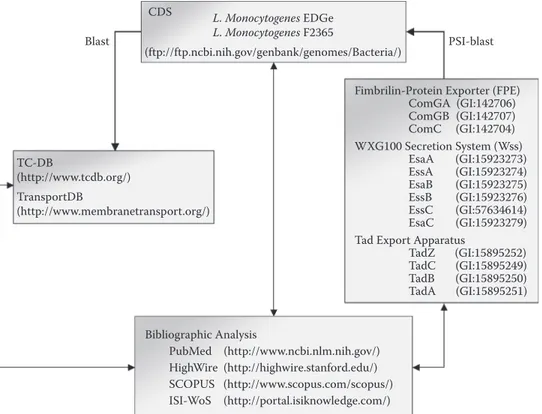
As depicted in Figure 12.1, identification of protein secretion systems in Listeria involved screening of genome coding sequences (CDS) against various databases as well as bibliographic analyses. From there, Sec, Tat, FPE, FEA, holins, and Wss were identified in L. monocytogenes37
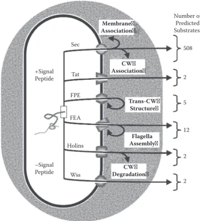
(Figure 12.2). While some components of these secretion systems have been experimentally inves tigated, in Listeria, protein translocation per se has never been ascertained in any of them yet. 12.2.1 Sec SyStem
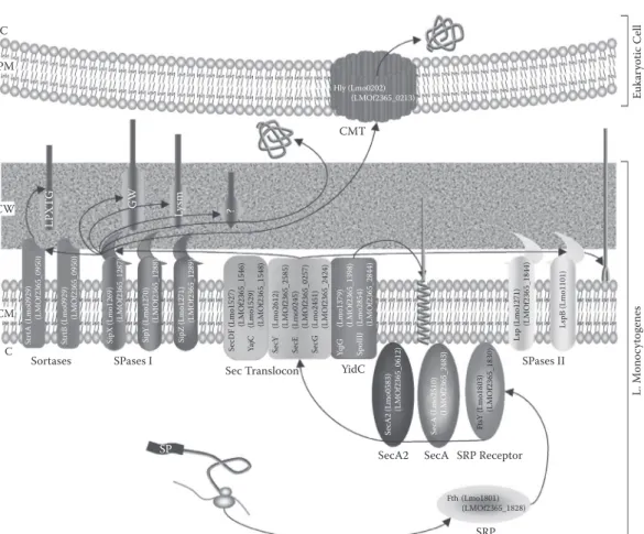
The presence, remarkable conservation, and essential nature of the Sec translocon in all living cells have given rise to the notion of a general secretory pathway (GSP) but also led to confusing state ments in the literature.38 As illustrated in Figure 12.3, all components of Sec translocon are encoded
CDS L. Monocytogenes EDGe L. Monocytogenes F2365
Blast PSI-blast
(ftp://ftp.ncbi.nih.gov/genbank/genomes/Bacteria/)
Fimbrilin-Protein Exporter (FPE) ComGA (GI:142706) ComGB (GI:142707) ComC (GI:142704) WXG100 Secretion System (Wss)
TC-DB EsaA (GI:15923273)
(http://www.tcdb.org/) EssA EsaB (GI:15923274)(GI:15923275)
TransportDB EssB (GI:15923276)
(http://www.membranetransport.org/) EssC (GI:57634614)
EsaC (GI:15923279) Tad Export Apparatus
TadZ (GI:15895252) TadC (GI:15895249) TadB (GI:15895250) TadA (GI:15895251) Bibliographic Analysis PubMed (http://www.ncbi.nlm.nih.gov/) HighWire (http://highwire.stanford.edu/) SCOPUS (http://www.scopus.com/scopus/) ISI-WoS (http://portal.isiknowledge.com/)
FIgure 12.1 Genomic identification of protein secretion systems in Listeria species.37 Prior to bioinfor
matic analysis, complete genome, coding sequences (CDS), and original annotation data sets were downloaded from GenBank. Each CDS was screened for the capacity to encode a component of a protein secretion system following BLAST against TCDB146 and TransportDB.248 These analyses revealed the presence of Sec com
ponents and partners as well as FEA subunits, Tat components and holins. MscL and ABC transporter truly implicated in protein secretion could not be identified. The identification of FPE was based on PSI-BLAST searches using GenBank amino acid sequences of ComGA, ComGB, and ComC from B. subtilis as queries. Similarly, Wss was identified using EsaA, EssA, EsaB, EssB, EssC, and EsaC from S. aureus as amino-acid sequence queries. Using protein sequences of Clostridium acetobutylicum as queries,33 Tad system compo
nents could not be identified. Overall, bibliographic analyses were also performed from various databases. particle (SRP) and the SRP receptor are ubiquitous and essential in all domains of life.39 In E. coli,
SRP interacts with nascent signal peptide for cotranslational translocation and specific integration of inner membrane proteins, whereas the targeting factor and chaperone SecB interacts with the mature part of the protein and allows post-translational translocation via Sec.40 As in all
Gram-positive bacteria,41 SecB and CsaA (analogous to SecB in B. subtilis30) are absent from L. monocy
togenes. In E. coli, three auxiliary proteins (SecD, SecF, and YajC) form a transmembrane complex loosely associated with SecYEG and increase the overall efficiency of protein translocation through the cytoplasmic membrane.42
Contrary to SecDF-YajC, the cytosolic ATPase SecA is essential to Sec-dependent translocation in bacteria as it provides the driving force for stepwise export of the protein.43 A SecA paralogue
(i.e., SecA2) has been identified in several Gram-positive bacteria including L. monocytogenes.44
Contrary to Streptococcus gordonii, for example,45 presence of SecA2 in L. monocytogenes is not
accompanied by duplication of SecY. While SecA2 is not essential and its relationship with SRP/ Sec is unknown, it clearly allows the secretion of a subset of proteins in L. monocytogenes (e.g., Iap,44 NamA,46 and FbpA47). Interestingly, the membrane protein FbpA lacks a putative N-terminal
signal peptide. As in B. subtilis,30 two paralogues of YidC could be identified in L. monocytogenes:
Number of
Membrane Predicted
Association Substrates:
Sec 508
CW
+Signal Tat Association
Peptide 2 FPE Trans-CW 5 Structure FEA 12 Flagella Holins Assembly 2 –Signal CW Peptide Wss Degradation 2
FIgure 12.2 Schematic overview of protein secretion pathways in L. monocytogenes EGD-e.37 Proteins
to be translocated can exhibit (+) or not (–) an N-terminal signal peptide (with the exception of Sec pathway, which can translocate proteins with or without signal peptide by alternative mechanisms). Proteins translocated via the Sec pathway remain membrane associated or cell wall associated, are released into the extracellular milieu, or would even be injected into an eukaryotic host cell. Proteins exported via Tat would most certainly be cell surfaced or released into the extracellular milieu. FPE would be involved in the formation of transcell wall structures. FEA is involved in flagella assembly. Proteins exported by holins seem secreted into the extra-cellular milieu or involved in cell wall degradation. WXG100 proteins would be secreted into the extraextra-cellular milieu. The number of translocated proteins by each pathway is given from most recent estimations. CW, cell wall; Sec, secretion; FPE, fimbrilin-protein exporter; Tat, twin-arginine translocation; FEA, flagella export apparatus; Wss, WXG100 (proteins with WXG motif of ~100 amino acyl residues) secretion system. insertion of all integral membrane proteins (IMPs).48 YidC is a versatile pathway since it can be
Sec-, SecA-, and/or SecB independent. In B. subtilis, studies have showed that SpoIIIJ and YqjG play a role in the folding of several secreted proteins and can work independently to insert integral membrane proteins.49
Signal peptide of translocated preprotein is cleaved off by a membrane-bound signal peptidase (SPase). Different classes of N-terminal signal peptide are recognized and are cleaved by different types of SPases. Signal peptides of proteins targeted to Sec are of two classes: class 1 and class 2. Class 2 signal peptides are present in lipoproteins and are cleaved off by SPase II (for further details, see section 12.3.1.2). As depicted in Figure 12.3, precursor proteins exhibiting a class 1 signal peptide meet different fates; that is, they can (1) insert in cytoplasmic membrane and thus become integral membrane proteins (for further details, see section 12.3.1.1), (2) remain attached covalently or noncovalently to cell wall components (for further details, see section 12.3.2), (3) be released into the extracellular milieu, or (4) be injected into a eukaryotic host cell via pore formed by Sec-secreted listeriolysin O in a process called cytolysin-mediated translocation (CMT).50,51 It is
C C PM Hly (Lmo0202) (LMOf2365_0213) CMT L. M on oc yt ogene s Euk ar yotic Cell St rtA (Lmo0929 ) (L MOf2365_0950) LPX TG Str tB (Lmo0929) (L MOf2365_0950) SipX (Lmo1269 ) (L MOf2365_1287) G W Sip Y (Lmo1270) (L MOf2365_1288) SipZ (Lmo1271) Ly sm (L MOf2365_1289) Se cDF (Lmo1527) ? (L MOf2365_1546) Ya jC (Lmo1529 ) (L MOf2365_1548) Se cY (Lmo2612 ) (L MOf2365_2585) Se cE (Lmo0245 ) (L MOf2365_0257) Se cG (Lmo2451) (LMOf2365_2424) YqjG (Lmo1379) (LMOf2365_1398) Sp olIIJ (Lmo2854) (LMOf2365_2844) CW CM Lsp (Lmo1271) (L MOf2365_1844) LspB (Lmo1101 ) Sortases SPases I Se cA2 (Lmo0583) (L MOf2365_0612) Se cA (Lmo2510) (L MOf2365_2483) Ft sY (Lmo1803) (L MOf2365_1830)
Sec Translocon YidC
SP
SPases II
SecA2 SecA SRP Receptor
Fth (Lmo1801) (LMOf2365_1828)
SRP
FIgure 12.3 Schematic representation of the Sec pathway in L. monocytogenes. 37 N-terminal signal pep
tide is recognized by SRP before cotranslational translocation of the protein through the Sec translocon in a SecA-dependent manner. Some proteins with or without a signal peptide can also be translocated in a SecA2-dependent manner. Integral membrane proteins integrate into the CM via YidC homologues in Sec dependent or -independent manner; such proteins bear stop-transfer sequence and can exhibit signal peptide or not, which can be cleaved or not. Lipoproteins, which bear signal peptide of class 2 cleavable by SPases II, are covalently attached to long-chain fatty acids of the CM. Proteins bearing class 1 signal peptide cleavable by SPases I are (1) secreted into the extracellular milieu or could even be injected into an eukaryotic host cell following CMT thanks to pores formed by oligomerization of listeriolysin O; (2) bound to CW components via cell binding motifs (i.e., GW, LysM, or uncharacterized motifs); or (3) covalently attached to CW by sortases because of the presence of C-terminal LPXTG motif. C, cytosol; PM, plasmic membrane; CW, cell wall; EM, extracellular milieu; CM, cytoplasmic membrane; SP, signal peptide; SPase, signal peptidase; SRP, signal recognition particle; CMT, cytolysin mediated translocation.
In Gram-positive bacteria, some Sec-dependent signal peptides exhibit a YSIRK motif (PF04650) present at the beginning of the H-domain. This motif is required for efficient protein secretion and is systematically associated with an LPXTG motif, even though the opposite is not true. Class 1 signal peptides are not always cleaved as the H-domain can serve of transmembrane anchor domain as observed in SPases I. Three SPases I have been uncovered and characterized in L. monocytogenes: SipX, SipY, and SipZ.52,53 Deletion of sipY genes had no detectable effect,
whereas SipX and SipZ had overlapping substrate specificity.52 lsp was demonstrated as encoding a
genuine SPase II54 and a second SPase II—LspB (Lmo1101)—was recently uncovered by genomic
analysis but only in L. monocytogenes EGD-e.37
While some proteins cleaved by SPases I can remain noncovalently bound by various cell wall binding domains (for further details, see section 12.3.2.2), covalent attachment of proteins to
AU: should “of” be “as”?
In Gram-positive bacteria, some Sec-dependent signal peptides exhibit a YSIRK motif (PF04650) present at the beginning of the H-domain. This motif is required for efficient protein secretion and is systematically associated with an LPXTG motif, even though the opposite is not true. Class 1 signal peptides are not always cleaved as the H-domain can serveof transmembrane anchor domain as observed in SPases I. Three SPases I have been uncovered and characterized in L. monocytogenes: SipX, SipY, and SipZ.52,53 Deletion of sipY genes had no detectable effect,
whereas SipX and SipZ had overlapping substrate specificity.52lspwas demonstrated as encoding a
genuine SPase II54and a second SPase II—LspB (Lmo1101)—was recently uncovered by genomic
analysis but only in L. monocytogenes EGD-e.37
While some proteins cleaved by SPases I can remain noncovalently bound by various cell wall binding domains (for further details, see section 12.3.2.2), covalent attachment of proteins to
cell wall requires sortases. Proteins emerging from the Sec apparatus and exhibiting an LPXTG like motif C-terminally located (for further details, see section 12.3.2.1) are recognized by mem-brane-associated sortase.55 Transpeptidase sortase attacks the TG bond of the LPXTG-like motif,
capturing cleaved polypeptide as a thioester-linked acyl enzyme at its active site cystein residue.56
Subsequently, this complex is resolved by the nucleophilic attack of the amino group of the cross-bridge within lipid II precursor. Based on phylogenetic analyses, sortases are now classified into four classes, designated A, B, C, and D.57 In L. monocytogenes, two sortases are present (SrtA and
SrtB; Figure 12.3).
As observed in other Gram-positive bacteria, sortase of class A (also called SrtA subfamily) in
L. monocytogenes is encoded only once in the genome, resembles a Type II membrane protein, and is necessary for the anchoring of the majority of LPXTG-containing proteins.58 Sortase of class B
(SrtB subfamily) recognizes a particular type of sorting signal (i.e., an NXZTN motif), which sug gests a lower stringency of the recognition motif of SrtB compared to SrtA.59 Captivatingly, from
investigations in Streptococcus pyogenes and Staphylococcus aureus, glycosylated LPXTGase, an enzyme that cleaves the C-terminal LPXTG motif, is the first enzyme found that is produced by nonribosomal peptide (NRP) synthesis.60,61 It is known that NRP synthesis (and similarly related
polyketide synthesis) occurs in Bacilli class, where NRPs are assembled in the cytoplasm by large megaproteins called NRP synthetases consisting of a series of active modules carrying out catalysis and modification of the tethered growing peptide chain.62 However, investigations in S. aureus sug
gest that enzymes responsible for cell wall assembly may also be involved in the construction of LPXTGase.61 Finally, it cannot be excluded that such a nonribosomally synthesized enzyme be also
present and involved in LPXTG-like protein anchoring in L. monocytogenes.37
Substrates of the Sec system are generally considered as exhibiting an N-terminal signal pep tide composed of three domains: (1) The N-domain contains positively charged amino terminus, (2) the H-domain is a hydrophobic core region, and (3) the C-domain contains the cleavage site.63
It must be emphasized, however, that it is not the case for all proteins (e.g., some SecA2-dependent and/or YidC-dependent proteins). Still, the presence of an N-terminal signal peptide indicates a protein is targeted to membrane. Despite lack of amino acid sequence similarity, signal peptides can be detected with good accuracy by various documented and publicly available applications (Table 12.1). The first methods developed were SigCleave and SPScan, which were implementations of a simple weight matrix approach.64 While SigCleave is part of the EMBOSS suite and also avail
able by an interface on the World Wide Web, SPScan is only available as part of the GCG suite and thus requires ability to work under Unix-like environment. Comparing the two programs, SPScan has clearly better predictive performance in terms of secretory protein and cleavage site recognition, especially for prokaryotic proteins.65 Nearly a decade later, SignalP, a promising method based on a
table 12.1
bioinformatic resources for Prediction of bacterial n-terminal signal Peptides
application method Webserver ref.
SigCleave Position weight matrix http://bioweb.pasteur.fr/seqanal/interfaces/sigcleave.html 64
SPScan Position weight matrix none 64
SignalP Neural network http://www.cbs.dtu.dk/services/SignalP/ 67
Hidden Markov model
PrediSi Position weight matrix http://www.predisi.de/ 69
SOSUIsignal Global physicochemical analysis http://bp.nuap.nagoya-u.ac.jp/sosui/sosuisignal/ 70
Phobius Hidden Markov model http://phobius.binf.ku.dk/ 71
PSORTb Support vector machine http://www.psort.org/psortb/ 74
Hidden Markov model
neural network, was released66 and has undoubtedly become the most popular method for predict
ing N-terminal signal peptide. Since the first available version 1.1, SignalP has been substantially improved up to the latest version 3.0.67
While version 1.1 is definitively out of date, both versions 2.0 and 3.0 use either a neural network (NN) or HMM. When comparing SignalP v2.0-NN, -HMM, and SPScan, it appears that (1) SPScan predicts correctly more proteins as secreted than SignalP v2.0-NN or -HMM; (2) SignalP v2.0-NN and -HMM are superior in predicting the correct cleavage site; (3) SignalP v2.0-NN lags behind SPScan and SignalP v2.0-HMM in classifying correctly the proteins, the latter providing the best prediction; and (4) SignalP v2.0-NN is the best for predicting of the correct cleavage site.65 In other
words, these methods are complementary in predicting an N-terminal signal peptide. The main improvement in SignalP v3.0 is increased accuracy in prediction of signal peptidase cleavage sites.67
In comparative analyses, SignalP3.0 performs significantly better than other machine learning and HMM methods. Despite performance improvement in the latest SignalP v3.0, however, it appears that SignalP v2.0-NN remains the best signal prediction program.68
A position weight matrix approach was improved by a frequency correction, which takes into consideration the amino acid bias (i.e., PrediSi).69 SOSUIsignal is a global structure analysis based
on physicochemical features of the three signal peptide domains—N-, H-, and C-domains—and discriminates between cleavable and anchoring signal sequences.70 Since a signal peptide contains
a hydrophobic H-domain, there is a risk of erroneously identifying a transmembrane α-helix as a signal peptide or, conversely, classifying a protein with a signal peptide H-domain region as an IMP. In order to discriminate between the two, a combined TM topology and signal peptide predictor has been developed: Phobius.71 Phobius significantly reduces false classifications of signal peptides
compared to SignalP. Another machine learning approach used for prediction of signal peptides is support vector machine (SVM), which can predict signal peptides with great accuracy.72 Such an
implementation of an SVM combined with an HMM is part of PSORTb,73 now applicable to both
Gram-positive and Gram-negative bacteria.74 Finally, SPdb, a repository of experimentally deter
mined and computationally predicted signal peptides, is also accessible via BLAST (basic local alignment search tool) search.75,76
It can be stressed again that these analyses only predict the presence of signal peptide, meaning that the protein is targeted to the cytoplasmic membrane. However, it does not necessarily mean the protein is translocated across the cytoplasmic membrane via Sec or released into the extracellular milieu. Indeed, proteins translocated via Tat or FPE also possess N-terminal signal peptides with additional features, which are not identified by the previous tools (Table 12.1). Thus, final prediction of a protein translocated via Sec requires additional inspections (see sections 12.2.2 and 12.2.3). Concerning proteins translocated by the Sec system and possessing a signal peptide, they can (1) be released into the extracellular medium or injected into a host cell, (2) remain associated to the cell wall by covalent or noncovalent interactions, or (3) remain associated to the cytoplasmic membrane by transmembrane domains (including H-domain of uncleaved signal peptide) or be lipoproteins (see section 12.3). Thus, final localization prediction of Sec substrates requires a combination of tools for prediction of function, motifs, and TMDs. It is also recommended to combine these results with those from tools dedicated to prediction of protein subcellular localization in Gram-positive bacteria (Table 12.2).
NNPSL was the first tool developed for such prediction and is based on an NN.77 SubLoc,78
PSORTb,73 CELLO79 (recently extended to prediction in Gram-positive bacteria80), and LOCtree81
are basically SVM. These tools have their own advantages and weaknesses,82 and some of them,
like PSORTb, combine a variety of individual predictors. Proteome Analyst is a novel type of machine-learning classifier that involves several steps in the prediction process, such as BLAST search against Swiss-Prot database and naïve Bayesian classifiers.83 From the most recent studies on
performance of prediction tools, PSORTb and Proteome Analyst achieve the highest overall preci-sion.84 Gpos-PLoc, another type of ensemble classifier, was recently developed where several basic
table 12.2
bioinformatic resources for Prediction of subcellular localization of Proteins in gram-Positive bacteria
application method Webserver ref.
NNPSL Neural network http://www.doe-mbi.ucla.edu/~astrid/astrid.html 77 SubLoc Support vector machine http://www.bioinfo.tsinghua.edu.cn/SubLoc/ 78
PSORTb Support vector machine http://www.psort.org/psortb/ 73
Ensemble classifier
CELLO Support vector machine http://cello.life.nctu.edu.tw/ 80
LOCtree Support vector machine http://cubic.bioc.columbia.edu/services/loctree/ 81 Proteome Analyst Ensemble classifier http://pa.cs.ualberta.ca:8080/pa/ 83 Gpos-PLoc Ensemble classifier http://202.120.37.186/bioinf/Gpos/ 85 DBSubLoc BLAST http://www.bioinfo.tsinghua.edu.cn/~guotao/intro.html 86
PSORTdb BLAST http://db.psort.org/ 87
PA-GOSUB BLAST http://www.cs.ualberta.ca/~bioinfo/PA/GOSUB/ 88
Augur Ensemble classifier http://bioinfo.mikrobio.med.uni-giessen.de/augur/ 80
classifiers were fused and optimized for predicting subcellular localization of Gram-positive bacte rial proteins.85 Finally, several databases (derived from previously described prediction tools) are
available following BLAST search (DBSubLoc,86 PSORTdb,87 and PA-GOSUB88). Augur is another
database especially dedicated to protein localization on cell surface of Gram-positive bacteria.89
Once again, final prediction of secreted proteins (and localization) should combine results from these various bioinformatic tools.84
Using SignalP v2.0 to predict signal peptide region and TopPred v2.0 to exclude other trans membrane domains, 86 proteins were predicted as secreted into the extracellular medium from genomic analysis of L. monocytogenes EGD-e.19 In L. monocytogenes F2365, 420 proteins were
predicted with a putative N-terminal signal peptide, including 2 with a YSIRK motif.18 Perform
ing extensive genomic analyses, which combined results from SignalP v2.0, SigCleave, SOSUI, PSORT, and TMPinGS, the number of proteins bearing an N-terminal signal peptide was estimated at 525 in L. monocytogenes EGD-e, including 255 IMPs and 270 exported proteins where 121 would be released into the extracellular milieu.90 All 14 virulence factors characterized so far in
L. monocytogenes are most likely translocated via the Sec translocon.37 Among the 121 proteins
originally predicted as secreted via Sec and released into the extracellular milieu, a closer look revealed that four prepilins—that is, ComGC (Lmo1345), ComGD (Lmo1344), ComGE (Lmo1343), and ComGG (Lmo1341)—should be removed from the output since they would form trans-cell-wall structure following translocation via FPE.37 Proteomic analysis of supernatant from liquid culture
of L. monocytogenes EGD-e allowed the identification of 54 out of 117 proteins predicted as extra-cellular, including virulence factors Hly, PlcA, and PlcB.90
12.2.2 tat Pathway
The term twin-arginine translocation (Tat) was coined from the systematic presence of RR motif in signal peptide of proteins translocated via this secretion system.91 The [ST]RRXFLK motif strad
dles the N-domain and H-domain of N-terminal signal peptide.92 Contrary to the Sec translocon, the
main feature of this pathway is its ability to translocate proteins in a folded state. General knowl edge on the precise succession and mechanistic events leading to protein secretion via this pathway remains rudimentary.93 The generally accepted translocation model was first proposed by Mori and
Cline,94 where Tat translocation follows a cycle in which TatBC functions in the specific recogni
(a) (b)
C CM RR
EM
TatA (Lmo0362) SPase I
TatC (Lmo0361)
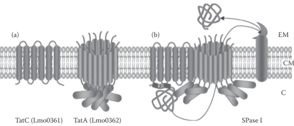
FIgure 12.4 Tat translocon in L. monocytogenes.37 During Tat secretion, the general model proposes a
cyclical assembly of components. (A) In resting state, Tat machinery components are separately present in the cytoplasmic membrane (i.e., TatB and TatA). (B) Once Tat substrate protein precursor binds to the TatC in an energy-independent step, this complex associates with TatA in a step driven by transmembrane proton electrochemical gradient. This association would persist until completion of protein transport across the mem brane driven by proton motive force. Tat signal peptide is subsequently cleaved by SPase I and Tat machinery components disassembled, as depicted in (A). RR, twin-arginine motif in Tat signal peptide; EM, extracellular milieu; CM, cytoplasmic membrane; C, cytoplasm.
that membrane integration could precede Tat-dependent translocation and the membrane targeting process may require ATP-dependent N-terminal unfolding-steps energy.95
Still, components of Tat translocon differ in number between Gram-negative and Gram-positive bacteria.30 The most baffling difference is the absence of TatB from all Gram-positive bacteria
sequenced so far, although it is an essential components of Tat translocon in E. coli, which is used as a paradigm.96 As in most Gram-positive bacteria, Tat translocon in L. monocytogenes is encoded
in one locus and is composed of only two proteins, TatA and TatC37 (Figure 12.4). TatC is a large
IMP generally considered as the primary site for signal-peptide recognition.97 TatA is a membrane
protein that oligomerizes to form a protein-conducting channel where the number of subunits would adjust in function of the Tat substrate size.98 In TatA, a cytoplasmic lid region acts as a gate and
would open following association of TatC–substrate complex with TatA, then inducing conforma tion change and protein translocation driven by proton motive force. Translocated protein is finally released after cleavage by SPAse I.96 A Tat translocon does not seem to be systematically present in
L. monocytogenes as no component could be identified in L. monocytogenes F2365. The Tat system has never been experimentally investigated in Listeria; thus, its expression, functionality, involve ment of one or three SPases I, or proteins secreted via this pathway remain unknown.37
Three tools are currently available to discern Tat substrates (Table 12.3) and TATFIND was the first program especially devoted to such identification.99 In its original available version, TATFIND
v1.2, prediction was based on two criteria: (1) presence of conserved Tat motif ZRRZZZ within the first 35 amino acid residues, where Z represents a defined set of permitted residues; and (2) presence
table 12.3
bioinformatic resources for Prediction of tat signal Peptides
application method Webserver ref.
TATFIND Physicochemical analysis and regular http://signalfind.org/tatfind.html 100 expression
TatP Neural network and regular expression http://www.cbs.dtu.dk/services/TatP/ 101 TATPred Naïve Bayesian network http://www.jenner.ac.uk/logP/JennerlogPcalc.htm 102
of an uncharged stretch of at least 13 amino acids downstream of the twin arginine. In the latest version, TATFIND v1.4, search for a single charged residue in positions +2 and +5 relative to the RR was included.100 TatP v1.0 incorporates signal peptide and cleavage site prediction based on a
combination of two artificial neural networks followed by a postfiltering of the output based on reg ular expression RR[FGAVML][LITMVF].101 Compared to TATFIND v1.2, TatP generates far fewer
false positive but slightly more false negative predictions. TATPred is the latest algorithm based on naïve Bayesian network developed for prediction of Tat substrates.102 Compared to TatP, TATPred
appears as the most robust and reliable predictor with higher sensitivity of prediction.
According to TATFIND search, only two Tat substrates could be identified in L. monocyto
genes EGD-e.99 One of these putative Tat substrates, however, is also present in L. monocytogenes
F2365, where the Tat system is not encoded.37 These substrates have never been reported as pres
ent in the extracellular milieu of L. monocytogenes. While it has been long assumed that the RR motif was highly specific and conserved in Tat substrates, it must be stressed that substitutions of one arginine, or in some cases both arginines, by lysine103 or that natural proteins harboring very
distantly related RR motifs104 could still permit targeting and translocation via the Tat pathway.105
This indicates that Tat system specificity is more flexible than originally thought and thus presence of Tat substrate cannot be systematically identified by bioinformatic analysis.
12.2.3 FPe
Components of fimbrilin-protein exporter (FPE) of Gram-positive bacteria are homologous to pro-teins required for secretion of substrate propro-teins in Gram-negative bacteria, namely, some ATPase and IMP components of the Type II protein secretion system (T2SS), Type 4 piliation sytem (Tfp), and Type IV protein secretion system (T4SS), as well as archaeal flagella.106 As in all Gram-positive
bacteria where it has been reported so far,33,107,108 components of FPE in L. monocytogenes are
encoded in a comG operon, except for ComC located elsewhere on the chromosome. Protein export ers of the FPE family consist of two constituents—ComGA and ComGB—that would function together in an ATP-hydrolysis-dependent export of proteins across the cytoplasmic membrane109,110
(Figure 12.5). ComGA is an ATPase localized to the cytoplasmic side of the membrane that could participate in modeling of pilus-like structure.109 As a homologue to PilC of Tfp and PulF of T2SS,109
ComGB is an IMP having three putative TMDs that could play the role of a protein-conducting channel.111 ComC is a Type 4 prepilin peptidase involved in cleavage of N-terminal signal peptide
of class 3;112 this signal peptidase belongs to the aspartic acid protease family.113
While ComC is required for maturation, translocation, and assembly of prepilins, an initial trans location event across the cytoplasmic membrane has not been clearly elucidated. As prepilin signal peptide is cleaved at the cytoplasmic side between the N- and H-domains, prepilins are certainly not translocated by the Sec or Tat pathways and the hypothesis of ComGAB involvement is favored. However, YidC contribution cannot be excluded30; prepilins were originally thought to insert sponta
neously in the membrane bilayer but with the current knowledge of membrane protein insertion this hypothesis should not be privileged (see section 12.3). Four Type 4 prepilins are encoded in comG locus by the comGC, comGD, comGE, and comGG genes110; ComGF is presumably an IMP. Once
maturated and translocated, pilins form a trans-cell-wall macromolecular complex where monomers are covalently linked by disulphide bonds.114 Since this structure is involved in bacterial competence
and does not form a proper Type 4 pilus, it was named competence pseudopilus.
In B. subtilis, Type 4 prepilins exhibit N-terminal signal peptides with a conserved motif [KR]G▼F[TSI][LTY][VLIP][EA] located between the N- and H-domains where ▼ indicates the predicted cleavage site.110 In Listeria, the motif is slightly different—that is, [NPRS][GA]▼F[TS]
L[VLP][EF]—and is found in five putative prepilins (i.e., ComGC, ComGD, ComGE, ComGF, and ComGG).37 In B. subtilis, the highly conserved phenylalanine at position +1 is aminomethylated
by ComC, which thus appears bifunctional as it is also involved in prepilin processing.30 Using
EM
CW
CM C
(Lmo1346)
(Lmo1347) ComC (Lmo1550)
GF TL XE GF E GFTL XE GFTL XE GF TL XE ComGB (LMOf2365_1363) ComGA (LMOf2365_1364) (LMOf2365_1570) TL X
ComGC ComGD ComGE ComGF ComGG
(Lmo1345) (Lmo1344) (Lmo1343) (Lmo1342) (Lmo1341)
(LMOf2365_1362) (LMOf2365_1361) (LMOf2365_1360) (LMOf2365_1359) (LMOf2365_1358)
FIgure 12.5 FPE in L. monocytogenes.37 The prepilins initially float in the CM; initial insertion into the
membrane is certainly Sec or Tat independent but the involvement of ComGAB remains to be ascertained. After processing by ComC signal peptidase, ComGA and ComGB would be involved in assembly of pseudo pilins to form a trans-cell-wall pilus-like structure. GFTLXE, conserved [GA]F[TS]LX[EF] motif in Type 4 prepilin signal peptide from Listeria; CW, cell wall; EM, extracellular milieu; CM, cytoplasmic membrane; C, cytoplasm.
H-domains of predicted signal peptide can thus be performed in order to identify putative FPE sub strates in Listeria. The FPE system has never been experimentally investigated in Listeria; thus, its expression, functionality, and involvement in bacterial competence remain to be established.108
12.2.4 Fea
L. monocytogenes produces up to six peritricheous flagella, which are down-regulated at 37°C, although variation from one strain to another was reported.116,117 Regulation of listerial flagella is
not entirely understood and appears rather complex since at least five regulators involved in its expression have been identified so far: FlaR,118 PrfA,119 DegU,120 MogR,121 and GmaR (Lmo0688
also called WcaA).122 Interestingly, the antirepressor GmaR is bifunctional since it also functions as
a glycosyltransferase for flagellin FlaA122 and glycosylation with β-O-linked N-acetylglucosamine
was indeed established for FlaA.123 This investigation constituted the first description of
β-O-GlcNac post-translational modification on a prokaryotic protein, though flagella glycosylation is not essential for motility in L. monocytogenes.124 As motility mediators, flagella are important in
colonization of abiotic surfaces and host cell invasion but do not function as adhesins.124,125
Interestingly, FlaA was also demonstrated as exhibiting a peptidoglycan-hydrolyzing activity that might play a role during flagella assembly.126,127 Indeed, some flagellar components are assembled
on the bacterial cell surface where local digestion of cell wall sacculus might be required—namely, for (1) the rod proteins (i.e., FlgB, FlgC, FliE, and FlgG), (2) the hook/junction proteins (i.e., FliK, FlgD, FlgE, FlgK, and FlgL), and (3) the filaments proteins FlaA and FliD. As in Gram-negative bacteria,128–132 these proteins lack a cleavable N-terminal signal peptide and are presumably translo
cated by the flagella export apparatus (FEA) composed of FlhA, FlhB, FliH, FliL, FliP, FliO, and FliH. In Listeria, all flagella components are encoded in a single flagellar–motility–chemotaxis cluster of 41 genes,37 where FEA and its potential substrates could be identified by homology search.
In Didermata, T3SS refers to a secretion system where translocation apparatus is homologous to injectisomes (T3aSS) and flagella (T3bSS),51,133 both of which are involved in secretion of extracel
lular proteins.134,135 As already stressed, however, this terminology is restricted to Gram-negative
bacteria. In monoderm bacteria, involvement of FEA in secretion of extracellular protein has only been suggested in Bacillus thuringensis.136
12.2.5 holinS
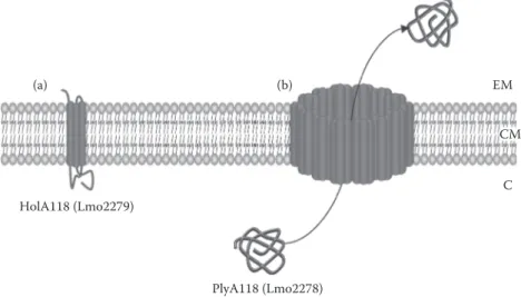
Holins (hole-formers) are small membrane proteins of phage origin that essentially control endolysin function in a process leading to bacterial apoptosis.137–139 A current model for the holin-endolysin
system proposes that holins accumulate in the cytoplasmic membrane, whereas endolysins accumu late in the cytoplasm140,141 (Figure 12.6). At a programmed time, holins oligomerized to form pores
in the cytoplasmic membrane, allowing release of endolysins into the extracytoplasmic space lead ing to cell lysis following cell wall degradation and membrane disruption. Homo-oligomeric pore complexes formed by holins would provide a passive but specific translocation system.142 Generally,
holin and its specific endolysin are genetically encoded in tandem. Some holin genes possess a dual start motif, which results in the expression of two distinct proteins with dramatically opposed function since one would promote autolysis (holin) and the other would inhibit it (antiholin).143 Such
regulation can also occur between proteins encoded at different loci (e.g., lrgAB/cidAB operons in
Staphylococcus aureus).144,145 Holins are an extremely diverse group of proteins with 23 distinct
families recognized in TC-DB (transport classification database),146 although they can be grouped
into three classes based on membrane topologies.147 Class 1 holins exhibit three helical TMDs,
whereas class 2 holins have two TMDs. Besides classes 1 and 2, which cover most holins, a third class was identified on the basis of T protein of phage T4 where only a single TMD is present.148
(a) HolA118 (Lmo2279) (b) CM C EM PlyA118 (Lmo2278)
FIgure 12.6 ϕA118 holin-endolysin system in L. monocytogenes EGD-e.37 (A) HolA118 is a class 1 holin
(i.e., with 3 TMDs). (B) HolA118 oligomerizes in the CM to form a pore allowing translocation and activation of endolysin Ply118. EM, extracellular milieu; CM, cytoplasmic membrane; C, cytoplasm.
The number of holins encoded in Listeria varies between strains and species37; only one holin
could be identified in L. monocytogenes F2365, whereas five holins were found encoded in non pathogenic strain L. innocua CLIP11262. However, only three distinct families of holins were iden tified in L. monocytogenes (i.e., as belonging to bacteriophage 118, TcdE, and bacteriophage 11 families). Holins of ϕA118 family (HolA118) were first identified and investigated in L. monocyto
genes although a homologue is at least also encoded in L. innocua.37,149 HolA118 is not encoded by
all L. monocytogenes species as it is absent from L. monocytogenes F2365. Native holin HolA118 is a 93-amino-acid-long protein belonging to class 1, but its encoding gene is subjected to dual trans lational initiation, which leads to a second 83-amino-acid-long protein called HolA118(83) acting as an antiholin.150 Gene encoding phage lysin of ϕA118 (PlyA118) systematically clusters with gene
encoding HolA118.18,151The endolysin PlyA118 is an L-alanoyl-D-glutamate peptidase hydrolyzing
the cross-linking bridges of cell wall peptidoglycan and thus responsible for bacterial lysis in a programmed cell death.
Holins belonging to TcdE family are encoded in all sequenced Listeria but as ϕ11 holins they have never been experimentally investigated. TcdE holin was investigated in Clostridium difficile, where toxigenic strains produce two large toxins, TcdA and TcdB, of major importance in bacte rial virulence, which would be translocated across the cytoplasmic membrane by TcdE.152,153 While
in C. difficile all these genes are encoded within a pathogenicity locus, no genes coding for tox ins or virulence factors could be identified in Listeria.37 However, a putative autolysin lacking a
signal peptide was systematically present (i.e., genes encoding Lmo0129 and LMOf2365_0147 in
L. monocytogenes EGD-e and F2365, respectively). Although this particular holin family has never been investigated per se in Listeria, proteomic analysis in L. monocytogenes EGD-e disclosed the presence of Lmo0129 in supernatant of bacterial cultures, suggesting this secretion pathway is active in this species.90 In Listeria, ϕ11 holins were only identified in unassembled genome of
L. monocytogenes F6854 and nonpathogenic L. innocua CLIP11262, where they systematically clustered with genes encoding amidases presumably involved in cell wall degradation.37
12.2.6 wSS
Wss stands for proteins with WXG motif of ~100 residues (WXG100) secretion system.154 WXG100
is a new superfamily of proteins around 100 amino acids long, possessing a coil–coil domain and bearing a conserved WXG motif located in the middle region. First identified members of this superfamily were paralogues ESAT-6 (early secreted antigen target of 6 kDa) and CFP-10 (cul ture filtrate protein 10) from Mycobacterium tuberculosis. While ESAT-6 and CFP-10 are specific and experimentally investigated proteins, WXG100 (PF06013) is an established and generic ter minology more appropriate to describe protein members of this family, especially those that have not been experimentally investigated yet. No generic terminology for the different components of Wss apparatus has been established yet. Presence of a novel protein secretion system was clearly suggested by bioinformatic analysis.154 In B. subtilis, genes encoding WXG100 proteins appeared
to cluster systematically with yukab, which are predicted to encode membrane bound ATPases with FtsK/SpoIIIE domains. Similar genetic organization was observed in some Corynebacterium,
Mycobacterium, Streptomyces, Bacillus, Clostridium, Listeria, and Staphylococcus species.31,154
YukAB homologues appear encoded as single or two CDS. To date, Wss seems phylogenetically restricted to Gram-positive bacteria and has been experimentally investigated only in M. tuberculo
sis, M. smegmatis, and S. aureus.
In Mycobacterium, two WXG100 proteins are secreted: ESAT-6 and CFP-10.155 Recently, a
C-terminal signal sequence required for secretion via Wss was unraveled in CFP-10.156 Mycobacte
rium Wss apparatus was named Snm (secretion in mycobacteria) and is composed at least of155,157:
(1) Snm1, Snm2, and Snm6 containing NTP-binding motifs (where Snm1 and Snm2 are homolo gous to YukAB); (2) Snm4, which is an IMP; (3) Snm5 and Snm7 with uncharacterized functions,
and Snm8 (i.e., a membrane anchored serine protease). Snm permits translocation of ESAT-6 and CFP-10 as well as their heterodimerization.158,159 In S. aureus, the Wss was named Ess (ESAT-6
secretion system) and is encoded in a locus composed of eight CDS including two WXG100 par-alogues—EsxA (Ess extracellular protein A) and EsxB—as well as:160 (1) EssC (Ess protein C)
homologous to YukAB; (2) EssA, EssB, and EsaA (ESAT-6 secretion accessory protein A), which are IMPs; and (3) EsaB and EsaC, which predict cytoplasmic chaperones. Compared to B. subtilis, where a putative Wss was primarily uncovered, EssB and EsaB appear homologous to YukC and YukD, respectively. In S. aureus, no homologue to Snm4, Snm5, Snm6, Snm7, or Snm8 was found, whereas in mycobacteria, no homologue to EssA, YukC, EsaA, and YukD could be identified. In both M. tuberculosis and S. aureus,160,161 Wss is important and critical for bacterial pathogenicity,
though the function of WXG100 proteins in virulence remains obscure.162
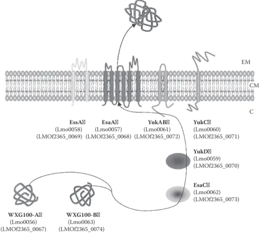
Synteny is highly conserved between Wss encoding loci of S. aureus and L. monocytogenes.160
However, compared to Mycobacterium species or S. aureus, only a single copy of Wss locus is pres ent in each sequenced Listeria genome.37 Following homology with S. aureus and Mycobacterium,
Wss in L. monocytogenes is represented in Figure 12.7. From one report,163 it seems that WXG100
protein is not required for virulence of L. monocytogenes. Still, protein expression, system function ality, and involvement in bacterial virulence of Wss remain to be established.
EM CM
C
EssA EsaA YukAB YukC
(Lmo0058)
(LMOf2365_0069) (LMOf2365_0068) (Lmo0057) (LMOf2365_0072) (Lmo0061) (Lmo0060)(LMOf2365_0071) YukD (Lmo0059) (LMOf2365_0070) EsaC (Lmo0062) (LMOf2365_0073) WXG100-A WXG100-B (Lmo0056)
(LMOf2365_0067) (LMOf2365_0074) (Lmo0063)
FIgure 12.7 Wss in L. monocytogenes.37 WXG100 proteins would interact with putative cytoplasmic chap
erones YukD and EsaC before being translocated by the Wss apparatus constituted of EssA, EsaA, YukC, and YukAB; in the course of translocation the two WXG100 proteins would finally form a heterodimer. EM, extracellular milieu; CM, cytoplasmic membrane; C, cytoplasm.
12.3 cell enveloPe-assocIated ProteIns
Cell envelope of Gram-positive bacteria is primarily composed of a single biological membrane (i.e., the cytoplasmic membrane) and a cell wall made up of peptidoglycan (which in turn con sists of linear polysaccharide chains cross-linked by short peptides).164 Besides peptidoglycan, the
rigid cell-wall of Gram-positive bacteria contains large amounts of wall-associated polymers, also called “secondary” cell wall polymers (SCWPs), which can be classified into three distinct groups: (1) teichoic acids (i.e., polyol phosphate polymers, including lipoteichoic acids), (2) teichuronic acids, and (3) other neutral or acidic polysaccharides that cannot be assigned to the two former groups (e.g., lipoglycans).55,165,166 The SCWPs, present in various proportions, are either covalently linked
to the peptidoglycan backbone (i.e., teichoic acids) or tethered to a lipid anchor moiety. Except for teichoic and teichuronic acids, the structure and biosynthesis of other SCWPs are largely unknown. It must be stressed that in almost all phylogenetic branches of Archaea and Bacteria, the cell enve lope is also constituted of a proteinaceous S-layer (regular crystalline surface layer), which forms the outermost cell-wall layer.167 The S-layer entirely coats the bacterial cell surface and is composed
of (glyco)proteins, which bind by noncovalent interactions to cell wall components and are arrayed in a two-dimensional lattice.167 S-layer, however, is not present in all Gram-positive bacteria as it
is absent from all members of Listeria genus. Within the cell envelope, proteins can associate with cytoplasmic membrane or cell wall components.8
12.3.1 membrane-aSSociated ProteinS
Membrane-associated proteins include membrane integrated proteins as well as peripheral membrane proteins. Being different from membrane integrated proteins, peripheral membrane proteins do not possess membrane spanning domains. Membrane integrated proteins are anchored within the lipid bilayer and thus systematically exhibit hydrophobic transmembrane domains (TMDs), which are normally α-helices for proteins found in the cytoplasmic membrane. Peripheral membrane proteins include (1) lipoproteins, (2) subunits of membrane-associated complexes, and (3) proteins interacting with membrane components due to electrostatic and/or hydrophobic/steric properties.168 Following
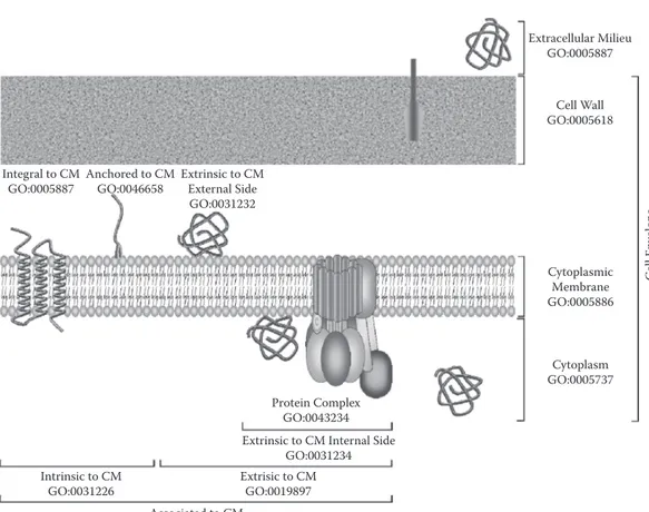
recommendations of the Gene Ontology (GO) Consortium for describing location of cellular com ponents (one of the three organizing principles of GO with biological process and molecular func-tion),169 two classes of membrane-related location are distinguished (Figure 12.8). First, intrinsic to
plasma membrane (GO:0031226) refers to proteins with covalently attached moiety embedded in the cytoplasmic membrane, which splits into (1) integral to plasma membrane (GO:0005887) corre sponding to membrane integrated proteins, where some part of the peptide sequence spans all or part of the cytoplasmic membrane; and (2) anchored to plasma membrane (GO:0046658) correspond ing to proteins tethered to the cytoplasmic membrane by a nonpolypeptidic covalently attached anchor: lipoproteins. Second, extrinsic to plasma membrane (GO:0019897) refers to proteins neither anchored by covalent bonds to any moiety nor directly embedded in the cytoplasmic membrane; some of these proteins can be (1) primarily present in the cytoplasm (GO:0005737) but interact with membrane components, or (2) subcomponents localized within protein complex (GO:0043234). 12.3.1.1 Integral membrane Proteins
As already mentioned, all bacterial IMPs are presumably inserted into the cytoplasmic membrane via YidC homologues48,170,171 (i.e., SpoIIIJ and YqjG in Gram-positive bacteria30; see section 12.2.1).
The Sec-independent function of YidC homologues is conserved and essential for bacterial cell growth as it works like a membrane protein insertase.172 YidC plays a major role in the folding
step of transmembrane-spanning domains but the exact mechanism of functioning is not fully understood.173 YidC would facilitate the insertion of membrane proteins by providing a special
amphiphilic surface, which would overcome the repulsion of the hydrophobic protein segments by polar head groups. In addition, polar residues seem to be protected against the hydrocarbon core of
Extracellular Milieu GO:0005887
Cell Wall GO:0005618 Integral to CM Anchored to CM Extrinsic to CM
GO:0005887 GO:0046658 External Side
GO:0031232 Cytoplasmic Membrane GO:0005886 Cytoplasm GO:0005737 Protein Complex GO:0043234 Extrinsic to CM Internal Side
GO:0031234
Intrinsic to CM Extrisic to CM
GO:0031226 GO:0019897
Associated to CM
FIgure 12.8 Description of protein localization following GO in Gram-positive bacteria.179 In monoderm
bacteria four subcellular compartments can be distinguished: (1) cytoplasm, (2) cytoplasmic membrane, (3) cell wall, and (4) extracellular milieu. A membrane-associated protein can be intrinsic or extrinsic. Proteins intrin sic to CM are either integral to membrane (i.e., integral membrane proteins) or anchored to CM, essentially lipoproteins with the restriction of lipoproteins having TMDs. Proteins extrinsic to CM can be located on the external or internal side of the CM (i.e., in exoplasmic or cytoplasmic compartment, respectively). They can interact more or less temporarily with membrane components or be part of membrane protein complex (e.g., F1F0ATP synthetase δ subunit) as indicated on the schema. A protein can also have multiple final localiza
tion. Importantly, because cell wall of Gram-positive bacteria is permeable, extracellular milieu penetrates it. CM, cytoplasmic membrane; GO, gene ontology.
the membrane by YidC. In the case of Sec-dependent translocation, the protein would be stabilized and then folded by contact with YidC after leaving the Sec YEG channel. It is quite possible that the transmembrane segments could fold and interact with each other even within SecYEG-YidC machinery. It has been suggest that YidC functions as an assembly site for hydrophobic domains, so it may be necessary for its attaching to the individual subunits of multisubunit membrane com-plex.174 It is worth noting that, in E. coli, targeting, translocation, and insertion of IMPs are consid
ered cotranslational and thus SRP dependent.175
Translocation of polypeptide chain is promoted by signal peptides and interrupted by another type of topogenic element called stop-transfer sequence176,177; both types of topogenic sequences act
as α-helical transmembrane domains. Multiple uncleaved signal peptides can be found all along the amino acid sequences; when located N-terminally, they can be cleaved or not. These types of topogenic elements have a Cout-Nin topology and when uncleaved are also called signal-anchors or
Type II signals.178 Similarly, one or more stop-transfer sequences with an N
out-Cin topology can be
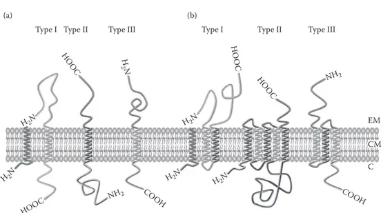
present in polypeptide chain and are also called Type II signals. Single-spanning membrane pro-teins are discriminated on the basis of Type I or Type II signal (Figure 12.9). Polytopic membrane
Cell En
velop
(a) (b)
Type I Type II Type III Type I Type II Type III
H H O HO OC OOC O C H 2N N
FIgure 12.9 Classification and topology of IMP in cytoplasmic membrane.179 (A) Three types of single
spanning membrane proteins can be discriminated: (1) Type I proteins possess a cleavable N-terminal signal peptide and thus have a Type I signal or stop-transfer sequence with Nout-Cin topology; (2) Type II proteins
have a Type II signal or signal-anchor sequence with a Cout-Nin topology, which can correspond to an uncleav
able N-terminal signal peptide; and (3) Type III proteins have reverse signal-anchor sequence (i.e., with a
out-Cin topology) and are sometimes described as Type I proteins without a cleavable signal peptide since the
reverse signal-anchor sequence is a Type I signal. (B) Three types of multispanning-membrane proteins (i.e., with a number of TMDs higher than 1) can be distinguished based on whether the most N-terminal TMD is either (1) cleaved by a SPase (i.e., Type I); (2) spans the membrane with an Nout-Cin orientation (i.e., Type II);
or (3) have a Cout-Nin orientation. Various numbers of TMDs are present in multispanning-membrane proteins
“where Type I and II signals alternate”? HOOC COOH COOH H2N H2N H2N H2N H2N NH2 NH2 EM CM C
where alternates Type I and II signals. EM, extracellular milieu; CM, cytoplasmic membrane; C, cytoplasm. AU: do you mean
proteins are built up of a series of Types I and II modules that initiate and halt the translocation of the polypeptide chain. Such IMPs are classified on the basis of the orientation of the most N-termi-nal TMD spanning the lipid bilayer179 (Figure 12.9).
dues).
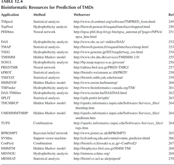
Numerous tools are available to predict IMPs and their topology. Table 12.4 is an attempt to review all of them. These tools are based on various approaches, such as (1) statistical analyses (e.g., TMpred or TMSTAT; (2) hydrophobicity analyses (e.g., SOSUI or TopPred); (3) NNs (e.g., PREDTMR or PHDhtm); (4) SVMs (e.g., SVMtm); or (5) HMMs (e.g., HMMTOP or THUMBUP). Some of them—for example, ConPred or TUPS—combine results of several models. Readers are invited to study related publications listed in Table 12.4 in order to get further insight into the methods used. The total number of IMPs encoded in L. monocytogenes genomes is estimated to be 1204 and 733 in L. monocytogenes EGD-e and F2365, respectively.18 Virulence factors ActA
and SvpA are cell surface exposed IMPs of L. monocytogenes exhibiting a hydrophobic tail (i.e., a carboxyl terminal region containing a hydrophobic domain followed by positively charged
resi-180,181 Following genomic analysis, a total of 11 surface proteins with hydrophobic tails have
been predicted in L. monocytogenes EGD-e.19 Contrary to what is sometimes assumed, cell surface
IMPs should not be restricted only to proteins with a hydrophobic tail182; indeed, depending on the
number and organization of Type I and Type II signals in IMPs, final protein topology can result in the cell surface exposure of functional domains located not only N- or C-terminally but also in loops. With FbpA as an example,47 it can be noticed that from experimental investigations, some
proteins appear located within the cytoplasmic membrane despite the absence of predicted signal peptide and TMD.
FIgure 12.9 Classification and topology of IMP in cytoplasmic membrane.179 (A) Three types of
single-spanning membrane proteins can be discriminated: (1) Type I proteins possess a cleavable N-terminal signal peptide and thus have a Type I signal or stop-transfer sequence with Nout-Cintopology; (2) Type II proteins
have a Type II signal or signal-anchor sequence with a Cout-Nintopology, which can correspond to an
uncleav-able N-terminal signal peptide; and (3) Type III proteins have reverse signal-anchor sequence (i.e., with a Nout-Cintopology) and are sometimes described as Type I proteins without a cleavable signal peptide since the
reverse signal-anchor sequence is a Type I signal. (B) Three types of multispanning-membrane proteins (i.e., with a number of TMDs higher than 1) can be distinguished based on whether the most N-terminal TMD is either (1) cleaved by a SPase (i.e., Type I); (2) spans the membrane with an Nout-Cinorientation (i.e., Type II);
or (3) have a Cout-Ninorientation. Various numbers of TMDs are present in multispanning-membrane proteins
where alternates Type I and II signals. EM, extracellular milieu; CM, cytoplasmic membrane; C, cytoplasm.
table 12.4
bioinformatic resources for Prediction of tmds
application method Webserver ref.
TMpred Statistical analysis http://www.ch.embnet.org/software/TMPRED_form.html 249
npsa_htm.html
thumbup.htm umdhmm.htm tups.htm
TopPred Hydrophobicity analysis http://bioweb.pasteur.fr/seqanal/interfaces/toppred.html 250 PHDhtm Neural network http://npsa-pbil.ibcp.fr/cgi-bin/npsa_automat.pl?page=/NPSA/ 251
DAS Hydrophobicity analysis http://www.sbc.su.se/~miklos/DAS/ 252
TMAP Statistical analysis http://bioweb.pasteur.fr/seqanal/interfaces/tmap.html 253 TSEG Hydrophobicity analysis http://www.genome.jp/SIT/tsegdir/tseg_exe.html 254
TMHMM Hidden Markov model http://www.cbs.dtu.dk/services/TMHMM-2.0/ 255
SOSUI Hydrophobicity analysis http://bp.nuap.nagoya-u.ac.jp/sosui/ 256
PREDTMR Neural network http://athina.biol.uoa.gr/PRED-TMR2/ 257
kPROT Statistical analysis http://bioinfo.weizmann.ac.il/kPROT/ 258
TMSTAT Statistical analysis http://bioinfo.mbb.yale.edu/tmstat/ 259
HMMTOP Hidden Markov model http://www.enzim.hu/hmmtop/ 260
TMFinder Hydrophobicity analysis http://www.bioinformatics-canada.org/TM/ 261
DAS-TMfilter Hydrophobicity analysis http://www.enzim.hu/DAS/DAS.html 262
SPLIT Statistical analysis http://split.pmfst.hr/split/ 263
THUMBUP Hidden Markov model http://sparks.informatics.iupui.edu/Softwares-Services_files/ 264 UMDHMMTMHP Hidden Markov model http://sparks.informatics.iupui.edu/Softwares-Services_files/ 264 TUPS Combination http://sparks.informatics.iupui.edu/Softwares-Services_files/ 264
BPROMPT Bayesian belief network http://www.jenner.ac.uk/BPROMPT/ 265
SVMtm Support vector machine http://ccb.imb.uq.edu.au/svmtm/svmtm_predictor.shtml 266
ConPred Combination http://bioinfo.si.hirosaki-u.ac.jp/~ConPred2/ 267
HMMTM Hidden Markov model http://biophysics.biol.uoa.gr/HMM-TM/ 268
MINNOU Hydrophobicity analysis http://minnou.cchmc.org/ 269
MEMSAT Statistical analysis http://bioinf.cs.ucl.ac.uk/psipred/ 270
12.3.1.2 lipoproteins
In monoderm bacteria, lipoproteins are attached to the outer surface of the cytoplasmic membrane via a covalently bound lipid anchor.32 Systematically, these proteins are first translocated in a Sec
dependent manner and thus possess N-terminal signal sequences. Such signal peptides, however, belong to class 2 as it exhibits a conserved lipobox motif in the C-domain.30 It can be noticed,
however, that in E. coli YidC plays an important role in targeting and translocation of some lipo-proteins.183 Lipobox includes invariably a cysteine residue located just after the cleavage site of
signal peptide. After translocation of the prolipoprotein, a common post-translational modification involves a prolipoprotein diacylglyceryl transferase (Lgt), which adds an N-acyl diglyceride group from a glycerophospholipid to the SH-group of the lipobox cysteine.184 This thioether linkage allows
protein anchoring to the membrane thanks to the insertion of the diacylglyceryl group into the lipid signal peptide and the cysteine becomes the N-terminal residue.185 In contrast to E. coli, however,
lipidation by Lgt (Lmo2482) in Listeria is neither essential for bacterial growth nor a prerequisite for activity of Lsp.186 Once signal peptide is cleaved off, the amino-terminal cysteine residue is usu
ally acylated at the free amino group by a phospholipid/apolipoprotein transacylase (Lnt), resulting bilayer. Subsequently, SPase II (also called Lsp for lipoprotein signal peptidase) cleaves off the