HAL Id: hal-02346653
https://hal.archives-ouvertes.fr/hal-02346653
Submitted on 16 Nov 2020HAL is a multi-disciplinary open access archive for the deposit and dissemination of sci-entific research documents, whether they are pub-lished or not. The documents may come from teaching and research institutions in France or abroad, or from public or private research centers.
L’archive ouverte pluridisciplinaire HAL, est destinée au dépôt et à la diffusion de documents scientifiques de niveau recherche, publiés ou non, émanant des établissements d’enseignement et de recherche français ou étrangers, des laboratoires publics ou privés.
Distributed under a Creative Commons Attribution - NonCommercial - NoDerivatives| 4.0 International License
Glycoproteomic Analysis of MGL-Binding Proteins on
Acute T-Cell Leukemia Cells
Martina Pirro, Esmee Schoof, Sandra van Vliet, Yoann Rombouts, Alexandre
Stella, Arnoud de Ru, Yassene Mohammed, Manfred Wuhrer, Peter van
Veelen, Paul Hensbergen
To cite this version:
Martina Pirro, Esmee Schoof, Sandra van Vliet, Yoann Rombouts, Alexandre Stella, et al.. Glyco-proteomic Analysis of MGL-Binding Proteins on Acute T-Cell Leukemia Cells. Journal of Proteome Research, American Chemical Society, 2019, 18 (3), pp.1125-1132. �10.1021/acs.jproteome.8b00796�. �hal-02346653�
Glycoproteomic Analysis of MGL-Binding Proteins on Acute T
‑Cell
Leukemia Cells
Martina Pirro,
†Esmee Schoof,
†Sandra J. van Vliet,
‡Yoann Rombouts,
§Alexandre Stella,
§Arnoud de Ru,
†Yassene Mohammed,
†Manfred Wuhrer,
†Peter A. van Veelen,
†and Paul J. Hensbergen
*
,††Center for Proteomics and Metabolomics, Leiden University Medical Center, 2300 RC Leiden, The Netherlands
‡Amsterdam UMC, Vrije Universiteit Amsterdam, Dept. of Molecular Cell Biology and Immunology, Cancer Center Amsterdam,
Amsterdam Infection & Immunity Institute, 1007 MB Amsterdam, The Netherlands
§Institut de Pharmacologie et de Biologie Structurale, Université de Toulouse, CNRS, UPS, Toulouse 31062, France
*
S Supporting InformationABSTRACT: C-type lectins are a diverse group of proteins involved in many human physiological and pathological processes. Most C-type lectins are glycan-binding proteins, some of which are pivotal for innate immune responses against pathogens. Other C-type lectins, such as the macrophage galactose-type lectin (MGL), have been shown to induce immunosuppressive responses upon the recognition of aberrant glycosylation on cancer cells. MGL is known to recognize terminal N-acetylgalactosamine (GalNAc), such as the Tn antigen, which is commonly found on malignant cells. Even though
this glycan specificity of MGL is well described, there is a lack of
understanding of the actual glycoproteins that bind MGL. We present a
glycoproteomic workflow for the identification of MGL-binding proteins,
which we applied to study MGL ligands on the human Jurkat leukemia cell
line. In addition to the known MGL ligands and Tn antigen-carrying proteins CD43 and CD45 on these cells, we have
identified a set of novel cell-surface ligands for MGL. Importantly, for several of these, O-glycosylation has hitherto not been
described. Altogether, our data provide new insight into the identification and structure of novel MGL ligands that presumably
act as modulatory molecules in cancer immune responses.
KEYWORDS: glycoproteomics, lectin, Tn antigen, leukemia, O-glycosylation
■
INTRODUCTIONC-type lectins belong to a family of glycan-binding proteins
(GBPs) that interact with their ligands in a Ca2+-dependent
manner through a carbohydrate recognition domain (CRD).1,2
They exist as monomers or can oligomerize to enhance the avidity by multivalent glycan interactions. Their activation on
immune cells, such as dendritic cells (DCs), triggers different
immune responses depending on the crosstalk with other receptors, the ligand/carbohydrate-specific signaling, and the
cell subset they are expressed in.3Within the CLR family, the
macrophage galactose-type lectin (MGL) is expressed on
tolerogenic DCs and macrophages4 and has the capability to
alter DC and macrophage phenotypes.5
MGL is the only lectin that exclusively binds terminal GalNAc residues, including three well-known tumor-associated
glycan epitopes:6 the sialylated and nonsialylated
Thomsen-nouvelle (Tn) antigens (GalNAcα1-Ser/Thr) and LacdiNAc
(GalNAcβ1-4GlcNAc).4 In normal cells, the Tn antigen is
usually elongated with other carbohydrate residues, for example, a galactose, to form the core 1 T antigen. This step is mediated by the enzyme T-synthase with the assistance of its chaperone Cosmc. Mutations in Cosmc are responsible for the
abortive elongation of O-glycans and higher expression of the
Tn antigen,7as observed in Tn syndrome patients.8High levels
of Tn antigen have also been found in multiple human cancer
types such as colon, cervix, stomach, ovary, and breast.9
Moreover, higher levels of this surface-truncated glycan were found in cancer cells with high metastatic behavior and
consequently poor prognosis for cancer patients.8Interestingly,
the interaction of MGL with aberrant glycosylation on the cell surface of cancer cells is associated with the activation of
immunosuppressive responses, immune tolerance,10and poor
survival of stage-III colorectal cancer patients,11suggesting a
role of MGL in mediating cancer progression.
Notwithstanding our current knowledge on the role of MGL and the glycans it binds, the proteins carrying the MGL ligands are hitherto largely unknown. Therefore, in this article, we sought to identify MGL-binding proteins in a T-cell leukemia model cell line, Jurkat, which is known to have high levels of Tn antigen due to a single nucleotide deletion in Cosmc and
Received: October 9, 2018
Published: December 24, 2018
Article pubs.acs.org/jpr
Cite This:J. Proteome Res. 2019, 18, 1125−1132
Derivative Works (CC-BY-NC-ND) Attribution License, which permits copying and redistribution of the article, and creation of adaptations, all for non-commercial purposes.
Downloaded via 91.169.6.110 on November 16, 2020 at 12:09:32 (UTC).
has been used as a model system to study immune modulation
mediated by MGL.1,12
■
EXPERIMENTAL SECTIONCell Lines Culture and Lysis
Jurkat (provided by S. J. van Vliet, VUMC, Amsterdam, The Netherlands) cells were cultivated in RPMI-1640 medium containing 10% fetal bovine serum (FBS) (Invitrogen) and
streptomycin/penicillin (Sigma-Aldrich) at 5% CO2 and 37
°C. Cells were harvested upon ∼70% confluency. A total of 2
× 107cells were obtained, harvested, washed, and centrifuged
at 1500 rpm to obtain cell pellets. Cell pellets were stored at
−20 °C until use. Then, cells were lysed as described before12
for 20 min on ice in lysis buffer (10 mM triethanolamine pH
8.2, 150 mM NaCl, 1 mM MgCl2, 1 mM CaCl2, and 1%
(volume/volume) Triton X-100 containing EDTA-free pro-tease inhibitor (Roche Diagnostics)). Protein concentration
was quantified by BCA assay (BCA Protein Assay Kit, Pierce).
Antibodies and Reagent
Chimeric MGL-Fc was provided by S. J. van Vliet (VUMC, Amsterdam, The Netherlands). The following antibodies were used for immunoblotting: mouse IgG1 anti-human CD43 (eBio84-3C1, eBioscience) and goat anti-mouse immunoglo-bulins/HRP (Dako).
Pull-Down Assay and Immunoblot Analysis
MGL ligands were pulled down from 1 mg of protein extracts
with 2μg of chimeric MGL-Fc coupled to 50 μL of Dynabeads
protein G (Invitrogen). The specific ligands were then eluted
by the addition of 100 mM EDTA and concentrated under vacuum. The addition of 100 mM EDTA, prior to the addition of MGL-Fc, was used as a negative control. Captured products
were separated by SDS-PAGE (NuPAGE 4−12% Bis-Tris
protein gels, Thermo Fisher Scientific) and transferred to a
PVDF membrane (Amersham Hyband P 0.45). Five % low-fat
milk (Campina) in 0.1% phosphate-buffered saline with Tween
20 (PBS-T) was used to block the blots for 1 h.
Immunoblotting was performed with specific antibodies,
followed by secondary antibody peroxidase-conjugated goat anti-mouse (Dako). Immunodetection was done by enhanced chemiluminescence (ECL) using Clarity Western ECL substrate (Bio-Rad) and an Amersham Imager 600.
Mass Spectrometry
The samples obtained with the MGL pull-down assay were
cleaned up by a short SDS-PAGE run (NuPAGE 4−12%
Bis-Tris protein gels, Thermo Fisher Scientific). For MS analysis,
in-gel trypsin digestion was performed using a Proteineer DP
digestion robot (Bruker). Prior to digestion, proteins werefirst
reduced and alkylated using dithiothreitol (10 mM) and iodoacetamide (50 mM), respectively.
Tryptic peptides were extracted from the gel slices, lyophilized, dissolved in solvent A (95/3/0.1 water/acetoni-trile/formic acid (FA) v/v/v), and subsequently analyzed by online C18 nano-HPLC MS/MS with a system consisting of an Easy nLC 1000 gradient HPLC system (Thermo, Bremen, Germany) and a LUMOS mass spectrometer (Thermo).
Fractions were injected onto a homemade precolumn (100μm
× 15 mm; Reprosil-Pur C18-AQ 3 μm, Dr. Maisch, Ammerbuch, Germany) and eluted via a homemade analytical
nano-HPLC column (15 cm× 50 μm; Reprosil-Pur C18-AQ 3
μm). The gradient was run from 10 to 40% solvent B (20/80/ 0.1 water/acetonitrile/FA v/v/v) in 20 min. The nano-HPLC
column was drawn to a tip of ∼5 μm and acted as the
electrospray needle of the MS source. The LUMOS mass spectrometer was operated in data-dependent MS/MS (top-10
mode) with collision energy at 32 V and recording of the MS2
spectrum in the Orbitrap. In the master scan (MS1) the
resolution was 120 000, and the scan range was m/z 400−1500
at an AGC target of 400 000 with maximumfill time of 50 ms.
Dynamic exclusion after n = 1 with exclusion duration of 10 s
was applied. Charge states 2−5 were included for MS2. For
this, precursors were isolated with the quadrupole with an
isolation width of 1.2 Da. The MS2scan resolution was 30 000
with an AGC target of 50 000 with maximumfill time of 60 ms.
During acquisition, a product ion trigger was set on the HexNAc oxonium ion at m/z 204.087. Upon the detection of
the oxonium ion, three additional data-dependent MS2scans of
the same precursor were executed with higher-energy colli-sional dissociation (HCD) collision energies of 32, 37, and 41 V, respectively, at an AGC target of 500 000 with a maximum fill time of 200 ms. In addition, a collision-induced dissociation (CID) spectrum of the same precursor was recorded.
In a post-analysis process, raw data were analyzed with MaxQuant 1.5.1.2 using the Andromeda search engine and the
Homo sapiens (2015) database. Modifications taken into
account in the search were oxidation (M), acetylation (N-term), and HexNAc (ST) as variable and carbamidomethyl
(C) asfixed. Trypsin was selected as the enzyme, allowing up
to two miss cleavages. A maximum of 12 modifications per
peptide was allowed. The mass tolerance used for precursor ions was 10 ppm, whereas it was 0.05 Da for fragment ions. Subsequently, only proteins that were either never found in the negative control experiment but were found in at least two of
the MGL pull-downs or proteins that were identified in all
three MGL pull-downs and identified a maximum of one time
in the negative control samples were subsequently considered MGL-binding proteins. Subsequently, these MGL-binding
proteins were furtherfiltered based on data on the subcellular
location from the UniProtKB database, thereby selecting only cell-surface membrane proteins. The surface location was subsequently manually curated by a literature search.
Alternatively, MS2spectra containing the specific HexNAc
oxonium ions at m/z 204.087 (HexNAc, [C8H14NO5]+) and
186.076 (HexNAc-H2O, [C8H12NO4]+) were selected and
written to an MGF file (in-house software), which was
subsequently submitted to Byonic version 2.13.2 (
proteinme-trics.com) using default settings. Thefixed modification was
carbamidomethyl (Cys). Variable modifications set were
oxidation (Met) and “N-glycan 309 mammalian no sodium”
or “O-glycan 78 mammalian” databases as glycosylation
parameters. Only glycopeptides with a Byonic score >200 were further selected.
Manual interpretation of spectra was done using Xcalibur (Thermo) for the spectrum visualization. The corresponding peptide fragment masses were calculated using Protein
Prospector (prospector.ucsf.edu).
Data Availability
The mass spectrometry proteomics data have been deposited
to the ProteomeXchange Consortium via the PRIDE13partner
repository with the data set identifier PXD011307.
Journal of Proteome Research Article
DOI:10.1021/acs.jproteome.8b00796
J. Proteome Res. 2019, 18, 1125−1132
■
RESULTSWorkflow of the MGL Pull-Down Assay
To identify novel binding partners of MGL on Jurkat T-cells, we developed a protocol where we used Fc-coupled MGL as a
bait in pull-down assays (Figure 1A). MGL-Fc is a chimeric
molecule formed by the extracellular domains of MGL fused to
the human immunoglobulin G1 Fc tail,14 allowing binding to
magnetic Protein G beads. Because the binding to the CRD of MGL is calcium-dependent, captured proteins were eluted using EDTA and subsequently analyzed by SDS-PAGE and
processed for mass-spectrometry-based protein identification.
CD43 and CD45 are hitherto the only described MGL-binding
proteins on Jurkat cells.12Therefore, to test the effectiveness of
our workflow, we first determined the capturing of CD43 by
Western blot. This clearly showed that CD43 was captured by
MGL-Fc and could be eluted with EDTA (Figure 1B).
Likewise, the addition of EDTA during the pull-down assay
prevented the binding of CD43 to MGL (Figure 1B).
MS-Based Identification of MGL-Binding Proteins from Jurkat Cells
Next, we performed three biologically independent pull-down experiments with MGL-Fc and analyzed the bound proteins by
LC−MS/MS following trypsin digestion. As a negative control,
pull-down assays in the presence of EDTA were performed.
Altogether, these experiments resulted in the identification of
775 proteins (data not shown), of which 540 were identified in
at least two experiments. To filter for proteins specifically
binding to MGL, we selected proteins that were not observed in the negative controls and at least two times in the downs with MGL or proteins that were found in all three pull-downs with MGL and at most once in a negative control. This
resulted in a list of 85 proteins (Table S1). The candidate
MGL-binding proteins included intracellular, plasma mem-brane, and predicted secreted proteins. Because we used total cell lysates for the pull-down assays, some of these proteins
may be physiologically less relevant. To filter for cell-surface
proteins, which may be expected to be in direct contact with MGL, we next selected only those proteins that were annotated as cell-surface proteins in the UniProtKB database,
resulting in afinal list of 17 MGL-binding cell-surface proteins
(Table 1). Importantly, these proteins were not found in the
negative control samples (see also Table S1), making them
strong candidate MGL-binding proteins. As expected, CD45 (PTPRC) and CD43 (SPN) were the two main MGL-binding
proteins identified based on the total number of
peptide-spectrum matches (summed for the three biological replicates) for these two proteins, respectively.
Characterization of Glycopeptides
MGL is known to bind terminal residues of
N-acetylgalactos-amine. To validate the specificity of the interaction of the
proteins listed inTable 1 with MGL, we next evaluated our
data for the presence of glycopeptides carrying this motif.
During our LC−MS/MS analyses of the tryptic digests, we
used a method that triggered additional MS/MS acquisitions once the MS/MS spectrum showed the presence of the characteristic HexNAc oxonium ion at m/z 204.087
([C8H14NO5]+). First of all, for these additional MS/MS
events, we used a higher number of ions for fragmentation, resulting in a higher signal-to-noise for the fragment ions.
Moreover, to improve the confidence of the glycopeptide
identifications, we collected Orbitrap spectra using different
HCD collision energies in addition to an ion trap CID spectrum.
Next, we searched for glycopeptides carrying the Tn antigen following database searches using MaxQuant and Byonic. With
these approaches, we could confirm the presence of the Tn
antigen on CD45 and CD43 (Table 1) with peptides
containing a maximum of four HexNAcs (Table 2). However,
it is known that certain regions in these two proteins contain a high density of O-glycans, which may have been missed by the automatic data analysis due to limitations of the particular software used, for example, due to the high number of occupied glycosylation sites in one individual peptide. Indeed, upon manual inspection of our data, we observed peptides
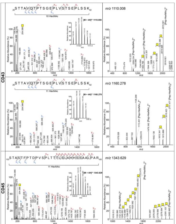
containing up to 11 HexNAcs (Figure 2). Of note, one peptide
with an extended O-glycan on the CD45 tryptic peptide
LNPTPGSNAISDVPGER (HexNAc2Hex1NeuAc1) was found
using Byonic, which, considering the Cosmc mutation in Jurkat
cells, could correspond to a core 6 O-glycan, a specific
MGL-binder (GlcNAcβ1-6GalNAcαSer/Thr).14
In addition to CD43 and CD45, our database searches
confirmed the presence of the Tn antigen on another 11
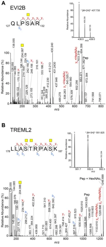
proteins (Table 2). We confirmed the Tn antigen on all of
these peptides by manual interpretation of the data. As an example, two glycopeptides from EVI2B and TREML2, Figure 1.Schematic representation and validation of the experimental
workflow for the identification of MGL ligands in Jurkat cells. (A) Pull-down workflow to capture MGL-binding proteins. As negative control, EDTA was added to the sample prior to incubation with MGL-Fc to prevent the binding to MGL. (B) Western blot analysis using monoclonal anti-CD43 staining of the unbound fraction and captured proteins (elution) from the MGL-Fc pull-down assay in the absence (−) and presence (+, negative control) of EDTA (100 mM). Fc: Fragment crystallizable region; Prot G: protein G; TCL: total cell lysate.
respectively, are shown in Figure 3. Between the peptides
presented inTable 2, one peptide (GLFIPFSVSSVTHK) with
three HexNAcs was identified from P-selectin glycoprotein
ligand 1 (SELPLG), which was not identified as a specific
binder in the proteomics data (Table 1). Inspection of the data
showed that, unexpectedly, it wasfiltered out due to the fact
that normal tryptic peptides of SELPLG were observed in only one of the three MGL pull-downs.
Terminal acetylgalactosamines can also be part of N-glycans, for example, as part of the LacdiNAc epitope. Hence we also searched our data for the presence of N-glycopeptides
using Byonic. Only for CD45 presented inTable 1 we could
identify N-glycopeptides (on N234, 278, 337, 380, 421, and 470, respectively), but none of these appear to contain a terminal N-acetylgalactosamine, as judged on the basis of the glycan compositions as well as the absence of the LacdiNAc
(GalNAcβ1-4GlcNAcβ1) marker ion at m/z 407.166 (Table
S2).
■
DISCUSSIONWe developed a workflow for the identification of MGL
ligands, which resulted in the identification of 17 cell-surface
proteins of Jurkat cells that bind to MGL. For most of these
proteins, we confirmed the specificity of the interaction with
MGL through the identification of O-glycopeptides carrying
the Tn antigen.
The glycopeptide, mediating the interaction with MGL,
could not be identified for all MGL ligands. These peptides
may have been missed due to insufficient sensitivity or low
MS/MS spectrum quality, which would hamper identification
by both search algorithms as well as manual inspection. Although in the past years, we and others have made considerable progress in the mass-spectrometry-based
identi-fication of glycopeptides,15−20
several issues related to their
fragmentation behavior still often impede straightforward (data) analysis, even though the Tn antigen represents a
relatively simple post-translational modification. We combined
multiple strategies for data acquisition and analysis to maximize our glycopeptide identification. Other methods, such as electron-transfer dissociation (ETD), have also been
valuable tools for the identification of glycopeptides,16
especially in cases where the localization of the GalNAc(s) was ambiguous. However, for this study, we were primarily interested in identifying MGL ligands, and not necessarily the
site-specific glycan assignment. Moreover, for several peptides,
we had full occupancy of all serine and threonine residues
present in the identified glycopeptide.
From the 17 potential MGL-binding proteins (Table 1),
some could have been obtained through an interaction with a genuine MGL ligand. For example, protein tyrosine phosphatase receptor type C-associated protein (PTPRCAP),
for which we could notfind the corresponding glycopeptide,
was captured during the MGL pull-down probably because it is
a binding partner of CD45.21Similarly, for only one of the four
LRRC8 subunits (LRRC8D) that we identified could a specific
O-glycopeptide be found. Because it is known that the LRRC8
subunits form heterodimers,22,23 it is conceivable that the
whole LRRC8 complex was captured.
As expected, CD43 and CD45 were among the top proteins carrying the Tn antigen in Jurkat cells. A recent study
presented a workflow where a combination of enzymatic and
chemical methods was used to selectively tag terminal GalNAc,
and not GlcNAc residues,24 allowing the enrichment of the
corresponding glycopeptides. To study proteins carrying the Tn antigen, this method was applied to total cell lysates of Jurkat cells. As a result, a total of 97 proteins harboring the Tn
antigen were identified, of which 27 were consistently found in
all three independent biological replicates. From the total of 97
Table 1. MGL-Binding Cell-Surface Proteins from Jurkat Cellsa
protein name gene name
UniProt entry
mol. weight (kDa)
exp 1:
#pep exp 2:#pep exp 3:#pep sum MS/MScount glycopeptide receptor-type tyrosine-protein phosphatase C
(CD45)
PTPRC P08575 147 73 60 62 1125 yes
leukosialin (CD43) SPN P16150 40 11 9 6 145 yes
leucine-rich repeat-containing protein 8D LRRC8D Q7L1W4 98 21 22 10 73 yes
podocalyxin PODXL O00592 59 7 8 6 66 yes
leucine-rich repeat-containing protein 8A LRRC8A Q8IWT6 94 15 16 6 53 no
protein EVI2B EVI2B P34910 49 7 10 4 35 yes
dyslexia-associated protein KIAA0319-like protein
KIAA0319L E7EN73 113 12 9 7 30 no
leucine-rich repeat-containing protein 8C LRRC8C Q8TDW0 92 8 11 4 28 no Protein tyrosine phosphatase receptor type
C-associated protein
PTPRCAP Q14761 21 5 3 0 11 no
semaphorin-4D SEMA4D Q92854 96 7 2 2 10 yes
TREM-like transcript 2 protein TREML2 Q5T2D2 35 3 2 2 6 yes
protein HEG homologue 1 HEG1 Q9ULI3 147 1 2 1 6 no
lysosome-associated membrane glycoprotein 3 LAMP3 E7ETP9 42 3 1 1 5 yes
chloride intracellular channel protein 1 CLIC1 O00299 27 2 3 0 5 no
low-density lipoprotein receptor LDLR H0YM92 34 2 1 1 4 no
leucine-rich repeat-containing protein 8B LRRC8B Q6P9F7 92 1 3 0 4 no
tumor necrosis factor receptor superfamily member 8
TNFRSF8 P28908 64 2 3 0 3 yes
aUsing total cell lysates from Jurkat cells, three independent pull-down assays with MGL-Fc were performed, and captured proteins were identified
by LC−MS/MS. Shown are the MGL-binding cell-surface proteins; for a full list, seeTable S1. Each protein is represented with the number of unique peptides identified in the three pull-down assays and the summed spectral count from all three assays. mol. weight: molecular weight; exp: experiment number;#pep: number of unique peptides per protein; sum MS/MS count: summed spectral count from the three assays; and glycopeptide: O-glycopeptide(s) of the respective protein found (yes) or not (no).
Journal of Proteome Research Article
DOI:10.1021/acs.jproteome.8b00796
J. Proteome Res. 2019, 18, 1125−1132
proteins, 11 were transmembrane signaling molecules, including CD45, Semaphorin-4D, and TNFRSF8, which
were also identified in our study. Some other
Tn-antigen-bearing glycoproteins were not found in our experiments. The observed differences may relate to the different experimental approaches because several Tn-bearing glycoproteins were
clearly identified in our experiments, such as, CD43, EVI2B,
and TREML2, but not in the above-mentioned study.24On the
contrary, it could be due to the fact that for MGL binding the
presence of Tn is necessary but not sufficient.12,25
For example, all CD45 isoforms (CD45ABC/AB/BC/B) except for one (CD45RO) bind MGL. Unlike the others, which are highly glycosylated, CD45RO has only two O-linked glycan epitopes. Although it is known that MGL can have
immune-modulatory activities, the specific proteins involved in the
cellular response elicited by MGL are largely unknown. Tolerogenic antigen presenting cells (APCs) express high levels of MGL on the cell surface, and the binding of MGL to CD45 suppresses TCR-mediated T-cell activation. This results
in an anti-inflammatory response characterized by the reduced
production of pro-inflammatory cytokines and lower
prolifer-ation of T-cells and induction of cell death in Jurkat.12
For the new MGL ligands identified in this study, the
outcome of the interaction with MGL remains to be determined. Semaphorin 4D, also known as CD100, is a
homodimeric transmembrane protein of 150 kDa that is highly expressed in secondary lymphoid organs and constitutively on
naı̈ve T-cells. It binds its ligand CD72, a C-type lectin
expressed on the surface of APCs, such as B cells and DCs,26
but also macrophages and some subpopulation of T-cells.27
Our data show that, at least in tumor cells with aberrant glycosylation, Semaphorin 4D also binds to MGL.
For several of the newly identified MGL ligands, literature
provides limited information about their function. For example, although KIAA0319L has been demonstrated to be an essential receptor for adeno-associated virus infection, the cellular function, especially in immune responses, is unknown.
However, our data support previous findings that suggested
high levels of mucin-type O-glycosylation on KIAA0319L.28
Our data also provide evidence of O-glycosylation on proteins that hitherto were unknown to be O-glycosylated, such as EVI2B and TREML2. Although the cellular function of these proteins is largely unknown, it has been demonstrated that EVI2B is the target for the transcription factor CCAAT/
enhancer-binding protein alpha (C/EBPα),29 and TREML2
binding by CD276 leads to enhanced T-cell responses.30
Altogether, our data warrant further exploration of the functional implications of the interaction of MGL with the
newly identified ligands. Notwithstanding the importance of
this, some responses elicited by MGL might also be the result
Table 2. Identified O-Glycopeptides from MGL-Binding Cell-Surface Proteins in Jurkat Cellsa
protein name gene name
UniProt
entry glycopeptide sequence
MaxQuant# HexNAc byonic# HexNAc manual# HexNAc lysosome-associated membrane glycoprotein 3
LAMP3 E7ETP9 QAPHQTLAAR 1 1 1
DYSQPTAAATVQDIK 3 3
leukosialin (CD43) SPN P16150 MYTTSITSDPK 3, 4 3 3, 4, 5
MOxYTTSITSDPK 3, 4
STTAVQTPTSGEPLVSTSEPLSSK 9, 10, 11
podocalyxin PODXL O00592 TPSPTVAHESNWAK 2 2, 3 2, 3
ANEILASVK 1 1
CEDLETQTQSEK 1 1
protein EVI2B EVI2B P34910 QLPSAR 1 1
STPGFILDTTSNK 4 4, 5
STPGFILDTTSNKQTPQK 4, 5
QITVHNPSTQPTSTVKNSPR 7
P-selectin glycoprotein ligand 1 SELPLG Q14242 GLFIPFSVSSVTHK 3 3 3
receptor-type tyrosine-protein phosphatase C (CD45) PTPRC P08575 LNPTPGSNAISDVPGER 2 1, 2, 3 2, 3 LNPTPGSNAISDVPGER HexNAc(2) Hex(1)Neu Ac(1) STASTFPTDPVSPLTTTLSLAHHSSAALPAR 10, 11 NGSAAMOxCHFTTK 1 1
semaphorin-4D SEMA4D Q92854 VVPKPVVAPTLSVVQTEGSR 2 1, 2 1, 2
IVINTVPQLHSEK 1 1, 2 1,2
TREM-like transcript 2 protein TREML2 Q5T2D2 LLASTRPASK 3 2, 3 3
NIPFTHLDNILK 2 2
tumor necrosis factor receptor superfamily member 8
TNFRSF8 P28908 LAQEAASKLTR 2 2 2
transferrin receptor protein 1 TFRC P02786 LAGTESPVREEPGEDFPAAR 1 1
voltage-dependent calcium channel subunit alpha-2/delta-4
CACNA2D4 Q7Z3S7 YKDVESSLK 2 2
glycosyltransferase 8 domain-containing protein 1
GLT8D1 Q68CQ7 YTEISNIK 2 2
volume-regulated anion channel subunit LRRC8D
LRRC8D Q7L1W4 TDFALPNQEAK 1 1
aO-glycosylated peptides were annotated using three different approaches (Maxquant, Byonic, manual annotation). # HexNAc: number of
N-acetylhexosamine carried by each glycopeptide; Hex: hexose; NeuAc: N-acetylneuraminic acid; and Ox: oxidation of methionine.
of the interaction of MGL with glycolipids,14 which was not the topic of our study.
The high level of Tn antigen is not restricted to leukemias but is frequently observed in other tumors as well. The best characterized ligand carrying Tn or sialyl-Tn (sTn,
Neu5A-cα2,6-GalNAc-O-Ser/Thr) in epithelial cells is the
glycopro-tein MUC1. MUC1-derived glycopeptides bind to MGL on DCs and induce the activation of the extracellular
signal-regulated kinases 1 and 2 (ERK1,2) and the nuclear factor-κB
(NF-κB) pathways.31 In another study, it was demonstrated
that these pathways are crucial for IL-10 production, which
regulates the DC maturation phenotype.32
The higher level of Tn in other cancers is not necessarily due to Cosmc mutations, as in the Jurkat T-cell used in our study, but may also be the result of other genetic alterations. For example, in colorectal cancer, higher levels of several GALNTs
have recently been linked to the BRAFV600E mutation.33
A positive correlation between this oncogenic mutation and
MGL ligands on tumor cells was previously identified.11
Hence, it will be interesting to study the proteins carrying MGL ligands in these tumor cells as well.
Figure 2.Highly O-glycosylated peptides of CD43 and CD45. MS/MS spectra of glycopeptides from CD43 carrying 10 and 11 GalNAcs and CD45 carrying 11 GalNAcs. Left panels: HCD MS/MS spectra. b and y ions represent fragments without the GalNAc, unless indicated otherwise. The inset shows the MS spectrum of the corresponding precursor with charge state and m/z value. Right panels: CID MS/MS spectra. Yellow square: GalNAc.
Journal of Proteome Research Article
DOI:10.1021/acs.jproteome.8b00796
J. Proteome Res. 2019, 18, 1125−1132
In conclusion, here we provide an optimized method to capture MGL-binding proteins followed by glycoproteomic analysis. The application of this procedure on the Jurkat cell line provides important novel insights into previously unknown MGL ligands. However, further investigations should evaluate
the functional immune responses triggered by MGL-specific
recognition of those proteins, as has been previously reported for the already known interaction partner CD45 by van Vliet et
al.12 This will provide a deeper understanding of the MGL
involvement in cancer progression and the glycan-specific
immune responses mediated by this lectin.
■
ASSOCIATED CONTENT*
S Supporting InformationThe Supporting Information is available free of charge on the
ACS Publications website at DOI: 10.1021/acs.jproteo-me.8b00796.
Table S1: MGL-binding proteins from Jurkat cells. Table
S2: N-Glycosylated peptides of CD45 (XLSX)
Figure S1: SDS-PAGE of MGL pull-down (PDF)
■
AUTHOR INFORMATIONCorresponding Author
*E-mail:P.Hensbergen@lumc.nl. Tel.: +31-71-5266394. Fax:
+31-71-5266907.
ORCID
Martina Pirro:0000-0001-9562-2395 Sandra J. van Vliet: 0000-0003-1811-2687 Yoann Rombouts:0000-0003-4482-2199 Yassene Mohammed:0000-0003-3265-3332 Manfred Wuhrer:0000-0002-0814-4995 Paul J. Hensbergen:0000-0002-3193-5445
Notes
The authors declare no competingfinancial interest.
The mass spectrometry proteomics data have been deposited
to the ProteomeXchange Consortium via the PRIDE13partner
repository with the data set identifier PXD011307.
■
ACKNOWLEDGMENTSThis work was supported by the European Commissions
Horizon 2020 programme“GlyCoCan” project, grant number
676421, and by the research programme Investment Grant NWO Medium with project number 91116004, which is
(partially)financed by ZonMw.
■
REFERENCES(1) Brown, G. D.; Willment, J. A.; Whitehead, L. C-type lectins in immunity and homeostasis. Nat. Rev. Immunol. 2018, 18 (6), 374− 389.
(2) Cummings, R. D.; McEver, R. P. C-Type Lectins. In Essentials of Glycobiology; Varki, A., Cummings, R. D., Esko, J. D., Stanley, P., Hart, G. W., Aebi, M., Darvill, A. G., Kinoshita, T., Packer, N. H., Prestegard, J. H., Schnaar, R. L., Seeberger, P. H., Eds.; Cold Spring Harbor Laboratory Press: Cold Spring Harbor, NY, 2015; pp 435− 452.
(3) Geijtenbeek, T. B.; Gringhuis, S. I. Signalling through C-type lectin receptors: shaping immune responses. Nat. Rev. Immunol. 2009, 9 (7), 465−79.
(4) van Kooyk, Y.; Ilarregui, J. M.; van Vliet, S. J. Novel insights into the immunomodulatory role of the dendritic cell and macrophage-expressed C-type lectin MGL. Immunobiology 2015, 220 (2), 185−92. (5) Zizzari, I. G.; Napoletano, C.; Battisti, F.; Rahimi, H.; Caponnetto, S.; Pierelli, L.; Nuti, M.; Rughetti, A. MGL Receptor and Immunity: When the Ligand Can Make the Difference. J. Immunol. Res. 2015, 2015, 1.
Figure 3. Tn-bearing O-glycosylated peptides of EVI2B and TREML2. (A) Manually assigned HCD MS/MS spectrum of the tryptic peptide QLPSAR from EVI2B carrying one GalNAc. The inset shows the MS spectrum of the precursor ion at m/z 437.735 [M + 2H]2+. (B) Manually assigned HCD MS/MS spectrum of the tryptic
peptide LAQEAASKLTR from TREML2 carrying three GalNAcs. The inset shows the MS spectrum of the precursor ion at m/z 531.950 [M + 3H]3+.Yellow square: GalNAc. b and y ions represent
fragments without the GalNAc, unless otherwise indicated.
(6) Reis, C. A.; Osorio, H.; Silva, L.; Gomes, C.; David, L. Alterations in glycosylation as biomarkers for cancer detection. J. Clin. Pathol. 2010, 63 (4), 322−9.
(7) Ju, T.; Lanneau, G. S.; Gautam, T.; Wang, Y.; Xia, B.; Stowell, S. R.; Willard, M. T.; Wang, W.; Xia, J. Y.; Zuna, R. E.; Laszik, Z.; Benbrook, D. M.; Hanigan, M. H.; Cummings, R. D. Human tumor antigens Tn and sialyl Tn arise from mutations in Cosmc. Cancer Res. 2008, 68 (6), 1636−46.
(8) Ju, T.; Otto, V. I.; Cummings, R. D. The Tn antigen-structural simplicity and biological complexity. Angew. Chem., Int. Ed. 2011, 50 (8), 1770−91.
(9) Fu, C.; Zhao, H.; Wang, Y.; Cai, H.; Xiao, Y.; Zeng, Y.; Chen, H. Tumor-associated antigens: Tn antigen, sTn antigen, and T antigen. HLA 2016, 88 (6), 275−286.
(10) Rabinovich, G. A.; Croci, D. O. Regulatory circuits mediated by lectin-glycan interactions in autoimmunity and cancer. Immunity 2012, 36 (3), 322−35.
(11) Lenos, K.; Goos, J. A.; Vuist, I. M.; den Uil, S. H.; Delis-van Diemen, P. M.; Belt, E. J.; Stockmann, H. B.; Bril, H.; de Wit, M.; Carvalho, B.; Giblett, S.; Pritchard, C. A.; Meijer, G. A.; van Kooyk, Y.; Fijneman, R. J.; van Vliet, S. J. MGL ligand expression is correlated to BRAF mutation and associated with poor survival of stage III colon cancer patients. Oncotarget 2015, 6 (28), 26278−90.
(12) van Vliet, S. J.; Gringhuis, S. I.; Geijtenbeek, T. B.; van Kooyk, Y. Regulation of effector T cells by antigen-presenting cells via interaction of the C-type lectin MGL with CD45. Nat. Immunol. 2006, 7 (11), 1200−8.
(13) Vizcaino, J. A.; Csordas, A.; Del-Toro, N.; Dianes, J. A.; Griss, J.; Lavidas, I.; Mayer, G.; Perez-Riverol, Y.; Reisinger, F.; Ternent, T.; Xu, Q. W.; Wang, R.; Hermjakob, H. 2016 update of the PRIDE database and its related tools. Nucleic Acids Res. 2016, 44 (22), 11033. (14) van Vliet, S. J.; van Liempt, E.; Saeland, E.; Aarnoudse, C. A.; Appelmelk, B.; Irimura, T.; Geijtenbeek, T. B.; Blixt, O.; Alvarez, R.; van Die, I.; van Kooyk, Y. Carbohydrate profiling reveals a distinctive role for the C-type lectin MGL in the recognition of helminth parasites and tumor antigens by dendritic cells. Int. Immunol. 2005, 17 (5), 661−9.
(15) Hinneburg, H.; Stavenhagen, K.; Schweiger-Hufnagel, U.; Pengelley, S.; Jabs, W.; Seeberger, P. H.; Silva, D. V.; Wuhrer, M.; Kolarich, D. The Art of Destruction: Optimizing Collision Energies in Quadrupole-Time of Flight (Q-TOF) Instruments for Glycopeptide-Based Glycoproteomics. J. Am. Soc. Mass Spectrom. 2016, 27 (3), 507−19.
(16) Stavenhagen, K.; Hinneburg, H.; Kolarich, D.; Wuhrer, M. Site-Specific N- and O-Glycopeptide Analysis Using an Integrated C18-PGC-LC-ESI-QTOF-MS/MS Approach. Methods Mol. Biol. 2017, 1503, 109−119.
(17) Stavenhagen, K.; Kayili, H. M.; Holst, S.; Koeleman, C. A. M.; Engel, R.; Wouters, D.; Zeerleder, S.; Salih, B.; Wuhrer, M. N- and O-glycosylation Analysis of Human C1-inhibitor Reveals Extensive Mucin-type O-Glycosylation. Mol. Cell. Proteomics 2018, 17 (6), 1225−1238.
(18) Liu, G.; Cheng, K.; Lo, C. Y.; Li, J.; Qu, J.; Neelamegham, S. A Comprehensive, Open-source Platform for Mass Spectrometry-based Glycoproteomics Data Analysis. Mol. Cell. Proteomics 2017, 16 (11), 2032−2047.
(19) Stadlmann, J.; Taubenschmid, J.; Wenzel, D.; Gattinger, A.; Durnberger, G.; Dusberger, F.; Elling, U.; Mach, L.; Mechtler, K.; Penninger, J. M. Comparative glycoproteomics of stem cells identifies new players in ricin toxicity. Nature 2017, 549 (7673), 538−542.
(20) Joshi, H. J.; Jorgensen, A.; Schjoldager, K. T.; Halim, A.; Dworkin, L. A.; Steentoft, C.; Wandall, H. H.; Clausen, H.; Vakhrushev, S. Y. GlycoDomainViewer: a bioinformatics tool for contextual exploration of glycoproteomes. Glycobiology 2018, 28 (3), 131−136.
(21) Leitenberg, D.; Falahati, R.; Lu, D. D.; Takeda, A. CD45-associated protein promotes the response of primary CD4 T cells to low-potency T-cell receptor (TCR) stimulation and facilitates CD45
association with CD3/TCR and lck. Immunology 2007, 121 (4), 545− 54.
(22) Gradogna, A.; Gavazzo, P.; Boccaccio, A.; Pusch, M. Subunit-dependent oxidative stress sensitivity of LRRC8 volume-regulated anion channels. J. Physiol. 2017, 595 (21), 6719−6733.
(23) Voss, F. K.; Ullrich, F.; Munch, J.; Lazarow, K.; Lutter, D.; Mah, N.; Andrade-Navarro, M. A.; von Kries, J. P.; Stauber, T.; Jentsch, T. J. Identification of LRRC8 heteromers as an essential component of the volume-regulated anion channel VRAC. Science 2014, 344 (6184), 634−8.
(24) Zheng, J.; Xiao, H.; Wu, R. Specific Identification of Glycoproteins Bearing the Tn Antigen in Human Cells. Angew. Chem., Int. Ed. 2017, 56 (25), 7107−7111.
(25) Marcelo, F.; Garcia-Martin, F.; Matsushita, T.; Sardinha, J.; Coelho, H.; Oude-Vrielink, A.; Koller, C.; Andre, S.; Cabrita, E. J.; Gabius, H. J.; Nishimura, S.; Jimenez-Barbero, J.; Canada, F. J. Delineating binding modes of Gal/GalNAc and structural elements of the molecular recognition of tumor-associated mucin glycopeptides by the human macrophage galactose-type lectin. Chem. - Eur. J. 2014, 20 (49), 16147−55.
(26) Duran-Struuck, R.; Tawara, I.; Lowler, K.; Clouthier, S. G.; Weisiger, E.; Rogers, C.; Luker, G.; Kumanogoh, A.; Liu, C.; Ferrara, J. L.; Reddy, P. A novel role for the semaphorin Sema4D in the induction of allo-responses. Biol. Blood Marrow Transplant. 2007, 13 (11), 1294.
(27) Kumanogoh, A.; Kikutani, H. The CD100-CD72 interaction: a novel mechanism of immune regulation. Trends Immunol. 2001, 22 (12), 670−6.
(28) Velayos-Baeza, A.; Toma, C.; Paracchini, S.; Monaco, A. P. The dyslexia-associated gene KIAA0319 encodes highly N- and O-glycosylated plasma membrane and secreted isoforms. Hum. Mol. Genet. 2008, 17 (6), 859−71.
(29) Zjablovskaja, P.; Kardosova, M.; Danek, P.; Angelisova, P.; Benoukraf, T.; Wurm, A. A.; Kalina, T.; Sian, S.; Balastik, M.; Delwel, R.; Brdicka, T.; Tenen, D. G.; Behre, G.; Fiore, F.; Malissen, B.; Horejsi, V.; Alberich-Jorda, M. EVI2B is a C/EBPalpha target gene required for granulocytic differentiation and functionality of hematopoietic progenitors. Cell Death Differ. 2017, 24 (4), 705−716. (30) Hashiguchi, M.; Kobori, H.; Ritprajak, P.; Kamimura, Y.; Kozono, H.; Azuma, M. Triggering receptor expressed on myeloid cell-like transcript 2 (TLT-2) is a counter-receptor for B7-H3 and enhances T cell responses. Proc. Natl. Acad. Sci. U. S. A. 2008, 105 (30), 10495−500.
(31) Napoletano, C.; Zizzari, I. G.; Rughetti, A.; Rahimi, H.; Irimura, T.; Clausen, H.; Wandall, H. H.; Belleudi, F.; Bellati, F.; Pierelli, L.; Frati, L.; Nuti, M. Targeting of macrophage galactose-type C-type lectin (MGL) induces DC signaling and activation. Eur. J. Immunol. 2012, 42 (4), 936−45.
(32) van Vliet, S. J.; Bay, S.; Vuist, I. M.; Kalay, H.; Garcia-Vallejo, J. J.; Leclerc, C.; van Kooyk, Y. MGL signaling augments TLR2-mediated responses for enhanced IL-10 and TNF-alpha secretion. J. Leukocyte Biol. 2013, 94 (2), 315−23.
(33) Sahasrabudhe, N. M.; Lenos, K.; van der Horst, J. C.; Rodriguez, E.; van Vliet, S. J. Oncogenic BRAFV600E drives expression of MGL ligands in the colorectal cancer cell line HT29 through N-acetylgalactosamine-transferase 3. Biol. Chem. 2018, 399 (7), 649−659.
Journal of Proteome Research Article
DOI:10.1021/acs.jproteome.8b00796
J. Proteome Res. 2019, 18, 1125−1132