Case report
Small size new silastic drains: life-threatening hypovolemic shock after
thoracic surgery associated with a non-functioning chest tube
Gregory Clark
a,*
, Marc Licker
b, Daniel Bertin
a, Anastase Spiliopoulos
b aCentre Hospitalier du Centre du Valais, Sion, SwitzerlandbService d’Anesthe´sie et de chirurgie, Hoˆpital Universitaire de Gene`ve, Geneva, Switzerland
Received 1 September 2006; received in revised form 10 December 2006; accepted 12 December 2006; Available online 9 January 2007
Abstract
We report a case of a massive haemothorax following bilateral surgical resection of apical bullae. Occult bleeding was not recognized until the onset of a life-threatening circulatory collapse associated with metabolic acidosis and a fall in haemoglobin level. Using a thoracotomy, large amounts of blood were evacuated from the thoracic cavity and bleeding originating from ruptured pleural adhesion was easily controlled. Thrombotic material with talc particles was found to obstruct the 19-French 4-channel Blake drain. Although this new silastic Blake tube has been recommended in cardiac surgical patients, extending its indication in thoracic surgery, particularly when talc pleurodesis is used, should be questioned given the enhanced postoperative prothrombotic state and risk of drain obstruction. In conclusion, caution should be exercised when new small-sized material is introduced in clinical practice, especially after talc pleurodesis following thoracic surgery.
#2007 European Association for Cardio-Thoracic Surgery. Published by Elsevier B.V. All rights reserved. Keywords: Thoracic surgical procedures; Chest tubes/complications; Silastic; Haemothorax
1. Introduction
Following thoracic surgery, large bore semi-rigid chest tubes are commonly inserted to allow lung re-expansion and drainage of residual air and blood.
Recently, a small and flexible silastic drain (Blake drain, 19F) was introduced to alleviate pleural irritation and attenuate chest pain while providing excellent drainage capacity. Safety and efficacy were only reported in retro-spective uncontrolled series[1—4].
In contrast with standard rigid chest drains, the Blake drains are more flexible, — although non-collapsible —, and allow drainage of fluid and air over the whole distal third, being divided into four ‘open channels’ designed to avoid clot obstruction and dynamic collapse (Fig. 1).
We report a case of bilateral bullous resection compli-cated by severe postoperative intra-thoracic bleeding that was not suspected until the onset of life-threatening circulatory collapse.
2. Case report
A 22-year-old patient was admitted to our chest centre with a spontaneous left pneumothorax. Apart from a marfanoı¨d morphotype (weight/height: 62 kg/182 cm; BMI = 18.7), his general health status was unremarkable and he had undergone major spine surgery through right video-assisted thoracoscopic surgery for cyphoscoliotic deformation 3 years previously. Clinical examination, blood laboratory, lung function tests were normal and a thoracic CT-scan showed large bilateral apical emphysematous bullae. The indication for pulmonary resection of bullous areas was deemed necessary as the patient profession involved regular flights in non-pressurized planes.
On the day of surgery, a bilateral axillary minithoracotomy with wedge resection of the apical bullous lesions was performed under general anaesthesia and selective one-lung ventilation. No adhesions were found on chest exploration. Talc was dispersed within the thoracic cavities (2 g each side), and the lungs were manually re-expanded. A standard silicon chest tube (24 F; Oriplast, Neunkirchen-Saar, Germany) was inserted on the left side, whereas a new Blake drain (19-F; Ethicon, Johnson & Johnson Company, Issy-Les-Moulineaux, France) was placed on the right side; chest tubes were connected to vacuum units ( 10 cmH2O) on each side and
intercostal anaesthetic blockade was performed on both sides. The early postoperative course was uneventful: cardi-opulmonary variables measured every 20 min were www.elsevier.com/locate/ejcts European Journal of Cardio-thoracic Surgery 31 (2007) 566—568
* Corresponding author. Address: Centre Valaisan de Pneumologie, CHCVs, 3963 Crans-Montana, Switzerland. Tel.: +41 27 603 81 80; fax: +41 27 603 81 81.
E-mail address:[email protected](G. Clark).
1010-7940/$ — see front matter # 2007 European Association for Cardio-Thoracic Surgery. Published by Elsevier B.V. All rights reserved. doi:10.1016/j.ejcts.2006.12.010
satisfactory (blood pressure [BP] of 130/70 mmHg, heart rate 70/min, pulsed oxygen saturation [SaO2] > 92%),
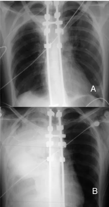
haemoglobin levels were 13 g/dl and chest X-ray showed lungs re-expansion and correct location of the drains (Fig. 2A). No air leak was observed and minimal amounts of blood were drained from the thoracic cavity (100 ml left side, 60 ml right side) over the first 18 h. The patient slept quietly whereas tachycardia (pulse rate > 120/min) and progressive reduction in BP (90/60 mmHg) were noted by the nurses on the morning of the first postoperative day. When moved on a sitting position, the patient became confused and nauseous as BP felt at 70/40 mmHg and heart rate increased over 140 beats/min. We immediately suspected some chest haemorrhage, and haemodynamic resuscitation was initiated. The chest X-ray showed a ‘white’ right lung (Fig. 2B) and the CT scan demonstrated lung compression by a massive haemothorax. We extracted 550 ml of blood after acute desobstruction of the right drain with 100 ml saline. No additional liquid was drained through the left chest tube. Meanwhile, blood tests showed a decrease in haemoglobin (Hb 66 g/l), acute renal failure (creatinine 258 mmol/l) and a severe meta-bolic acidosis (pH 7.18).
A thoracotomy was urgently carried out to control active bleeding originating from apical pleural adhesion; the Blake drain was found to be obstructed by thrombotic clots and talc particles. The postoperative course was uneventful and the patient was discharged from the hospital 4 days later.
3. Discussion
Acute bleeding is one of the most feared complications after thoracic surgery leading to re-intervention for hae-mostasis, transfusion and, if left unrecognized, to haemor-rhagic shock. Efficient chest drainage should allow recognition of ongoing bleeding and air leak. Occasionally, non-functioning drain due to malposition, kinking or thrombosis prevents recognition of intra-thoracic bleeding G. Clark et al. / European Journal of Cardio-thoracic Surgery 31 (2007) 566—568 567
Fig. 1. (A) Blake drain 19F. (B) Blake drain tip with specially designed grooves on every side.
Fig. 2. (A) Postoperative chest X-ray. (B) Chest X-ray with massive right haemothorax.
or bronchopulmonary air leaks. Risk factors for thrombotic occlusion include small drain size, prothrombogenic material and patient-related factors such as inflammatory post-operative hypercoagulability.
Based on retrospective studies in cardiac surgery, chest drainage using small-sized silastic Blake drains is considered safe and efficient[1—4]. In this type of surgery, Blake and conventional drains showed comparable drainage capacity, similar incidence of major complications and even shorter hospital length of stay[2], although outcome and safety data have only been gathered from uncontrolled and retrospective case-controlled studies[1—4].
In our case report, development of hypovolemic shock without clear evidence of intra-thoracic bleeding resulted from the thrombotic occlusion of the chest tubing and this critical event delayed a life-saving re-intervention. We recently published a review of our prospective data bank in thoracic surgery[5]. Out of 1145 patients undergoing various surgical procedures (pneumonectomy, lobectomy, bilobect-omy, wedge resection), our global mortality and hae-mothorax rates (1.9 and 0.8%, respectively) compare favourably with other large series [6]. Importantly, we had never experienced drain occlusion when using conventional chest drains.
Indeed, experience with small Blake chest drains (19F) in thoracic surgery is limited. Strong evidence is, however, lacking because of non-randomized design, poor definition of outcome, non-standardized management and the small number of patients[7—9]. This case suggests, at least, that the good results obtained with small Blake chest drains in cardiac surgery cannot be directly transposed in lung surgery, especially when talc pleurodesis is used. Unlike cardiac surgery patients, the risk for blood clotting within the drainage system is increased in thoracic surgical patients for many reasons: first, the general prothrombotic state after surgery is unopposed by anticoagulation and antiaggregation; second, talc pleurodesis induces a pleural state of hyper-coagulability [10]; third, talc itself extracted through the chest tube increases the risk of mechanical obstruction. Therefore, we ought to use either conventional drains or at
least larger-sized Blake drains in this kind of thoracic surgery. Clinicians should also keep in mind that any drain may become occluded regardless of its design, and nurse staff should regularly check that negative pressure is transmitted to the water-valve.
Acknowledgment
We would like to thank Professor Jean-Marie Tschopp for his help in writing this manuscript.
References
[1] Obney JA, Barnes MJ, Lisagor PG, Cohen DJ. A method for mediastinal drainage after cardiac procedures using small silastic drains. Ann Thorac Surg 2000;70:1109—10.
[2] Frankel TL, Hill PC, Stamou SC, Lowery RC, Pfister AJ, Jain A, Corso PJ. Silastic drains vs conventional chest tubes after coronary artery bypass. Chest 2003;124:108—13.
[3] Lancey RA, Gaca C, Vander Salm TJ. The use of smaller, more flexible chest drains following open heart surgery: an initial evaluation. Chest 2001;119:19—24.
[4] Sakopoulos AG, Hurwitz AS, Suda RW, Goodwin JN. Efficacy of Blake drains for mediastinal and pleural drainage following cardiac operations. J Card Surg 2005;20:574—7.
[5] Licker M SA, Frey JG, Morel P, Rochat T, Widerseiner JM, Widikker I, Tschopp JM. Traitement chirurgical du cancer du poumon: Un mode`le de collaboration entre hoˆpitaux universitaitres et pe´riphe´riques. Bulletin des me´decins suisses 2004; 85:2559—2569.
[6] Sirbu H, Busch T, Aleksic I, Lotfi S, Ruschewski W, Dalichau H. Chest re-exploration for complications after lung surgery. Thorac Cardiovasc Surg 1999;47:73—6.
[7] Ishikura H, Kimura S. The use of flexible silastic drains after chest surgery: novel thoracic drainage. Ann Thorac Surg 2006;81:331—3.
[8] Stolz AJ, Lischke R, Simonek J, Schutzner J, Pafko P. Comparison study on the use of tubular and spiral thoracic drains following lung resections. A prospective study. Rozhl Chir 2005;84:529—32.
[9] Kejriwal NK, Newman MA. Use of a single silastic chest drain following thoracotomy: initial evaluation. ANZ J Surg 2005;75:710—2.
[10] Rodriguez-Panadero F, Segado A, Martin Juan J, Ayerbe R, Torres Garcia I, Castillo J. Failure of talc pleurodesis is associated with increased pleural fibrinolysis. Am J Respir Crit Care Med 1995;151:785—90.
G. Clark et al. / European Journal of Cardio-thoracic Surgery 31 (2007) 566—568 568