2010/112
Short Communication
Zinc supplement greatly improves the condition of parkin
mutant Drosophila
Nidhi Saini and Walter Schaffner*
Institute of Molecular Life Sciences, University of Zu¨rich,
Winterthurerstrasse 190, CH-8051 Zu¨rich, Switzerland
* Corresponding author
e-mail: walter.schaffner@imls.uzh.ch
Abstract
Parkinson’s disease (PD) is a progressive neurodegenerative
disorder in which oxidative stress is implicated as a major
causative factor. Mutations in the gene encoding Parkin, a
ubiquitin ligase, are responsible for a familial form of PD.
In a
Drosophila disease model lacking Parkin (park
25null
mutant), we tested the effect of zinc supplementation. Zinc
is an essential trace metal and a component of many enzymes
and transcriptional regulators. Unlike copper and iron, zinc
is not redox-active and under most conditions serves as an
antioxidant. We find that the condition of
parkin mutants
raised on zinc-supplemented food is greatly improved. At
zinc concentrations where controls begin to show adverse
effects as a result of the metal supplement,
parkin mutants
perform best, as manifested in a higher frequency of reaching
adulthood, extended lifespan and improved motoric abilities.
Keywords: antioxidant; metal homeostasis;
metallothioneins; MTF-1; Parkinson’s disease; zinc
transporters.
Parkinson’s disease (PD), characterized by the loss of
dopa-minergic (DA) neurons in the substantia nigra, is a
progres-sive neurodegenerative disorder with the second highest
incidence rate and is the most common age-related
move-ment disorder (Olanow and Tatton, 1999; Dawson and
Daw-son, 2003; Greene et al., 2003). Both genetic and
envi-ronmental factors contribute to its pathogenesis. Oxidative
stress is considered to be a major factor in the pathogenesis
of PD, as evidenced by an elevated content of redox-active
iron and lipid peroxides in the diseased brain, impaired
mito-chondrial function, and alterations in the antioxidant defense
mechanisms (Dexter et al., 1989; Jenner and Olanow, 1996;
Greene et al., 2003; Pesah et al., 2004). Mutations in six
genes, including
parkin which encodes an E3 ubiquitin
ligase, have been associated with rare, early-onset, familial
forms of PD (West and Maidment, 2004; Gasser, 2005; Sang
et al., 2007). Interestingly, some alleles of these genes might
be susceptibility factors for environmental toxins (Choi et
al., 2000; Warner and Schapira, 2003; Bueler, 2009).
In our study, we used a
Drosophila melanogaster line in
which the ortholog of the human
parkin gene is disrupted
by transposition of a P-element (Greene et al., 2003; Pesah
et al., 2004). Parkin mutant flies present with male and
female sterility (Riparbelli and Callaini, 2007), mitochondrial
and muscle abnormalities, locomotor defects, an inability to
fly owing to degeneration of indirect flight muscles,
in-creased sensitivity to multiple stresses, including oxidative
stress, and a severely reduced lifespan (Palacino et al., 2004;
Greene et al., 2005; Whitworth et al., 2005). Some of these
defects arise because
parkin mutants have dysfunctional
mitochondria with disturbances in the electron transport
chain. In mice, in contrast to its pivotal role in humans (Choi
et al., 2000), Parkin function does not seem to be critical for
the survival of DA neurons (Goldberg et al., 2003; Itier et
al., 2003; Palacino et al., 2004). Similarly,
Drosophila that
are null mutants for
parkin do not generally display DA
neu-ron loss (Greene et al., 2003; Pesah et al., 2004), although a
partial loss of DA neurons in the PPL1 cluster of the brain
has been reported (Whitworth et al., 2005).
In the late 1980s, the antioxidant function of the
redox-inert metal zinc was recognized and proposed to be mediated
by the protection of protein sulfhydryl groups and/or by
com-peting against redox-active metals (Bray and Bettger, 1990).
Additionally, zinc ions can upregulate the expression of
metallothioneins, which owing to their high cysteine content
can serve as antioxidants. Zinc has an established
antiapop-totic function that minimizes ROS-induced cellular oxidative
damage (Suzuki et al., 1991). This also occurs in the central
nervous system, particularly in the brain (Kocaturk et al.,
1996).
More than 70 different enzymes involved in the
metabo-lism of biomolecules require zinc as a cofactor (Parkin,
2004). Zinc is an integral part of the hundreds of
transcrip-tion factors that contain zinc finger domains (Berg and Shi,
1996), and it plays a role in cellular signal transduction and
in modulation of synaptic neurotransmission. Zinc is critical
for the growth and regulation of cells and alterations in zinc
metabolism have been implicated in causing neurological
dysfunctions on the one hand, and on the other hand
provid-ing neuroprotection. Maintenance of intracellular zinc
homeo-stasis is thus an essential requirement in all living organisms
(Valko et al., 2005).
The best characterized zinc-activated transcription factor
is the metal response element-binding transcription factor-1,
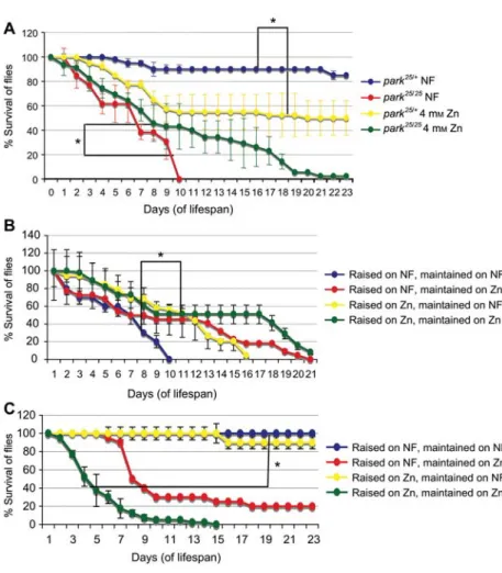
Figure 1 Drosophila parkin mutants show an enhanced lifespan on zinc-supplemented food.
(A) Zinc supplementation (4 mMZnCl2) increased the longevity of theparkin mutant flies (park25/25). The maximum lifespan of 11 days
on normal food (NF) increased to 23 days. Control flies (park25/q) showed an opposite effect with a reduction of lifespan on zinc food. (B)
Survival ofpark25/25
flies and (C) ofpark25/q
control flies raised and maintained on NF or zinc-supplemented food. Error bars indicate standard deviation. The significance between survival curves was analyzed using the Kaplan-Meier log-rank statistical test (*p-0.01).
Methods: for survival assays, NF was supplemented with zinc chloride to a final concentration of 4 mM. One- to two-day-old flies (20 per
vial) were maintained at 258C on a 12:12 h light/dark cycle for each genotype in triplicate vials. Surviving flies were transferred to fresh food vials every 2 days and counted daily. As a control, flies of the same genotype were grown on food without metal supplement. In each lifespan assay testing different conditions, the controls ofpark25/qandpark25/25flies raised on NF were the same. The variations in the
median lifespan of control flies in different experiments can be attributed to subtle experimental variations.
Table 1 Zinc increases the frequency of parkin mutant Drosophila reaching
adulthood.
Food condition Genotype park25/25
/total progeny % ofpark25/25
Normal food Set 1park25/25 31/1116 2.8%
Set 2park25/25 10/450 2.2%
4 mMZn Set 1park25/25
110/611 18% Set 2park25/25 186/956 19%
For the analysis of eclosure frequency, egg laying was allowed for 6 days and the parent population (10 males and 10 females each ofpark25/q) was the same in all
vials of normal food (NF) or zinc (Zn), and progeny flies were counted at the same time.
also referred to as metal-responsive transcription factor-1
(MTF-1). In response to zinc, MTF-1 translocates from the
cytoplasm to the nucleus where it regulates the expression
of several genes, notably metallothioneins and some zinc
transporters (Andrews, 2000; Smirnova et al., 2000; Lichtlen
and Schaffner, 2001; Saydam et al., 2001). MTF-1 requires
elevated zinc concentration for DNA binding. This property
was exploited for the activation of MTF-1-dependent
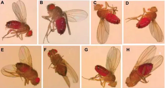
pro-Figure 2 Gustatory assay with adult flies on normal or zinc-supplemented food with acid red dye as a marker. (A, B) park25/q
control flies and (C, D) park25/25
flies show the same feeding behavior on normal food, exemplified by the fully red abdomens. A food supplement of 4 mMZn (E, F) altered feeding behavior ofpark25/q(only partially red abdomens) but no change in the
behavior ofpark25/25flies was observed (G, H) (fully red abdomens). The mutants are recognized by white eyes because of the loss of wq
(red eyes) which are present in control flies.
Methods: the adult gustatory assay was essentially carried out as described by A. Hilliker and colleagues (Bahadorani, 2008). Briefly, newly eclosed flies were reared on normal food for 2–3 days, then starved for 18 h on Whatman paper soaked with distilled water. After this treatment, starved flies (20 per vial) were transferred onto zinc-supplemented food with 0.2% sulforhodamine B sodium salt (acid red) for 2 h. For control flies, culture medium was supplemented with 0.2% acid red, without metal supplement. After 2 h of feeding at optimum temperature (258C) and relative humidity, flies were anesthetized and the degree of abdomen redness was visually inspected. Abdomen redness was used as an indicator of the amount of food taken up.
moters by zinc and other metals in a cell-free transcription
system (Zhang et al., 2003).
Zinc is not redox-active, but nevertheless toxic when in
excess (Beyersmann and Haase, 2001). Acute zinc toxicity
is rare but has been reported (Duncan et al., 1992; Whittaker,
1998; Prasad et al., 1999). If the extracellular concentration
of zinc exceeds the capacity of zinc homeostasis
mecha-nisms, it becomes cytotoxic and an excess of free
intracel-lular zinc can trigger apoptosis (Choi et al., 1988; Duncan
et al., 1992; Kim et al., 1999; Beyersmann and Haase, 2001;
Wilhelm et al., 2001; Beyersmann, 2002; Walther et al.,
2003). Zinc transport in
Drosophila, as in vertebrates, is
mediated by two families of solute linked carrier proteins:
zinc importers (ZIPs), which function in the uptake of zinc
to the cytoplasm, and zinc exporters (ZnTs), which reduce
cytoplasmic zinc concentrations by promoting zinc efflux
(Liuzzi and Cousins, 2004; Yepiskoposyan et al., 2006).
More than 10 zinc transporter genes are annotated in
Dro-sophila melanogaster based on their sequence similarities to
vertebrate zinc transporters. The ZIP family gene
foi (fear of
intimacy) was characterized in Drosophila and shown to be
a zinc importer that is critical during development (Moore
et al., 1998; Mathews et al., 2005). Transcriptional responses
to zinc in
Drosophila larvae were analyzed in our laboratory
(Yepiskoposyan et al., 2006). Apart from the expected
upreg-ulation of metallothioneins and the zinc exporters
ZnT35C
and
ZnT63C, there was also an induction of
neurotransmit-ters, detoxification enzymes (such as glutathione
S-transfer-ase), ferritin and chaperone encoding genes.
We found that whereas
parkin mutant flies readily feed on
high-zinc food, their wild type counterparts avoid
zinc-load-ed food. The mutants also had an increaszinc-load-ed survival rate on
zinc-supplemented food, which prompted us to investigate
their response to zinc in more detail.
With standard ‘normal food’ (NF),
parkin mutant flies
have a median lifespan of 6 days with a maximum of 11 days
(Figure 1A). Zinc supplementation in the form of zinc
chlo-ride increased the lifespan of
parkin mutants. When
main-tained on supplements of 4 m
MZn, the mutant flies survived
up to 23 days with a median lifespan of 8 days (Figure 1).
This increase was as a result of zinc and not chloride ions,
as similar survival assays on NaCl-supplemented medium
did not extend lifespan (data not shown). Zinc
supplemen-tation also increases the eclosion frequency of
parkin
mutants, from 2.5% to 19%, i.e., close to 25% which is the
expected frequency of
parkin mutants in the progeny of
par-kin heterozygous parents (Table 1). In contrast, heterozygous
control flies did not draw any benefit from
zinc-supple-mented food: if kept on food with 4 m
MZnCl
2, the median
lifespan reduced significantly to 17 days, whereas on NF,
80% were still alive at the end of the experiment (23 days)
(Figure 1A). Similar adverse effects of zinc load were
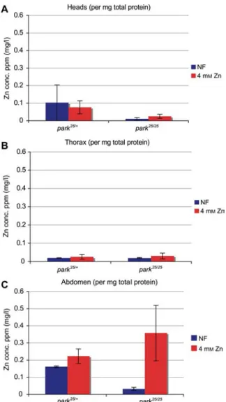
Figure 3 Quantification of Zn content in the different body parts ofpark25/qcontrol flies andpark25/25flies.
Flies were raised on normal food (NF) or zinc-supplemented food (4 mMZn). (A) Heads, (B) thoraces, (C) abdomens.
Methods: female flies were allowed to lay eggs on zinc-supple-mented (4 mMZnCl2) fly food for 6 days and removed afterwards.
The resulting progeny was collected at regular intervals and the percentage of eclosingparkin homozygous mutants and
heterozy-gous controls were calculated each time. These progeny were then frozen and stored until the required number of 200 flies was obtained in triplicate for each genotype. Frozen flies were then dis-sected to separate their heads, thoraces and abdomens which were collected separately. Each sample set was then subjected to homog-enization using cold protein homoghomog-enization buffer (0.32 mM
sucrose, 20 mM HEPES, 1 mM MgCl2, 0.5 mM PMSF protease
inhibitor at pH 7.4) and the samples were normalized for protein content. A highly sensitivity flame atomic absorption spectropho-tometer (FAAS; GTA-120/PSD-120, Varian Australia Pty Ltd, Mul-grave, VIC, Australia) was used to detect the zinc content in each body part of each genotype assessed.
observed with wild type
yw and OregonR flies (data not
shown). To determine if zinc has a stronger effect during
development from eggs to adults or during the adult feeding
stage, we followed the lifespan of both
parkin mutant and
parkin heterozygous flies under four conditions: (i)
devel-opment and adult maintenance in zinc supplement, (ii)
rais-ing the flies on NF until adult stage and then maintainrais-ing
them on zinc-supplemented food post-eclosion, (iii) raising
the flies on zinc food but maintaining them on NF after
eclo-sion, and (iv) raising and maintaining the flies on NF. We
observed that the strongest positive effect of zinc on
park
25/25flies was when they were both raised and maintained on
4 m
MZn-supplemented food (Figure 1B). The strongest
neg-ative effect of zinc on
park
25/qcontrol flies was also
observed under these conditions (Figure 1C). Development
of
park
25/qand
OregonR wild type flies was delayed but
generally less affected by zinc than the survival of adults;
egg transfer (200 each) from NF to vials containing
increas-ing concentrations of zinc resulted in an equal percentage of
eclosing adults in NF, 4 and 6 m
MZn but none developed
in 10 m
MZn (data not shown; see also Egli et al., 2003).
parkin mutants might sense a deficiency in and/or a
res-cuing effect of zinc, and in response to this eat normal
amounts of zinc-supplemented food unlike heterozygous or
wild type flies. This was indicated by a visual inspection of
adult
Drosophila in a gustatory test and was quantitatively
confirmed by the measurement of total zinc uptake by flies
(Figures 2 and 3). On NF, the zinc content in heads of
parkin
mutant flies was much lower than in controls, consistent with
a zinc deficiency in the mutants. Zinc supplementation
indeed resulted in an, albeit minor, increase of zinc content
in mutant heads (Figure 3A), whereas in thoraces the zinc
content was similar in mutant and control flies (Figure 3B).
Interestingly, the abdomens of
park
25/25flies fed on zinc food
showed a 10-fold increase in zinc levels compared with flies
fed on NF (Figure 3C). Such an effect was, however, not
observed in the control flies which only showed a minor
increase in zinc levels upon zinc feeding, which is in line
with their tendency to avoid zinc-loaded food.
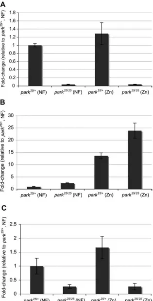
We also determined the transcript levels of
parkin,
MTF-1 and some other genes involved in zinc import/export
(Figure 4). As expected,
parkin mutants have no detectable
parkin transcripts. In comparison to control flies, transcript
levels of the metallothionein
MtnB are higher in parkin
mutants (Figure 4A,B). Zinc supplementation induced
MtnB
transcripts 24-fold in
parkin mutants; in control flies the
increase was 14-fold (Figure 4B). Boosting expression of
metallothioneins, which act as ROS scavengers, is one way
zinc could play an antioxidant role. The elevated
metallo-thionein levels could also explain at least in part why an
increased zinc concentration is not toxic to the
parkin
mutants. We also determined the transcript levels of three
zinc transporters, the exporters
ZnT35C and ZnT63C and the
importer
foi (Mathews et al., 2005; Yepiskoposyan et al.,
2006) both in
parkin mutants and in the heterozygous
con-trols. The most conspicuous difference was observed with
the exporter gene
ZnT35C, a known target of MTF-1. In
parkin mutants, expression was dramatically reduced
com-pared to controls and not responsive to zinc. In heterozygous
controls, zinc supplement resulted in a 1.5-fold upregulation,
which indicates a response to zinc overload (Figure 4C). An
increased expression of zinc exporters in control flies is in
.
Figure 4 Real-time analysis ofparkin transcript levels.
(A) Inpark25/25
flies noparkin transcripts are detectable. In park25/q parkin transcripts are, if anything, elevated in flies raised on zinc
food. (B) park25/25
flies have increased levels of metallothionein
MtnB transcripts in NF compared to control flies. Zinc induces MtnB in both genotypes, but to a higher level in park25/25. (C) The
zinc exporter,ZnT35C, shows increased transcript levels in park25/q
on zinc food, which is not observed inpark25/25
flies.
Methods: total RNA was purified from the adultDrosophila tissue
using the nucleospin RNA II protocol and eluted in 60
m
l RNase-free water. cDNA was prepared using the transcriptor high fidelity cDNA synthesis kit from Roche (Mannheim, Germany). The cDNA obtained was further purified using the AM 1906 DNA-free kit (Ambion, Rotkreuz, Switzerland). This was then used for analysis by real-time PCR on the Tecan Genesis 200/8 robot (Tecan, Ma¨n-nedorf, Switzerland) using the Mesa Green qPCR Mastermix Plus (Eurogentec, Seraing, Belgium) for SYBR assays. The qPCR run was performed on an Applied Biosystems machine (ABI Prism SDS 7900 HAT; Seraing, Belgium) in a 384-well format with a reaction volume of 10m
l. All fold-change values are normalized to corre-spondingpark25/qvalues on normal food (NF). Housekeeping genes used for reference wereactin5C, sec24 and GAPDH. The primer
sequences used for the transcripts quantified were: forparkin,
59-AAG ATC ATA TTT GCC GGT 59-AAG GAA-39 and 59-CGC TTT GCT GAC CCA AGT C-39 amplify a 73-bp fragment only from the
parkin heterozygous control flies; for MtnB, 59-TTG CAA GGG
TTG TGG AAC AAA-39 and 59-TGC AGG CGC AGT TGT CC-39 amplify a 65-bp fragment, and forZnT35C, 59-GCT CTC GCC
GAT CTG AAA GT-39 and 59-CGC ACC GAC AAG TGT TTC TTA TA-39 amplify a 75-bp fragment.
agreement with their avoidance of zinc-supplemented food.
Transcripts of
ZnT63C and, somewhat unexpectedly of foi,
were also lower in
parkin mutants compared to parkin
het-erozygous controls (data not shown). Taken together, these
data suggest that zinc homeostasis is distorted in
parkin
mutants, with the effect that zinc supplement in food
pro-duces a strong phenotypic rescue effect. It remains to be seen
whether zinc supplementation also has a positive effect in a
mouse model of familial PD and ultimately in Parkinson’s
patients.
Acknowledgments
We thank Drs. Jessica Greene and Leo Pallanck (University of Washington) for providing the park25/25
flies, Drs. Eva Freisinger and Augusto Cabral for the metal quantification experiments, Martin Moser for the RT-PCR analysis, Ivan Ostojic (Friedrich Miescher Institute, Basel, Switzerland) for statistical analyses, Till Strassen for maintenance of fly stocks and Dr. George Hausmann for critical
reading of the manuscript. This work was supported by the Kanton Zu¨rich and the Swiss National Science Foundation.
References
Andrews, G.K. (2000). Regulation of metallothionein gene expres-sion by oxidative stress and metal ions. Biochem. Pharmacol.
59, 95–104.
Berg, J.M. and Shi, Y. (1996). The galvanization of biology: a grow-ing appreciation for the roles of zinc. Science271, 1081–1085.
Beyersmann, D. (2002). Effects of carcinogenic metals on gene expression. Toxicol. Lett.127, 63–68.
Beyersmann, D. and Haase, H. (2001). Functions of zinc in signal-ing, proliferation and differentiation of mammalian cells. Bio-metals14, 331–341.
Bray, T.M. and Bettger, W.J. (1990). The physiological role of zinc as an antioxidant. Free Radic. Biol. Med.8, 281–291.
Bueler, H. (2009). Impaired mitochondrial dynamics and function in the pathogenesis of Parkinson’s disease. Exp. Neurol. 218,
235–246.
Choi, D.W., Yokoyama, M., and Koh, J. (1988). Zinc neurotoxicity in cortical cell culture. Neuroscience24, 67–79.
Choi, H.Y., Song, J.H., Park, D.K., and Ross, G.M. (2000). The effects of ascorbic acid on dopamine-induced death of PC12 cells are dependent on exposure kinetics. Neurosci. Lett.296,
81–84.
Dawson, T.M. and Dawson, V.L. (2003). Molecular pathways of neurodegeneration in Parkinson’s disease. Science 302, 819–
Dexter, D.T., Carter, C.J., Wells, F.R., Javoy-Agid, F., Agid, Y., Lees, A., Jenner, P., and Marsden, C.D. (1989). Basal lipid peroxidation in substantia nigra is increased in Parkinson’s dis-ease. J. Neurochem.52, 381–389.
Duncan, M.W., Marini, A.M., Watters, R., Kopin, I.J., and Markey, S.P. (1992). Zinc, a neurotoxin to cultured neurons, contaminates cycad flour prepared by traditional guamanian methods. J. Neu-rosci.12, 1523–1537.
Egli, D., Selvaraj, A., Yepiskoposyan, H., Zhang, B., Hafen, E., Georgiev, O., and Schaffner, W. (2003). Knockout of ‘metal-responsive transcription factor’ MTF-1 inDrosophila by
homol-ogous recombination reveals its central role in heavy metal homeostasis. EMBO J.22, 100–108.
Gasser, T. (2005). Genetics of Parkinson’s disease. Curr. Opin. Neu-rol.18, 363–369.
Goldberg, M.S., Fleming, S.M., Palacino, J.J., Cepeda, C., Lam, H.A., Bhatnagar, A., Meloni, E.G., Wu, N., Ackerson, L.C., Klapstein, G.J., et al. (2003). Parkin-deficient mice exhibit nigrostriatal deficits but not loss of dopaminergic neurons. J. Biol. Chem.278, 43628–43635.
Greene, J.C., Whitworth, A.J., Kuo, I., Andrews, L.A., Feany, M.B., and Pallanck, L.J. (2003). Mitochondrial pathology and apop-totic muscle degeneration in Drosophila parkin mutants. Proc. Natl. Acad. Sci. USA100, 4078–4083.
Greene, J.C., Whitworth, A.J., Andrews, L.A., Parker, T.J., and Pal-lanck, L.J. (2005). Genetic and genomic studies ofDrosophila parkin mutants implicate oxidative stress and innate immune
responses in pathogenesis. Hum. Mol. Genet.14, 799–811.
Itier, J.M., Ibanez, P., Mena, M.A., Abbas, N., Cohen-Salmon, C., Bohme, G.A., Laville, M., Pratt, J., Corti, O., Pradier, L., et al. (2003). Parkin gene inactivation alters behaviour and dopamine neurotransmission in the mouse. Hum. Mol. Genet.12, 2277–
2291.
Jenner, P. and Olanow, C.W. (1996). Oxidative stress and the patho-genesis of Parkinson’s disease. Neurology47, S161–S170.
Kim, E.Y., Koh, J.Y., Kim, Y.H., Sohn, S., Joe, E., and Gwag, B.J. (1999). Zn2qentry produces oxidative neuronal necrosis in
cor-tical cell cultures. Eur. J. Neurosci.11, 327–334.
Kocaturk, S., Kocaturk, P.A., Kavas, G.O., and Mutluer, N. (1996). Antioxidant defence system in a patient with cerebrovascular accident. J. Int. Med. Res.24, 376–380.
Lichtlen, P. and Schaffner, W. (2001). Putting its fingers on stressful situations: the heavy metal-regulatory transcription factor MTF-1. Bioessays23, 1010–1017.
Liuzzi, J.P. and Cousins, R.J. (2004). Mammalian zinc transporters. Annu. Rev. Nutr.24, 151–172.
Mathews, W.R., Wang, F., Eide, D.J., and Van Doren, M. (2005).
Drosophila fear of intimacy encodes a Zrt/IRT-like protein (ZIP)
family zinc transporter functionally related to mammalian ZIP proteins. J. Biol. Chem.280, 787–795.
Moore, L.A., Broihier, H.T., Van Doren, M., and Lehmann, R. (1998). Gonadal mesoderm and fat body initially follow a com-mon developmental path in Drosophila. Development 125,
837–844.
Olanow, C.W. and Tatton, W.G. (1999). Etiology and pathogenesis of Parkinson’s disease. Annu. Rev. Neurosci.22, 123–144.
Palacino, J.J., Sagi, D., Goldberg, M.S., Krauss, S., Motz, C., Wac-ker, M., Klose, J., and Shen, J. (2004). Mitochondrial dysfunc-tion and oxidative damage in parkin-deficient mice. J. Biol. Chem.279, 18614–18622.
Parkin, G. (2004). Synthetic analogues relevant to the structure and function of zinc enzymes. Chem. Rev.104, 699–767.
Pesah, Y., Pham, T., Burgess, H., Middlebrooks, B., Verstreken, P., Zhou, Y., Harding, M., Bellen, H., and Mardon, G. (2004). Dro-sophila parkin mutants have decreased mass and cell size and
increased sensitivity to oxygen radical stress. Development131,
2183–2194.
Prasad, A.S., Beck, F.W., Kaplan, J., Chandrasekar, P.H., Ortega, J., Fitzgerald, J.T., and Swerdlow, P. (1999). Effect of zinc supple-mentation on incidence of infections and hospital admissions in sickle cell disease (SCD). Am. J. Hematol.61, 194–202.
Riparbelli, M.G. and Callaini, G. (2007). TheDrosophila parkin
homologue is required for normal mitochondrial dynamics dur-ing spermiogenesis. Dev. Biol.303, 108–120.
Sang, T.K., Chang, H.Y., Lawless, G.M., Ratnaparkhi, A., Mee, L., Ackerson, L.C., Maidment, N.T., Krantz, D.E., and Jackson, G.R. (2007). A Drosophila model of mutant human parkin-induced toxicity demonstrates selective loss of dopaminergic neurons and dependence on cellular dopamine. J. Neurosci.27,
981–992.
Saydam, N., Georgiev, O., Nakano, M.Y., Greber, U.F., and Schaff-ner, W. (2001). Nucleo-cytoplasmic trafficking of metal-regula-tory transcription factor 1 is regulated by diverse stress signals. J. Biol. Chem.276, 25487–25495.
Smirnova, I.V., Bittel, D.C., Ravindra, R., Jiang, H., and Andrews, G.K. (2000). Zinc and cadmium can promote rapid nuclear trans-location of metal response element-binding transcription factor-1. J. Biol. Chem.275, 9377–9384.
Suzuki, T., Umeyama, T., Ohma, C., Yamanaka, H., Suzuki, K., Nakajima, K., and Kimura, M. (1991). Immunohistochemical study of metallothionein in normal and benign prostatic hyper-plasia of human prostate. Prostate19, 35–42.
Valko, M., Morris, H., and Cronin, M.T. (2005). Metals, toxicity and oxidative stress. Curr. Med. Chem.12, 1161–1208.
Walther, U.I., Czermak, A., Muckter, H., Walther, S.C., and Fichtl, B. (2003). Decreased GSSG reductase activity enhances cellular zinc toxicity in three human lung cell lines. Arch. Toxicol.77,
131–137.
Warner, T.T. and Schapira, A.H. (2003). Genetic and environmental factors in the cause of Parkinson’s disease. Ann. Neurol. 53
(Suppl. 3), S16–S23; discussion S23–S25.
West, A.B. and Maidment, N.T. (2004). Genetics of parkin-linked disease. Hum. Genet.114, 327–336.
Whittaker, P. (1998). Iron and zinc interactions in humans. Am. J. Clin. Nutr.68, 442S–446S.
Whitworth, A.J., Theodore, D.A., Greene, J.C., Benes, H., Wes, P.D., and Pallanck, L.J. (2005). Increased glutathione S-trans-ferase activity rescues dopaminergic neuron loss in a Drosophila model of Parkinson’s disease. Proc. Natl. Acad. Sci. USA102,
8024–8029.
Wilhelm, B., Walther, U.I., and Fichtl, B. (2001). Effects of zinc chloride on glutathione and glutathione synthesis rates in various lung cell lines. Arch. Toxicol.75, 388–394.
Yepiskoposyan, H., Egli, D., Fergestad, T., Selvaraj, A., Treiber, C., Multhaup, G., Georgiev, O., and Schaffner, W. (2006). Trans-criptome response to heavy metal stress inDrosophila reveals a
new zinc transporter that confers resistance to zinc. Nucleic Acids Res.34, 4866–4877.
Zhang, B., Georgiev, O., Hagmann, M., Gunes, C., Cramer, M., Faller, P., Vasak, M., and Schaffner, W. (2003). Activity of met-al-responsive transcription factor 1 by toxic heavy metals and H2O2in vitro is modulated by metallothionein. Mol. Cell. Biol. 23, 8471–8485.