Nanoparticle–Cell Interaction: A Cell Mechanics Perspective
Dedy Septiadi,* Federica Crippa, Thomas Lee Moore, Barbara Rothen-Rutishauser,
and Alke Petri-Fink*
1. Introduction
Nanoparticles (NPs) have emerged in recent decades as exciting materials for biomedical applications (e.g., drug delivery, imaging contrast, theranostic medicine, and biosensing), and in parallel the study of fundamental bio–nano interactions has grown.[1–3] For the most part, researchers have focused on
the effect of specific physicochemical properties on NP cel-lular uptake, cytotoxicity, and in vivo biokinetics, systemic tox-icity and NP fate.[4–8] However, fewer studies have focused on
studying the effect of particles on cell mechanics, i.e., study on force–cell structure relationship and how it relates to cellular functions.[9] Cell mechanics are critical indicators for cell
func-tionality and health, and these processes drive important bio-logical activities such as cell migration, differentiation, wound
Progress in the field of nanoparticles has enabled the rapid development of multiple products and technologies; however, some nanoparticles can pose both a threat to the environment and human health. To enable their safe implementation, a comprehensive knowledge of nanoparticles and their bio-logical interactions is needed. In vitro and in vivo toxicity tests have been con-sidered the gold standard to evaluate nanoparticle safety, but it is becoming necessary to understand the impact of nanosystems on cell mechanics. Here, the interaction between particles and cells, from the point of view of cell mechanics (i.e., bionanomechanics), is highlighted and put in perspec-tive. Specifically, the ability of intracellular and extracellular nanoparticles to impair cell adhesion, cytoskeletal organization, stiffness, and migration are discussed. Furthermore, the development of cutting-edge, nanotechnology-driven tools based on the use of particles allowing the determination of cell mechanics is emphasized. These include traction force microscopy, colloidal probe atomic force microscopy, optical tweezers, magnetic manipulation, and particle tracking microrheology.
Dr. D. Septiadi, F. Crippa, Dr. T. L. Moore, Prof. B. Rothen-Rutishauser, Prof. A. Petri-Fink
Adolphe Merkle Institute University of Fribourg
Chemin des Verdiers 4, 1700 Fribourg, Switzerland E-mail: dedy.septiadi@unifr.ch; alke.fink@unifr.ch Prof. A. Petri-Fink
Department of Chemistry University of Fribourg
Chemin du Musée 9, 1700 Fribourg, Switzerland
healing, and tissue integrity (i.e., adhesion of cells to the extracellular matrix (ECM) and even the ECM production). Thus, it is worthwhile to investigate how NPs may affect cell mechanics and influence spe-cific cellular functions, physiological con-ditions, or development of diseases.
The field of cell mechanics has been heavily studied and is well-described.[10] In
general, the fundamental understanding of cell and tissue mechanics are con-structed by two major cellular constitu-ents, namely cellular adhesion system and the cytoskeleton. The cellular adhesion system comprises different structures (e.g., cell–cell and cell–ECM junctions) and proteins (e.g., integrins, cadherins, and selectins), and enables the cells to probe external force from their environ-ment (both the ECM and neighboring cells). This system mechanically connects the cell surface, the cytoskeleton, and the cell’s external surrounding. It has been long understood that cells can “feel” their external environment, and thus mechan-ical inputs to a cell can modulate the most basic cellular pro-cesses, such as cell migration, cell division, and differentia-tion.[11–14] On the other hand, the cytoskeleton exerts force on
its surrounding to reshape the ECM via dynamic assembly and disassembly of cytoskeleton proteins (e.g., actin and tubulin) and motor protein complexes. The cytoskeleton is also involved in cell migration, for example, during wound healing and cancer metastasis, and in part regulates cell elastic properties, as broadly investigated by cell mechanics measurements.[15–19]
As particles can interact with cellular organelles and cytoskeletal structures, it is intuitive to imagine that particles (i.e., nano and micrometer size) can affect or inhibit their asso-ciated cellular functions, or be used to probe the environments in which cells interact. Moreover, the improvement of nano-fabrication techniques has resulted in increasing application of micro- and nanoparticles towards probing cellular mechanics. Here, we aim to explore from two different perspectives this emerging field of bionanomechanics, the study of biomech-anics using nanoscience tools. Specifically, we explore (i) how nanoparticles influence cell mechanics, and (ii) how nano-science tools can incorporate particles to investigate cell mechanics (Figure 1).
Inherently, bionanomechanics is an interdisciplinary field built on the expertise of biologists, toxicologists, physicists, bio-engineers, materials scientists, and chemists. Thus, we aim to emphasize the interdisciplinary nature involved in this research and facilitate communication between fields. First, we survey
http://doc.rero.ch
Published in "Advanced Materials doi: 10.1002/adma.201704463, 2018"
which should be cited to refer to this work.
the effect of NPs on cell adhesion and cytoskeleton organiza-tion, which influences cell stiffness and migration dynamics. Second, we elaborate on the application of particulate materials (i.e., microparticles and NPs) to study the mechanical property of the cells—this includes the use of engineered particles as extracellular and intracellular probes to study force exerted by the cells on their environment, and the corresponding mecha-notransduction pathways as well as rheological properties of the cell.
This Progress Report is intended and written for broad scien-tific community with the aim to provide a global understanding of particle–cell interactions in the context of cell mechanics. Future direction in bionanomechanics research is expected to lead to a new standardized protocol especially for scientists and industries in addressing the influence of NPs on human and animal’s toxicity from point of view of cell mechanics.
2. Effects of Intracellular Nanoparticles on
Adhesion, Cytoskeleton Organization, Cell
Stiffness, and Cell Migration
2.1. Introduction to Cell Mechanics
Cell mechanics is a subfield of biophysics that focuses on mechanical properties of cells and their relationship with cer-tain biological behaviors.[20] It has been long understood that
cell mechanics play an important role, not only in basic cellular functions such as cell proliferation, differentiation, apoptosis, polarization, adhesion, and migration, but also in more com-plex activities including embryonic development, cancer metas-tasis, as well as wound healing. Moreover, it has been reported that the progression and development of certain diseases (e.g., atherosclerosis, heart failure, asthma, pulmonary fibrosis, preeclampsia) is directly associated to cell and tissue mechanics (also reviewed in ref. [21]), highlighting the importance of cell mechanics in development, physiology, and diseases.
Tissue/organ development is perhaps considered as the “oldest” field describing cell mechanics. The concepts of cell mechanics are generally related to two distinct processes: force generation by the cell body and force transmission through adhesive complexes.[22] Changes in the size and shape of the
cell during development (e.g., motility and reorganization), for example, depends on mechanical activities of cytoskel-eton, an interconnected intracellular network of filamentous polymers and regulatory proteins.[10] In addition, cells interact
mechanically with their surrounding (i.e., extracellular matrix and neighboring cells) through cell–cell and cell–matrix adhe-sions. These adhesions, which involve many different proteins and their complexes, together with the cytoskeleton compo-nents provide important mechanical functions: (i) allowing the cell to exert mechanical forces on its environment, and (ii) detecting mechanical stimulation from the environment and transducing these mechanical signals (mechanotransduction) into a biological response (or responding through cytoskeletal reorganization and force generation). Cellular adhesions and cytoskeletons have been shown to drive development and tissue self-assembly. In particular, adhesion has been shown to drive
cellular sorting (i.e., the rearrangement of scattered mixtures of two or more cell types into homogeneous clusters[23]) during
tissue assembly to specific locations based on the type and den-sity of adhesion proteins.[24]
It has been shown that environmental effects, genetic muta-tions, pathogens, or perturbations to the mechanical properties can disrupt the cytoskeletal architecture of the cells and further lead to the alteration of cell mechanics.[20] For instance, it was
demonstrated that adhesion behavior,[25] cytoskeleton
organi-zation (including cell polariorgani-zation and elongation)[26] and
trac-tion forces[27] from different types of cells varied when the cells
were grown on substrates with different stiffness. Micro-[28] and
nanotopography[29] have also been described to induce changes
in cell adhesion, cytoskeletal organization, and the mechanical properties in human cells.
Due to their nanosize combined with unique physicochem-ical properties, internalized NPs are expected to interact with the cell membrane,[30] cytoskeletal structures,[31] and
orga-nelles[32] and as a consequence alter specific cellular functions.
Contrary to the extensive number of cell viability studies, the impact of NPs on cell mechanics has been less thoroughly investigated. Therefore, an understanding of any possible alteration of cell mechanics following NP exposure is needed to enhance our comprehension of nanomaterial–cell interac-tions. This particular chapter focuses on the impact of NPs in cell–cell and cell–matrix adhesion, cytoskeletal building blocks (actin/tubulin/intermediate filament system), cell stiffness and cell migration. Substrate environment parameters, which have been shown to influence cell mechanics,[33,34] are not
con-sidered in the discussion of Section 2, since for most in vitro experiments the cells were cultured in similar conditions (i.e., on commercial glass cover slips), hence the cell mechanics responses which are described are only associated to the NP– cell interaction.
2.2. Influence of NPs on Cellular Adhesions
When the cells contact a substrate (i.e., the ECM, other cells, or tissues), the first cellular response will be adhesion or
adherence. In a biophysical term, adhesion can be described as the bonding of two distinct entities (e.g., cell to cell/tissue, or cell to the ECM) in a manner that resists their subsequent separation, normally through either homotypic or heterotypic protein-protein interactions.[23] Transmembrane adhesion
pro-teins (also called cell adhesion molecules or CAMs) such as immunoglobulin superfamily CAMs (IgSF CAMs), selectins, integrins, and cadherins have important functions in adhe-sion. In particular integrins form connections between cells and the ECM, while cadherins connect cells with other cells/ tissues. The ECM itself is mainly composed of a variety of ver-satile polysaccharides (such as proteoglycan) and fibrous pro-teins (e.g., collagens, elastin, fibronectin, and laminin) which are secreted locally by the cells and assemble into an organized meshwork whose purpose is mainly to provide support for the surrounding cells (i.e., to hold cells and tissues together) and an organized lattice through which cells migrate and interact with each other.[35] Depending on their functional classification, cell
adhesions or junctions can be classified into three classes. First, occluding junctions (also called tight junctions) which form an impermeable barrier across the epithelial cell layer, second, communicating junctions (or gap junctions) which allow the transport of small molecules between the cells, and third, anchoring junctions which connect the cytoskeleton of the cell (e.g., F-actins and intermediate filaments) to its neighbors or to the ECM.[35] Anchoring junction includes focal adhesion (FA)
for cell–ECM adhesion, hemidesmosomes for cell-basal lamina adhesion, adherens junctions and desmosomes for cell–cell adhesion (Figure 2). Cell adhesion plays an important role in homeostasis and regulation of cell biological behavior including cell communication (reviewed in ref. [36]).
The presence of cell-associated NPs (either internalized or strongly attached to the cell membrane) has been shown to alter many cellular processes, e.g., provoking cell death. In particular, the examples discussed below reveal that NPs could indeed induce damage to cell adhesions. It is important to underline that there is no clear proof or explanation regarding the mechanism by which NPs might interact with cell adhesion proteins or alter adhesion-related signaling pathways. Mostly these conclusions are based on experimental observations,
Figure 1. Bionanomechanics encompasses two different topics: (i) the influence of NP on cell mechanics (i.e., cell adhesion, cytoskeleton, cell stiffness,
and migratory property of the cells) and (ii) the application of particles to probe cell mechanics, including rheological properties and mechanotrans-duction pathways.
hence the observed impact can be understood as a result of a complex, unknown processes. To the best of our knowledge, only limited investigations have been performed and mostly focused on cell–ECM adhesion, adherence, and tight junction.
The disruption of tight junction by NPs was observed by Xu et al.[37] Using a coculture blood brain barrier (BBB) model of
rat brain consisting of microvascular endothelial cells, peri-cytes, and astroperi-cytes, they were able to demonstrate that silver NPs (AgNPs) could induce destruction of the tight junctions and allow the particles to trancytosis through the BBB. They showed that after 24 h treatment, AgNPs at initial concentration 10 μg mL−1 significantly decreased transendothelial electrical
resistance (TEER) values compared to a control “no particle” sample (TEER for control experiments and with AgNPs were ≈200 and 160 Ω cm2, respectively). Further analysis using
immunostaining and fluorescence microscopy revealed the reduction of tight junction protein expression, namely zonula occludens-1 (ZO-1), as characterized by the absence of con-tinuous, smooth, pericellular, belt-like structures which were detected in the control cells. They also observed severe mito-chondrial shrinkage, vacuolations, and endoplasmic reticulum expansion in the astrocytes.
Setyawati et al.[38] performed investigations into detrimental
impact of NPs on adherens junctions, demonstrating that titania (TiO2) NPs caused endothelial cell leakiness by
dis-turbing the hemophilic interaction of adherens junction protein, VE-cadherin. They observed that NP treatment on human lung microvascular endothelial cells (HMVEC) resulted in tyrosine phosphorylation of VE-cadherin at intracellular residues (i.e., Y658 and Y731), loss of interaction between VE-cadherin and p120 as well as β-catenin, and degradation of VE–cadherin (Figure 3a). Similarly, Long et al.[39] showed that a negatively
charged mercaptoundeonic acid-capped AgNPs affected retinal vascular permeability through activation of the plasma contact system, and disruption of the adherens junctions. In vitro exper-iment using human retina endothelial cells (HREC) revealed
that these NPs activated the plasma kallikrein–kinin system (KKS) by triggering Hageman factor autoactivation. The activa-tion of KKS led to the release of bradykinin, which further acti-vated B2 receptors and induced the shedding of VE-cadherin.
IgSF CAM family proteins, such as vascular cell adhesion molecule VCAM-1,[40] intercellular adhesion molecule
ICAM-1,[41] or selectin family member, endothelial cell leukocyte
adhesion molecule ELAM-1[42] are some adhesion molecules
which mediate endothelial adhesiveness in endothelial cells. Increasing endothelial adhesiveness itself is commonly associ-ated to an increase of endothelial cell dysfunction, which is fre-quently observed in the development of cardiovascular diseases such as atherosclerosis.[43] Previous studies have shown higher
induction (expression) of these molecules in alumina NP-treated porcine arterial endothelial cells (PEC) and human umbilical vein endothelial cells (HUVEC), resulting in the increase of the cell adhesiveness. Increased adhesiveness was further shown to instigate the recruitment of monocytes on the endothelial layer, indicative of an inflammatory response initiation.[44]
The effect of intracellular NPs on cell–ECM adhesion can be monitored from the formation of FAs, clusters of integrin receptors which are formed through the binding of the trans-membrane protein integrin and integrin binding motifs on the ECM. FAs are mechanically linked to F-actin cytoskeleton (see dedicated section for actin cytoskeleton in Section 2.3). The presence of FAs in the cells can be directly visualized by staining cytoskeletal FA adaptor proteins, e.g., vinculin, talin, paxillin, α-actinin, to name some (Figure 2). To understand the dynamics of cellular adhesion upon NP interaction, FA parame-ters such as FA number and FA size/area (for studying FA mat-uration) warrant consideration. De Cuyper and co-workers[45]
observed a linear decrease of vinculin FA area and number, as well as an increase in cell polarization upon increasing the concentration of intracellular superparamagnetic iron oxide NPs (SPIONs) in human blood outgrowth endothelial cells (HBOEC). In the presence of NPs, the FA area was reduced by
Figure 2. Left: Schematic representation of the cell cytoskeleton (i.e., F-actins, microtubules, and intermediate filaments), structures, and cell adhesion
junctions and molecules (i.e., gap junction, adherens junction, tight junction, desmosome, hemidesmosome, and focal adhesion). Right: Different FA adaptor proteins.
half with respect to nontreated cell samples. Small FA size was attributed to newly formed adhesion points due to high levels of actin cytoskeleton remodeling. In addition, a significant reduc-tion of focal adhesion kinase (FAK) expression, a cytoplasmic tyrosine kinase that plays a critical role in integrin-mediated signal transductions,[46] was also observed.[45] Similarly, using
quantitative fluorescence imaging, Hou et al.[47] reported the
amount of vinculin in mesenchymal stem cells (MSC) was lower after incubation with TiO2 NPs, which is in agreement
results previously reported by Wu et al.,[48] who showed the
dis-appearance of vinculin spots in HUVEC and a decrease in FAK expression after treatment with SPIONs. Moreover, Leong and co-workers[49] observed higher occurrences of long vinculin near
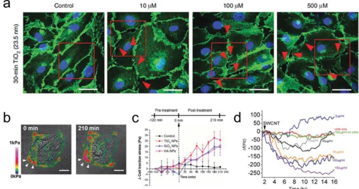
the vicinity of the cell–cell boundary (Figure 3b), indicating that oral mucosa cells (TR146) are highly contractile upon treatment with silica (SiO2), TiO2, and hydroxyapatite (HA) NPs.
The physicochemical properties of particles might be expected to dictate particular cell mechanics response(s). How-ever, to the best of our knowledge, there are no systematic reports accounting for the influence of NPs properties apart from size/dimension and dose on cell mechanics. Thus, it will be critical to introduce these parameters in the future bionano-mechanics research. Previously, the reduction of cell adhesion after exposure to TiO2 particles was reported in 2010 by Saldaña
and Vilaboa.[50] They demonstrated the reduction of FA area
and colocalization of paxillin FAs with phospho-focal adhesion kinase p-FAK (Tyr-397), which further led to a decrease in cell adhesive strength (i.e., a very rapid detachment of TiO2-treated
human osteoblastic Saos-2 cells during cell detachment assays).
However, in their in vitro experiment, only micrometer-size particles were used. In addition, Hou et al.[47] observed
size-dependent effects on adhesion dynamics, where less vinculin FAs were detected after MSC were treated with large (108 nm) TiO2 particles compared to smaller NPs (14 nm). In the other
direction, Pöttler et al. showed the dose-dependent adhesion after NP treatment. Cell adhesion was determined by counting the number of attached cells. Cells treated with SPIONs up to 20 μg cm−2 showed no significant alteration of adhesion for
rabbit vocal fold fibroblasts (VFF). However, exposure of 40 and 80 μg cm−2 SPIONs resulted in a significant decrease in VFF
adhesion.[51]
As briefly mentioned, exact mechanisms of how NPs alter the adhesion property of cells are still debatable. Mokhtari et al.[52] reported the significant inhibitory effects of
SPIONs-polyethylene glycol (PEG)-cisplatin on the adhesion of the human mammary gland T47D cells may be due to the inhi-bition of α2β1 integrin mRNA expression. Vieira et al.[53] and
Pan et al.[54] showed a reduction of ECM-associated protein
production (i.e., collagen and laminin) due to the presence of AgNPs, gold nanoparticles (AuNPs), and TiO2 NPs. Collagen
itself is an abundant ECM protein which provides binding site for FAs. Disruption of the expression of collagen might have direct influence on the structural integrity and function of actin stress fibers (nonmuscle cell contractile actin bundles) since these fibers are generally anchored to integrins which bind to secreted ECM proteins. Another possible explanation is the induction of cell death by NPs, since it is well known that dying cell lose their adherence.
Figure 3. NPs induce damage in cell adhesions. a) Confocal microscopy image of the VE-cadherin of HMVECs (green). Leaking between the cells (red
arrow) was observed in the TiO2 NP-treated sample in contrast to the control cells which showed no leaks. Reproduced with permission.[38] Copyright 2013,
Springer Nature. b) Traction force map of TR146 epithelial cells post SiO2 NPs exposure and c) real-time cell traction stress profile as a function of time
and type of NP treatments. Adapted with permission.[49] Copyright 2014, American Chemical Society. d) Time-dependent response of adherence of
mac-rophages on gold-coated QCM surface in the presence of different concentrations of SWCNTs. Reproduced with permission.[57] Copyright 2011, Springer.
Traction force microscopy (TFM) can be used to determine the strength of cell–ECM adhesion (or cell traction force/stress, CTF; see dedicated TFM part in section 3.1.2). In the absence of NPs, human dermal fibroblasts (HDF) exerted robust traction forces along their periphery (CTF > 500 Pa), indicating stronger adhesion. However, TiO2 NPs exposure resulted in reduced
cell area with weaker traction force (CTF < 300 Pa).[54] This
finding however, was not supported by results published later by Leong and co-workers.[49] They observed relatively strong
traction force applied by TR146 epithelial cells in the presence of SiO2, TiO2, and HA NPs (CTF > 100 Pa) in comparison to
the control sample (CTF < 75 Pa). Furthermore, they simulta-neously observed maturation of FAs and the promotion of an adhesive cell phenotype (Figure 3c,d).[49] Different values of the
traction force observed for those two cases can be attributed to the difference in cell type. For example, it has been shown that metastatic cells such as MDAMB231 human breast ade-nocarcinoma, PC3 human prostatic cell carcinoma, and A549 human lung epithelial carcinoma possess higher traction stress compared to their nonmetastatic counterparts, e.g., MCF10A human breast epithelial cell line, PrEC human primary prostate epithelial cells, and BEAS-2B human bronchial epithelial cells (CTF > 400 Pa vs CTF < 120 Pa).[55]
Distraction of cellular adhesion upon NPs treatment can be monitored using quartz crystal microbalance (QCM) measure-ments. QCM is a very sensitive nanogram mass sensing device and has been widely used to monitor and quantify adsorption and deposition of materials on the surface of a piezoelectric crystal. Any change of the crystal’s oscillation frequency due to dynamic processes occurring on the surface, including cellular adhesion and readhesion, can be detected. In the presence of human hepatoma HepG2 cells, Wei et al.[56] observed a quite
large shift of QCM frequency (≈700 Hz) due to a strong adhe-sion between the cells and the QCM surface, as well as the changes in mass and viscoelasticity of cells on the gold sub-strate at 0–4 h. Within the next phase (4–18 h), a slight decrease in frequency shift of the crystal indicated cell detachment, spreading and re-(adhesion). Introduction of AuNPs or a mix-ture of AuNPs and paclitaxel, a microtubule stabilizing drug, to the cells resulted in the decrease of the frequency shift (to ≈150 or 500 Hz for AuNPs and AuNPs and paclitaxel, respec-tively). This indicated more cell detachment. Wang et al.[57]
similarly used QCM to study the adhesion properties and cyto-toxic response of DH82 macrophages upon incubation with single-walled carbon nanotubes (SWCNTs). Without SWCNTs, no change in oscillation frequency was detected while for SWCNTs-treated macrophages a significant frequency decrease was observed (Figure 3e). They suggested that the difference of adhe-sion and migration properties of nonphagocytotic mode (in the absence of nanotubes) and phagocytotic macrophages (in the pres-ence of SWCNTs) could be responsible for the frequency change.
2.3. Effects of NPs on Cellular Cytoskeletal Structures
The cytoskeleton has several basic functions: (1) to organize the contents of the cell, (2) to physically and biochemically connect the cell to the environment, (3) to generate forces enabling the cell to move and change shape, and (4) to provide contractility
and help in cell division.[10,35,58] In eukaryotic cells, the
cytoskel-eton is made of three filamentous proteins, namely actin micro-filaments (filamentous actins or F-actins), microtubules (MT) and intermediate filaments (IF). F-actins are formed through polymerization of actin monomers (also called globular actin or G-actin), while MT are constructed from polymerization of alpha and beta tubulin, in which the nucleation of the polymerization is mediated by gamma tubulin.[35] IF on the other hand are
com-posed of different proteins that are expressed in variety types of cells, including keratins, vimentin, neurofilament proteins, nuclear lamins, and nestin.[59] In particular, nuclear lamins are
important for the structural properties of the nucleus (especially nuclear membrane/nuclear envelope), and the regulation of numerous nuclear processes including DNA replication, tran-scription and chromatin organization.[60]
In order to understand the effect of NPs on cytoskeletal integ-rity, we must first understand any possible physicochemical interactions between NPs and the main cytoskeletal proteins. Indirect interaction of NPs and cytoskeleton has been shown by Nienhaus and co-workers[31] where they demonstrated that
NP-carrying vesicles (i.e., intracellular quantum dots trapped in endosomes/lysosomes of HeLa cells) were actively trans-ported along MT with a speed of ≈0.4 μm s−1, similar to the
transport rate of kinesin. However, it is not known how NPs, which are normally compartmentalized inside endosomes/lys-osomes rather than freely “swimming” in the cytoplasm, could affect cytoskeletal structures. The only evidence comes from fibers such as carbon nanotubes (CNTs) which, depending on their aspect ratio, can be found inside the cytoplasma[61]
and could possibly directly interact with F-actin as proven by computer simulations.[62,63] Furthermore, Dawson and
co-workers[64] revealed the presence of intracellular tubulin (i.e.,
alpha and beta tubulin) in the protein corona formed around SiO2 NPs, and this finding has brought a growing interest in
the domain of NP-protein interaction. For example, by using different techniques such as dynamic light scattering, UV–vis spectroscopy, circular dichroism (CD) spectroscopy, hyperspec-tral imaging, and transmission electron microscopy (TEM), Wen et al.[65] probed the interaction between cytoskeletal
pro-teins and citrate-coated 30 nm AgNPs. Most likely, the NPs interact with these structural proteins through electrostatic, van der Waals, and hydrophobic interactions or hydrogen bonding. They also observed an increase in hydrodynamics size and zeta potential, a red-shift of the plasmonic band of AgNPs upon binding, as well as changes to the secondary structures of actin and tubulin which might affect their polymerization properties (Figure 4a,b). Similarly, Choudhury et al.[66] investigated the
binding and aggregation properties of tubulin heterodimers and the inhibition of MT polymerization in the presence of AuNPs in cell-free systems using UV–vis, CD, Raman and Fou-rier transform-infrared spectroscopies.
The interaction of actin/tubulin with NPs observed in vitro was found to have a rather negative impact on cytoskel-etal integrity. Although the numbers of studies is limited, the majority of findings showed destabilization and degradation of actin filaments in cell samples containing NPs. Pernodet et al.[67] demonstrated that “bioinert” AuNPs induced aberrant
F-actin formation. In vitro experiments using HDF showed the formation of actin “dots” rather than long stress fibers, a
reduction of F-actin fiber size and density, as well as a decrease in cell area by nearly a factor of two at all concentrations of AuNPs used (up to 800 μg mL−1). Next, Vieira et al.[53] reported
an alteration in cytoskeletal organization, including a change of cell polarity and the increase of F-actin fiber formation, in HDF treated with AuNPs and AgNPs. Another result sug-gested that exposure of zinc oxide (ZnO) NPs to RAW 264.7 mouse macrophages resulted in F-actin depolymerization and a decrease in the level of F-actins.[68] Fanarraga and co-workers[69]
observed a reorganization of actin microfilaments into cell bun-dles and formation of aberrant F-actin trails affecting prolifera-tion and viability of human keratinocytes (HaCat) and cervical cancer cells (HeLa) after ZnO NPs treatment (Figure 4c). It was speculated that the dissolution of ZnO in the lysosome to Zn2+
allowed the binding of Zn2+ to the actin network caused this
perturbation, since it is known that actin contain Zn2+ binding
sites and this binding could disturb the self-assembly of actin microfilaments.[69] Moreover, exposure of HBOEC to SPIONs
has caused significant F-actin remodeling, thus leading to a
decrease in cell area and polarization.[45] This study is in
agree-ment with previous reports by Wu et al.[48] and Gupta et al.[70]
in HUVEC and human fibroblast following treatment with SPIONs and gelatin NPs, respectively. Decrease or loss of F-actins itself could lead to destabilization of the cytoskeleton, cell architecture and furthermore induce permanent injury to the cells or even cell death.[71]
Apart from detrimental effects on the F-actin microfilament networks, NPs have been similarly shown to influence MT network polymerization and further induce MT destabiliza-tion and dysfuncdestabiliza-tion. For example, disorganized structures (including straightening, thickening, and shortening of MT) were observed in ZnO NP-treated HaCat and HeLa cells,[69]
and SPION-exposed endothelial cells.[45] A similar result
was found by Leong and co-workers,[49] whereby
internaliza-tion of SiO2, TiO2, and HA NPs by TR146 cells was shown
to lower the level of MT acetylation, destabilize the MT net-works and further affect the homeostatic regulation of intracel-lular tension. These impacts included the loss of intracelintracel-lular
Figure 4. Probing the interaction between NPs and cytoskeletal proteins. a) The red shift of AgNPs’ plasmonic band was induced by the formation of two
main cytoskeletal protein–AgNP corona. Plasmonic peak of naked AgNPs is 406 nm. b) Alteration in the secondary structures of actin and tubulin upon their binding with AgNPs. Adapted with permission.[65] Copyright 2013, The Royal Society of Chemistry. c) Confocal microscopy image showing aberrations in the
organization of the actin microfilaments (red channel) and microtubules (green channel), including F-actin spikes (red arrows) and MT straightening, thick-ening, and shortening (green arrows) post zinc oxide (ZnO) NP treatment. Reproduced with permission.[69] Copyright 2016, The Royal Society of Chemistry.
filamentous MT, an increase in F-actin remodeling, and the maturation (elongation) of vinculin FAs. Previously, it has been demonstrated that cell exposure to TiO2 particles led to
a disorderly arrangement of β-tubulin and acetylated α-tubulin fibers, reduction in the number of actin ventral stress fibers and a reduction in overall cell adhesion area.[50] Additionally,
NPs induced damage to the MT network not only through inhibition of MT polymerization, but also through aggrega-tion of tubulin heterodimers. Fluorescence microscopy data of NP-treated A549 and MCF-7 human breast cells showed that AuNP treatment destroyed MT, disrupted cell morphology, and shrank the cellular periphery while the control cells dis-played normal structure.[66] Through in situ MT
de-polymer-ization/re-polymerization experiments and quantification of fluorescence intensity of stained MTs in A549 cells, Gonzalez et al.[72] have demonstrated the decrease in MT acetylation
fol-lowing treatment with SiO2 NPs. In agreement with previous
result,[49] they demonstrated that MT were less acetylated after
NPs treatment, thereby indicating a lower level of MT stability. The change of the MT network in the presence of NPs, how-ever, does not always correspond to lower levels of microtubule acetylation. Apopa et al.[73] reported increased levels of MT
acetylation in human microvascular endothelial cells (HMEC) due to the induction of reactive oxygen species following expo-sure to SPIONs.
In neuronal cells, microtubule-associated proteins, namely tau proteins, play a major role in MT polymerization and axon growth.[74] In vitro interaction between tau proteins and NPs
such as iron NPs[75] and CNTs[76] have been previously described
in the literature. Based on a recent study in SH-SY5Y neuroblas-toma cells by Mao et al.,[77] the interaction between TiO
2 NPs and
tau proteins resulted in a decrease in MT density, disorganiza-tion, and disruption of MT. Furthermore, treatment of primary rat cortical neurons (PRCN) with AgNPs resulted in the loss of cytoskeleton components such as β-tubulins and F-actins, as well as the reduction of a number of synaptic clusters of the presynaptic vesicle protein synaptophysin, and the postsynaptic receptor density protein PSD-95. Exposure to AgNPs caused a reduction in β-tubulin branching and overlapping, a decrease in the degree and intensity of fluorescent phalloidin (a tool in the study of actin networks at high resolution), and finally cell death.[78]
2.4. Effect of NP Uptake on Cell Stiffness
Cell stiffness (or the opposite of cell elasticity) can be defined in simple terms as the resistance of the cell to an externally induced deformation. It is commonly expressed by the Young’s modulus (E), the ratio between the stress (force sustained by the sample over its cross section area) and the applied strain (with unit in Pascals).[79] Increase or decrease in cell
stiff-ness is often associated with a change in cell physiology and leading to a diseased state. Previous findings have revealed the role of the cytoskeleton in conferring stiffness and con-tractility to the cells,[80] as well as the relationship between the
cytoskeleton/cell stiffness in context of tumors/cancer malig-nancy. Malignant tumor cells for example, are found to be softer than benign and healthy ones (reviewed in ref. [9]). In healthy cells with a relatively high degree of stiffness, actin microfilaments are generally well organized, resulting in a larger Young’s modulus. In cancerous cells these organ-ized structures are not apparent or less observed, hence the cells are softer and more flexible.[18,79] Usually, cell stiffness
is measured through indentation experiments using atomic force microscopy (AFM; see dedicated part of AFM in Section 3.2.1).[81] Earlier we have described the detrimental effects of
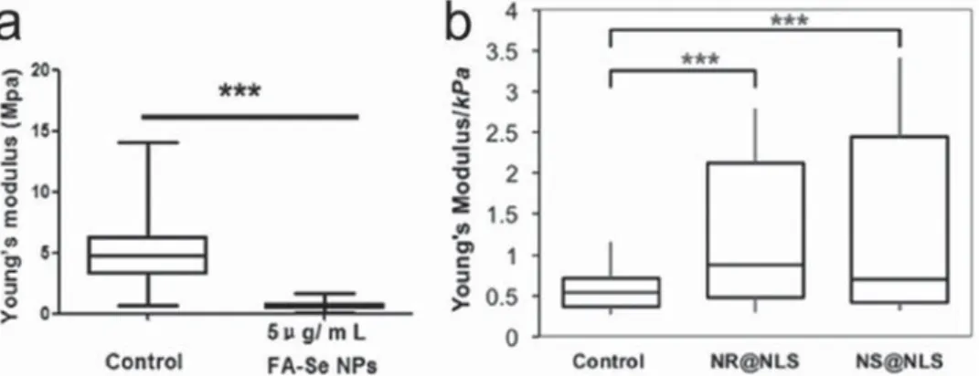
NPs on the F-actin microstructure, and as expected, the stiff-ness of the cells will be reduced by NPs. Pi et al.[82]
demon-strated that intracellular selenium (Se) NPs could induce membrane bio-mechanical property changes in MCF-7 cells by disturbing actin microfilaments and the transmembrane adhesion protein CD44. AFM experiment clearly indicated that control MCF-7 cells had a much higher Young’s modulus than SeNPs-treated MCF-7 cells (5.05 ± 2.43 MPa vs 0.69 ± 0.31 MPa), indicating that MCF-7 cells were much softer after Se NPs treatment (see Figure 5a).
The following studies, however, suggest the opposite effect. Thus, NP-induced changes to cell stiffness actually involve mechanisms that are more complex. Ogneva et al.[83]
reported an increase in cell stiffness of about 2.5 times for SiO2 NP-treated MSC compared to control cells (≈1 pN/nm
for control cell and 2.66 pN nm−1 for SiO
2 NP-treated MSC).
They claimed the changes might arise due to alterations in protein content (i.e., F-actin/G-actin ratio), followed by the reorganization of the cortical cytoskeleton and modification of
Figure 5. NPs impair cell stiffness. a) Comparison of Young’s modulus (E) values between control and SeNPs-treated MCF-7 cells. Reproduced with
permission.[82] Copyright 2013, Elsevier. b) Stiffness plots from single cells post nuclear-targeted AuNP and AuNR treatment. Reproduced with
permis-sion.[85] Copyright 2017, American Chemical Society.
cell surface. A transient increase of G-actin in the cytoplasm was attributed to the dissociation of F-actin to G-actin, which in turn initiated different signaling pathways. Using single cell compression-based AFM, Zimmer et al.[84] determined the
cel-lular stiffening of human aortic endothelial cells (HAEC) in the presence of ZnO NPs. The parameter for cell stiffening was expressed as relative deformation (ε) which is defined as the change in height of the cell over the initial height or rela-tive volume deformation (RVD), which is denoted as displaced volume over the initial cell volume. The lower the ε or RVD value, the higher the observed stiffness. They found out that at a low ZnO NP dose (i.e., 10 μg mL−1), cells were stiffer in
comparison to untreated cells. Increasing the concentration to 50 μg mL−1, however, led to significant changes in cell
mor-phology (i.e., cell swelling) and surprisingly a right-shifted RVD value indicating softening. They suggested that the higher uptake of ZnO by membrane and intracellular components would lead to heterogeneity of the cell membrane, ion flux dys-function (swelling), as well a weakening of the cytoskeleton. As opposed to ZnO, SiO2 NPs did not induce any similar response
even after the concentration was increased (50 μg mL−1) and
no detectable changes for both height and volume of the cells were observed upon NP treatment. These data however, contra-dict the previous conclusion by Ogneva et al.[83] Another related
example was shown very recently by El-Sayed and co-workers.[85]
They reported that nuclear membrane targeted AuNPs enhanced cell nucleus stiffness when introduced to HEY A8 ovarian cancer cells (Figure 5b). The localization of AuNPs and gold nanorods (AuNRs) in the nuclear membrane of the cells, due to targeting of the nuclear localization signal (NLS), led to the increased expres-sion of IF inner nuclear membrane lamin A/C protein, and fur-ther added mechanical stiffness to the cell nucleus. Berntsen et al.[86] also measured changes to the stiffness and contractility
of human airway smooth muscle cells (HASMC) after different NP exposure using a different technique, namely optical mag-netic twisting cytometry. They concluded that cell contractility was decreased by ZnO (40–100 nm) and copper(II) oxide (CuO; 50 nm) NPs treatment, while TiO2 NPs (25 nm) caused no effect.
2.5. Effect of NPs in Cell Motility
Cell migration/locomotion/movement/motility plays an impor-tant role in tissue formation during embryogenesis, wound
healing or immune response such as white blood cells move-ment to inflammation/injury sites. Impaired cell migration during all stages of development lead to severe embryonic malformations ranging from early embryonic lethality to birth defects and multiple human syndromes (e.g., neurological dis-orders, congenital heart diseases, and physical and mental retar-dation).[87] In order to migrate, there are four major steps cells
have to perform: (1) polarization of cell body (establishment of a front-to-rear polarity axis/leading-to-trailing edge), (2) protru-sion/extension of cellular membranes or lamellipodia, thin, sheet-like membrane protrusions located at the leading edge, (3) formation of new adhesions on the underlying ECM, and (4) translocation/cell body retraction. It has been shown that NPs are able to inhibit or alter the speed of the cell locomotion through different mechanisms. Previously, we have briefly mentioned that the cytoskeleton, mainly F-actins and MT, provides important roles and functions in cell motility; hence, any modification in cytoskeletal structure and function will affect the migration properties of the cells. Gonzalez et al.[72]
demonstrated that a decrease in MT acetylation reduced the speed of A549 cell movement following SiO2 NP treatment.
Disruption of F-actin and MT further retarded cell migration in cells following incubation with SPIONs[48] or ZnO NPs.[68]
A second pathway by which NPs inhibit cell migration is associated with cell adhesion. Strong traction forces and matu-ration of FAs will not only promote stable anchorage of the cells on the substrate, but also lead to a retardation of cell migration. Leong and co-workers[49] reported that SiO
2, TiO2 and HA NPs
treatment of TR146 cells promoted the destabilization of MT networks and the formation of mature vinculin FAs, increased cell traction and retarded collective cell migration. By using a scratch-based wound healing assay, they showed that NP-treated cells reduced cell migration in a dose-dependent manner by 60–70% with respect to nontreated cells (Figure 6a,b). Interest-ingly, cells pre-treated with monodansylcadaverine, a substance used to block the uptake of NPs, almost completely restored their migration ability indicating that the internalized NPs signifi-cantly retard cell migration.[49] However, it is worth mentioning
that with low adhesion, cells will not be able to exert enough force to pull the cells forward during the body retraction step.[88]
The latter has been shown by Hou et al.,[47] where treatment of
MSC cells with TiO2 significantly decreased the adhesion of the
cells (i.e., reduced vinculin FAs) and reduced cell migration. By using a matrigel invasion assay, they observed that the cells
Figure 6. NP influence on cell migration. a) Brightfield images show retardation of cell sheet migration of TR146 epithelial cells in the presence of SiO2
NPs. b) Displacement profile of the cells in the presence of three different types of NPs. Reproduced with permission.[49] Copyright 2014, American
Chemical Society.
treated with 14 nm NPs had a higher relative migration in com-parison with 108 nm TiO2 NP-treated cells.
The final mechanism by which NPs can inhibit cell migra-tion is by altering the expression of cell migramigra-tion-related proteins/molecules. Vieira et al.,[53] reported the reduction of
human fibroblast migration speed after AuNP and AgNP treat-ment, even at low concentration (1–10 μg mL−1): both particles
decreased the deposition of ECM by fibroblasts (i.e., a decrease in collagen and laminin deposition was observed), change the expression of ECM receptors in particular integrin-type receptors (very late antigen 2, α2β1 integrin (VLA-2) and very late antigen 6, α6β1 integrin (VLA-6)), and alter the cytoskeletal organization. AgNPs have been also shown to downregulate the expression of epidermal growth factor receptor (EGFR). Over-expression of EGFR is often associated to cancer prognosis and metastasis, and has been shown to enhance cell migration in human breast cancer cells and NIH3T3 fibroblasts.[89] Analysis
of EGFR expression in AgNP-treated A549 cells after over-stimulation with external EGF showed a significant decrease of EGFR mRNA expression, resulting in the retardation of cellular migration in a wound healing assay.[90] NPs were also able to
upregulate expression of microRNA miR-34a which has itself been shown to target and downregulate Ras-related C3 botu-linum toxin substrate 1 (Rac1) protein,[91] a protein involved in
actin polymerization and cytoskeletal organization, including the formation of lamellipodia.[92] Bhattacharya et al.[93] have
shown the thymoquinone-encapsulated PEG NPs could sig-nificantly increase the expression of miR-34a through tumor protein p53, and further down-regulated Rac1 expression fol-lowed by actin depolymerization. They observed a significant reduction in lamellipodia or filopodia (i.e., thin, spiky actin-rich plasma-membrane protrusions) formation on the surface of human breast cancer cell line MCF-7 and HBL-100, thus retarding cell motility. This finding however, did not support the previous results by Kang and co-workers[94] where they reported
that ZnO NPs caused activation of Rac1 and cell division control protein 42 homolog (Cdc42), the latter related to formation of filopodia in HUVEC.[95] Production of adenosine triphosphate
(ATP) in the mitochondria was also influenced by the presence of intracellular NPs. A recent report has described a mecha-nism whereby bovine serum albumin-coated AuNRs inhibited mitochondrial oxidative phosphorylation and glycolysis, which resulted in a major reduction of ATP production and subse-quently inhibited F-actin assembly. Formation of lamellipodia in AuNRs-treated NIH-3T3 fibroblast, B16F10 melanoma, and PC3 prostate cancer cells was repressed, and cell migration and invasion ability was reduced. Similar behavior was confirmed in vivo in a nude mouse model.[96] Expression of putative
ATP-dependent RNA helicase DEAH (Asp-Glu-Ala-His) box helicase 15 (or DHX15) in human gastric cancer cell lines (MKN28 and BGC823) has been found to increase after the cells were treated with cerium oxide NPs.[97] The increased expression of DHX15
could activate p38 mitogen-activated protein kinases (MAPK) signaling pathway,[98] further leading to an inhibition of
pro-liferation and metastasis of the cells.[99] Abrogation of MAPK
signaling pathway in AuNP-treated A2780 human ovarian car-cinoma cells was already observed previously by Mukherjee and co-workers,[100] and a reduced activation of vascular endothelial
growth factor receptor 2 (VEGFR2) which regulates endothelial
migration and proliferation[101] was also reported. Du and
co-workers[102] proposed that AuNPs were able to bind VEGF165
protein and indirectly reduce VEGFR-2 activation in HUVEC. Using a Transwell wound healing assay, they showed that cells which were treated with VEGF165 in the presence or absence of AuNPs possessed different migration properties: inhibition in cell migration in NPs-treated cells was monitored.
Contrary to many reported studies regarding cell motility retardation, Liu et al.[103] observed an increase in A549 and 95D
lung cancer cell migration activity after treatment with less than 10 nm size citrate-capped AuNPs. This may be associated with the increased expression of two key modulators of cell invasion, namely matrix metalloproteinase 9 and ICAM-1. High expres-sion of both proteins generally is connected to advanced stages of cancer. Separately, increased motility speed of Schwann cells (SC), a glial cell in the peripheral nervous system in the pres-ence of SPIONs was reported by Huang et al.,[104] however it
was only possible when alternating magnetic field was turned on. They observed an enhanced migration along the direction of magnetic field. SPIONs stimulated SC to cross the astro-cytes-SC boundary via integrin-mediated mechanotransduction.
2.6. Lessons Learned from NPs and Cellular Mechanics
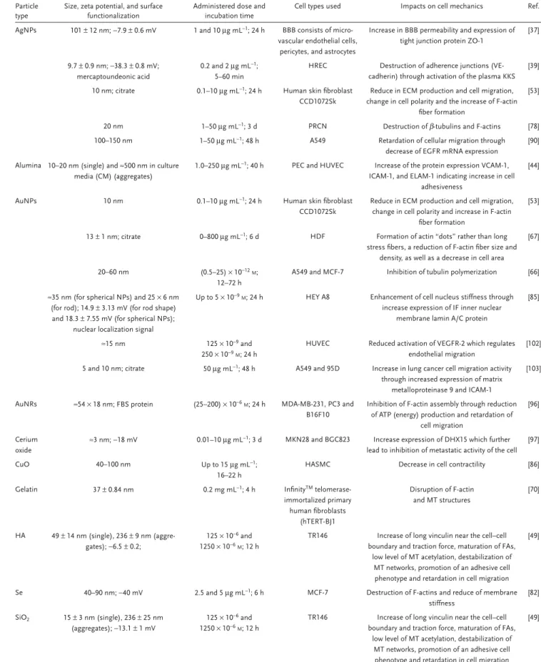
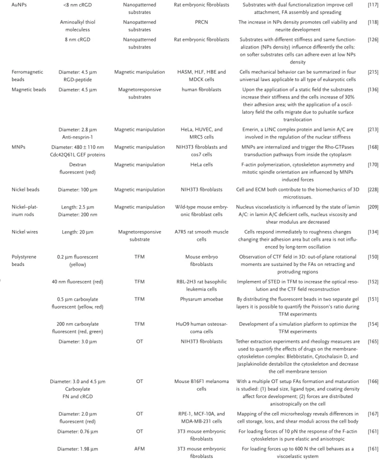
The number of in vitro and in vivo toxicity studies of NPs has risen dramatically over the years; however, the availability of infor-mation regarding alteration of cell mechanics due to interaction with NPs is still limited. Nonetheless, recent findings have shown that NPs can modulate the mechanical behavior of cells. The influence of different parameters including particle type, physico-chemical properties and cell culture conditions influencing cell mechanics in vitro is summarized in Table 1. The similarity we could observe is the fact that most of the administered NPs were mostly particles without any surface functionalization, and it is well-known that many of these nonfunctionalized particles have been shown to induce toxic effects in different cell types.[105–108]
In addition, we noticed that there is still not any systematic study regarding this topic. Moreover, most of our current knowledge comes from nanotoxicity studies where researchers are inves-tigating potentially adverse effects of NPs on cells/organisms. Therefore, there is potential for bias to view NPs as harmful, but in reality there is a necessity to perform more research about pos-sible effects of NPs on cell mechanics at subtoxic concentrations. In fact, the exact mechanisms of toxicity for each NP type are still incomplete,[105] and perhaps bionanomechanics could provide a
mechanistic understanding of this phenomenon. Even though it is too early to predict, we are convinced that in the future the effect of NPs in the context of cell mechanics can be combined with classical viability study to better understand and asses the hazard and safety of nanomaterials.
The field of bionanomechanics is in its nascent phases, therefore plenty of investigations and studies are possible. From the materials chemistry point of view, the influence of, e.g., surface functionalization, size, shape, concentration, etc. on cell mechanics still needs to be thoroughly investigated. From a biological point of view, the cell type needs to be con-sidered, especially to address whether effects on cell mechanics are cell-dependent or not. Information on any possible effects
Table 1. Cell mechanics responses depending on particle type.
Particle type
Size, zeta potential, and surface functionalization
Administered dose and incubation time
Cell types used Impacts on cell mechanics Ref.
AgNPs 101 ± 12 nm; −7.9 ± 0.6 mV 1 and 10 μg mL−1; 24 h BBB consists of
micro-vascular endothelial cells, pericytes, and astrocytes
Increase in BBB permeability and expression of tight junction protein ZO-1
[37]
9.7 ± 0.9 nm; −38.3 ± 0.8 mV; mercaptoundeonic acid
0.2 and 2 μg mL−1;
5–60 min
HREC Destruction of adherence junctions (VE-cadherin) through activation of the plasma KKS
[39] 10 nm; citrate 0.1–10 μg mL−1; 24 h Human skin fibroblast
CCD1072Sk
Reduce in ECM production and cell migration, change in cell polarity and the increase of F-actin
fiber formation
[53]
20 nm 1–50 μg mL−1; 3 d PRCN Destruction of β-tubulins and F-actins [78]
100–150 nm 1–50 μg mL−1; 48 h A549 Retardation of cellular migration through
decrease of EGFR mRNA expression
[90] Alumina 10–20 nm (single) and ≈500 nm in culture
media (CM) (aggregates)
1.0–250 μg mL−1; 40 h PEC and HUVEC Increase of the protein expression VCAM-1, ICAM-1, and ELAM-1 indicating increase in cell
adhesiveness
[44]
AuNPs 10 nm 0.1–10 μg mL−1; 24 h Human skin fibroblast
CCD1072Sk
Reduce in ECM production and cell migration, change in cell polarity and increase in F-actin
fiber formation
[53]
13 ± 1 nm; citrate 0–800 μg mL−1; 6 d HDF Formation of actin “dots” rather than long
stress fibers, a reduction of F-actin fiber size and density, as well as a decrease in cell area
[67]
20–60 nm (0.5–25) × 10−12M;
12–72 h
A549 and MCF-7 Inhibition of tubulin polymerization [66] ≈35 nm (for spherical NPs) and 25 × 6 nm
(for rod); 14.9 ± 3.13 mV (for rod shape) and 18.3 ± 7.55 mV (for spherical NPs);
nuclear localization signal
Up to 5 × 10⁻⁹M; 24 h HEY A8 Enhancement of cell nucleus stiffness through
increase expression of IF inner nuclear membrane lamin A/C protein
[85]
≈15 nm 125 × 10−9 and
250 × 10−9M; 24 h
HUVEC Reduced activation of VEGFR-2 which regulates endothelial migration
[102] 5 and 10 nm; citrate 50 μg mL−1; 48 h A549 and 95D Increase in lung cancer cell migration activity
through increased expression of matrix metalloproteinase 9 and ICAM-1
[103]
AuNRs ≈54 × 18 nm; FBS protein (25–200) × 10−6M; 24 h MDA-MB-231, PC3 and B16F10
Inhibition of F-actin assembly through reduction of ATP (energy) production and retardation of
cell migration
[96]
Cerium oxide
≈3 nm; −18 mV 0.01–10 μg mL−1; 3 d MKN28 and BGC823 Increase expression of DHX15 which further
lead to inhibition of metastatic activity of the cell [97]
CuO 40–100 nm Up to 15 μg mL−1;
16–22 h
HASMC Decrease in cell contractility [86]
Gelatin 37 ± 0.84 nm 0.2 mg mL−1; 4 h InfinityTM
telomerase-immortalized primary human fibroblasts (hTERT-BJ1 Disruption of F-actin and MT structures [70] HA 49 ± 14 nm (single), 236 ± 9 nm (aggre-gates); −6.5 ± 0.2; 125 × 10−6 and 1250 × 10−6M; 12 h
TR146 Increase of long vinculin near the cell–cell boundary and traction force, maturation of FAs,
low level of MT acetylation, destabilization of MT networks, promotion of an adhesive cell phenotype and retardation in cell migration
[49]
Se 40–90 nm; −40 mV 2.5 and 5 μg mL−1; 6 h MCF-7 Destruction of F-actins and reduce of membrane
stiffness [82] SiO2 15 ± 3 nm (single), 236 ± 25 nm (aggregates); −13.1 ± 1 mV 125 × 10−6 and 1250 × 10−6M; 12 h
TR146 Increase of long vinculin near the cell–cell boundary and traction force, maturation of FAs,
low level of MT acetylation, destabilization of MT networks, promotion of an adhesive cell phenotype and retardation in cell migration
[49]
on cell-basal lamina adhesion (i.e., hemidesmosomes, FAs) and cell–cell adhesion (i.e., desmosomes, adherens junctions) are needed to draw a general idea of the impact of NPs on cell adhesion. The study of NP interactions with IF and cytoskeletal mechanosignaling pathways needs to be addressed. In addition, theoretical studies using suitable biophysical models for reca-pitulating NPs and cell mechanics should be performed.
A general conclusion of this previous chapter is presented as follows:
Take-Home Messages:
• Cell mechanics plays important role in organism develop-ment, physiology and diseases.
• Ag,[37,39] alumina,[44] HA,[49] SPIONs,[45,51] SiO
2,[49] and TiO2
NPs[38,47,49] can induce detrimental effects on cell adhesion
including destruction of tight and adherens junctions and modulation of cell–ECM adhesion (i.e., reduction and matu-ration of FAs).
Particle type
Size, zeta potential, and surface functionalization
Administered dose and incubation time
Cell types used Impacts on cell mechanics Ref.
59–174 nm; −7.74 to −17 mV 7.5–211 μg mL−1; up
to 40 h
A549 Decrease in MT acetylation and migration velocity
[72]
7 nm 50–100 μg mL−1; 1–24 h MSC Increase in cell stiffness ≈2.5-fold than control
cell
[83]
SPIONs 132.4 nm; −25.37 mV; 20, 40, and 80 μg cm−2;
24 h
VFF At higher concentration (e.g., 40 and 80 μg cm−2
SPIONs) resulted in a significant decrease in VFF adhesion
[51]
4–14 nm; −31.3 ± 7.3 mV; lipids, dextran, carboxydextran and citrate
500–1000 μg mL−1;
4 and 24 h
HBOEC Decrease of vinculin FA area and number as well as increase in cell polarization, reduction of focal adhesion kinase (FAK) and F-actin remodeling
[45]
<10 nm 12.5–100 μg mL−1; 5 h HMEC Increase in MT acetylation and endothelial cell
permeability
[73] TiO2 23.5 nm (single) and 57.1 nm (aggregates) 10 × 10−6M; 30 min HMVEC Endothelial cell leakiness through degradation
of VE–cadherin
[38] 14, 108, and 196 nm 50, 100, and 200 μg
mL−1; 3, 7, and 14 d
MSC Reduce of expression of vinculin FA adaptor and cell migration [47] 21 ± 8 nm (single), 272 ± 4 nm (aggregates); −7.1 ± 1 mV 125 × 10−6 and 1250 × 10−6M; 12 h
TR146 Increase of long vinculin near the cell–cell boundary and traction force, maturation of FAs,
low level of MT acetylation, destabilization of MT networks, promotion of an adhesive cell phenotype and retardation in cell migration
[49]
3.32 ± 2.39 μm 0.5–2.5 mg mL−1; 1–24 h Human osteoblastic
Saos-2 cells
Reduction of paxillin FA area and decrease in cell adhesive strength, disorderly arrangement of
β-tubulin and acetylated α-tubulin fibers, reduc-tion in the number of actin ventral stress fibers
and in overall cell adhesion area
[50]
15.0 ± 3.5 nm (single NP) and 200 ± 13 nm (aggregates)
0.4 and 0.8 μg mL−1; 30 min to 2 d
HDF Decreases in cell area, traction force, cell prolif-eration, mobility, and ability to contract collagen
[54] 20.90 ± 3.57 nm (single) and
110.0 ± 72.9 nm (aggregates in CM); −0.73 ± 1.27 mV in CM and −142.56 ± 19.80 mV in water
0.1–100 μg mL−1; 24 h SH-SY5Y Disorder, disruption,
retraction, and decreased intensity of MT
[77]
ZnO 200–250 nm; −0.56 mV Up to 500 μg mL−1; 24 h Mouse macrophage RAW
264.7
Depolymerization and degradation of F-actin and inhibition of cellular migration
[68] 86 ± 3 nm 15–100 μg mL−1; 24–96 h HaCat and HeLa Reorganization of actin microfilaments into cell
bundles, formation of aberrant F-actin trails and rigid macrotubes (i.e., straightening, thickening
and shortening) of MT
[69]
100–200 × 20–70 nm 10 and 50 μg mL−1; 4 h HAEC Increase of cell stiffness at low dose
(e.g., 10 μg mL−1) and decrease
of cell stiffness at higher dose (50 μg mL−1)
[84]
40–100 nm Up to 15 μg mL−1 HASMC Decrease in cell contractility [86]
93.35 ± 14.53 nm (single) and 126.2 ± 120.4 nm (aggregates)
0.1–100 μg mL−1; 24 h HUVEC Activation of Rac1 and Cdc42 protein which
induce formation lamellipodia and filopodia [94]
Table 1. Continued.
• Interaction of Ag,[37,53] HA,[49] SPIONs,[45] TiO
2,[49] and
ZnO[68,69] NPs and cytoskeletal proteins (actin and tubulin)
can promote destabilization of the cytoskeleton including re-modeling and destruction of MT and F-actins structures. • Cell stiffness increases due to the presence of Au[85] and SiO
2
NPs.[83]
• NPs can retard cell migration through destruction of the cytoskeleton,[68,72] increase in adhesions[49] and
modulating the expression of cell migration-related proteins/ molecules.[53,90,93,96,102]
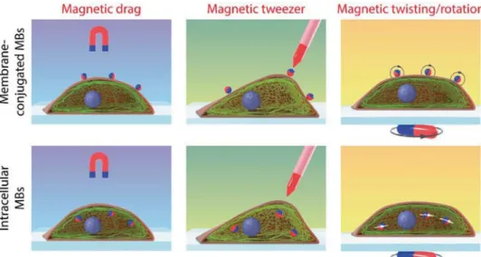
3. Particles as a Tool to Study Cells Mechanics
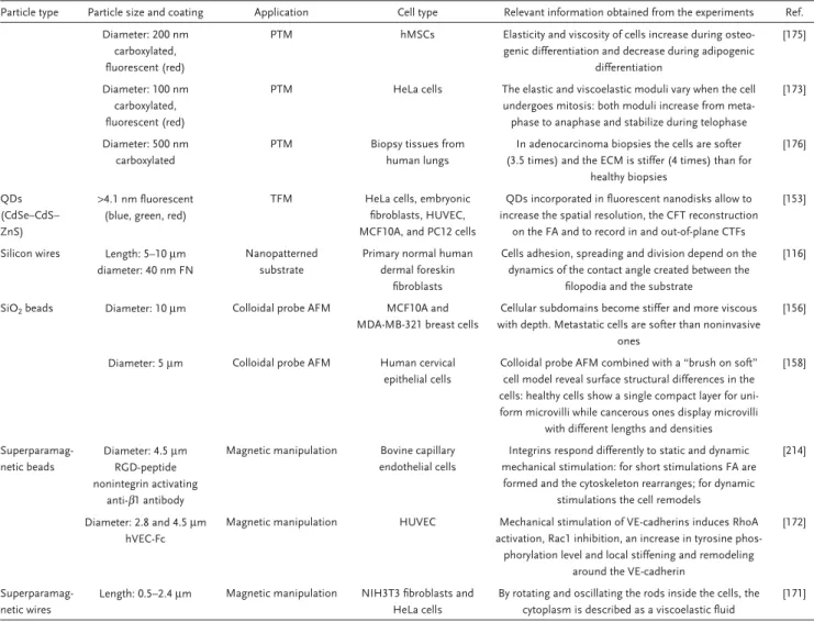
Because the study of how particle properties can have an impact on cell mechanics is an emerging field, there is a need for tools to investigate cell mechanics behavior. To this point, rapid progress in the field of particle-based technolo-gies has enabled the utilization of particles as tools to study a myriad of cellular phenomena: Particles, because they can directly interact with subcellular structures, are ideal candidates to study cell mechanics. Fluorescent, metallic/ plasmonic or magnetic micro- and NPs are widely used materials for sensing, and current efforts have been dedi-cated to implementing particles in cells as in vitro probes. Particle technologies have enabled researchers to develop new methods and probe cellular phenomena/mechanical properties (e.g., cell forces exerted on the ECM, cell stiffness and cytoplasmic viscosity, strength of cellular interactions, and translation of mechanical forces into biochemical sig-nals). To be certain, studying the influence of particles on cell mechanics is tied to the utilization of particles to study these very same processes. For example, particle-based tech-niques offer a valid method to directly investigate how inter-nalized NPs alter cell stiffness and modify cellular forces. Thus, we describe here the (emerging) methods by which particles can act as tools to study cellular mechanics and cell mechanical properties.3.1. Micro and NPs to Study the Interaction between Cells and the ECM
Nanotechnology-driven tools including microscopy and (micro and nano)particles-based characterization techniques have been widely used to probe cell–substrate/ECM interac-tion, i.e., the effect of the substrate on cellular behavior and the force that the cells exert on the substrate (traction force). Synthetic surfaces/substrates are used to mimic the ECM, thus allowing concomitant study of the interaction of the cells with their external environment and how this affects cell physiology and behaviors. The forces generated by the cells during adhesion, spreading and migration can be quantified, for example, by using TFM, a technique that strongly relies on the displacement of micro and nanotrackers embedded in elastic substrates. To ease the understanding in TFM, we wish first to provide information regarding the use of NPs in and as cell substrates.
3.1.1. NPs in Cell Substrates
In tissues, cells attach to the ECM through cell–ECM adhesion. Apart from its biochemical and structural properties, physical features of ECM such as its rigidity, density, porosity, insolu-bility and topography (i.e., spatial arrangement and orienta-tion)[109] are very important aspects which dictate cell–ECM
response.
In fact, it is well-known that the nanotopography of a surface can play a significant role in cell development, cell mechanics and migration.[29,110] It is therefore intuitive that
nanoscale objects can be regarded as an alternative platform to mimic the ECM structures. Furthermore, by incorporating NPs onto a surface, it is possible to control and engineer spe-cific surface geometry/physical characteristics. To this point, ECM-like surfaces can be fabricated by creating synthetic substrates with defined patterns of specifically functional-ized NPs. Thus, beyond exploiting the possible benefits of nano and micro particle-decorated surfaces, these particles can be used as a tool to understand the mechanism behind cell adhesion and spreading,[111–119] migration[120,121] and
differentiation.[122–125]
In order to realize particle-based substrates as a surface for studying cell mechanics, particles must frequently be function-alized to provide selective adhesion. ECM ligands (arginine-gly-cine-aspartic acid (RGD)-based motifs) linked to the NPs surface are often used to promote cell adherence.[114–116,120,122,126]
How-ever, a single binding motif alone is not guaranteed to promote cell–substrate interaction, FA assembly, and spreading. Rather, this action is mediated by ligand interaction, particle spacing and ligand density. Schenk et al.[117] developed a substrate
func-tionalized with both cyclic RGD (cRGD)-coated AuNPs and Pro-His-Ser-Arg-Asn (PHSRN) peptides (interspaced on the substrate via a PEG molecule) which synergistically enhanced cell spreading. In vitro experiment using rat embryonic fibro-blasts showed differences in the adhesion behavior on sub-strates functionalized with only PHSRN, cRGD-coated AuNPs, or both ligands. In particular, the two ligands alone did not promote cells-substrate interaction while their combination boosted cells attachment, FA assembly, and spreading.[117]
Another basic aspect that regulates the interaction between the cells and the external environment is the spacing and density of the surface ligand. To obtain stable FAs, the distance between the integrins and ECM ligands must be close enough. This dis-tance varies among different cell lines: for example by using functionalized AuNPs as the ECM-like substrates for rat embry-onic fibroblast cells, this distance was shown to be ≤58 nm,[114]
while for human breast epithelial metastatic cancer cells the distance between cRGDfk-functionalized AuNPs was relatively large (≈1.7 μm).[115]
Apart from a specific surface functionalization, particle density is also essential. Recently Li et al.[118] investigated the
effect of substrates’ particle density and their composition on neuron adhesion and neuritogenesis. They observed an increase in viability and neurite development for PRCN upon an increase in the concentration of amine-functionalized AuNPs (from 1 to 490 particles μm−2; Figure 7a). Rigidity of
the substrate on which particles are patterned on has been similarly shown to affect cell adhesion. Advanced work by
Platzman et al.[126] demonstrated that cells which were cultured
on two substrates having different stiffnesses, but similar sur-face functionalization, possessed different mechanical behav-iors. In this work, 8 nm cRGD coated AuNPs were patterned on PEG-passivated glasses and soft PEG-diacrylate hydrogels. The patterns were hexagonal and the lateral distance between the particles was varied between 62 and 161 nm. They observed that ligand spacing and stiffness of the substrate influenced the adhesion of rat embryonic fibroblast cells, in particular the cells only attached on the substrate with a spacing of 62 nm for both glass and hydrogel substrates, while for higher spacing distances they could adhere and spread only on the hydrogel. They hypothesized that the observation came from the ability to deform the hydrogel substrate and reduce the distance between particles.
Another important aspect for cell adhesion is the interac-tion between filopodia and ECM-induced nanotopography, as recently addressed by Albuschies and Vogel.[116] In their work,
nanoengineered substrates were used to understand how the filopodia interact with nanometric structures. Flexible
hairy silicon nanowires were grown from gold seeds on a micropatterned glass surface (Figure 7b). The wires and the flat surface were both functionalized with fibronectin to avoid results biased by different ligand densities. By analyzing the adhesion dynamic of primary normal human dermal fore-skin fibroblast, they concluded that filopodia steered adhe-sion, spreading, and division of the cells depending on the dynamics of the contact angle formed between filopodia and the substrate. On the semiflexible nanowires the filopodia aligned and adhered strongly while on the rigid flat glass, they were more prone to peel off. In the latter case, only the small portion of filopodia that created a low angle with the surface could be stabilized. A new biophysical model called the molec-ular zipping model was proposed to explain how filopodia adapt to the topography (Figure 7c): the filopodia probe the surface by applying traction forces on the FA, and the contact angle between the filopodium and the substrate determines whether there is adhesion or if the filopodium peels off. In particular they calculated a maximum contact angle in order to still have a strong adhesion (<12°); at higher contact angles
Figure 7. NPs mimicking the ECM are used as cell culture substrates. a) A scheme of the surface functionalization with AuNPs; scanning electron
microscopy (SEM) images of surface functionalization at different coating densities and live-and-dead fluorescent staining image of neurons seeded on the surfaces. Reproduced with permission.[118] Copyright 2015, The Royal Society of Chemistry. b) SEM image of islands patterned with silicon
nanowires. Scale bar 5 μm. c) The molecular zipping model schematic of the force distributions on integrins as a function of adhesion angle (α). Adapted with permission.[116] Copyright 2013, Springer.