Original article
Short fluorodeoxyuridine exposure of different human
glioblastoma lines induces high-level accumulation of S-phase
cells that avidly incorporate
125I-iododeoxyuridine
Florence Perillo-Adamer1, Angelika Bischof Delaloye1, Céline S. Genton2, Andreas O. Schaffland1, Yves M. Dupertuis3,
Franz Buchegger1, 4
1Service of Nuclear Medicine, University Hospital of Lausanne, Rue du Bugnon 46, CH-1011 Lausanne, Switzerland 2Department of Biochemistry, University of Lausanne, Epalinges, Switzerland
3Service of Nutrition, University Hospital of Geneva, Geneva, Switzerland 4Service of Nuclear Medicine, University Hospital of Geneva, Geneva, Switzerland
Received: 6 June 2005 / Accepted: 15 September 2005 / Published online: 1 February 2006 © Springer-Verlag 2006
Abstract. Purpose: Radio-iododeoxyuridine (IdUrd) is a potential Auger radiation therapy agent incorporated into DNA during the synthesis phase. In this study we sought to optimise S-phase targeting by modulating cellular cycling and radio-IdUrd DNA incorporation using short non-toxic fluorodeoxyuridine (FdUrd) incubations. Methods: Three human glioblastoma cell lines with different p53 expression were pre-treated with various
FdUrd conditions. After different intervals, 125I-IdUrd
DNA incorporation was measured. Fluorescence-activated cell sorter cell cycle analysis was performed after identical intervals post FdUrd pre-treatment.
Results: The highest increase in 125I-IdUrd DNA
incor-poration was induced by 1-h incubation with 1μM FdUrd.
Increase in radio-IdUrd DNA incorporation was greatest
16–24 h after FdUrd, reaching factors of ≥7.5 over
baseline incorporation in the three cell lines. Furthermore, cell synchronisation in S phase was observed with a peak
of ≥69.5% in the three cell lines at 16 and 24 h post
FdUrd, corresponding to an increase of 2.5–4.1 over baseline.
Conclusion: FdUrd-induced thymidine synthesis inhibi-tion led to S-phase accumulainhibi-tion that was maximal after an
interval of 16–24 h and time-correlated with the highest
radio-IdUrd DNA incorporation. These observations might allow the rational design of an Auger radiation therapy targeting a maximal number of S-phase cells in single treatment cycles.
Keywords: Iododeoxyuridine– Fluorodeoxyuridine –
S phase – Glioblastoma – Auger radiation
Eur J Nucl Med Mol Imaging (2006) 33:613–620 DOI 10.1007/s00259-005-0009-y
Introduction
Iododeoxyuridine (IdUrd), a thymidine (dThd) analogue,
labelled with123I or125I, is an interesting Auger radiation
therapy agent [1]. However, two major limitations restrict
its DNA incorporation and therapeutic efficacy: firstly, radio-IdUrd DNA incorporation is low because of compe-tition from the endogenous nucleotide pool and a short biological half-life attributable to rapid in vivo catabolism
and dehalogenation [2,3], and secondly, therapeutic IdUrd
targeting is restricted to cells in the synthesis phase (S phase) of the cell cycle. Thus, for the short period of its in vivo availability, only cells in S phase can be efficiently targeted with radio-IdUrd.
In order to favour DNA incorporation of IdUrd, different groups, including ours, have studied inhibition of the de novo synthesis of dThd with agents such as
fluorodeoxyuridine (FdUrd) [4–6], 5-fluorouracil [7] or
methotrexate [7]. FdUrd, a dThd analogue, inhibits
thymidylate synthase (TS) and depletes the TTP pool by
acting as non-convertible substrate [8]. The depletion of the
cellular TTP pool favours incorporation into DNA of
exogenous dThd and dThd analogues such as IdUrd [4–7].
FdUrd has been shown to have an extended effect on TS inhibition even after its removal in the human colon
carcinoma cell line HT 29 [9]. Clinically, push
adminis-tration of FdUrd, used in the early 1960s, is also well
tolerated at high doses [10]. However, prolonged exposure
to low doses of FdUrd leads to clonogenic death of HT29 Florence Perillo-Adamer ())
Service of Nuclear Medicine, University Hospital of Lausanne, Rue du Bugnon 46,
CH-1011 Lausanne, Switzerland e-mail: Florence.Adamer@chuv.ch
cells [11]. Our recent studies [6,12] have shown that short i.v. treatment of animals with FdUrd allows a significant improvement in tumour DNA incorporation of radio-IdUrd. However, while different groups have shown that FdUrd pre- or co-treatment results in the expected increase
in incorporation of radio-IdUrd into DNA [4–7], a
corresponding improvement in the efficacy of treatment
has not been observed [4].
Due to the absence of improved efficacy of the different Auger radiation therapy approaches with radio-IdUrd (also evident in personal unpublished observations), we ad-vanced the hypothesis that the limitation of S-phase targeting has not been addressed adequately. In fact, our previous observation of increased radio-IdUrd DNA
incorporation immediately post FdUrd treatment [6, 12]
still targeted only the very limited number of cells in S phase at radio-IdUrd incubation in vitro or injection in vivo. Therefore, in this study we investigated whether S-phase accumulation might be achieved in different glio-blastoma cell lines. If S-phase accumulation and simulta-neous high efficacy of radio-IdUrd DNA incorporation could be obtained, we would expect these combined effects to allow the design of a more rational Auger radiation therapy approach.
Materials and methods Reagents
125
I-IdUrd of high radiochemical purity was prepared from the precursor 5-tri-n-butyl-stannyl-2'-deoxyuridine and purified by SepPak RP-18 cartridges using a modified method [13] described originally by Foulon et al. [14]. Radiochemical purity was repeatedly verified with thin-layer chromatography using polyethyleneimine cellulose F strips (Merck, Darmstadt, Germany) with a mobile phase of 0.2 M ammonium bicarbonate and radioactivity reading on a Tracemaster 20 (Berthold, Bad Wildbad, Germany). In all experi-ments,125I-IdUrd represented >98% of overall125I activity. In the production of125I-IdUrd, we were able to avoid contamination with unlabelled IdUrd [13]. It was calculated that the standard activity of 5 kBq 125I-IdUrd in 0.5 ml used in the incorporation experiments represented a concentration of 0.2 nM, though this is only an indicative value as it has not been experimentally verified. FdUrd and dThd were obtained from Sigma-Aldrich Chemie GmbH (Steinheim, Germany). Stock solutions were prepared in distilled water at a concentration of 5 mM and stored at −80°C. Unlabelled IdUrd (Sigma-Aldrich Chemie GmbH) was dissolved in physiological NaCl using NaOH at minimally required concentration and stock solution (50 mM) stored at−80°C
Cell lines and culture
Three human glioblastoma cell lines were chosen for their different p53 expression [15] that could potentially provide them with different repair capacities or FdUrd resistance [16]: U251 and U87 (American Type Culture Collection, ATCC) homozygous mutant and homozygous wild type, respectively, and the heterozygous line LN229 (gift from Prof. N. de Tribolet, Service of Neurosurgery, University Hospital, Geneva, Switzerland). Cells were cultured at 37°C and 5% CO2 in RPMI 1640 with glutamax I, supplemented
with 10% heat-inactivated fetal bovine serum (Chemie Brunschwig AG, Basel, Switzerland), penicillin (50 units/ml) and streptomycin (0.05 mg/ml; Life Technologies Inc., Grand Island, NY, USA), which was changed twice a week. Cell doubling times in culture flasks as measured by daily counting over 1 week were 24.6±2.3 h, 33.7±3.4 h and 34.0±0.2 h for U251, LN229 and U87, respectively (n≥3). Because mycoplasma contamination is a potential source of in-creased nucleoside turnover [17,18] and could therefore modulate FdUrd efficacy, all cells were tested negative for four major poten-tial mycoplasma contaminations (Mycoplasma Detection Kit, F. Hoffmann-La Roche Ltd, Basel, Switzerland), and maximal cell culture time after testing was 6 months.
125
I-IdUrd incorporation assay
To assess the effect of FdUrd on125I-IdUrd incorporation into DNA, 24-well culture dishes were plated with 20,000 cells for LN229 and U87, and with 15,000 cells for U251. After 4 days, the exponentially growing cells were pre-treated for varying times and with varying doses of FdUrd, and washed twice with PBS (NaCl 0.15 M, phosphate 0.01 M, pH 7.4) before fresh medium was added. After different intervals of 0–72 h, cells were incubated for 2 h at 37°C with 5 kBq125I-IdUrd in 0.5 ml medium. In a time course experiment with LN229 and U251 cells, we had observed that after incubation of cells for 2 h, the large majority of intracellular radio-IdUrd was incorporated into DNA (results not shown). In accordance with published protocol [4,19], cells were washed once with complete medium, twice with PBS and then recovered in 5-ml plastic tubes after incubation for 10 min with 200 μl of trypsin-EDTA (Life Technologies Inc.). Wells were washed with 200 μl complete medium and this wash was also added to the cells. Thereafter, cells were lysed with 400μl of 1 M NaOH. The DNA was precipitated with the addition of 400 μl of 10% trichloroacetic acid (Sigma-Aldrich Chemie GmbH) and the total volume of 1.2 ml centrifuged for 30 min at 3,500 g. A half volume was separated as one-half supernatant, and the remaining half volume (0.6 ml) was considered as sediment (containing DNA) plus another half supernatant. Radioactivity was determined in a gamma counter (Wizard 3” 1480 automatic gamma counter, Wallac Oy, Turku, Finland) and results expressed for total cellular uptake (sediment + supernatant) of radio-IdUrd and for DNA-incorporated activity (sediment alone). All results given here represent DNA-incorporated radioactivity that was generally >85% of cell-bound activity. In all timing experiments, cells were counted in identically treated control wells and DNA-incorporated activity was normalised according to the number of cells.
Fluorescence-activated cell sorter (FACS) analysis
Cells were analysed by two-parameter flow cytometry measuring both DNA content by propidium iodide (PI) (Fluka Chemie AG, Buchs, Switzerland) and labelling index with IdUrd [6]. Exponen-tially growing cells were either pre-treated or not with 1μM FdUrd for 1 h, washed with PBS, incubated in fresh medium for 0–72 h and labelled with 10μM non-radioactive IdUrd for 1 h. For LN229 and U251 cells, one of three experiments was performed with 3 μM IdUrd. Cells were then washed twice with PBS, trypsinised, centrifuged for 15 min at 500 g, resuspended in PBS, diluted and fixed as aliquots of 106cells in 4 ml 70% cold ethanol for 30 min on ice. Cells were centrifuged for 10 min at 500 g and 4°C. The DNA was denatured by incubating the cells in 1 ml 2 M HCl/0.5% BSA/ PBS for 30 min at room temperature. Cells were centrifuged, and acid cell sediment was neutralised by resuspension in 1 ml of 0.1 M sodium borate at pH 8.5. After centrifugation for 10 min at 500 g,
cells were either stored at −20°C or directly prepared for FACS analysis by incubating them for 30 min at room temperature in 50μl 0.5% Tween 20/0.5% BSA/PBS containing 0.5 μg of mouse monoclonal antibody against anti-BrdUrd (Dako A/S, Glostrup, Denmark) cross-reacting with IdUrd. After incubation, 1 ml PBS was added before centrifugation (10 min at 500 g and 4°C). Then, the pellet was resuspended in 50μl of 0.5% Tween 20/0.5% BSA/PBS with 1μg of anti-mouse IgG conjugated with FITC (Sigma-Aldrich Chemie GmbH) and incubated for another 30 min at room temperature. Cells were washed with 1 ml PBS, centrifuged and resuspended in 1 ml of PBS containing 2.5μg/ml PI. Samples were incubated for 16–20 h at 4°C before analysis on a flow cytometer (FACScan; Becton Dickinson, Franklin Lakes, NJ, USA) with a laser excitation at 488 nm for PI and FITC fluorescence.
External beam irradiation and cell survival assay
Twenty-four well culture dishes were plated with 20,000 and 15,000 cells for LN229 and U251, respectively, 4 days prior to treatment. After pre-treatment for 1 h with 1μM FdUrd, cells were washed, re-incubated with fresh medium and irradiated at the times indicated in a photon beam (6 MeV Primus Linear Accelerator, Siemens AG, Munich, Germany) at doses of 2 or 6 Gy. The colony-forming assay was then used to measure the cell survival fraction: cells were trypsinised, resuspended in RPMI 1640 and counted. The cell suspension was serially diluted and 200 or 2,000 viable cells were plated into 100×20 mm tissue culture dishes in 10 ml fresh culture medium. After 14 days of incubation at 37°C and 5% CO2, grown colonies were fixed and stained with 0.5% crystal violet in methanol/ acetic acid (3:1, v/v). Colonies containing≥50 cells were counted. Each experiment was performed using triplicates and repeated at least three times. The survival fraction (S/S0) was calculated as the plating efficiency of irradiated pre-treated cells divided by the plating efficiency of corresponding unirradiated pre-treated cells. The colony-forming assay cannot be performed in liquid medium with U87 cells because they form spheroids and can detach. For this reason, this line was not tested in this assay.
Statistical analysis
Statistical multi-group comparisons were performed with the ANOVA multiple range Tukey-HSD test at different confidence levels using the UNISTAT 5.5 statistical package for Windows (2002 edition, Unistat Ltd, London, UK, run on Windows XP). The Tukey-HSD test applies the largest range value independent of the effective subset size; it is more conservative than the Student-Newman-Keuls test but less conservative than the Scheffé or the Bonferroni test.
10 0.001 1 10 30 100 + dThd 0 1 2 3 4 5 6 7 8 9 0 0.003 0.01 0.03 0.1 0.3 3 100 0 + dThd LN229 U251 U87
Relative increase in cooperation (Io/I)
FdUrd concentration (µM)
Fig. 1. Increase in DNA incorporation (I/Io) of125I-IdUrd according to the concentration of FdUrd. Cells were co-incubated with 5 kBq 125I-IdUrd and with/without FdUrd (0.001–100 μM). Results (mean±1 SD) are from three independent experiments, each performed with three samples per test condition. Results are normalised to control samples incubated for 4 h with radio-IdUrd alone. Additional controls of incorporation were performed by competition with 50μM thymidine (+dThd) 15 ** ** ** ** ** ** ** ** ** ** ** ** * * 0 5 10 0 5 10 15 0 5 10 15 0 24 48 72
Normalised relative increase in
125
I-IdUrd DNA incorporation
Time after FdUrd pre-treatment (h) ** ** ** ** ** ** ** ** ** ** ** ** * * A B C
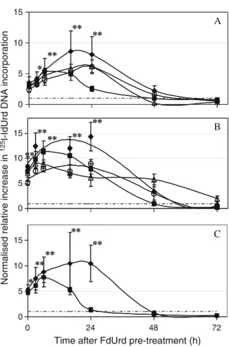
Fig. 2. Incorporation kinetics of125I-IdUrd according to the interval after FdUrd pre-treatment, for LN229 (a), U251 (b) and U87 (c) cell lines. The different pre-treatments were: 1μM FdUrd for 1 h (♦), 10μM FdUrd for 1 h (∆), 1 μM FdUrd for 10 min (■) and 10 μM FdUrd for 10 min (○). After various intervals post FdUrd pre-treatment, cells were incubated for 2 h with 5 kBq125I-IdUrd. The mean values ±1 SD of three independent experiments are shown, each performed with three to four measurements per condition. The results represent relative increase in DNA incorporation of radio-IdUrd for pre-treated cells divided by untreated samples (I/Io) and normalised to the number of cells per well. Baseline relative DNA incorporation is represented by a broken line. * and ** denote results that were significantly (p<0.05) or highly significantly (p<0.001) different compared with untreated control cells
Results
Dependence of 125I-IdUrd DNA incorporation on FdUrd
concentration
First experiments were performed on the three glioblasto-ma cell lines in exponential growth using co-incubation for
4 h with 5 kBq125I-IdUrd and different concentrations of
FdUrd. Depending on the FdUrd concentration, increased DNA incorporation of radio-IdUrd up to a plateau was observed compared with untreated cells. A maximum increase in incorporation of 4.0- to 8.5-fold was obtained in the three cell lines with FdUrd concentrations between 0.01
and 3 μM (Fig. 1). A high percentage incorporation of
radio-IdUrd in absolute amount (results not shown) might have been partially responsible for the observed plateau. At
concentrations of 10 μM FdUrd or higher, the DNA
incorporation of radio-IdUrd decreased in all cells and co-incubation with dThd almost completely abolished it
(Fig. 1). Compared with previous experiments [6], the
results presented here show a higher efficacy of FdUrd and a higher absolute amount of radio-IdUrd incorporated. This might be related to mycoplasma contamination in previous experiments, though this has not been formally demon-strated. The cells used here, however, were checked for the absence of mycoplasma contamination.
Delayed effect of FdUrd pre-treatment on DNA
incorporation of125I-IdUrd
Shorter incubation times of 10 and 60 min with 1 and
10μM FdUrd were further evaluated because of the known
low toxicity and efficacy of such treatments [6, 10].
Though some growth delay was observed with direct cell counting, absence of cytotoxicity of these treatments was confirmed in LN229 and U251 cells with the colony-forming assay (results not shown). Cells were pre-treated with FdUrd, washed to eliminate trace amounts of FdUrd that could influence the cell cycle and re-incubated with
fresh medium for 0–72 h, following which the radio-IdUrd
incorporation rate was measured in a 2-h incubation.
Incubation with 1μM FdUrd for 1 h was the pre-treatment
which gave the highest increase in 125I-IdUrd DNA
incorporation and the most prolonged effect for the three
glioblastoma cell lines (Fig. 2). Under this condition, the
incorporation rate increased to a maximum after an interval
of 16–24 h post FdUrd pre-treatment (all results are
corrected for the number of counted cells). Incorporation increased by factors of 7.5, 10 and 12 or more for LN229, U87 and U251, respectively, compared with un-pretreated cells (p>0.001). This corresponded, at 24 h post FdUrd, to 16.4%, 35.1% and 50.7% DNA incorporation in absolute amount calculated on the total radio-IdUrd present during the 2-h incubation for the LN229, U87 and U251, respectively (results not shown). The immediate increase
in incorporation post FdUrd exposure (time 0, Fig.2) was
similar to that seen in the 4-h co-incubation experiments shown above, but it was significantly less (p<0.05 for the three cell lines) than at 16 and 24 h post FdUrd. The other
pre-treatments also favoured 125I-IdUrd DNA
incorpora-tion, but the increase in incorporation was not as high as
with 1 μM FdUrd for 1 h. In particular, the shorter
incubation period of 10 min was insufficient to produce a similar effect, despite the use of a higher concentration of FdUrd. Furthermore, the maximum increase in incorpora-tion tended to occur earlier post FdUrd for the other
pre-treatments (Fig.2).
Cell cycle distribution according to the interval after FdUrd pre-treatment
Cell cycle distribution was then assessed by double staining FACS analysis after the optimal pre-treatment
for 1 h with 1μM FdUrd. Highly reproducible results were
observed in independent experiments performed with
LN229, U251 and U87 cells. Incubation with 3μM (one
experiment performed on LN229 and U251 cells) or 10μM
IdUrd (two experiments on the three cell lines) gave
identical results, which were pooled (Table1). As controls,
antibody anti-IdUrd staining was negative for cells
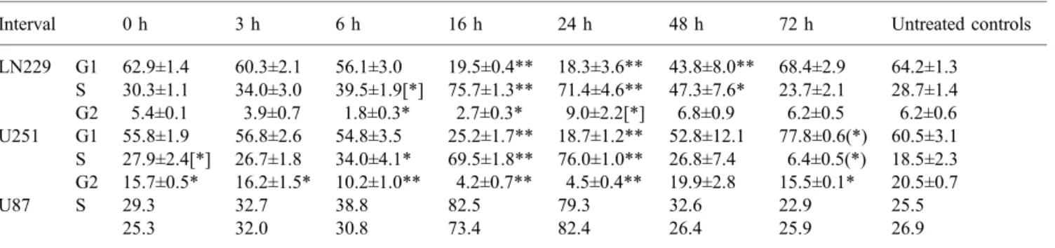
Table 1. Cell cycle FACS analysis
Interval 0 h 3 h 6 h 16 h 24 h 48 h 72 h Untreated controls
LN229 G1 62.9±1.4 60.3±2.1 56.1±3.0 19.5±0.4** 18.3±3.6** 43.8±8.0** 68.4±2.9 64.2±1.3 S 30.3±1.1 34.0±3.0 39.5±1.9[*] 75.7±1.3** 71.4±4.6** 47.3±7.6* 23.7±2.1 28.7±1.4 G2 5.4±0.1 3.9±0.7 1.8±0.3* 2.7±0.3* 9.0±2.2[*] 6.8±0.9 6.2±0.5 6.2±0.6 U251 G1 55.8±1.9 56.8±2.6 54.8±3.5 25.2±1.7** 18.7±1.2** 52.8±12.1 77.8±0.6(*) 60.5±3.1 S 27.9±2.4[*] 26.7±1.8 34.0±4.1* 69.5±1.8** 76.0±1.0** 26.8±7.4 6.4±0.5(*) 18.5±2.3 G2 15.7±0.5* 16.2±1.5* 10.2±1.0** 4.2±0.7** 4.5±0.4** 19.9±2.8 15.5±0.1* 20.5±0.7 U87 S 29.3 32.7 38.8 82.5 79.3 32.6 22.9 25.5 25.3 32.0 30.8 73.4 82.4 26.4 25.9 26.9
Flow cytometry was performed at the indicated intervals post FdUrd pre-treatment (1μM FdUrd for 1 h). Mean values ±1 SD of three
independent FACS analyses are given for LN229 and U251 cells. S-phase results of two FACS analyses are given for U87 cells. * and ** denote results that were significantly (p<0.05) or highly significantly (p<0.001) different compared with untreated control cells. (*) and [*] indicate differences that were close to significance, at p<0.07 and p<0.15, respectively
incubated with FdUrd alone, dThd inhibited IdUrd DNA incorporation and the absence of primary antibody also gave a negative result (data not shown).
As shown (Fig.3and Table1), the rate of S-phase cells
increased to 69.5% or higher at 16–24 h after FdUrd
pre-treatment, compared with baseline rates of 28.7%, 18.5% and 25.5% for un-pretreated LN229, U251 and U87 cells, respectively. The S-phase increase at 16 and 24 h was highly significant (p<0.001) compared with both untreated cells and cells evaluated immediately after FdUrd pre-treatment (t=0). G1 and G2 phases decreased at these times to the benefit of the S phase. Interestingly, while the cell cycle distribution of LN229 and U87 cells had returned to
about baseline 72 h after FdUrd removal (Table1), results
in respect of U251 cells showed an under- and over-reaction of the cell phases which, compared with baseline results, was either significant (G2 phase) or close to being significant (p<0.07), suggesting that these cells remained partially synchronised.
S-phase cells were further analysed by plotting the
number of cells against DNA content (Fig. 4 shows a
representative experiment). This sub-analysis of the S-phase fractions showed that LN229 and U87 accumulated in early S phase up to 16 h and U251 cells up to 24 h post FdUrd pre-treatment. Release of the early S-phase block was observed at 24 h for LN229 and U87 and at 48 h for U251 cells.
125I-IdUrd DNA incorporation rate in the presence of 3
μM unlabelled IdUrd
In order to directly compare radio-IdUrd DNA incorpora-tion and cell cycling, a comparison at identical IdUrd
concentrations (3μM unlabelled IdUrd) was performed on
LN229 and U251 cells. In the FACS analysis, the use of 3
or 10 μM unlabelled IdUrd gave identical cell cycle
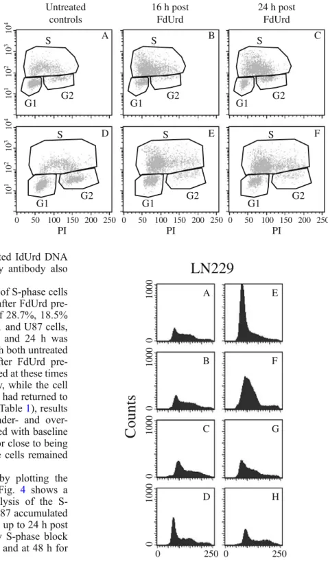
10 1 10 2 10 3 10 4 10 1 10 2 10 3 10 4 0 50 100 150 200 250 0 50 100 150 200 250 0 50 100 150 200 250 PI PI PI IdUrd immunofluorescence Untreated controls 16 h post FdUrd 24 h post FdUrd A B C D E F G1 G2 S G1 G2 S G1 G2 S G1 G2 S G1 G2 S S G1 G2
Fig. 3. Cell cycle distribution in untreated controls and 16 and 24 h after pre-treatment with 1μM FdUrd for 1 h. Represen-tative FACS results for LN229 (a–c) and U251 (d–f) are shown. The incorporated IdUrd, revealed by FITC immunofluo-rescence, is plotted against DNA content of cells, revealed by PI staining. Note the accumulation in early S phase (and depletion of G2) in b, e and f. LN229 cell block-release occurs at 24 h post FdUrd (c). Results of the complete cycle analysis at different inter-vals post FdUrd are given in Table1
Counts
PI
LN229
PI
E F G H 0 250 A 0 1000 B 0 1000 C 0 1000 D 0 1000 0 250Fig. 4. Sub-analysis of S-phase cells according to interval after 1 h pre-treatment with 1μM FdUrd. IdUrd immunofluorescence-stained S-phase cells have been further analysed by plotting cell counts against DNA content, revealed by PI staining. Analyses of a rep-resentative experiment for LN229 are shown. a Untreated control; b 0 h post FdUrd; c 3 h post FdUrd; d 6 h post FdUrd; e 16 h post FdUrd; f 24 h post FdUrd; g 48 h post FdUrd; h 72 h post FdUrd. Note that the block-release of cells occurs at 24 h in LN229 (f)
distributions with notably similar S-phase rates after FdUrd
pre-treatment (Table 1). 125I-IdUrd DNA incorporation
measurements were therefore performed using incubation
of pre-treated cells with125I-IdUrd in the presence of 3μM
unlabelled IdUrd. The presence of the additional 3 μM
unlabelled IdUrd partially decreased the radio-IdUrd DNA incorporation compared with experiments with radio-IdUrd alone. In both cell lines, however, the optimal
DNA incorporation increase was still observed at 16–24 h
post FdUrd (Fig.5). For both cell lines, LN229 and U251,
the increase in uptake at 16 and 24 h was highly significant (p<0.001) compared with untreated cells, and also com-pared with the incorporation immediately after FdUrd pre-treatment. These results therefore show that, under iden-tical IdUrd concentrations, S-phase accumulation and the
highest DNA incorporation rate of 125I-IdUrd remained
time-correlated.
Radiosensitivity after FdUrd pre-treatment
These experiments were performed in order to test whether the S-phase arrested cells would exhibit any radio-resistance. LN229 and U251 cells were pre-treated or not
with 1μM FdUrd for 1 h. Cells were then irradiated with 2
or 6 Gy immediately after FdUrd exposure or after an interval of 20 h, and survival evaluated by colony-forming assay. Results showed reproducibly that pre-treated cells
were equally or more radiosensitive than un-pretreated
cycling control cells (Fig.6). An increased cytotoxic effect
(statistically significant, p<0.05) after FdUrd pre-treatment was observed in LN229 cells at both 0 and 20 h post irradiation with 2 and 6 Gy, while in U251 cells such a significant difference was only observed at 20 h post FdUrd and was limited to the 6-Gy irradiation. These results thus show the absence of any radio-resistance of the S-phase-enriched cell populations.
Discussion
A rational approach to radio-IdUrd Auger radiation therapy requires targeting of the highest possible number of cells and high DNA incorporation of radio-IdUrd. Previously, we have shown that short-term, non-toxic concentrations of FdUrd are able to induce high percentage DNA
incorpo-ration of radio-IdUrd in vitro and in vivo [6]. However, the
number of targeted cells, namely the cells in S phase, remained low at the time of incubation/administration of radio-IdUrd immediately after FdUrd.
In this study, we therefore investigated cellular cycling and radio-IdUrd DNA incorporation after different inter-vals post FdUrd pre-treatment in three glioblastoma cell lines, aimed particularly at optimising S-phase targeting. We were able to show with highly reproducible FACS
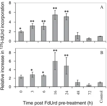
Control A B ** ** ** ** ** ** * * * 0 2 4 6 8 0 2 4 6 8 0 3 6 16 24 48 72 Relative increase in 125 I-IdUrd incorporation
Time post FdUrd pre-treatment (h)
** ** ** ** ** ** * * *
Fig. 5. Radio-IdUrd incorporation in the presence of 3 μM unlabelled IdUrd is shown for LN229 (a) and U251 (b) cells. The incorporation rate of125I-IdUrd co-incubated for 2 h with 3μM un-labelled IdUrd is shown as relative increase in DNA incorporation of radio-IdUrd in FdUrd-pre-treated cells compared with un-pretreated control cells and normalised to the number of cells per well. Mean values ±1 SD of three independent experiments are given, each performed with three to four measurements per condition. * and ** denote results that were significantly (p<0.05) or highly significantly (p<0.001) different compared with untreated control cells
4 6
Radiation dose (Gy)
A
B
0.001 0.01 0.1 1 0.01 0.1 1 0 2Survival rate (S/So)
A
B
Fig. 6. Survival fractions of LN229 (a) and U251 (b) cells after external beam irradiation. Cells were irradiated after 0- and 20-h intervals following pre-treatment or not with 1μM FdUrd for 1 h: (♦), un-pretreated controls; (☐), 0 h post FdUrd; (▲), 20 h post FdUrd. Mean values ±1 SD of three independent experiments are shown; each experiment was performed with triplicate measure-ments per condition. Survival in the colony-forming assay has been normalised with unirradiated but otherwise identically treated control cells (S/So). Note that the increased cytotoxic effect following FdUrd pre-treatment was statistically significant (p<0.05) in LN229 cells at both 0 and 20 h post irradiation with 2 and 6 Gy, while in U251 cells such a significant difference was observed only at 20 h and 6-Gy irradiation
results that cell cycle evolution was remarkably similar in the three different glioblastoma cell lines. Baseline S-phase rates of untreated cells were between 18.5% and 28.7%. S-phase cells then increased gradually up to 69.5% or more
16 and 24 h after 1 h pre-treatment with 1μM FdUrd. This
corresponds to an increase by a factor of between 2.5 and 4.1 over the baseline S-phase rates in the three cell lines. We also observed a similar S-phase rate accumulation after FdUrd pre-treatment in the ovarian cancer cell line SKOV3 (data not shown). These data confirm and extend a similar observation after FdUrd pre-treatment that was described
for the human colon carcinoma cell line HT 29 [9]. HT 29
showed a particularly short doubling time of 22–24 h, and
it was not obvious that cell lines with a longer cycling time would show similar S-phase accumulation. Furthermore, in the three glioblastoma cell lines explored, the difference in
p53 expression [15], as observed frequently in recurrent
tumour [20, 21], had no major impact on S-phase
accumulation post FdUrd. In fact, the accumulation peaked to similar degrees in the three glioblastoma cell lines 16 h post FdUrd exposure.
The results reported here indicate that the observed S-phase increase corresponds to a synchronisation of cells rather than just an accumulation, since a grouped release from the block in early S phase was observed in the three
glioblastoma lines after 16–24 hours. Furthermore, U251
cells remained partially synchronised as late as 72 h after FdUrd pre-treatment. The high S-phase rate in the three cell lines at 16 and 24 h post FdUrd is remarkable since the measured doubling times of these cells varied between 24 and 34 h.
Cells accumulated in S -phase as late as 16 and 24 h after FdUrd pre-treatment continued to take up and in-corporate IdUrd as soon as it became available. This was shown by their ability to incorporate unlabelled IdUrd in micromolar concentrations in the FACS analysis and also
in nanomolar concentrations in the125I-IdUrd DNA
incor-poration assay. Indeed, the radio-IdUrd DNA incorincor-poration peaked at the time of maximal S-phase accumulation.
A further interesting observation was the high percent-age of radio-IdUrd incorporated into tumour cells. In fact, the short availability of radio-IdUrd in vivo is a significant limitation for therapy. In these in vitro experiments, the uptake in absolute amount of radio-IdUrd rose from
baseline incorporations of 2.4%–3.6% to between 16.4%
and 50.7% at 24 h post FdUrd pre-treatment. Again, we observed a similar increase in uptake in SKOV3 ovarian cancer cells (results not shown). In vivo, without taking advantage of any S-phase increase after an interval post FdUrd, we observed that FdUrd pre-treatment enhanced the tumour uptake of radio-IdUrd in LN229 cells after intratumoural injections: tumour uptake reached 19.8% of the injected dose compared with only 4.1% in animals not
pre-treated with FdUrd [12]. These data demonstrate that
the short bio-availability of radio-IdUrd might be at least partially circumvented by the use of FdUrd or other dThd synthesis-blocking agents.
Evidence derived from radiobiology indicates that cell radiosensitivity is maximal in the M, late G1 and early S
phases and minimal in the early G1 and G2 phases [22].
However, arrested cells might respond differently to irradiation than cycling cells. It has been reported that fluoropyrimidine-mediated radiosensitisation is dependent on progression of cells into S phase under conditions of
altered deoxynucleotide triphosphate pools [23]. The
observation that the S-phase-enriched population 20 h post FdUrd treatment remained as, or slightly more, sensitive than untreated cycling cells therefore appears
favourable. However, Auger radiation therapy with123
I-or 125I-IdUrd will be active over one or several days,
respectively. Efficacy of Auger radiation treatment, there-fore, might be less dependent on cell cycling than external beam photon therapy, since Auger radiation induces a high percentage of double strand breaks that cannot be repaired efficiently.
These data might pave the way for treatment of high percentages of tumour cells in a single therapy cycle with
Auger radiation pharmaceuticals such as 123I- or 125
I-IdUrd. Furthermore, it has recently been shown that Auger
radiation therapy can also produce a bystander effect [24].
This might allow treatment in a single cycle of more than just the S-phase cells targeted 16 h after the FdUrd pre-treatment. However, the capacity to induce S-phase synchronisation in vivo remains to be demonstrated.
In conclusion, our results confirm and extend earlier observations in a colorectal cancer line to three human glioblastoma cell lines, with the observation of high percentage accumulation in early S phase delayed after FdUrd pre-treatment. We have further shown that these S-phase cells remained accessible to radio-IdUrd targeting at nanomolar up to micromolar concentrations and incorpo-rated high percentages. Thus, highest radio-IdUrd DNA incorporation and S-phase accumulation were observed
16–24 h following FdUrd pre-treatment. The
S-phase-enriched cell population was also shown to be as, or more, radiosensitive than un-pretreated cycling cells. The short FdUrd pre-treatment itself was non-toxic. Viewing these considerations in conjunction, it appears that the combina-tion of FdUrd and radio-IdUrd could allow the racombina-tional design of a local Auger radiation treatment of cancer at optimised timing post FdUrd.
Acknowledgements. We thank Dr. Marek Kosinki for the preparation of high-purity125I-IdUrd and Mrs. Frances Godson for reviewing the manuscript. This study was supported by a grant from the Swiss National Science Foundation (grant No 31–67112.01).
References
1. Commerford SL. Biological stability of 5-iodo-2'-deoxyuridine labelled with iodine-125 after its incorporation into the deoxyribonucleic acid of the mouse. Nature 1965;206:949–50 2. Blasberg RG, Roelcke U, Weinreich R, Beattie B, von Ammon K, Yonekawa Y, et al. Imaging brain tumor proliferative activity with [124I]iododeoxyuridine. Cancer Res 2000;60:624–35
3. Klecker RW, Jr., Jenkins JF, Kinsella TJ, Fine RL, Strong JM, Collins JM. Clinical pharmacology of 5-iodo-2'-deoxyuridine and 5-iodouracil and endogenous pyrimidine modulation. Clin Pharmacol Ther 1985;38:45–51
4. Kassis AI, Guptill WE, Taube RA, Adelstein SJ. Radiotoxicity of 5-[125I]iodo-2'-deoxyuridine in mammalian cells following treatment with 5-fluoro-2'-deoxyuridine. J Nucl Biol Med 1991;35:167–73
5. Lawrence TS, Davis MA, McKeever PE, Maybaum J, Stetson PL, Normolle DP, et al. Fluorodeoxyuridine-mediated modu-lation of iododeoxyuridine incorporation and radiosensitization in human colon cancer cells in vitro and in vivo. Cancer Res 1991;51:3900–5
6. Dupertuis YM, Vazquez M, Mach JP, De Tribolet N, Pichard C, Slosman DO, et al. Fluorodeoxyuridine improves imaging of human glioblastoma xenografts with radiolabeled iododeoxy-uridine. Cancer Res 2001;61:7971–7
7. Mester J, DeGoeij K, Sluyser M. Modulation of [5-125I] iododeoxyuridine incorporation into tumour and normal tissue DNA by methotrexate and thymidylate synthase inhibitors. Eur J Cancer 1996;32A:1603–8
8. Armstrong RD, Diasio RB. Metabolism and biological activity of 5'-deoxy-5-fluorouridine, a novel fluoropyrimidine. Cancer Res 1980;40:3333–8
9. Miller EM, Kinsella TJ. Radiosensitization by fluorodeoxyuri-dine: effects of thymidylate synthase inhibition and cell synchronization. Cancer Res 1992;52:1687–94
10. Serlin O, Wolkoff JS, Amadeo JM, Keehn RJ. Use of 5-fluorodeoxyuridine (FUDR) as an adjuvant to the surgical management of carcinoma of the stomach. Cancer 1969; 24: 223–8
11. Parsels LA, Parsels JD, Tai DC, Coughlin DJ, Maybaum J. 5-Fluoro-2'-deoxyuridine-induced cdc25A accumulation corre-lates with premature mitotic entry and clonogenic death in human colon cancer cells. Cancer Res 2004;64:6588–94 12. Buchegger F, Adamer F, Schaffland AO, Kosinski M,
Grannavel C, Dupertuis YM, et al. Highly efficient DNA incorporation of intratumourally injected [125 I]iododeoxyuri-dine under thymiI]iododeoxyuri-dine synthesis blocking in human glioblasto-ma xenografts. Int J Cancer 2004;110:145–9
13. Schaffland AO, Delaloye AB, Kosinski M, Dupertuis YM, Buchegger F. The preparation of clinical grade 5-[123 I]iodo-2'-deoxyuridine and 5-[125I]iodo-2'-deoxyuridine with high in vitro stability and the potential for early proliferation scintig-raphy. Nucl Med Commun 2004;25:461–8
14. Foulon CF, Adelstein SJ, Kassis AI. Kit formulation for the preparation of radiolabeled iododeoxyuridine by demetallation. J Nucl Med 1996;37:1S–3S
15. Van Meir EG, Kikuchi T, Tada M, Li H, Diserens AC, Wojcik BE, et al. Analysis of the p53 gene and its expression in human glioblastoma cells. Cancer Res 1994;54:649–52
16. Parsels LA, Zellars RC, Loney TL, Parsels JD, Clarke MF, Merchant AK, et al. Prevention of fluorodeoxyuridine-induced cytotoxicity and DNA damage in HT29 colon carcinoma cells by conditional expression of wild-type p53 phenotype. Mol Pharmacol 1997;52:600–5
17. Sinigaglia F, Talmadge KW. Inhibition of [3H]thymidine incorporation by Mycoplasma arginini-infected cells due to enzymatic cleavage of the nucleoside. Eur J Immunol 1985; 15:692–6
18. Wang L, Westberg J, Bolske G, Eriksson S. Novel deoxynu-cleoside-phosphorylating enzymes in mycoplasmas: evidence for efficient utilization of deoxynucleosides. Mol Microbiol 2001;42:1065–73
19. Xiao WH, Dupertuis YM, Mermillod B, Sun LQ, de Tribolet N, Buchegger F. Unlabelled iododeoxyuridine increases the cyto-toxicity and incorporation of [1251]-iododeoxyuridine in two human glioblastoma cell lines. Nucl Med Commun 2000; 21:947–53
20. Sidransky D, Mikkelsen T, Schwechheimer K, Rosenblum ML, Cavanee W, Vogelstein B. Clonal expansion of p53 mutant cells is associated with brain tumour progression. Nature 1992; 355:846–7
21. Hayashi Y, Yamashita J, Yamaguchi K. Timing and role of p53 gene mutation in the recurrence of glioma. Biochem Biophys Res Commun 1991;180:1145–50
22. Terasima T, Tolmach LJ. Variations in several responses of HeLa cells to x-irradiation during the division cycle. Biophys J 1963;3:11–33
23. Hwang HS, Davis TW, Houghton JA, Kinsella TJ. Radiosen-sitivity of thymidylate synthase-deficient human tumor cells is affected by progression through the G1 restriction point into S-phase: implications for fluoropyrimidine radiosensitization. Cancer Res 2000;60:92–100
24. Xue LY, Butler NJ, Makrigiorgos GM, Adelstein SJ, Kassis AI. Bystander effect produced by radiolabeled tumor cells in vivo. Proc Natl Acad Sci U S A 2002;99:13765–70