Offprint trom
Trace Element
Analytical Chemistry in
Medicine and Biology
Volume 5
Proceedings of the Fifth International Workshop
Neuherberg, Federal Republic of Germany,
April 1988
Editors
Peter Brâtter . Peter Schramel
Walter de Gruyter . Berlin . New York 1988
This offprint is not for sale
or
reproductionTHE REGULATION OF LIVER IRON METABOLISM
Y. Beguin, H.A. Huebers, C.A. Finch
Division of Hematology, University of Washington, Seattle, USA
G. Weber, I. Roelandts
Department of Experimental Nuclear Physics, University of Liege, Belgium
Abstract
SSFe-transferrin and s9Fe-ferritin were injected simultaneously in rats and storage iron release by the hepatocyte was calculated from the cumulative incorporation of the two isotopes in the red cell mass over 2 weeks. About 6 % of hepatocyte storage iron was released daily in normal rats, but a non-mobilizable pool was identified in iron overload. Iron release was regulated by the rate of erythropoiesis and iron status of the animal, while acute inflammation blocked iron release only transiently.
Introduction
Hepatocyte iron release to the plasma is less well characterized than iron uptake by the liver (1). Two phases of iron release can be distinguished, i.e. (a) an immediate release during the initial processing of
transferrin, hemoglobin, ferritin, or "freell iron (early release, ER), and (b) iron release from stores (late release, LR). We present the results of studies of storage iron release in normal rats and animais with various disorders of iron status and erythropoiesis, using s9Fe-labelled ferritin as a selective tag of hepatocytes (2,3,4).
Trace Element Analytîcal Chemistry in Medicine and Biology
541
56a. Flynn, A., S.I. Miller, S.S. Martier, N.L. Goldern, R.J. SakaI, and
B.e.
Del Villano. 1981. Lancet.l, 572-575. 57. Turnlund, J.R., C.A. Swanson, and J.C. King. 1983. J.Nutr. ~, 2346-2352.
58. Cornmittee on Dietary Allowances, Food and Nutrition Board. 1980. Recommended Dietary Allowances, 9th Edition.
National Academy of Sciences, Washington DG, USA. 59. Freidman, S., C. Baharg, B. Eckerling, and B. Gans.
1969. Obstet. Gynecol.
11,
189-194.60. Schenker, J.G., E. Jiengies, and W.Z. Polishiek. 1969.
Am. J. Obstet. Gynecol. 105, 933-937.
61. Q' Leary, J .A., G.S. Novalis, and G.J. Vosburgh. 1966. Obstet. Gynecol. ~, 112-117.
62. Mirzakarimov, M.G. 1953. Akush. Ginekol.
l,
55-85.63. Rasuli, 2.M. 1963. Akush. Ginekol.
1.2.,
63-66. 64. Linares, A.,Diaz-Perez.
J.J. Larranz, J. Rodrlguez-Aluscon, and J.L.
1979. Lancet~, 43-44.
65. Mjolnerod, O.K., K. Rasmus~en, S.A. Dommerad, and S.T. Gjeraldsen. 1971. Lancet~, 673-675.
66. Henkin, R.I., J.R. Marshall, and S. Meret. 1971. Am. J. Obstet. Gynecol. 103, 320-344.
67. Swanson, C.A., D.C. Reames, C. Veillon, J.C. King, and O.A. Levander. 1983. Am. J. Clin. Nutr. 38, 169-180. 68. Rudolph, N., and Wong, S.L. 1978. Ped. Res.
g,
789-792. 69. Behne, D., and W. Walters. 1979. J. Clin. Chem. Clin.Biochem.
11,
133-135.70. Levander, O.A., P.B. Moser, and V.C. Morris. 1987. Am. J. Clin. Nutr. i§., 694-698.
71. Butler, J.A., P.D. Whanger, M.J. Tripp. 1982. Am. J. Clin. Nutr. ~, 15-23.
72. Anderson, R.A. 1986. Clin. Physiol. Biochem. ~, 31-41. 73. Davidson, I.W.F., and R.C. Burt. 1973. Am. J. Obstet.
Gynecol. ~, 601-608.
74. Hambidge, K.M., and W. Dioegemueller. 1974. Obstet. Gynecol.
ii,
666-672.75. Mahalko, J.R., and M. Bennion. ~, 1069-1072.
1976. Am. J. Clin. Nutr.
76. Krebs, 1985.
N.F., K.M. Hambidge, M.A. Jacobs, and J.O. Rasback. Am. J. Clin. Nutr .
.::...l.,
560-570.77. Vuori, E., S.M. Makinen, R.Kara, and P. Kuitunen. 1980. Am. J. Clin. Nutr.ll, 227-231.
78. Mannan, S., and M.F. Picciano. i§., 95-100.
~3
Methods
Rat transferrin was purified by saturating plasma transferrin with ferrous sulfate, followed by ammonium sulfate precipitation, ion-exchange
chromatography using a DEAE Sephacel column and gel chromatography using a S-200 Sephacryl column. Apotransferrin was prepared by incubating
diferric transferrin with desferrioxamine, which was removed afterwards by ultrafiltration. Labeling of apotransferrin with 5sFe ferrous sulfate was achieved in the presence of 0.05 M Na bicarbonate and 0.1 M Tris/Hel. The transferrin solution was brought ta the point of saturation by adding cold ferrous sulfate. Ferritin was purified fram rat liver by heating at 75°C for 10 minutes, ammonium sulfate precipitation, gel chromatography uslng a Sepharose 6B column, and ultracentrifugation (3). In vitro tagging of ferritin was achieved by adding s9FeSO~ to a normal rat ferrltin preparation in 0.1 M Hepes buffer (pH 7.5). The labelled ferritin was purified by ion-exchange chromatography using a DEAE Sephacel column (3). Male Sprague-Dawley rats 8 to 10 weeks of age were used in most
experiments. AnimaIs to be made iron deficient were placed at 4 weeks on a diet containing 4-8 mg of iron/Kg, or alternatively were given an iron deficient diet at 6 weeks of age and bled 2-3 ml 4 to 5 times over two weeks to accelerate iron depletion. Female rats to be iron-Ioaded were placed on a 1% carbonyl iron diet for> 1 year with occasional
reintroduction of a normal diet to maintain animaIs in good health. In order to suppress erythropoiesis, rats were transfused with 3 ml packed red cells approximately twice a week ta maintain an hematocrit of 60-65 %,
and S9Fe ferritin was injected on the 5th day after the first transfusion. Hemolytic anemia was produced by the intraperitoneal injection of 50 mg acetylphenylhydrazine per Kg body weight, and S9Fe ferritin was injected 5 days later. Inflammation was produced by injecting 0.25 ml turpentine oil in each thlgh, and 59 Fe ferritin was injected 16 hours later.
Plasma was drawn from a rat prepared in the sarne way as the experimental animaIs and labelled by adding 0.5 ~Ci S 5Fe as FeS04 in 0.9% saline (pH 2) to 0.5 ml plasma. The rate of radioiron disappearance (t-1/2) was
determined and the plasma iron turnover (PIT) was calculated in ~g/Kg/day!
~4
PIT
SeFe (~g/dl) x (100 - Ret x 0.92) x 7 t-l/2 (min)
Release of storage iron by the hepatocyte (i.e. late release) was studied over a peried of 14 days. The cumulative incorporation of S9Fe (injected as ferritin) and 5sFe (injected as diferric transferrin) in the red celi mass was determined over a 2 wk period. The ratio of 59Fe ta sSFe
activity in the red cell mass at any tirne represents the total fraction of 59Fe released by the hepatocyte (total release at time t = early release +
late release at time t). When the complement of this ratio was plotted against time on a semilogarithmic paper, late release appeared ta be a single exponential curve. The proportion of the injected dose released in the early and late phases was obtained by extrapolating the curve ta zero tirne. The t-1/2 of the curve, determined by least squares, represents the speed of late release .and was expressed as % of the dose/day. Formal proof of this method has been published (5). Labelled compounds were injected into a tail vein and blood samples were obtained from the tail vein on the opposite side. At the end of each experiment, rats were exsanguinated from ~he abdominal aorta, and perfused with 20-30 ml saline. Whole blood, plasma, and liver activities were counted. Total plasma and total red cell activities were calculated from the hematocrit and assuming a blood volume of 0.07 ml/g body weight. Non-herne iron was extracted from rat livers and iron was determined by Proton-Induced X-ray Emission
(PIXE). Assuming that ferritin s9Fe uniformly labelled non-heme iron stores in the hepatocyte, the amount of iron released daily by the hepatic parenchyma was calculated as the product of radioiron release (%/day) and hepatocyte iron stores (~g).
Results
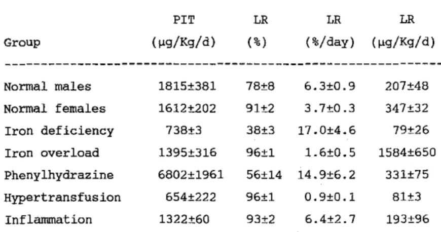
Table 1. ,Plasma iron turnover and storage iron release. Group Normal males Normal fernales Iron deficiency Iron overload Phenylhydrazine Hypertransfusion Inflannnation PIT (~g/K9/d) 1815±381 1612±202 738±3 1395±316 6802±1961 654±222 1322±60 LR (%) 78±8 91±2 38±3 96±1 56±14 96±1 93±2 LR LR (%/day) (~g/K9/d) 6.3±O.9 207±48 3.7±O.3 347±32 17.0±4.6 79±26 1.6±D.5 1584±650 14.9±G.2 331±75 O.9±O.1 BI±3 6.4±2.7 193±96 545
Normal males re!eased 17.5% of the ferritin s9Fe in the early phase.
Thereafter, 6.3% of the residual hepatocyte radioiron was re!eased daily,
representing a total of 207 ~g Fe/Kg/day, i.e. about 11% of the PIT. Normal fernales had a slower rate of late release. However, as their iron
stores were consistently higher, the contribution of hepatocyte iron ta the PIT was found ta he higher than in males (21%). Iron turnover in the hepatocyte of iran deficient rats is extrernely rapid (56% ER and
thereafter 17%/day of LR) but invalves relatively small amounts of iron, a little more than 10% of the PIT. By contrast, iron overloaded rats had a turnover about 10 times slower (6% ER and 1.6%/day of LR), amounting to a calculated value of 1584 ~g/kg/day, which is an overestimation. Rats treated with phenylhydrazine had an increased amount and rate af iron turnover in the hepatocyte. However, the participation of the parenchyrnal cell in the PIT was rather small (5%). On the other hand, hypertransfused rats had a slow turnover involving small amounts of iron which represented 12% of the PIT. Inflammation reduced considerably the early release of radioiron. However, release of hepatocyte iron in the following days was normal. There was a very significant correlation between the percent ER and the rate of LR (R=0.90, P<O.OOl). Iron release was inversely related ta the hematocrit and positively to the PIT. Plasma iron and radioiron t-1/2, as weIl as hepatic iron, correlated with the rate but not the
~6
Discussion
After uptake hy the hepatocyte, ferritin is catabolized and its iron enters an intermediate pool before being incorporated into stores (3,4). Because it i8 based on the assurnptions that ferritin 59Fe labels aIl hepatocyte iron stores uniformly and that aIl non-herne iron is mobilizable in the sarne way, the calculation of the amount of hepatocyte iron released daily ta the plasma i8 less precise than the measurement of radioiron release. The rate of radioiron re!ease was considerably increased in iron deficient rats but 6.2% of the ferritin 59Fe remained in the liver after 2 wks, indicating that not aIl hepatic iron i8 rnobilizab!e by increased marrow requirements. Another pool of non-exchangeable iron, presumably hemosiderin, was identified in iron overloaded rats in which the
calculation of iron released by the liver was grossly overestimated. In iron overload, hemosiderin iron represents a large proportion of hepatic iron, which is not exchangeable at least over the time and in the conditions of the present study. Nevertheless, the values obtained in non-deficient and non-overloaded rats are reasonable. Iron release in normal animaIs amounted to 207 ~g Fe/Kg/day, which represents 11.4% of the PIT. These findings are similar to values reported by others (4). This is also in accordance with hepatocyte iron uptake, as approxLffiately 10% of transferrin radioiron is localized in the liver after the early release phase. with iron deficient and iron overloaded rats not included in the analysis, there was a good correlation between hepatic storage iron and the rate of LR, but not with the absolute amount of iron released daily. This observation confirms that s9Fe and cold iron are released in a
similar fashion. Inflammation produced a significant decrease in the rate of early radioiron release. However, late re1ease was not affected. These observations confirm a previous study in which the effect of turpentine-induced inflammation on storage iron mobilization was a1so transient and was no longer apparent beyond 48 hrs (6). However, human studies have shown that chronic inflammation is associated with a b10ckade of iron release from RE stores (7). The effect of erythropoiesis on
547
hepatocyte iron mobilization was examined in phenylhydrazine-injected and hypertransfused rats. Iron release was considerably modified in parallel ta changes
in
erythropoiesis. The amount of iron re!eased by the hepatic parenchyrna seemed appropriate (12% of the PIT) in hypertransfused rats. However, it accounted for only 4.9% of the PIT in phenylhydrazine-injected animaIs, indicating that recycling of red cell iron through thereticuloendothelial system contributed more than hepatocyte iron mobilization ta increased marrow iron requirements.
Acknowledgement
Supported in part by NIH (USA) Grant No. HL06242 and by IISN (Belgium) Grant No. 4450865. Y. Beguin was a Research Assistant of the FNRS
(Belgiurn) and supported in part by a Fogarty International Research Fellowship from the NIH. G. Weber is a Research Associate of the FNRS.
References
1. Morgan, E.H., E. Baker. 1986. Fed.Proc. 45, 2810.
2. Kim, B.-K., H.A. Huebers, C.A. Finch. 1987. Am.J.Hematol. 24, 277. 3. Pippard, M.J., O.K. Johnson, C.A. Finch. 1982. Brit.J.Haematol. 52,
211.
4. Unger, A., C. Hershko. 1974. Brit.J.Haematol. 28, 169.
5. Fillet, G., J.O. Cook, C.A. Finch. 1974. J.Clin.lnvest. 53, 1527. 6. Hershko, C., J.O. cook, C.A. Finch. 1974. Brit.J.Haematol. 28, 67. 7. Oresch, C., Y. Najean. 1972. Eur.J.Clin.Biol.Res. 17, 930.
NEW
DATA ON
THE
HYPOTHESIS OF
THE
BRAIN PARTICIPATION IN
IRON HOMEOSTASIS
B. Ribas, J.F. Pelayo, N. L. Roorigues.
Institute
of
Biochemistry,
CSIC-Complutense
University,
Faculty of Pharmacy, 28040-Madrid
Abstract
Ferropeni c posthemorrhagi c rats were used wh en hematocri te
reached 30%
and
hemoglobin was
lower than 8g/l00ml. The
brain
;5
homogenized (1:4.w:v), sonicated, centrifuged andthe supernatant, is incubated with 2pCi 59 Fe for a period
of 20h.
The
subsequent elution of two peaks,
supposedly
ferritin
and
metallothionein
marked
with
59 Fe
justifies
the hypothesis before exposed of the brain participation
in iran homeostasis.
Introduction
t·1etallothionein, a well known low relative molecular weight
protein, with approximately 6000 D, 30% cystein and without
aromatic
aminoacid
residues,
associates
between
5 ta
11
atoms of metals (1,2).
lts signification in metabolism is
not well understood, it is suggested that it could participate
as regulatory protein (3). Sorne data are published before,
comparing the increase of transferrin and metallothioneinin the intestinal mucosa of ferropenic rats ;n the brain
(5,6) .
The object of this work is to furnish new data which justify
the hypothesis of the brain participation in ;ron homeostasis(7,8), exposed after the experimental results on the
partici-Trace Element Analytical Chemistry in Medicine and Biology