HAL Id: hal-01858228
https://hal.archives-ouvertes.fr/hal-01858228
Submitted on 6 Sep 2018
HAL is a multi-disciplinary open access
archive for the deposit and dissemination of
sci-entific research documents, whether they are
pub-lished or not. The documents may come from
teaching and research institutions in France or
abroad, or from public or private research centers.
L’archive ouverte pluridisciplinaire HAL, est
destinée au dépôt et à la diffusion de documents
scientifiques de niveau recherche, publiés ou non,
émanant des établissements d’enseignement et de
recherche français ou étrangers, des laboratoires
publics ou privés.
N-Terminal Cu-Binding Motifs (Xxx-Zzz-His, Xxx-His)
and Their Derivatives: Chemistry, Biology and
Medicinal Applications
Paulina Gonzalez, Karolina Bossak, Ewelina Stefaniak, Christelle Hureau,
Laurent Raibaut, Wojciech Bal, Peter Faller
To cite this version:
Paulina Gonzalez, Karolina Bossak, Ewelina Stefaniak, Christelle Hureau, Laurent Raibaut, et al..
N-Terminal Cu-Binding Motifs (Xxx-Zzz-His, Xxx-His) and Their Derivatives: Chemistry, Biology
and Medicinal Applications. Chemistry - A European Journal, Wiley-VCH Verlag, 2018, 24 (32),
pp.8029-8041. �10.1002/chem.201705398�. �hal-01858228�
N-terminal Cu Binding Motifs Xxx-Zzz-His (ATCUN) and Xxx-His and their
derivatives: Chemistry, Biology and Medicinal Applications.
Paulina Gonzalez
a,b, Karolina Bossak
c, Ewelina Stefaniak
c, Christelle Hureau
b,d,e, Laurent Raibaut
a,
Wojciech Bal
c*, Peter Faller
a,b*
Abstract:
Peptides and proteins with N-terminal amino acid sequences NH2-Xxx-His (XH) and NH2-Xxx-Zzz-His (XZH) form well established high affinity CuII-complexes. Key examples are Asp-Ala-His (in serum albumin) and Gly-His-Lys, the wound healing factor. This opens a straightforward way to add a high affinity CuII-binding site to almost any peptide or protein, by chemical or recombinant approaches. Thus, these motifs, NH2 -Xxx-Zzz-His in particular, have been used to equip peptides and proteins with a multitude of functions based on the redox activity of Cu, including nuclease, protease, glycosidase, or oxygen activation properties, useful in anticancer or antimicrobial drugs. More recent research suggests novel biological functions, mainly based on the redox inertness of CuII in XZH, like PET imaging (with 64Cu), chelation therapies (for instance in Alzheimer’s disease and other types of neurodegeneration), antioxidant units, Cu transporters and activation of biological functions by strong CuII binding. This review gives an overview on the chemical properties of Cu-XH and –XZH motifs and discusses the pros and cons for the vast and different biological applications, and how they could be improved depending on the application.1. Introduction
Peptides and proteins with N-terminal amino acid sequences Xxx-His (XH) and Xxx-Zzz-His (XZH) bind CuII with a high affinity. Adding XH or XZH by chemical or recombinant approaches is a straightforward way to add a high affinity CuII binding site to almost any peptide or protein. Thus, these motifs have been used to equip peptides and proteins with a multitude of functions. In the present review, we focus on the applications relevant for biology and medicine, which include nuclease, protease, glycosidase, or oxygen activation, properties useful in anticancer or antimicrobial drugs, imaging, chelation, design of antioxidant units and Cu transporters.
2. The two motifs and metal ions they bind.
2.1. Short description of the motifs:
The two motifs considered here, H2N-Xxx-His (XH) and H2 N-Xxx-Zzz-His (XZH), are N-terminal sequences of peptides and proteins which have a His residue at the second or third position with respect to the free N-terminal amine. CuII can bind to XH in
a tridentate fashion, with three nitrogens from the N-terminal amine (NH2), the first amide (N-) and the imidazole nitrogen at the delta ring position (Nδ) being involved (Figure 1, left). The fourth equatorial binding site is occupied by an external molecule such as water, buffer, etc. In the case of the XZH motif, CuII binds to four nitrogens: the N-terminal amine, the first two amides and the Nδ imidazole nitrogen (Figure 1, right). Such coordination modes are denoted as 3N (NH2; N-; Im) and 4N (NH2; 2×N-; Im), respectively, throughout the text. Their structures are known from several X-ray studies[1–3] and from the analysis of their spectroscopic properties (Table 1)
Figure 1: Scheme of the equatorial CuII
-coordination of the two motifs, Xxx-His (left) and Xxx-Zzz-His (right). L depicts an external ligand, could be solvent or other ligand.
2.2.Spectroscopic characteristics of 3N and 4N motifs
The CuII complexes of XH and XZH motifs can be readily identified upon their spectroscopic properties, collected in Table 1. The split pattern of bands in the visible range of CD spectra is particularly characteristic for 4N complexes of XZH peptides. This split is less visible for the XH pattern, where the higher energy negative band is very weak and not always visible.
Table 1. Spectroscopic features of CuII
-XH and CuII
-XZH complexes
Motif
Absorption CD EPR
d-d transition d-d transition Charge
transfer gǁ g Aǁ (MHz) XH ~ 600 nm ~590 nm (+) ~490 nm (-)* ~300 nm, ~330 nm ~ 2.22 ~ 2.05 ~ 560 [1.8 x 10⁻² cm-1] XZH ~520 nm ~480 nm (+) ~560 nm (-) ~310 nm ~ 2.19 ~ 2.04 ~ 600 [2.0 x 10⁻² cm-1 ]
* weak band, not always discernible
2.3. Significance of copper complexes, and XH and XZH motifs
a) Identity of the amino acid: The CuII ion exhibits high affinity towards nitrogen donors, and is one of very few metal ions able to deprotonate and coordinate the peptide bond nitrogen.[4–7] Moreover, binding with the Nδ of the His side chain creates a stable 6-membered chelate ring with the preceding amide nitrogen. Such rings are also achievable via Asp and Cys residues. Asp is a hard acid, and therefore it binds CuII weaker than His. Cys, on the other hand, can form strong bonds similarly to His, but due to its redox reactivity CuII undergoes reduction to CuI by the thiol group, which is oxidized to a disulfide, resulting in a motif loss. Other amino acids (Glu, Met, etc.) would form less favored, larger rings. Therefore, in terms of
[a] Dr. Paulina Gonzalez, Dr. L. Raibaut,Prof. Dr. Peter Faller Institut de Chimie, UMR 7177,CNRS-Université de Strasbourg 4 rue Blaise Pascal, 67000, Strasbourg, France.
E-mail: pfaller@unistra.fr
[b] Dr. Paulina Gonzalez, Prof. Dr. Peter Faller, Dr. Christelle Hureau University of Strasbourg Institute for Advanced Study (USIAS), Strasbourg, France
[c] Dr. Karolina Bossak, Ewelina Stefaniak, Prof. Dr. Wojciech Bal Institute of Biochemistry and Biophysics, Polish Academy of Sciences, Pawińskiego 5a, 02-106 Warsaw, Poland. E-mail: wbal@ibb.waw.pl
[d] Dr. Christelle Hureau
CNRS; LCC (Laboratoire de Chimie de Coordination) 205, route de Narbonne, F-31077 Toulouse, France [e] Dr. Christelle Hureau
Université de Toulouse
CuII binding, the His residue provides the strongest bond and the most stable complex.
b) Influence of His position on metal binding: With a His residue at position 4 and beyond, the advantage of the formation of successive 5 or 6 chelate-rings cannot occur. Thus the CuII -binding becomes much weaker.[7] His at position 1 provides a stable bidentate ligand (N-terminal amine and Nδ of His). The coordination sphere can be completed by another His-1 peptide molecule, resulting in a 2:1 4N complex (2 x NH2; 2 × Im), able to provide high affinity binding only at a high concentration. At lower concentrations and ratios ~1:1, XH and XZH motifs bind CuII much stronger. Notably, His analogues ((4-thiazolyl)-L-alanine, (2-pyridyl)-L-((4-thiazolyl)-L-alanine, and (pyrazol-1-yl)-L-alanine) can also form stable 4N motifs with affinities similar to His.[7] c) Significance of CuII: In order to form Cu-XH and Cu-XZH complexes, one or two amides have to be deprotonated, respectively. Thus, a strong Lewis acid, and a metal ion that favors three or four ligands in an equatorial plane are needed (the latter condition stems from structural properties of peptides composed of α-amino acids). Those requirements are met well by CuII and somewhat poorer (at higher pH) by NIII ions. PdII, AuIII and CoII also bind ATCUN motifs, although for PdII and AuIII the kinetics is slow and those metals are less important biochemically. CoII requires high pH for efficient binding, while ZnII seems not to be able to support the 4N form (at least not at pH below 9), due to its lower Lewis acidity and its preference for tetrahedral geometry. It was reported that ZnII can bind to the 3N form in XH at higher pH.[7–9]
d) Influence of the neighboring His residues: In principle, these amino acids can be any, but Pro as Zzz. Pro is a secondary amine, hence its nitrogen does not carry a dissociable H that could be replaced by the metal ion, when it participates in the peptide bond. Cys is easily oxidized (2CuII + 2CysS ---> 2CuI + CysS-SCys), but the disulfide formation does not preclude its participation in Xxx-His and Xxx-Zzz-His motifs.
2.4. Relative affinities of the two motifs and its pH dependence:
2.4.1 Comparison of XZH and XH:
Cu(XH) 3N complexes recruit one external ligand to complete equatorial coordination sphere, whereas the Cu(XZH) 4N complexes have it already completed (Figure 1). This produces an affinity gain in Cu(XZH) due to the entropic chelate effect, caused by the additional amide donor. However, the entropic gain by the chelate effect of an additional coordination site is counteracted by the enthalpic cost of deprotonating the amide at pH 7.4. Table 2 presents a selection of conditional stability constants at pH 7.4 (cK7.4) for both motifs, calculated from the original using the competitivity index (CI) method.[10,11] These data serve to illustrate the general ranges of affinities, K of 1011.3 – 1013.2
M-1 for 3N XH complexes and 1012 – 1014.7
M-1 for 4N XZH complexes. Thus, despite some overlap, Cu(XZH) complexes are typically about one order of magnitude stronger at physiological pH than Cu(XH) complexes. Moreover, these affinities are generally lower than the affinities reported for Cu(II)-binding enzymes, such as superoxide dismutase.[12]
2.4.2 The pH dependence:
The metal-binding affinities of the XZH and XH motifs are pH dependent. Increasing the pH increases the affinity, because in order to bind, the metal ion must replace peptide’s hydrogen ions. For CuII this process occurs at pH as low as ~3-4 for XH motifs and at slightly higher pH for XZH, due to the higher number of amides involved. This means that below a certain pH the 3N Cu(XH) complexes become more stable than the 4N Cu(XZH) complexes. The coordination process is very cooperative. The intermediate complexes with the less than
maximal number of nitrogen donors are virtually absent for XH and seldom and minor if present for XZH[13]
Table 2. Conditional affinity constants for CuII complexes of XH and XZH
motifs, obtained by recalculation of original potentiometric data by the CI method [10,11] or by calorimetric (a) or spectroscopic (b) titration.
peptide/protein Cu II binding sequence log c K7.4 ref. to original data XH GH-COO- GH 11.3 12.3 [14][15]* RHA-COO -RH 12.6 [16] α-factor model WH 12.8 [17] GHK-COO- GH 12.7 13.2a [18][19] XZH HSA DAH 12.0b [20] GGH-COO- GGH 12.4 [21] des-angiotensinogen N-term VIH 13.0 [4]
Aβ4-16 RFH 13.5 [22] DAHK-amide DAH 13.8 [4] YYH-COO- YYH 14.4 [23] HP2 N-term RTH 14.5 [13] Endostatin N-term HSH 14.5 [24] Hepcidin N-term DTH 14.7 [25] * this apparent discrepancy may be due to different background salts used, KNO3 in ref. 15 and Cl- in ref. 16, the latter being a possible auxiliary CuII
ligand.
2.4.3 Influence of Xxx/Zzz:
Side chains of amino acids surrounding His residues within XH and XZH motifs have an impact on the CuII affinity, although they do not directly participate in a metal ion coordination.[6,26–28] For the XH motif, residues located further away from the N-terminus or coming from another molecule can bind to the vacant fourth coordination site, but this case will not be discussed here. A systematic analysis by Kozłowski et al. indicated that amino acid residues at positions 1 and 2 have the significant influence on CuII coordination of the XZH motif, with partial linear correlations between the pKa values of amine nitrogens of the amino acids and log K of the 4N complex formation.[29] Recently, Miyamoto et al. compared several XZH complexes (X = Z = Asn, Phe, Arg, Lys, Val, Asp, Glu, Tyr, Thr) and found that the pKa of the N-terminal amine is the most determinant factor in this series, conforming this view.[23] The pKa varied from 6.83 (Asn) to 8.18 (Asp). An explanation for Asp having the highest pKa is due to the stabilization of the protonated amine by the H-bond from the carboxylate side chain (formation of a 6-membered ring) (Figure 2A). As CuII-binding involves the deprotonation of this amine, the CuII affinity will decrease, since CuII must compete with the stabilized amine proton. In contrast, Asn could stabilize the deprotonated form by an H-bond, by donating a H-bond from its amide, thus enhancing CuII binding (Figure 2B). Also, pK
a of His residue changed with the sequence (pKa change from 5.19 to 6.64) but this had little correlation with the thermodynamic stability. This seems reasonable as His is already deprotonated at pH 7.4. It is also in line with the small effect of pKa of His and its analogues on the stability of DSUAK-amide pentapeptides derived from histatin-5 (U = His, 4-thiazolyl)-L-alanine, (2-pyridyl)-L-alanine, and (pyrazol-1-yl)-L-alanine).[30]
Figure 2: A: Asp stabilizing the protonated form of the N-terminal amine by forming a H-bond resulting in creation of a 6-memeberd ring. B: Asn stabilizing the deprotonated form of the N-terminal amine via a H-bond with a formation of a 6-membered ring. C: Arg could decrease the pKa of the amide via an
electrostatic interaction with the oxygen of the amide bond. This should facilitate the deprotonation and hence the CuII binding. D: Asp could stabilize the protonated form of the amide by forming a H-bond. This should decrease the affinity of CuII, because CuII binding requires the deprotonation of this amide.
As indicated above,[29] the pK
a of the coordinating peptide bond nitrogens has a significant effect on CuII affinity (assuming that the amide pKa is strongly correlated with the amine pKa of the source amino acid). However, due to the high amide pKa values (>15) they are not measurable by experimental titration.[31] Nevertheless, based on the discussion presented above, one may propose that positively charged and/or H-bond donor side chains could interact with the amide carbonyl oxygen, hence decreasing the peptide group pKa (Figure 2C). Thus, side chains like Lys and Arg at positions 1 and 2 should increase the affinity of CuII. In contrast, negatively charged and H-bond acceptors could interact (via H-bond) with the H-N of the amide bond, increasing its pKa (Figure 2D), and consequently decreasing its affinity for CuII. For example, Asp is more efficient than Glu in this respect, as the H-bond created forms a 7-membered vs. a 9-membered ring.
Moreover, hydrophobic amino acids may increase the stability of complexes of XZH and XH motifs through a hydrophobic fence formation. Although the CuII complex of Val-Ile-His-Asn is just 4-fold stronger than that of Gly-Gly-His, the effect for the structurally related NIII complex is 60-fold.[4–6,32] However, the increased stability could be partially due to a sequence difference at position 4 (which is absent in GGH). Additionally, aromatic side chains were proposed to increase CuII complex stability with XZH and XH motifs due to their direct d-π interaction.[27]
With all these observations and correlations listed, an inspection of Table 2 reveals a disturbing fact that essentially the same XZH motif yields complexes placed all over the affinity range: the Cu(DAH) complex in HSA is among the weakest known, the same sequence when isolated in a tri- or tetrapeptide provides a 100-fold higher stability, while a similar Cu(DTH) complex (also present in several mammalian albumins) formed by the hepcidin N-terminal hexapeptide is the most stable one known so far.[25] The reasons for this effect remain unknown and there is not enough data to even speculate responsibly in this respect.
2.4.4 Cyclisation:
Kritzer and coworkers[33,34] recently investigated cyclic XZH motifs, by linking the side chain of Lys at position 1 with the C-terminus of His3 via a -peptide bond. For such structure, the
CuII complex formation was recorded at a much higher pH compared to the linear XZH peptide, i.e. with a midpoint near 6.0 vs. 4.5. This indicates that the cyclisation decreases thermodynamic stability, although a kinetic effect cannot be excluded (not studied). Plausibly, perturbation of square planar arrangement within a ring created a conformation unfavorable for equatorial CuII ion coordination.
2.4.5 Chelate ring size:
Different chelate ring sizes in the XZH motif have been studied in the past, but the impact of the ring size was recently studied more systematically, by introducing β-Ala at different positions.[35,36] The 4N coordination in Cu-XZH consists of three chelate rings (5,5, and 6-membered) as shown in the right-side panel of Figure 2. Thermodynamic stability ranked the natural form (without β-Ala) as a most stable. The general ranking of chelate ring sizes was: 5,5,6 > 6,5,6 (β-Ala in position 1) > 5,6,6 (β-Ala in position 2) ≫ 6,6,6 (β-Ala in both positions).
2.5 Kinetic stability
In contrast to the thermodynamic stability, still little is known about the kinetic stability of XH and XZH complexes. Recently, the lifetime in the blood plasma was measured for 64Cu complexes of XXH tripeptides mentioned above (X = Asn, Phe, Arg, Lys, Val, Asp, Glu, Tyr, Thr).[1,23] It was concluded that the kinetic stability depends mostly on the bulkiness and hydrophobicity of the side chain, with YYH being the most stable, followed by VVH. The orders of kinetic and thermodynamic stabilities were significantly different in this series of peptides. This finding raises a barely explored issue about the reaction pathways in formation and decomposition of these complexes, casting a shadow on the general assumption that the kon rates are similarly high, near the diffusion limit, and the stability of CuII peptide complexes is controlled by koff rates.[29] On the other hand, we measured the rate of CuII transfer from HSA to the hepcidin N-terminal hexapeptide which supports the nearly diffusion-controlled formation of the Cu(HSA) complex.[25] NMR studies compared the self-exchange of CuII ions bound to canonic XZH and XH peptides, DAHK and GHK. The CuII exchange from Cu(DAHK) to DAHK (at a substoichiometic Cu:DAHK ratio) was slower than the NMR time scale, i.e. of the order of minutes or slower. In contrast, substoichiometric CuII was exchanged between GHK molecules very rapidly, i.e. within ms or faster. The amide (de)protonation can be quite slow,[37] thus the CuII binding and release from a peptide is very dependent on the number of amides involved in coordination, stipulating a higher kinetic stability for XZH peptides. The incomplete coordination sphere of GHK, and its ability to form self-ternary GHK-Cu-GHK complexes under substoichiometric Cu:GHK ratios was suggested to command rate exchange acceleration.[2] This clearly shows that although the thermodynamic affinities of CuII complexes of XZH and XH motifs are quite similar at pH 7.4, the complexes exhibit divergent kinetic features.
3. Redox chemistry
3.1 Electrochemical studies
CuII-XH and CuII-XZH exhibit very different properties in cyclic voltammetry (CV), mirroring the different CuII coordination site in the two kinds of peptides. CuII-XZH can be electrochemically oxidized to CuIII with anodic potential ranging from 0.87 to 1.07 V vs. NHE depending on the nature of X and Z. The reversibility is also dependent on the nature of the X and Z and of other
amino-acid residues present upstream of the ATCUN motif. [2,22,34,36,38– 40]
Cu-XZH could not be electrochemically reduced down to -1.0 V vs. NHE.[2] In contrast, CuII-XH complexes cannot be oxidized up to 1.2 V vs. NHE, but can be reduced at a cathodic potential ~ -0.2 V vs. NHE.[2,41] The reduction is electrochemically irreversible leading to two anodic peaks on the reverse scan at ~ 0.1 and 0.3 V. vs. NHE, corresponding to oxidation of uncoordinated CuI and of CuI bound to two His residues, respectively. In the presence of more than one His or with an exogenous imidazole source, the first cathodic peak is no more observed. The electrochemical properties of both systems are summarized in Figure 3, where they are compared with the chemical redox properties of both system.
3.2. Chemical redox reactivity
Most of the non-electrochemical redox reactivity studies of CuII -XH and CuII-XZH complexes concern reactions with ascorbate and/or O2 or H2O2 as substrates. The main driving force for these studies was the idea to induce cleavage of DNA and other biomolecules by CuII-XZH moieties, as described below. As indicated by CV studies, CuII-XZH can support the CuII/CuIII, but not the CuI/CuII couple, and conversely, the CuII/CuIII couple is not accessible for CuII-XH, but the CuII/CuI couple is.
i) Cu-XZH: In line with the redox potentials evaluated by electrochemistry, the addition of excess of ascorbate is not able to diminish the intensity of the d-d band of CuII-DAHK complex,[42] supporting that ascorbate is not able to reduce CuII in DAHK effectively. The same was observed for the complex of peptide Aβ4-16 containing the FRH motif.[43] This agrees with most of the reported studies showing that Cu-XZH complex are not able to catalyze the oxidation of ascorbate efficiently. This is confirmed by the measurements of the HO° production by CuII -XZH in the presence of ascorbate/O2: compared to CuII in buffer, or Cu-peptide complexes like CuII-Aβ1-16 or CuII-α-synuclein, the HO° production by CuII-XZH was virtually absent.[2,22,39,42,44–46] However, introducing a His-His motif nearby the ATCUN site can help CuII-reduction by ascorbate, via coordination and stabilization of CuI in the His-His motif.[47–49]
Specifically, in the
N-terminal peptide of the Ctr1 membrane copper receptor, containing an XZH motif followed by a HH couple (MDHSHHMG…), the addition of stoichiometric amounts of ascorbate resulted in a partial reduction of the d-d band intensity. This effect was assigned to the HH motif, which is able to bind CuI, and hence to stabilize it after reduction.[43,50–52]
Thus ascorbate is able to reduce the CuII -XZH only very slowly and hence it is not clear if the CuII can be reduced in the tight 4N coordination, or if a very low populated, different CuII -coordination sphere (like Cu with one more ligand(s) uncoordinated) undergoes the reduction. Also CuII bound to human serum albumin (HSA), bearing the DAHK motif could be slowly and only partially reduced by ascorbate.[42] The reason why HSA acts differently from DAHK is not known, but indeed might be due to a presence of CuI stabilizing coordination site elsewhere in the HSA molecule, analogously to the mechanism postulated for the Ctr1 peptide.
Cowan and coworkers analyzed several CuII- complexes (including XZH) for their ascorbate oxidation capacity in the presence of O2 or H2O2.[53] They found that only Cu-complexes with reduction potentials between -250 and +500 mV (NHE) were able to oxidize ascorbate efficiently. CuII-XZH complexes do not fall into this range (having their potentials between 0.87 – 1.07 V, see above), and hence ascorbate oxidation was slow and could only be observed in presence of H2O2. This correlates with previous results by Margerum and coworkers,[54] and more recent results on the N-terminal peptide of the Ctr1 membrane copper receptor.[47] It was proposed that Cu-XZH induces the formation of alkene peptides via an intermediate CuIII species, the nature of which is not known yet. However, even when H2O2 was added, the ascorbate oxidation was slow compared to other Cu-peptides of certain inorganic Cu complexes (see above). ii) Cu-XH: this motif has been less investigated compared to the XZH one. Tested Cu-XH complexes were not able to perform ascorbate oxidation and associated HO° production efficiently.[2,46] Although the reduction to CuI is accessible as shown in the electrochemistry, CuI will bind differently to XH (does not bind to amidates) and with a much lower affinity.
Figure 3. Scheme of the electrochemical (blue arrow) and chemical (orange arrow) redox properties of Cu-XZH and Cu-XH.3. Occurrence and Possible Roles in
Biology
4. Occurrence and Possible Roles in Biology
4.1. CuII-XZH
A multitude of proteins and peptides have the N-terminal motif XZH. This includes serum albumins of humans and most mammals, salivary histatins, Ctr1, neurokinin B,[55] hepcidin,[56,57] sperm protamine 2 of humans and several other mammals,[13] and others. However, in humans only serum albumin (HSA) clearly binds CuII under physiological conditions. For other
proteins and peptides, evidence for in vivo CuII-binding is mostly indirect. HSA is the most abundant protein in the blood, present at a concentration of about 0.6 mM (40 mg/ml). Only a minor fraction, about 0.5-1%, is loaded with CuII. Whether other peptides can bind CuII in vivo is still not clear, requiring further research. Factors to consider include:
i) CuII-binding affinity and concentration: the higher, the more likely. If the peptide/protein is present/exposed to the blood, they are in a CuII-binding competition with the highly abundant HSA (containing the DAH motif).
ii) Localization: intracellular Cu is mostly in the reduced CuI state, thus CuII binding is more likely to occur extracellularly, or in more
oxidative compartments, as a result kinetic stability may be more important than thermodynamic stability for intracellular CuII binding.
iii) CuII availability: It is also possible that XH or XZH motifs do not bind CuII under regular conditions, but do that when CuII concentrations increase, like due to intoxication, Cu-misregulation etc.
4.2. Cu-XH
As for XZH, XH occurs in a multitude of proteins/peptides, e.g. in the Immunosuppressant Protein from tick,[58] and in
Saccharomyces cerevisiae mating α-factor.[59] When or whether
they bind CuII in vivo is not certain. The most prominent peptide of this class is GHK, present in blood plasma at submicromolar concentrations. Indication that GHK binds Cu in vivo comes from reports stating that GHK is isolated at least partially as a CuII complex and that CuII is required for its biological activity.[60,61] Likewise, α-factor, which has a WHW N-terminal sequence, was co-isolated from cell culture media as CuII complex,[59] and its CuII affinity indicates that its CuII complexed form may be the actual biological species.[17]
5. Applications
XZH and XH motifs have been used for a vast variety of applications. Here, we focus on the relevant biological and medicinal applications, which are the most abundant ones. For other applications, particularly regarding very intriguing non-biological catalytic properties see e.g. [62] or [63] for similar motifs.
5.1. Catalytic activity: cleavage of biomolecules: DNA, RNA, proteins and sugars
Studies on catalytic and stoichiometric reactivity of XZH and XH complexes have been largely inspired by biology. Initial research was focused on potential mechanisms of metal toxicity, with particular attention turned to oxidative DNA damage catalyzed by redox active central metal ions.[64] Quite naturally, the flip side of this research was testing the application of this newly discovered reactivity to create artificial nucleases. More recent research is largely inspired by attempts to develop novel metal bearing antimicrobial peptides (AMPs) and artificial proteases.
5.1.1 DNA and RNA cleavage by ATCUN complexes
A remarkable 1983 investigation by Pauling and coworkers[65] of antitumor activity of the CuII-GGH complex against Ehrlich ascites tumor cells[65] raised interest in ATCUN motifs as DNA cleavage agents with a therapeutic potential. Since then several groups have been working towards a better understanding of CuII-XZH nuclease activity, often accompanied by parallel studies of NiII complexes.[66] In general, metal complexes with oligopeptides are attractive candidates for metallodrugs because they are easy to synthesize in high quantities and can be easily tuned towards specific DNA fragments by ATCUN amino acid substitutions or addition of DNA recognition motifs.
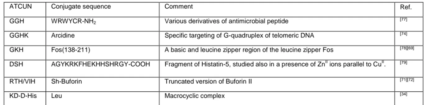
Fragments of proteins or peptides that were already known to exhibit specific DNA binding abilities, such as Hin recombinase,[67] leucin zipper Fos,[68,69] histatin-5,[70] or Buforin II[71,72] have been applied in such a role. Non-protein modifiers or scaffolds can be also used as nucleic acid recognition motifs. Literature includes bis-PNA[73] and acridine[74] as examples. Long et al. proposed a “tandem array” with GGH motifs incorporated within repetitive linear sequences through the Gly-to-δ-Orn substitution.[75] Neupane et al.[34,76] embedded an ATCUN motif inside a peptidic macrocycle created by coupling Lys and Asp side chains together. Such tight structure exhibited more efficient DNA cleavage than its linear counterpart.[34] The conjugates of CuII-XZH complexes exhibiting nuclease activity are listed in Table 3.
Table 3. List of conjugates of CuII-XZH complexes exhibiting nuclease activity
ATCUN Conjugate sequence Comment Ref.
GGH WRWYCR-NH2 Various derivatives of antimicrobial peptide [77]
GGHK Arcidine Specific targeting of G-quadruplex of telomeric DNA [74]
GKH Fos(138-211) A basic and leucine zipper region of the leucine zipper Fos [78][69]
DSH AGYKRKFHEKHHSHRGY-COOH Fragment of Histatin-5, studied also in a presence of ZnII ions parallel to CuII. [79]
RTH/VIH Sh-Buforin Truncated version of Buforin II [71][72]
KD-D-His Leu Macrocyclic complex [34]
Even though the other part of such conjugate may provide a crucial selectivity in DNA recognition, the ATCUN motif is essential for the nuclease activity. It was proposed that the complexed metal ion is a redox center capable of formation of metal-bound hydroxyl radicals that act as intermediate reactive species, causing the H-atom abstraction from the C4 of the 2-deoxyribose, which ultimately leads to a DNA strand break.[38,80] Although historically the Ni(GGH) complex was the first one studied as a potential nuclease,[67,80] most recent studies focused on CuII complexes. Jin and Cowan proposed that the redox activity of CuII/CuIII coordinated to the XZH motif under physiological conditions could generate reactive oxygen species (ROS) that contribute to the localized DNA damage.[81] This reaction required ascorbate in addition to molecular oxygen to proceed and a Fenton-like mechanism was proposed for it. Amino acid replacement, including the usage of D-amino acids in the X and Z positions of the motif improved the binding and
recognition of the DNA minor groove (usually within A/T rich regions), and eventually the nuclease activity.[72] CuII-XZH complexes generated not only single, but also double-strand DNA breaks. This activity was attributed to a long residency time at DNA and a diffusible nature of ROS generated by the complex.[38,72]
Cleavage of RNA was studied mostly for the genetic material of Human Immunodeficiency (HIV)[45,81–83] and hepatitis C (HCV) viruses,[33,84] and yeast tRNAPhe.[85,86] CuII(GGH) and CuII(KGHK) motifs conjugated with Rev1 and Rev2 domains were demonstrated to target their cognate Rev Response Element (RRE) in HIV virus RNA in the presence of ascorbate. The site-selectivity rather than diffused cleavage was attributed to the formation of copper-bound non-diffusible ROS.[82] The activity was preserved under physiological (cellular) conditions in
E. Coli and mammalian Jurkat cells. Both the ATCUN and the
expanded the knowledge concerning the cleavage chemistry showing: (i) irreversibility of RRE RNA cleavage, (ii) possibility of regeneration of reduced CuII-ATCUN-Rev, and (iii) identification of cleavage products attributable to the catalyst-mediated oxidative hydrogen abstraction.[45,83]
The cleavage of HCV RNA was achieved by adopting an analogous strategy, using the CuII complex of GGH as the cleaving unit and assuring RNA sequence recognition by specific peptides identified in separate studies. The IC50 of these complexes in an HCV cellular replicon assay was in the ~1 μM range, yielding promise for further development as anti HCV drugs.[33,84]
The Cu(KGHK) complex was highly selective towards isolated tRNAPhe, cleaving it within D and TψC loops[86]
with a higher specificity than NIII-XZH nucleases, tested previously against the same target.[85] Both DNA and RNA nuclease activities require further studies since many DNA cleaving agents work in vitro, but not under physiological conditions. The underlying redox activity is quite low, due to the high stability of the CuII complexes. Moreover, there are ubiquitous natural competitors for CuII ions and reducing agents (such as GSH), that could potentially “deactivate” metallopeptidic nuclease by reducing and/or pulling the metal out of the XZH complex. Therefore, the design of such molecules should focus also on the complex kinetic stability in order to reach the target DNA cleavage site in an active form.
5.1.2 Protein cleavage by ATCUN complexes
Cowan et al. used the concept of XZH motif attached at the N-terminus of a ligand-binding domain, presented above in the context of nuclease activity, to design artificial proteases. Their first study reported inactivation of human angiotensin converting enzyme (ACE, peptidyl dipeptidase A) by the Cu(KGHK)+ motif, with redox cycling provided by ascorbate. The observed rate constant kobs of 2.9 ± 0.5 × 10⁻² min-1 (t1/2 ~24 min.). This was much slower than compared to the native enzyme ascorbate oxidase with a rate of 1.7 104 M-1s-1.[87] Thus, further development of this novel class of catalytic enzyme inactivators is warranted.[68] Interestingly, the CuII and CoIII(NH
3)2 complexes of KGHK inhibited the enzyme ACE competitively in the absence of the redox agent (ascorbate). In a follow-up study the same authors tested several XZH motifs to select the one best suited for inactivation of rabbit angiotensin-converting enzyme (rACE) and human endothelin-converting enzyme (hECE-1). These results are proposed as a novel strategy for the development of antihypertension agents.[69]
Analogous constructs have also been tested as irreversible inactivators of activity of SrtA, a transpeptidase that anchors surface adherence proteins to the bacterial cell wall, thus being a target for novel antibiotics.[65] The GGH motif was thus linked to amidated peptides LPET and LPETG, cell wall sorting signals which act as sortase-targeting moieties. The LC-MS/MS analysis showed oxidation of Cys-184 and Arg-197 residues at the SrtA active site as the mechanism of inactivation. This result provides a link to section 4.2, dealing with peptides designed to combat pathogenic microorganisms.
5.1.3 Sugar cleavage by ATCUN complexes
A recent study by Yu et. al proposed a novel artificial fucosidase (Cu-GGH-tOL), in the form of a conjugate of the CuII-GGH complex with a truncated form of odorranalectin (tOL: KCFRYPNGVLACT) to produce artificial Bombay phenotype blood (lacking the functional H2 antigen, also known as Oh, or h/h). Their study showed that such conjugate cleaves L-fucose more efficiently than D-glucose. Therefore Cu-GGH-tOL can act as a potential agent that could specifically regress the polysaccharide Fucα1-2Galβ1-4GlcNAc, a residue of regular glycolipids and glycoproteins present on the surface of
erythrocytes, into a disaccharide Galβ1-4GlcNAc, thus forming a H2 antigen depleted analogue present in a Bombay phenotype blood. Cleavage was observed upon addition of mild reducing agents (ascorbate or hydrogen peroxide) that are natural agents under physiological conditions. The importance of this research is driven by the rarity of such blood phenotype and its limited supply in a blood bank reserves.[74]
5.2 Combination with antimicrobial and antifungal peptides
The inspection of Antimicrobial Peptide Database (APD) revealed the presence of nearly 50 antimicrobial peptides (AMP) that contain the N-terminal XZH motif within a total of 2813 AMPs. Only a few example (<5) of AMP containing the N-terminal XH metal-binding motif were listed. Copper has been long known for its antimicrobial activity. Indeed, a few cases of an increase in activity of AMPs with a native XZH motif (such as hepcidin or ixosin[88]) by addition of CuII have been reported towards several microorganisms.[71] Although the mechanisms of activity enhancement by copper have not been well understood, it has been suggested to include Cu-catalyzed ROS production leading to DNA cleavage[89] or other oxidative degradation of biomolecules, like phospholipids.[88] Based on these results, XZH motifs were also grafted to short AMPs (such as anoplin or buforin), which act by interacting with the cell membrane[71] or inhibiting intracellular targets.[90] This can be obtained by elongation of the AMP N-terminus with the XZH sequence or by mutating the 3rd amino acid to His. N-terminal elongation by XZH of the AMP anoplin, pro-apoptotic peptide, and sh-buforin showed an about 2-8 fold increase in activity (without coordinated CuII), but the pre-formed CuII-XZH-AMP complexes did not show any increase in activity compared to XZH-AMP.[71]
Very recently, the impact of Cu-binding to histatin-5, a salivary peptide with a XZH motif, on the fungus Candida albicans was tested. A five-fold decrease of the EC50 from ~5 to ~1 µM was obtained by the Cu-histatin 5 compared to the apo-form. The presence of a bis-His site (two adjacent His) was crucial for the activity and underlined the importance of a CuI-binding site (different from XZH) for the increased activity.[49]
5.3. XZH and XH as chelators to remove and/or redox-silence Cu
Based on the high stability of CuII(XZH) complexes towards oxidation and reduction described above, the motif has been used to redox silence copper. In aqueous solutions CuII is a very efficient catalyst of ascorbate oxidation in the presence of dioxygen and hence produces efficiently ROS like H2O2 and HO°. Thus, addition of peptides bearing XZH motifs abolished almost completely this ROS production by Cu. An early example of such concept is provided by quenching of deleterious oxidative chemistry of the CuII/H
2O2 couple by the N-terminus of human protamine 2 (HP2). Protamines are peptides replacing histones in sperm cells. Notably, impairment of HP2 production is one of major causes of male infertility in humans.[13,91,92] A similar protection against Cu catalyzed ROS production was shown against the complex of Cu with the Aβ1-x peptide. Aβ1-x forms aggregates containing a high Cu content in Alzheimer’s disease brains and can quite efficiently catalyze the production of ROS. Addition of XZH to Cu(Aβ1-x) resulted in a Cu transfer from Aβ1-x to XZH and a concomitant abolishment of ROS production and related cell toxicity.[2,42,93,94] Moreover, as such, XZH was also able to suppress Cu-induced Aβ1-x aggregation. It is tempting to speculate that protection from copper toxicity is a biological function of some endogenous XZH motifs for which no other function has been found.
Following the idea, Franz[48] and co-workers designed a pro-chelator peptide that gets activated enzymatically. The beta-secretase enzyme cleaves the pro-chelator in a designed site,
yielding the XZH motif at the site of Aβ production and a supposed Cu-induced oxidative stress. On the other hand, a strong critique of the chelation approach to treat Alzheimer’s disease was presented recently.[95]
5.4 CuII-XZH for biological imaging
Miyamoto et al.[23] reported the imaging of tumor-bearing mouse by using the N-terminal sequence YYH attached to the peptide octreotide, a somatostatin mimic that binds to the somatostatin receptor. The latter is expressed at relatively high levels in many tumors. The YYH-Octreotide was labelled with 64Cu, an isotope emitting a positon, and hence useful for positon emission tomography (PET). The YYH motif was selected based on its highest kinetic stability of the tested XXH peptides. Further studies are needed to see if the kinetic stability of XZH complexes can be further exploited to find a lead for medicinal applications.
5.5 Cu-sensing
In the past two decades, different sensors have been developed to serve as tools to study the homeostasis and transport of metals in biology. Different strategies have been used but the most common involve the use of fluorescence. Generally, a “turn-on” switch is favorable, in other words, the fluorophore would not be active until it binds the desired metal ion. Unfortunately, this is very difficult to reach for CuII, due to its paramagnetic character resulting in a universal fluorescence quenching. Thus, most of the sensors reported in the literature have a “turn-off” character. One of the first sensors that used an XZH motif for selective CuII sensing was designed by Torrado et.al,[96] constituting a series of pentapeptides based on the XZH motif with the introduced a Dns fluorophore (5-(dimethylamino)naphthalene-1-sulfonamide) at the X position. CuII binding resulted in a strong and selective fluorescence quenching. The selectivity of this sensor was assured by the CuII ability to deprotonate peptide nitrogens, and the lack of quenching by the isostructural, but diamagnetic NIII complex. Using the same premise Jung and Chung[97] designed a CuII selective reporter protein using the Gly-Gly-His sequence added next to the fluorophore in the green fluorescent protein (GFP). The GFP fluorescence was quenched by about 85% upon the CuII binding, thus providing live imaging of CuII fluctuations on the surface of HeLa cells.
Utilizing the backbone of Imperiali and co-workers, Kulak group took the compound R-NH2-Dap-β-Ala-His-Ser-Ser-CONH2 and attached different fluorophores (R= fluorophore = Rhodamine B,
dansyl chloride, fluorescein isothiocyanate) to it.[98] They were able to show that their analogs cleaved DNA efficiently with a minimal concentration of 25 µM, but only in the presence of a reducing agent like ascorbic acid. The attached fluorophore allowed them to monitor the reduction of CuII to CuI, via the fluorescence turn-on, as CuI is diamagnetic and hence does not quench the fluorophore like CuII does.
5. Conclusions
The XH and XZH motifs have been studied concerning their metal-ion coordination chemistry, on the one hand due to the potential physiological role and on the other hand for different applications, e.g. in material science, catalysis, biology and medicine of complexes with CuII, NIII, CoII, AuIII, and PdII ions). For medicinal purposes, CuII-XH/XZH complexes have been studied most avidly, because out of all metals listed above, copper is the only essential metal ion in humans potentially able to bind (CoII is present only in cobalamin). A very convenient advantage of these two motifs is that they are very small (2 or 3 amino acids) and can be very easily added by either recombinant methods or chemical synthesis to any wanted peptide or protein.
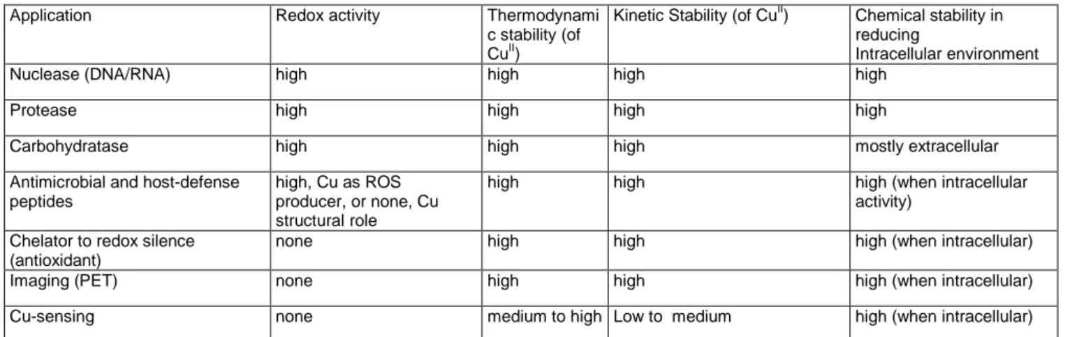
The ability of XH and XZH peptides to bind CuII has gained versatile interest in the literature, regarding mainly XZH peptides. The applications can be divided into two categories (Table 4). i) CuII-XZH complexes are used to produce ROS to cleave biological targets such as proteins, DNA, RNA, lipids or sugars. The efficiency of the cleavage is based on the redox-reactivity of the CuII-XZH complex, but whether it involves CuI/CuII or CuII/CuIII couple is not clear. (In a study of CuII in solution and H2O2 , the redox couple CuI/CuIII was suggested)[99] ii) High stability and redox inertness of CuII-XZH complexes empowers usage of XZH peptides to redox silence loosely bound CuII, and to image and sense CuII. These two categories are somehow contradictory, as on one side a highly efficient ROS production is warranted and on the other hand is the lowest possible. It seems that variations in the sequence can modulate the redox activity, however, these changes impact only the second coordination sphere and hence are limited. The XZH motif is very well suited for a stable CuII coordination, but is a weak ligand for CuI. Thus, the CuI/II redox activity is quite inefficient and a risk exists that during the redox cycle the CuI is released form the peptide. This is particularly the case intracellularly, where strong CuI chelators are present.
Table 4. List of applications of CuII
-XZH/XH applications and their chemical properties
Application Redox activity Thermodynami
c stability (of CuII)
Kinetic Stability (of CuII) Chemical stability in reducing
Intracellular environment
Nuclease (DNA/RNA) high high high high
Protease high high high high
Carbohydratase high high high mostly extracellular
Antimicrobial and host-defense peptides
high, Cu as ROS producer, or none, Cu structural role
high high high (when intracellular
activity) Chelator to redox silence
(antioxidant)
none high high high (when intracellular)
Imaging (PET) none high high high (when intracellular)
Cu-sensing none medium to high Low to medium high (when intracellular)
Concerning the redox activity of the CuII/III couple, CuIII can form a stable complex with XZH, without a large rearrangement compared to CuII. Therefore, the redox based ROS production is
potentially very efficient. However, this is limited by the high redox potential (~1V NHE), a value particularly difficult to achieve in the rather reducing environment of the cell.
The motif XH has been much less studied. A reason is that Cu-XH is generally less stable, particularly kinetically, but at pH 7.4 (and higher pH) also thermodynamically. Moreover, from the electrochemical studies, reduction of CuII-GHK seems accessible by physiological reductants (like superoxide, NADH etc.). This might seem to be of interest for catalytically activity, however, CuI binds very weakly to XH and hence such a CuI-XH complex would not be stable in a biological medium due to the presence of quite strong CuI ligands (like glutathione, methionine etc.).
7. Outlook
As pointed out, there are two different general approaches for the use of XH and XZH, one based on the redox inertness and the other on the redox cycling. Gathering all the results discussed above, it seems that these motifs are rather prone to redox inertness, including the application like detoxification, redox silencing, PET etc. For such application the motif XZH is more suited than XH, due to its higher thermodynamic and kinetic stability. It would be worthwhile to explore variants of XZH to improve both stabilities. There are a lot of thermodynamic binding constants known for XZH, but as discussed above there is no broad rationalization reported that would explain the differences and allow predictions of affinity constants. Up to now, the conditional Kd values at pH 7.4 span from log 12 – 14.7. There might be room to increase the affinities, although likely limited, because the first equatorial coordination sphere cannot be changed or expanded. Nonetheless, rationalization of the relation of sequence with Kd seems a primordial aim for the future.
In contrast to the thermodynamic stability, little work has been done on the kinetic inertness. This seems to be another important question to tackle in the future. For PET imaging and redox silencing kinetic stability is warranted, however for CuII sensing, faster kinetic might be of interest as well, hence making the XH motif of potential high interest.
For applications based on the stability and inertness of Cu-XZH, it is very important not only to explore stability against a competing chelator, but also the redox activity. As the CuII/CuIII potential is very high and CuIII binds strongly to XZH, the key point is the CuII/CuI couple. CuI cannot bind in the same coordination as CuII in XZH, and CuI binds only weakly to XZH. Thus it is very likely that once CuII is reduced to CuI, it will very rapidly be bound by other biomolecules in vivo and the Cu-XZH motif will be lost. Indeed very recently Santoro et al. showed that glutathione and cysteine can pull out CuII from Cu-Aβ4-16 (with XZH) via a reduction to CuI followed by CuI transfer to metallothionein (an abundant ZnII and CuI-binding protein).[100] This reaction is a potential disrupter for all Cu-XH/XZH, in particular intracellularly where mM concentrations of glutathione are present. This type of reactions are defined so far and merit a more detailed analysis.
In potential application based on the redox reactions of CuII -XH/XZH, mainly cleavage of biomolecules (DNA, RNA, proteins etc.) it is still not very clear which redox couple of Cu is involved, although the CuI/II is generally favored. The CuII/III couple has a redox potential above the range for efficient ascorbate oxidation, but has an advantage of retaining the coordination sphere. Thus, the reorganization energy is low and hence redox activity could be very efficient. For this reason, an aim would be to try to reduce the redox potential, by exploring various X, Z and R in the XZHR sequence. However, as this influences mainly the secondary coordination sphere of a quite rigid first equatorial sphere, it might be not possible to lower the redox potential enough. One should also consider that the electrochemical determination of the redox potential does not include the interaction of the substrates (such as O2 and ascorbate) and their binding could change the redox activity.
The other relevant redox couple is CuI/II, which is generally more available in a biological medium due to the presence of quite strong reducing agents. Here the inherent problem is that the
coordination chemistry is very different for CuI and CuII, and XH/XZH is well suited for CuII, but not for CuI. Two problems are involved here:
i) CuI binds very differently to XH/XZH (does not bind amidate) and hence a large reorganization has to occur, meaning that the redox reaction in not efficient. Indeed this is what is observed. This should be somehow improved. A step into this direction is the presence of a CuI binding site nearby, such as a His-His (see above). However, still the reorganization is quite important and it is questionable if this problem can be efficiently solved in the framework of XZH alone.
ii) The CuI affinity of XH/XZH is quite low. Thus there is a high risk that upon reduction of CuII–XH/XZH, the CuI is fast chelated by biomolecules. This is clearly expected as discussed above, and could only be avoided if the CuI is reoxidized faster than the complexation of CuI by a competing biomolecule. This is particular relevant for all intracellular application such as cleavage of DNA, RNA and cytosolic proteins, due to the high concentration of glutathione and other strong reducing agents (Table 4). Hence we consider it as outmost important that for such application their activity in the presence of glutathione is determined and considered as a standard experiment.
Apart from the here discussed improvements and understanding of the motif, concerning the already known application, one can also expect new applications in the future. Most of the work has been done with XZH, which seems to be the most suitable motif for application which needs high thermodynamic and kinetic stabilities (Table 4). However, the motif XH deserves more attention, not only for applications that would need more labile complexation, but also because Cu-XH can form ternary complexes that could change its chemical properties. So far the cleavage of biomolecules has not been applied to lipids. Another interesting and so far unexplored application would be Cu-transporters. So far only solidly confirmed function of Cu-XZH in humans is Cu transport by serum albumin. Hence the motif could be useful for Cu-transporting application concerning distortions in Cu-metabolism such as Wilson’s or Menkes disease.[101,102] Moreover, XH/XZH bind also other metals including medicinally relevant ones, such as AuIII, PtII, PdII that might also expand the applications.
Acknowledgements
This work was sponsored by University of Strasbourg Institute for Advanced Study (USIAS), Strasbourg, France (P.F. and C.H.), installation grant of the Frontier Research in Chemistry Foundation, Strasbourg (P.F.), the ERC StG-638712 (C.H.), and the National Science Centre of Poland grant Nos. 2013/09/B/ST5/03398 and 2016/23/B/ST5/02253 (W.B.)
References
[1] N. Camerman, A. Camerman, B. Sarkar, Can. J. Chem 1976, 54, 1309–1316.
[2] C. Hureau, H. Eury, R. Guillot, C. Bijani, S. Sayen, P.-L. Solari, E. Guillon, P. Faller, P. Dorlet, Chemistry 2011, 17, 10151–10160. [3] I. Sóvágó, C. Kállay, K. Várnagy, Coord. Chem. Rev. 2012, 256,
2225–2233.
[4] M. Sokolowska, A. Krezel, M. Dyba, Z. Szewczuk, W. Bal, Eur. J.
Biochem. 2002, 269, 1323–31.
[5] W. Bal, G. N. Chmurny, B. D. Hilton, P. J. Sadler, A. Tucker, J. Am.
Chem. Soc 1996, 118, 4727–4728.
[6] L. D. Pettit, S. Pyburn, W. Bal, H. Kozlowski, M. Bataille, H. Kozlowski, J. Chem. Soc. Dalt. Trans. 1990, 240, 3565. [7] H. Kozlowski, W. Bal, M. Dyba, T. Kowalik-Jankowska, Coord.
[8] D. L. Rabenstein, S. A. Daignault, A. A. Isab, A. P. Arnold, M. M. Shoukrys, 1985, 107, 6435–6439.
[9] M. Foerster, H. Vahrenkamp, Chem. Ber. 1995, 128, 541–550. [10] A. Krężel, J. Wójcik, M. Maciejczyk, W. Bal, R. W. Jeanloz, D.
Beyersmann, Chem. Commun. 2003, 95, 704–705.
[11] M. Jezowska-Bojczuk, P. Kaczmarek, W. Bal, K. S. Kasprzak, J.
Inorg. Biochem. 2004, 98, 1770–7.
[12] J. P. Crow, J. B. Sampson, Y. Zhuang, J. a Thompson, J. S. Beckman, J. Neurochem. 1997, 69, 1936–1944.
[13] W. Bal, M. Jeżowska-Bojczuk, K. S. Kasprzak, Chem. Res. Toxicol.
1997, 10, 906–914.
[14] G. Brookes, L. D. Pettit, Dalt. Trans. 1975, 2112–2117. [15] I. Sóvágó, E. Farkas, A. Gergely, Dalt. Trans. 1982, 2159–2163. [16] K. Bossak, A. . Protas, W. Bal, Manuscr. Prep. n.d.
[17] K. Bossak, M. Mital, J. Poznański, A. Bonna, S. Drew, W. Bal, Inorg.
Chem. 2016, 55, 7829–7831.
[18] C. Conato, R. Gavioli, R. Guerrini, H. Kozlowski, P. Mlynarz, C. Pasti, F. Pulidori, M. Remelli, Biochim. Biophys. Acta 2001, 1526, 199–210.
[19] A. Trapaidze, C. Hureau, W. Bal, M. Winterhalter, P. Faller, J. Biol.
Inorg. Chem. 2012, 17, 37–47.
[20] M. Rózga, M. Sokołowska, A. M. Protas, W. Bal, J. Biol. Inorg.
Chem. 2007, 12, 913–918.
[21] R. W. Hay, M. M. Hassan, C. You-Quan, J. Inorg. Biochem. 1993, DOI 10.1016/0162-0134(93)85619-J.
[22] M. Mital, N. E. Wezynfeld, T. Fraczyk, M. Z. Wiloch, U. E.
Wawrzyniak, A. Bonna, C. Tumpach, K. J. Barnham, C. L. Haigh, W. Bal, et al., Angew. Chemie Int. Ed. 2015, 54, 10460–10464. [23] T. Miyamoto, Y. Fukino, S. Kamino, M. Ueda, S. Enomoto, Dalt.
Trans. 2016, 45, 9436–9445.
[24] A. Kolozsi, A. Jancsó, N. V Nagy, T. Gajda, J. Inorg. Biochem. 2009,
103, 940–947.
[25] D. Płonka, W. Bal, Inorg. Chim. Acta 2017, DOI 10.1016/j.ica.2017.06.051.
[26] H. Kozłowski, W. Bal, M. Dyba, T. Kowalik-Jankowska, Coord.
Chem. Rev. 1999, 184, 319–346.
[27] I. Sovago, K. Osz, Dalt. Trans. 2006, 3841–3854.
[28] I. Sovago, K. Varnagy, N. Lihi, A. Grenacs, Coord. Chem. Rev.
2016, 327–328, 43–54.
[29] P. Młynarz, N. Gaggelli, J. Panek, M. Stasiak, G. Valensin, T. Kowalik-Jankowska, M. L. Leplawy, Z. Latajka, H. Kozłowski, Dalt.
Trans. 2000, 0, 1033–1038.
[30] I. Zawisza, M. Rózga, W. Bal, Coord. Chem. Rev. 2012, 256, 2297– 2307.
[31] T. Branch, P. Girvan, M. Barahona, L. Ying, Angew. Chem. Int. Ed.
Engl. 2015, 54, 1227–30.
[32] H. Sigel, R. B. Martin, Chem. Rev. 1982, 82, 385–426. [33] M. J. Ross, S. S. Bradford, J. A. Cowan, E. Drouet, M. Jamin, S.
Das, M. R. Conte, Y. A. Jang, E. H. Ko, Dalt. Trans. 2015, 44, 20972–20982.
[34] K. P. Neupane, A. R. Aldous, J. A. Kritzer, Inorg. Chem. 2013, 52, 2729–2735.
[35] J. Nagaj, K. Stokowa-Sołtys, I. Zawisza, M. Jeżowska-Bojczuk, A. Bonna, W. Bal, J. Inorg. Biochem. 2013, 119, 85–89.
[36] M. Z. Wiloch, I. Ufnalska, A. Bonna, W. Bal, W. Wroblewski, U. E. Wawrzyniak, J. Electrochem. Soc. 2017, 164, G77–G81. [37] L. F. Wong, J. C. Copper, D. W. Margerum, J. Am. Chem. Soc.
1976, 98, 7268–74.
[38] Y. Jin, J. A. Cowan, J. Am. Chem. Soc. 2005, 127, 8408–8415. [39] M. Mital, N. E. Wezynfeld, T. Frączyk, M. Z. Wiloch, U. E.
Wawrzyniak, A. Bonna, C. Tumpach, K. J. Barnham, C. L. Haigh, W. Bal, et al., Angew. Chemie - Int. Ed. 2015, 54, 10460–10464. [40] M. Z. Wiloch, U. E. Wawrzyniak, I. Ufnalska, A. Bonna, W. Bal, S. C.
Drew, W. Wroblewski, J. Electrochem. Soc. 2016, 163, G196–G199. [41] P. Gonzalez, B. Vileno, K. Bossak, Y. El Khoury, P. Hellwig, W. Bal,
C. Hureau, P. Faller, Inorg. Chem. n.d.
[42] L. Perrone, E. Mothes, M. Vignes, A. Mockel, C. Figueroa, M.-C. Miquel, M.-L. Maddelein, P. Faller, ChemBioChem 2009, 11, 110– 118.
[43] N. E. Wezynfeld, E. Stefaniak, K. Stachucy, A. Drozd, D. Plonka, S. C. Drew, A. Krezel, W. Bal, Angew. Chemie Int. Ed. 2016, 55, 8235–8238.
[44] M. Mital, I. A. Zawisza, M. Z. Wiloch, U. E. Wawrzyniak, V. Kenche, W. Wróblewski, W. Bal, S. C. Drew, Inorg. Chem. 2016, 55, 7317– 7319.
[45] J. C. Joyner, J. A. Cowan, J. Am. Chem. Soc. 2011, 133, 9912– 9922.
[46] M. Jensen, A. Canning, S. Chiha, P. Bouquerel, J. T. Pedersen, J. Østergaard, O. Cuvillier, I. Sasaki, C. Hureau, P. Faller, Chem. - A
Eur. J. 2012, 18, 4836–4839.
[47] S. Schwab, J. Shearer, S. E. Conklin, B. Alies, K. L. Haas, J. Inorg.
Biochem. 2016, 158, 70–76.
[48] K. L. Haas, A. B. Putterman, D. R. White, D. J. Thiele, K. J. Franz, J.
Am. Chem. Soc. 2011, 133, 4427–4437.
[49] S. E. Conklin, E. C. Bridgman, Q. Su, P. Riggs-Gelasco, K. L. Haas, K. J. Franz, Biochemistry 2017, 56, 4244–4255.
[50] R. A. Himes, G. Y. Park, A. N. Barry, N. J. Blackburn, K. D. Karlin, J.
Am. Chem. Soc. 2007, 129, 5352–5353.
[51] C. Hureau, Coord. Chem. Rev. 2012, 256, 2164–2174.
[52] R. A. Himes, G. Y. Park, G. S. Siluvai, N. J. Blackburn, K. D. Karlin,
Angew. Chemie 2008, 120, 9224–9227.
[53] J. C. Joyner, J. Reichfield, J. A. Cowan, J. Am. Chem. Soc. 2011,
133, 15613–26.
[54] S. K. Burke, Y. Xu, D. W. Margerum, Inorg. Chem. 2003, 42, 5807– 5817.
[55] D. Russino, E. McDonald, L. Hejazi, G. R. Hanson, C. E. Jones,
ACS Chem. Neurosci. 2013, 4, 1371–1381.
[56] S. Melino, L. Garlando, M. Patamia, M. Paci, R. Petruzzelli, J. Pept.
Res. 2008, 66, 65–71.
[57] K. Kulprachakarn, Y.-L. Chen, X. Kong, M. C. Arno, R. C. Hider, S. Srichairatanakool, S. S. Bansal, J. Biol. Inorg. Chem. 2016, 21, 329–38.
[58] D. K. Bergman, R. N. Ramachandra, S. K. Wikel, J. Med. Entomol.
1998, 35, 505–509.
[59] W. Duntze, D. Stötzler, E. Bücking-Throm, S. Kalbitzer, Eur. J.
Biochem. 1973, 35, 357–65.
[60] L. Pickart, J. H. Freedman, W. J. Loker, J. Peisach, C. M. Perkins, R. E. Stenkamp, B. Winstein, Nature 1980, 288, 715–717.
[61] L. Pickart, J. Vasquez-Soltero, A. Margolina, Cosmetics 2015, 2, 236–247.
[62] B. Kandemir, L. Kubie, Y. Guo, B. Sheldon, K. L. Bren, Inorg. Chem.
2016, 55, 1355–1357.
[63] J. S. Pap, Ł. Szyrwiel, Comments Inorg. Chem. 2016, 3594, 1–19. [64] Z. Yu, J. A. Cowan, Chem. Eur.J 2017, 23, 14113–14127. [65] E. Kimoto, H. Tanaka, J. Gyotoku, F. Morishige, L. Pauling, Cancer
Res. 1983, 43.
[66] C. M. Agbale, M. H. Cardoso, I. K. Galyuon, O. L. Franco,
Metallomics 2016.
[67] D. P. Mack, P. B. Dervan, Biochemistry 1992, 31, 9399–9405. [68] C. Harford, S. Narindrasorasak, B. Sarkar, Biochemistry 1996, 35,
4271–4278.
[69] T. Mahmoudi, B. Sarkar, Biopolymers 1999, 50, 273–286. [70] S. Melino, C. Santone, P. Di Nardo, B. Sarkar, FEBS J. 2014, 281,
657–672.
[71] M. D. Libardo, J. L. Cervantes, J. C. Salazar, A. M. Angeles-Boza,
ChemMedChem 2014, 9, n/a-n/a.
[72] M. D. J. Libardo, T. J. Paul, R. Prabhakar, A. M. Angeles-Boza,
Biochimie 2015, 113, 143–155.
[73] M. Footer, M. Egholm, S. Kron, J. M. Coull, P. Matsudaira,
Biochemistry 1996, 35, 10673–10679.
[74] Z. Yu, M. Han, J. A. Cowan, Angew. Chemie Int. Ed. 2015, 54, 1901–1905.
[75] M. A. Lewis, K. M. Williams, Y.-Y. Fang, F. A. Schultz, E. C. Long,
Curr Bioact Compd 2014, 10, 13–20.
[76] K. P. Neupane, A. R. Aldous, J. A. Kritzer, J. Inorg. Biochem. 2014,
139, 65–76.
[77] J. C. Joyner, W. F. Hodnick, A. S. Cowan, D. Tamuly, R. Boyd, J. A. Cowan, J. Bürck, C. Muhle-Goll, A. Ulrich, S. Keller, et al., Chem.
Commun. 2013, 49, 2118.
[78] C. Harford, S. Narindrasorasak, B. Sarkar, Biochemistry 1996, 35, 4271–4278.
[79] S. Melino, M. Gallo, E. Trotta, F. Mondello, M. Paci, R. Petruzzelli,
Biochemistry 2006, 45, 15373–15383.
[80] Q. Liang, D. C. Ananias, E. C. Long, J. Am. Chem. Soc 1998, 120, 248–257.
[81] Y. Jin, J. A. Cowan, JBIC J. Biol. Inorg. Chem. 2007, 12, 637–644. [82] Y. Jin, J. A. Cowan, J. Am. Chem. Soc. 2006, 128, 410–411. [83] J. C. Joyner, K. D. Keuper, J. A. Cowan, Chem. Sci. 2013, 4, 1707–
1718.
[84] S. Bradford, J. A. Cowan, Chem. Commun. 2012, 48, 3118. [85] I. J. Brittain, X. Huang, E. C. Long, Biochemistry 1998, 37, 12113–
12120.
[86] S. Bradford, Y. Kawarasaki, J. A. Cowan, J. Inorg. Biochem. 2009,
103, 871–875.
[87] P. Kroneck, F. Armstrong, H. Merkle, A. Marchesini, Advances in
Chemistry, Ascorbic Acid:Chemistry, Metabolism, and Uses,
American Chemical Society, Washington, D.C., 1982.
[88] M. D. J. Libardo, V. Y. Gorbatyuk, A. M. Angeles-Boza, ACS Infect.
Dis. 2016, 2, 71–81.
[89] C. A. Álvarez, F. Guzmán, C. Cárdenas, S. H. Marshall, L. Mercado,
Fish Shellfish Immunol. 2014, 41, 93–101.
[90] C. B. Park, K. S. Yi, K. Matsuzaki, M. S. Kim, S. C. Kim, Proc. Natl.
Acad. Sci. U. S. A. 2000, 97, 8245–50.
[91] W. Bal, J. Lukszo, K. S. Kasprzak, Chem. Res. Toxicol. 1997, 10, 915–921.
[92] R. Liang, S. Senturker, X. Shi, W. Bal, M. Dizdaroglu, K. S. Kasprzak, Carcinogenesis 1999, 20, 893–898.
[93] C. Hureau, I. Sasaki, E. Gras, P. Faller, ChemBioChem 2010, 11, 950–953.
[94] D. Russino, E. McDonald, L. Hejazi, G. R. Hanson, C. E. Jones,
ACS Chem. Neurosci. 2013, 4, 1371–1381. [95] S. C. Drew, Front. Neurosci. 2017, 11, DOI
10.3389/fnins.2017.00317.
[96] A. Torrado, G. K. Walkup, B. Imperiali, J. Am. Chem. Soc. 1998,
120, 609–610.
[97] Y.-A. Choi, J. O. Keem, C. Y. Kim, H. R. Yoon, W. Do Heo, B. H. Chung, Y. Jung, Chem. Sci. 2015, 6, 1301–1307.
[98] C. Wende, N. Kulak, Chem. Commun. 2015, 51, 12395–12398. [99] A. N. Pham, G. Xing, C. J. Miller, T. D. Waite, J. Catal. 2013, 301,
54–64.
[100] A. Santoro, N. Wezynfeld, M. Vasak, W. Bal, P. Faller, Chem.
Commun. 2017, 53, 11634–11637.
[101] B.-E. Kim, T. Nevitt, D. J. Thiele, Nat. Chem. Biol. 2008, 4, 176–185. [102] P. Delangle, E. Mintz, Dalt. Trans. 2012, 41, 6359–6370.

![Table 2. Conditional affinity constants for Cu II complexes of XH and XZH motifs, obtained by recalculation of original potentiometric data by the CI method [10,11] or by calorimetric (a) or spectroscopic (b) titration](https://thumb-eu.123doks.com/thumbv2/123doknet/13659932.429415/4.892.457.821.219.598/conditional-constants-complexes-recalculation-potentiometric-calorimetric-spectroscopic-titration.webp)