Author miss-numbered some pages.
Page numbers for page 103 & 104
are repeated. Numbering goes back
to normal at page 150. Author
Distal Hydrogen-Bonding Effects and
Cofacial Bimetallic Salen Architectures for
Oxygen Activation Chemistry
Jenny Yue-fon YangB. S., University of California, Berkeley (2001)
Submitted to the Department of Chemistry In Partial Fulfillment of the Requirements
For the Degree of
DOCTOR OF PHILOSOPHY IN INORGANIC CHEMISTRY at the
MASSACHUSETTS INSTITUTE OF TECHNOLOGY April 2007
© 2007 Massachusetts Institute of Technology. All rights reserved.
Signature of Author:
Department of Chemistry
SDepartmento
eApril,
2007
Certified by:
/
Daniel G. NoceraW. M. Kec ssor of Energy and Professor of Chemistry Thesis Supervisor
Accepted by:
Robert W. Field Haslam and Dewey Professor of Chemistry Chairman, Department Committee on Graduate Studies
LIBRARIES
ARCH,1,~
MASSACHUSETTS INSTITUTEOF TECHNOLOGY"
This doctoral thesis has been examined by a Committee of the Department of Chemistry as follows:
A .A t'
Professor Joseph P. Sadighi
S " " Professor of Chemistry
Chairman
A
Professor Daniel G. Nocera j--'-- "- "
-W. l. ck Professorf Vnergy and Professor of Chemistry Thesis Supervisor
Professor Stephen J. Lippard
0 Artur Anr Noyes Professor of Chemistry
Abstract
Distal Hydrogen-Bonding Effects and Cofacial Bimetallic Salen
Architectures for Oxygen Activation Chemistry
By Jenny Yue-fon Yang
Submitted to the Department of Chemistry on April 18, 2007 in partial fulfillment of
the requirements for the degree of Doctor of Philosophy in Inorganic Chemistry
Abstract
Two distinct structural scaffolds elaborated from Schiff-base macrocycles were designed to study the proton-coupled electron transfer chemistry of 0-0 bond forming and activation chemistry. The "Hangman" architecture is composed of hydrogen-bonding functionalities poised over a redox active manganese salophen or salen platform. The complexes proved to be proficient catalase mimics (disproportionation of hydrogen peroxide to water and oxygen). Detailed spectroscopic, computational, and structure-function relationship studies elucidated the key redox, steric, and secondary coordination sphere effects for optimal catalytic ability. The incorporation a chiral backbone into the macrocycle led to catalysts that perform enantioselective epoxidation of unfunctionalized olefins. A macrocycle with an amide, imine, and bisphenolic functionalities was also incorporated as the redox platform in the Hangman framework; the manganese complex also performed catalytic oxygen atom transfer to olefins. The second framework, dubbed "Pacman", is composed of two salen platforms linked cofacially by rigid pillars xanthene or dibenzofuran. A series of bimetallic complexes, including chromium, iron, manganese, cobalt, copper, and zinc were generated. Mossbauer spectroscopy was used in the characterization of the iron salen complexes, which were also examined for photolytic oxidation chemistry.
Thesis supervisor: Daniel G. Nocera
Contents
Table of contents
Title page . . . . . Thesis Committee . Dedication ... Abstract ... Table of contents . List of Figures . . . List of Schemes . . List of Charts . . . List of Tables . . . List of abbreviations Introduction1. Proton-Coupled Electron Transfer in Nature ...
Proton-Coupled Electron Transfer in Synthetic Systems Photolytic Generation of Metal Oxo Species
Schiff-base Macrocycles as Redox Platforms References .... ..
Chapter 2 Hangman Salophen Mediated Activation of 0-0 Bonds:
M echanistic Insights ...
1. Motivation and Specific Aims . ...
2. Background
3. Results and Discussion
3.1. Synthesis .... ... .
3.2. Stop-Flow Spectroscopy
3.3 Decomposition in the Presence of Hydrogen Peroxide 3.4. Mechanistic Implications
3.5. Generation of Manganese Salophen Oxo with Hydrogen P ero x id e . . . . . . . . . . . . . 4. Concluding Remarks. .. ... ... ... . . . . . . . . . . . . . . . . . . . I . . 1 . . . . . . . . 2 . . . . . . . . . . . . . . . . . . 3 . . . . . . . . 5 .. ..... .. .. .. . . . . . . . . . . . . . . . . . 7 ...... ... ... . . . 1 2 ...... ... . . . 1 7 ...... .. .... . . . 1 9 ... ... .. .... .. . 2 0 ...... .. ... 2 3 Chapter 1 26 27 29 20 32 .. 39 40 40 .. 42 42 . 43 47 47 50 53
Contents
5. Experimental5.1 General Methods ... .
5.2. Physical Methods ....
5.3. Synthesis ....
5.4. Stop-Flow Kinetic Studies ..
5.5. Hydrogen Peroxide Disproportionation Reactions 5.6. Independent Generation of High-Valent Mn(V) oxo 6. References and Notes
apter 3 Synthesis of Enantiopure Manganese Hangman Salens and
Their Epoxidation Activity . . . .
1. Motivation and Specific Aims ...
2. Background . .. .
3. Results and Discussion 3.1. Ligand Design
3.2. Synthesis of Hangman Salen Ligands (HSX*) ...
3.3. Synthesis of Benzoic Acid Functionalized Hangman Salen Ligands (HphSX*). . ... . . ....
3.4.
Epoxidation Activity of Manganese Hangman Salens
...
3.5.
Design of Sterically Protected Hangman Salens
3.6. Synthesis of Mn[HSX*tBu]CI and Mn[HphSX*tBu]Cl
3.7.
Epoxidation Activity of Sterically Protected Manganese
Hangman Salens ..
...
3.8.
Mechanistic Implications
4.
Concluding remarks
5. Experimental Section 5.1. Materials 5.2. Physical Measurements ... 5.3. Synthesis ... 5.4. Epoxidation of 1,2-Dihydronapthalene ....5.5. X-Ray Crystal Data Collection and Refinement Parameters 6. References
Catalase Activity and Epoxidation of Functionalized Olefins by
Manganese Hangman Salens ... . ...
53 53 54 54 58 59 60 62 Ch 65 66 66 67 67 68 72 75 75 77 78 79 80 81 81 82 82 97 98 102 105 Chapter 4
Contents
1. Motivation and Specific Aims .. ... . 106
2. Background ... . .... . .. .. 106
3. Results and Discussion . ... ... .. . 107
3.1. Synthesis . . . . 107
3.2. Catalase Activity of Manganese Hangman Salen Complexes 109 3.3. Mechanistic Insights ... 111
3.4. Synthesis of Methyl Esters of Sterically Protected Manganese Hangman Salen Complexes 113 3.5. Catalase Activity of Sterically Protected Manganese Hangman Salen Complexes . . .... .. ... 114
3.6. Mechanistic Insights into Steric Protection 115 3.7. Density Functional Theory of Manganese Hydroperoxide Hangman Salen Complexes ... . 116
3.8. Acid to Metal Distance Effects . 119 3.9. Synthesis of Dibenzofuran Analogues of Manganese Hangman S a len s . . . . . . . . . . . .. . . . ... 1 19 3.10. Density Functional Theory of Dibenzofuran Analogues of Manganese Hangman Salens 121 3.11. Catalase Activity: Acid to Metal Distance Effects 122 3.12 Epoxidation ofFunctionalized Olefins 124 4. Conclusions and Future Work .. . 126
5. Experimental Section .... 128
5.1. Materials 128 5.2. Physical Measurements .. ... .. 129
5.1. Synthesis ... ... . 129
5.2. Hydrogen Peroxide Disproportionation Reactions 143 5.1. Epoxidation of Functionalized Olefins 144 5.2. Density Functional Theory Calculations ... 145
7. References ... 148
Chapter 5 Synthesis of Amido-Imine Macrocycle Ligands ... 153
1. Motivation and Specific Aims .. . .. 154
2. Background ... .. .. .. ... .. .. .. 154
Contents
3.1. Synthetic Attempts towards a Hangman Salen Bearing a Single
Functionalized Xanthene Scaffold 155
3.2. Design and Synthesis of Amido-Imine Macrocycles and
Manganese Complexes 156
3.2. Epoxidation Activity of Manganese Complexes 158
4. Concluding Remarks 158 5. Experimental Section 159 5.1. Materials 159 5.2. Physical Methods 159 5.3. Synthesis 160 5.3. Epoxidation Measurements 167 6. References .. 168
Chapter 6 Synthesis of Cofacial Pacman Salen Architectures and Their
Iron Complexes 171
1. Motivation and Specific Aims 172
2. Background 172
3. Results and Discussion 174
3.1. Ligand Design 174
3.2. Synthesis of DiSalen Xanthene Ligand (DSX) 175 3.3. Synthesis of DiSalen Dibenzofuran Ligand (DSD) 178 3.4. Synthesis of Ferric Chloride Complexes 180
3.5. Synthesis of Diiron p-oxo Complexes 181
3.6. Synthesis of Ferrous Complexes 182
3.7. 57Fe M6ssbauer Spectroscopy of Iron Complexes 183
3.8. Photolysis Studies of Iron Complexes 186
4. Concluding remarks 187 5. Experimental Section 188 5.1. Materials 188 5.2. Physical Measurements 188 5.3. Synthesis 189 5.4. Photolysis Studies 195
5.5 X-Ray Crystal Data Collection and Refinement Parameters 196
Contents
Ch apter 7. Synthesis of Bimetallic Cofacial Pacman Salens
1. Motivation and Specific Aims ...
2. B ackground . . . . ... . . . . . . . . 3. Results and Discussion ...
3.1. Ligand Characteristics and Synthesis ... 3.2. Synthesis of Chromium Pacman Complexes ... 3.3. Epoxide Ring Opening with Chromium Pacman Co 3.4. Synthesis of Copper Pacman Complexes ... 3.5. Synthesis of Manganese, Cobalt, and Zinc Pacman 4. Concluding Remarks and Future Work ...
5. Experim ental Section ... ...
5.1. M aterials . . . . . . . 5.2. Physical M easurements ...
5 .3 . Synth esis . .. . . . 5.4. Epoxide Ring Opening Studies ...
6. References .... 207 .... .. 208 .... .. 208 . .. . . . 209 209 209 nplexes 210 . .. . . . 2 10 omplexes 211 212 . .. . . . 2 12 . .. . . . 2 12 213 . .. . . . 2 13 . .. . . . 2 17 219
Reaction Setup for Measuring Catalase Activity . ... B. Alternative Synthetic Procedures .. ...
B. 1. From Chapter 3 ... .. . .. . ... B.2. From Chapter 5 .. ...
C. Density Functional Theory Calculations ... C. 1. Input File and Output Coordinates for the hydroperoxide
complex of Mn(HSX*) ...
C.2. Input File and Output Coordinates for the hydroperoxide complex of Mn(HphSX*) with carboxylic acid dimers. C.3. Input File and Output Coordinates for the hydroperoxide
complex of Mn(HSD*) .
C.4. Input File and Output Coordinates for the hydroperoxide complex of Mn(HphSD*) 221 222 223 223 225 228 228 233 239 244 Acknowledgements .... Biographical note ... . Curriculum vitae .... 249 252 253 C Appendix A.
Contents List of Figures Chapter 1 Figure 1.1. Figure 1.2. Figure 1.3. Chapter 2 Figure 2.1. Figure 2.2. Figure 2.3. Figure 2.4.
Representation of the active sites (a) peroxidases, (b) catalases, and (c)
cytochrome P450 monooxygenases highlighting the proposed roles distal residues play in O--O bond cleavage.
Proposed catalytic cycle of the catalase-like disproportionation of hydrogen peroxide by iron (HPX-CO2H) and epoxidation by manganese (HPX-CO2H).
Proposed catalytic cycle for the photolytic oxidation catalysis performed by diiron Pacman porphyrin complexes, and summary of
substrates and turnover numbers observed.
Reaction profiles of 02 evolution over time from the dismutation of H202 from manganese Hangman salophen catalysts with X = OMe
(]), Br (0), H (o), t-Butyl (A), and NO2 ().
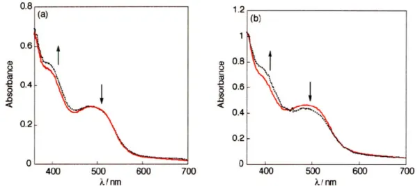
(a) Stopped-flow UV-vis spectra obtained from reaction of m-CPBA (3.3 x 10⁻⁴) with 4.0 x 10⁵ M 1 (red solid line) ca. 3 s post injection in
1:1 MeOH:MeCN at -20 'C. (b) UV-vis spectrum obtained from a solution of 5.0 x 10⁻⁵ M 1 in MeOH:MeCN at room temperature (red solid line). UV-vis spectrum obtained from a solution of 5.0 x 10⁻⁵ M
1 in 1:1 MeOH:MeCN at room temperature in the presence of benzoic acid (5.0 x 10⁻³ M) (black dotted line).
(a) Absorption spectra obtained from spectral global analysis of a stopped-flow reaction of4.0 x 10-5 M land 3.3 x 10- M m-CPBA in
1:1 MeOH:MeCN at -20 'C. Formation of Mn(V) oxo (green solid line) is immediately followed by a bleach, likely to a Mn" decay product (red solid line) over 800 s. (b) Calculated concentrations of the colored species with respect to time by spectral global analysis by Jake
Soper.
Observation of a common oxidized Hangman salophen intermediate by treating Mn[HSX(OMe)-COOH]CI (1) with three different
Contents
Figure 2.5. Studying high-valent Mn-salophens: (a) comparison of the UV-Vis spectra of Mn-oxo complexes derived from Mn[salophen(t-bu)] and Mn[salophen(OMe)], respectively; (b) comparison of the UV-Vis spectra of Mn(V)-nitrido complexes derived from Mn[salophen(t-bu)] and Mn[salophen(OMe)], respectively.
Figure 2.6. Reaction of Mn-oxo with a reductant leads to the regeneration of the Mn(III) starting material.
Chapter 3
Figure 3.1. Potential approaches of an olefin to Jacobsen's Catalyst. The high valent oxo bound to the manganese (not shown) is perpendicular to and coming out of the page.
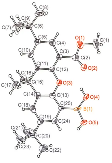
Figure 3.2. Thermal ellipsoid plot of 5-(boronic acid)-2,7-di-tert-butyl-9,9-dimethyl-9H-xanthene-4-carboxylic acid methyl ester (3). Thermal ellipsoids are drawn at the 50% probability level.
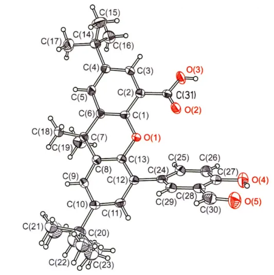
Figure 3.3. Thermal ellipsoid plot of
2,7-di-tert-butyl-5-(3-formyl-4-hydroxyphenyl)-9,9-dimethyl-9H-xanthene-4-carboxylic acid (5). Thermal ellipsoids are drawn at the 50% probability level.
Figure 3.4. Thermal ellipsoid plot of 4-Bromo-2,7-di-tert-butyl-9,9-dimethyl-5-(4-hydroxyborane)-9H-xanthene (19). Thermal ellipsoids are drawn at the 50% probability level.
Chapter 4 Figure 4.1.
Figure 4.2.
Turnover number (TON) of hydrogen peroxide dismutation catalyzed by manganese compounds [Mn(5-phsalen)Cl] (13) (13* in the
presence of 2 equivalents of benzoic acid), Mn[HSX*-COOH]CI, Mn[HSX*-COOMe]Cl (9), COOH]Cl and Mn[HphSX*-COOMe]CI (10) after 1 hour.
Oxygen release from hydrogen peroxide dismutation catalyzed by manganese compounds Mn(5-phsalen)Cl] (13) ( ), 13* (*in the presence of 2 equivalents of benzoic acid) (0), Mn[HSX*-COOH]Cl (o), Mn[HSX*-COOMe]Cl (9) (x), Mn[HphSX*-COOH]Cl (+), and Mn[HphSX*-COOMe]Cl (10) (o) over 1 hour. Inset shows reaction profile for the first five minutes excluding the methyl ester compounds 9 and 10.
Contents
Figure 4.3. Turnover number (TON) of hydrogen peroxide dismutation catalyzed by manganese compounds Mn(salen)C1, Mn(salen)Cl in the presence of 2 equivalents of benzoic acid), Mn[HSX*tBu-COOH]CI, Mn[HSX*
tBu-COOMe]C1 (18), Mn[HphSX* tBu -COOH]Cl and Mn[HphSX* tBu-COOMe]CI (19) after 1 hour.
Figure 4.4. Energy minimized structure obtained from DFT of the hydroperoxide complexes of Mn(HSX*-COOH), showing one of the carboxylic acids
is hydrogen bonded to the oxygen on the xanthene scaffold, while the other is hydrogen bonded to the hydroperoxide.
Figure 4.5 Energy minimized structure obtained from DFT of the hydroperoxide complexes of Mn(HphSX*-COOH) showing the two carboxylic acids
span the face of the salen macrocycle to make a -(COOH) 2- dimer that
interacts with the hydroperoxide via a hydrogen bond.
Figure 4.6 Energy minimized structure obtained from DFT of the hydroperoxide complexes of Mn(HphSX*-COOH) with the xanthenes splayed away from each other, precluding formation of the -(COOH)2- dimer. Each
of the carboxylic acids has a hydrogen- bonding interaction with the hydroperoxide.
Figure 4.7 Energy-minimized structures obtained from DFT of the hydroperoxide complex Mn[HSD*tBu]OOH.
Figure 4.8 Energy-minimized structures obtained from DFT of the hydroperoxide complex Mn[HphSD*tBu]OOH.
Chapter 5
Figure 5.1. Equilibrium representing the exchange reaction that produces symmetric impurities when salens containing two unique phenolate arms undergo hydrolysis. The two unique salicylaldehyde components are represented in blue (dashed) and red (solid).
Figure 5.2. ' H NMR of compound 17.
Figure 5.3. 'H NMR spectra of compound 19.
Chapter 6
Figure 6.1. Variable temperature 1H NMR spectra of DiSalen Xanthene ligand (4) from room temperature (20 °C) to -60 'C, taken in 10 'C intervals.
Contents
Figure 6.2. X-Ray crystal structure of DiSalen Xanthene (8). Crystals were grown from slow evaporation of a pentane-dichloromethane solution.
Carbons are depicted as gray, oxygens as red, and nitrogens as blue. H atoms are omitted for clarity.
Figure 6.3. X-Ray crystal structure of DSD impurity. Crystals were grown out of a pentane-dichloromethane solution. Carbons are depicted as gray. Figure 6.4. X-Ray crystal structure of the unpillared Fe2(salen)20 (15). Crystals
were grown from a pentane-dichloromethane solution. The angle of the Fe-O-Fe bond is 165.2(7)0, and the salens are by rotated 124.50 with respect to each other.
Figure 6.5. Infrared spectra displaying the fingerprint region of Fe2DSXO (13),
Fe2DSDO (14), and Fe2(salen)20 (15), highlighting the imine (C=N)
and Fe-O-Fe bond stretch.
Figure 6.6. Fitted 57Fe MLssbauer spectra of (a) Fe2DSXCI2 (10), (b) Fe2DSDC12
(11), (c) Fe(salen)Cl (12) at 4.2 K. Gray dots represent the
experimental data points and the black solid line represents the fit. The vertical axis is an arbitrary transmission scale.
Figure 6.7. Fitted 57Fe Mdssbauer spectra of (a) Fe2DSXO (13), (b) Fe2DSDO
(14), (c) Fe2(salen)20 (15) at 4.2 K. Gray dots represent the
experimental data points and the black solid line represents the fit. The vertical axis is an arbitrary transmission scale.
Figure 6.8. Fitted 57Fe M6ssbauer spectra of(a) Fe
2DSX (16), (b) Fe2DSD (17),
(c) Fe(salen) (18) at 4.2 K. Gray dots represent the experimental data points and the solid black line represents the fit. The vertical axis is an arbitrary transmission scale.
Figure 6.9. UV-vis absorption profiles of 30 imol solutions of Fe2DSXCl2 (13)
(---), Fe2DSDCl2 (14) ('"), and Fe2(salen)20 (15) (-) in acetonitrile. The
spectra is scaled to molar absorbtivity.
Figure 6.10 X-Ray crystal structure of 4,6-(4,4,5,5-Tetramethyl-[1,3,2]dioxaborolan-2-yl)-dibenzofuran (6).
Chapter 7
Figure 7.1. EPR spectra of compound 11 taken a chloroform-ethanol frozen solution at 4.5 K.
Contents
Appendix
Contents
List of Schemes
Chapter 1
Scheme 1.1. Hangman Architecture
Chapter 2 Scheme 2.1. Scheme 2.1. Chapter 3 Scheme 3.1. Scheme 3.2. Scheme 3.3. Scheme 3.4. Scheme 3.5. Scheme 3.6. Scheme 3.7. Scheme 3.8. Chapter 4 Scheme 4.1. Scheme 4.2. Scheme 4.3. Synthesis
Proposed catalytic cycle for iron Hangman porphyrin and manganese Hangman salophen compounds. The porphyrin compounds are indicated in green, and the salophen in black. The catalase reaction is represented in blue. The stop-flow kinetic studies were performed using the red reagents, as well as the rates that can be determined using the double-mixing setup.
Synthesis Synthesis Synthesis Synthesis Synthesis Synthesis Synthesis
Proposed catalytic cycle for substrate oxidation by a Hangman catalyst, highlighting the potential interaction of the carboxylic acid
functionality with the external oxidant O-X
Synthesis Synthesis
Proposed catalytic cycle for the dismutation of hydrogen peroxide by manganese salens, highlighting substrate assembly by the hanging groups to facilitate oxidation
Contents
Scheme 4.4. Scheme 4.5. Scheme 4.1. Scheme 4.1. Chapter 5 Scheme 5.1. Scheme 5.2 Scheme 5.3 Chapter 6 Scheme 6.1. Scheme 6.2 Scheme 6.3 Synthesis SynthesisProposed interaction between a functionalized olefin and the high valent metal oxo of a Hangman salen complex
Synthesis
Synthesis Synthesis Synthesis
Proposed catalytic cycle in the photocatalytic oxidation chemistry of diiron g-oxo Pacman porphyrin complexes.
Synthesis Synthesis
Chapter 7
Scheme 7.1. Proposed bimetallic intermediate in the nucleophilic epoxide ring opening reactions catalyzed by metallosalens.
Appendix
Contents
List of Charts
Chapter 1 Chart 1.1. Chapter 2 Chart 2.1. Chart 2.2. Chart 2.3. Chapter 3 Chart 3.1. Chart 3.2 Chart 3.3 Chapter 4 Chart 4.1. Chapter 5 Chart 5.1. Chapter 6 Chart 6.1. Chart 6.2. Chapter 7 Chart 7.1.Porphyrins and Schiff-base Macrocycles
Hangman Salophen Architecture and PCET Manganese Hangman Salophens
Manganese Salophens
Jacobsen's Catalyst and Epoxidation Hangman Salens
Sterically Protected Hangman Salens
Dibenzofuran Analogues of Hangman Salens
Hangman Salen Architecture and Salient Features
Xanthene and Dibenzofuran Pillared Pacman Salen Ligands Iron Complexes
Contents
List of Tables
Chapter 2
Table 2.1. Redox potential of manganese (HSX) chloride compounds with varying X functionalities in the 5 and 5' position, observed LMCT band in the UV-visible spectrum, and the amount of time after injection that half of 4388 equivalents of H202 was measured to be
consumed by monitoring the amount of oxygen evolved
Table 2.2. Rates of the formation of Mn(V) oxo intermediates and their decay for a series of Mn[salophen(X)] peroxyacid complexes
Table 2.3 Stability of Methoxy-Substituted Manganese Salophen Complexes Table 2.4 Stopped-Flow Kinetic Studies on the Formation of Mn(V)-oxo
Intermediates
Chapter 3
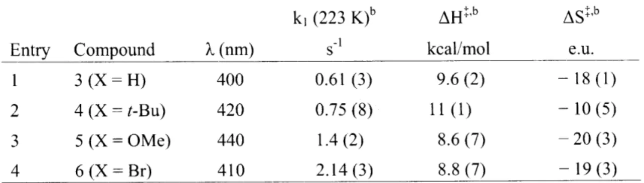
Table 3.1. Turnover numbers (TON) for epoxidation of 1,2-dihydronapthalene in dichloromethane
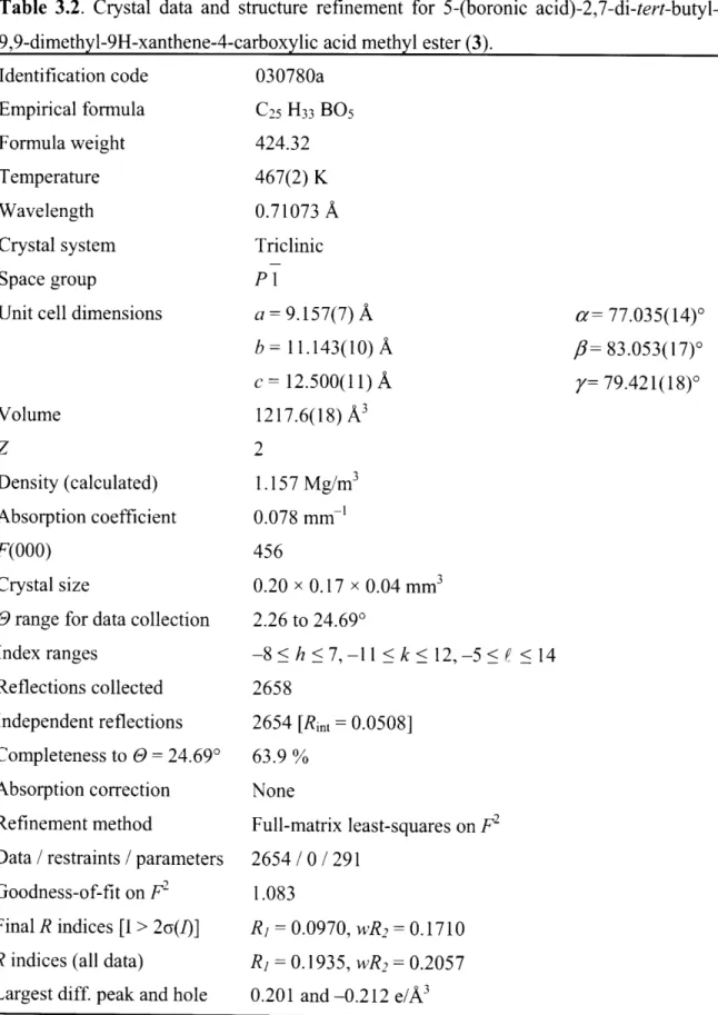
Table 3.2. Crystal data and structure refinement for 5-(boronic acid)-2,7-di-tert-butyl-9,9-dimethyl-9H-xanthene-4-carboxylic acid methyl ester (3).
Table 3.3. Crystal data and structure refinement 2,7-di-tert-butyl-5-(3- formyl-4-hydroxy-phenyl)-9,9-dimethyl-9H-xanthene-4-carboxylic acid (5).
Chapter 4
Table 4.1. Observed turnover number (TON) and initial rate constant (ki)
for Catalase-like H202 disproportionation catalyzed by
manganese catalysts in 2: 1 dichloromethane/methanol at 25 C. Table 4.2. Total bond energies of calculated manganese Hangman salen
compounds.
Table 4.3 Observed Turnover Number (TON) for H202 Disproportionation by
Contents
Table 4.4
Table 4.5
Chapter 6
Analysis of products and turnover numbers (TON) for epoxidation reactions as determined using GC/MS with 4-acetoxystyrene (R = H) and 2,2-dimethyl-propionic acid 4-vinyl-phenyl ester (R = t-bu) over I and 3 hours, using sodium hypochlorite as the oxidant.
Selected bond distance parameters from DFT calculated energy minimized structures of the hydroperoxide complexes of manganese Hangman salen compounds.
Table 6.1. Summary of M6ssbauer parameters for compounds 10 - 18 recorded at 4.2 K. 8 is relative to elemental iron. All samples were measured in the solid state (powdered). F is the half-width at half-maximum of the peaks, and the value presented is the same for both peaks.
Table 6.2 Crystal data and structure refinement for DSX (4). Table 6.3 Crystal data and structure refinement for
4,6-(4,4,5,5-Tetramethyl-[ 1,3,2]dioxaborolan-2-yl)-dibenzofuran (6). Table 6.4 Crystal data and structure refinement for structure shown in
Figure 6.3.
Table 6.5 Crystal data and structure refinement for structure shown in Figure 6.4.
Appendix
Table C. 1 Cartesian coordinates from the DFT calculated energy minimized structure of the hydroperoxide complex of Mn(HSX*). The structure is shown in Figure 4.4. Table C.2 Cartesian coordinates from the DFT calculated energy
minimized structure of the hydroperoxide complex of
Mn(HphSX*) with the xanthenes oriented so the carboxylic acids can form a hydrogen bonded dimer. The structure is shown in Figure 4.5.
Table C.3 Cartesian coordinates from the DFT calculated energy minimized structure of the hydroperoxide complex of Mn(HSD*). The structure is shown in Figure 4.7.
Contents
Table C.4 Cartesian coordinates from the DFT calculated energy minimized structure of the hydroperoxide complex of Mn(HphSD*). The structure is shown in Figure 4.8.
Contents
List of Abbreviations
Ac acetate
ADF Amsterdam Density Functional
anal analytical
bn benzyl
BSA bovine serum albumin
Bu butyl
C Celsius
calcd calculated
CcO Cytochrome c Oxidase
cm centimeteter
Cy cyclohexane
DCC 3-dicyclohexylcarbodiimide DFT Density functional theory DMAP 4-dimethylaminopyridine
DME dimethoxyethane
DMF dimethylforamide
DMS dimethyl sulfide
DMSO dimethyl sulfoxide
dppf (diphenylphosphino)ferrocene
ee enantiomeric excess
EPR electron paramagnetic spectroscopy A-EQ quadropole splitting value
ESI electrospray ionization
Et ethyl exec excitation FG functional group FT fourier transform g gram GC gas chromatography
GGA generalized gradient approximation HRMS high resolution mass spectroscopy
Hz hertz
init
initial
i-Pr iso-propyl IR infrared K Kelvin kJ kilojouleContents LMCT lwp M max Mb m-CPBA mg Me MeCN mL MeOH MHz mm mM mmol MS nm NMR OAT OEC OMe PCET Ph PSII ROS S SOD t T t-Bu THF TON TMP TS UV-vis V
ligand to metal charge transfer long wavelength pass
molar maximum myoglobin m-chloroperbenzoic acid milligram methyl acetonitrile milliliter methanol megahertz millimeter milimolar milimol mass spectroscopy nanometer
nuclear magnetic resonance oxygen atom transfer Oxygen Evolving Complex methoxy
proton-coupled electron transfer phenyl
Photosystem II
reactive oxygen species spin state Superoxide dismutase temperature time tert-butyl tetrahydrofuran turnover number tetramesitylporphyrin transition state ultraviolet-visible volume
Chapter 1
1.1 Proton Coupled Electron Transfer in Nature
The principal theme of the research presented is the elucidation of mechanistic details in the coupled transport of both protons and electrons in bond-making and bond-breaking catalysis.1-4 This is a recurring theme throughout a broad range of natural systems that encompass a diverse range of reactivity. Consummate examples are the four electron oxidation of water to oxygen by Photosystem II (PSII),5-9 and its microscopic reverse, the reduction of oxygen to water by cytochrome c oxidase (CcO),'o-17 which highlights our interests in 0-0 bond chemistry.
While electron transfer at active sites is typically regulated via a transition metal platform or cluster, proton transfer is mediated through non-covalent hydrogen-bonding interactions in the secondary coordination sphere. A prototypical example of the dual importance of electron and proton control by structure and function can be found in heme-dependent hydroperoxidases.18-2 2 A ferric protoporphyrin IX prosthetic group is
found at the active site of peroxidases, catalases, and the cytochrome P450 monooxygenases, as shown in Figure 1.1. Structurally oriented acidic or basic amino acid residues and/or locally hydrogen bonded water assist 0-0 bond cleavage of hydrogen peroxide in peroxidases and catalases, and oxygen in the cytochrome P450 monooxygenases. These interactions on the distal oxygen, frequently characterized as the "pull effect",18-22
promote heterolytic bond cleavage to form the two electron oxidized Compound I intermediate,2 0-24 as opposed to the one electron oxidized Compound II
Figure 1.1 Representation of the active sites (a) peroxidases, (b) catalases, and (c) cytochrome P450 monooxygenases highlighting the proposed roles distal residues play in O-O bond cleavage. Adapted from reference 67.
species that results from homolytic bond cleavage. The reactivity of the of Compound I species in these subclasses then diverge; peroxidases tend to behave as one electron oxidants,22,'25,26 while catalases are selective in their reactivity towards the two electron
oxidation of hydrogen peroxide,22'26-28 and cytochrome P450 monoxygenases perform
two electron oxygen atom transfer chemistry to a variety of substrates. 19 The tuned microenvironment guides proton and electron delivery within the active site, assisting in O-0O bond activation, and the subsequent chemistry of the oxidizing intermediate.
The importance of this microenvironment has successfully been demonstrated by modifying natural enzymes. By adjusting the distance and position of distal residue(s) over the heme environment in the oxygen carrier myoglobin (Mb), peroxidase, catalase, or enantioselective monooxygenase activity is observed.18,29-47 Additionally, the effect of the redox environment has been studied independently by inserting unnatural redox cofactors to generate artificial metalloenzymes.48-64 Alternatively, these individual factors
can be analyzed in greater detail using a synthetic system which incorporates the key factors that mediate reactivity in the natural systems we are targeting.
1.2 Proton Coupled Electron Transfer in Synthetic Systems
The complexity of tuning the precise factors which dictate PCET in biological systems has led to the use of molecular models to elucidate important non-covalent interactions in oxidation catalysis. A minimalist secondary coordination sphere is constructed onto a ligand platform which can support metals in a wide range of oxidation states, as shown in Scheme 1.1. This Hangman architecture in effect "hangs" an acid-base functional group over the redox platform using a rigid scaffold.65 Synthetic methods can be used to tune
the redox properties of the metal, while the scaffold can be modified to specify the spatial
location and pKa of the distal group. This allows structure-function relationships to be probed using more direct methods.
The initial redox platform incorporated into this architecture was a porphyrin macrocycle with a carboxylic acid functionality (HPX-CO2H), as shown in Figure 1.2.6568 The iron
porphyrin platform faithfully reproduces many of the essential features of heme-dependent hydroperoxidases, including a ferric hydroperoxide and Compound I and Compound II type intermediates. Additionally, crystallographic characterization of the ferric hydroxide complex reveals a water molecule oriented by the acid and bound hydroxide. This is reminiscent of water molecules situated by distal amino acid residues in heme peroxidases.66 The effect of the hanging group is highlighted in the reactivity of
the Hangman complexes. The iron Hangman complexes are effective catalase mimics, generating turnover numbers (TON) more than two orders of magnitude over the unfunctionalized redox platform (Figure 1.2).67,
68 Stop-flow spectroscopic studies of the
reaction using the oxidant m-CPBA found the presence of the hanging group both accelerated peroxy acid ligation and exclusively favored the proton-coupled two-electron 0-0 bond heterolysis to form a Compound I-like intermediate, as opposed to a less oxidizing Compound II-like intermediate.69 In the presence of olefin substrate and
Figure 1.2. Proposed catalytic cycle of the catalase-like disproportionation of hydrogen peroxide by iron (HPX-CO2H), and epoxidation by manganese
(HPX-CO2H). Adapted from reference 67.
TO~N >100C (>450) (02) R/ R R R (H)20,) R R mes H202 S 0--HO0. H 0 0 mes / Nt1 N e - N mM o t-Bu mes M = MnV M = FelV(por +) H20
hydrogen peroxide, the manganese Hangman complexes catalyzed epoxidation at improved TON over the manganese porphyrin lacking the hanging group.67 In both cases,
the exceptional improvement in catalytic activity due to the hanging acid group is due to the proton mediated formation of the more oxidizing high valent M=O species.
1.3 Photolytic Generation of Metal Oxo Species
In Nature, O-0O bond formation is performed by the oxygen evolving complex (OEC) in Photosystem II. In the proposed mechanism, 0-0 bond formation is initiated via a nucleophilic hydroxide or water attack onto an electrophilic manganese oxo.6
'9 Initially,
we are interested in studying the formation and electrophilic character of high valent metal oxo species. Both of these factors can be probed by their reaction with a variety of electron rich substrates. (Analogous to the use of olefin epoxidation in investigating high valent oxo formation in the manganese hangman porphyrins described above.) This approach has been successful in the cofacial bimetallic "pacman" porphyrin system that has also been explored in this research program.70-77 The Fe-O bond in diiron p-oxo
pacman porphyrin complexes can be photolytically cleaved to expose an iron(IV) oxo
Figure 1.3. Proposed catalytic cycle for the photolytic oxidation catalysis performed by diiron pacman porphyrin complexes, and summary of substrates and turnover numbers observed.78 82 subsbaf Tdh ONO DMS DMSO 9635 ± 165 0 0C dG H3 150 * 27 (10%) H 1609 340 (87%) 37 ± 6 (3%) 0 Ph-'*'Ph Ph p Ph 160 * 25 OH 116 14(45%) U6 143 101(56%)
0
S101
S
C"3 --- cr, 76*11capable of oxidation chemistry.78-82 Detailed mechanistic studies resulted in ligand architecture modifications that dramatically increased turnover numbers.81'82 This was
coupled with the use of electron withdrawing groups on the porphyrin to increase the electrophilic nature of the iron oxo intermediate and thus the ability to oxidize more difficult substrates, as shown in Figure 1.3. The photolytic generation of a high valent metal oxo from a thermally inert diiron i-oxo complex to perform oxidation chemistry using molecular oxygen as the oxidant was an additional avenue of reactivity that we were interested in expanding.
1.4 Schiff-base Macrocycles as Redox Platforms
The use of porphyrin macrocycles to support the redox platform in Hangman and Pacman porphyrin architectures was very successful in producing effective catalysts for oxidation chemistry. Furthermore, modulating functional groups around the porphyrin platform allowed tuning of the steric, redox, and photophysical properties in order to expand the scope of substrate and catalytic conditions. While synthetic preparation of variously functionalized porphyrins is well established, most procedures still require lengthy, multi-step synthesis, particularly in order to allow attachment to the xanthene and dibenzofuran spacers in constructing the Hangman and Pacman ligands. Alternatively, the replacement of the porphyrins with Schiff-base macrocycles as the redox platform in our architectures engenders several advantages. Firstly, they are easily assembled via condensation of two salicylaldehydes with a diamine, as shown in the retrosynthesis in Chart 1. lb. Secondly, a large variety of functionalized salicylaldehydes exists commercially and in the literature,
Chart 1.1 R* N Nj (a) "R N\/ R* N N RR R2 R2 R2 R2 R1. R, R R- H2N NH2 (b) -- N. _. _,N- 0O O -Sb 5 7H M HO 5' 3 3' 3 3'
and can typically be prepared in one or two steps.83-87 The 3 and 5 positions are
highlighted in Chart 1.1lb as they are activated and thus the easiest positions to functionalize. Combined with the scope of aliphatic and aromatic diamines that can be used, this constitutes a modular approach to a wide range of macrocycles with tunable steric and redox properties. Additionally, given the rich oxidation chemistry displayed by the porphyrin hangman and pacman complexes, one of the features that would be appealing to add to the catalytic chemistry is enantioselectivity. Although some metalloporphyrins catalysts have, through elaborate synthetic pathways, incorporated asymmetric functional groups to promote chiral induction of substrates, the enantiomeric excess (ee) of the products tend to be poor, and the scope of substrates limited.88-95 This is
partly due to the lack of sp3 carbons near the metal center; addition of chiral
functionalities has to occur on the perimeter of the macrocycle (Chart 1.1a). As shown in Chart 1.1.b, an aliphatic diamine provides a location for enantioselective induction proximate to the reactive metal center. Macrocycles of this type have already demonstrated excellent stereochemical communication to a wide variety of substrates in organic transformations.96-105 We are interested in integrating these features into the catalysts that we have studied thus far.
References
1. Chang, C. J.; Chang, M. C. Y.; Damrauer, N. H.; Nocera, D. G. Biophys. Biochim.
Acta 2004, 1655, 13-28.
2. Stubbe, J.; Nocera, D. G.; Yee, C. S.; Chang, M. C. Y. Chem. Rev. 2003, 103, 2167-2202.
3. Cukier, R. I.; Nocera, D. G. Annu. Rev. Phys. Chem. 1998, 49, 337-369.
4. Dempsey, J. L.; Esswein, A. J.; Manke, D. R.; Rosenthal, J.; Soper, J. D.; Nocera, D. G. Inorg. Chem. 2005, 44, 6879-6892.
5. Yachandra, V. K.; Sauer, K.; Klein, M. P. Chem. Rev. 1996, 96, 2927-2950. 6. Tommos, C.; Babcock, G. T. Acc. Chem. Res. 1998, 31, 18-25.
7. Yocum, C. G.; Pecoraro, V. L. Curr. Opin. Chem. Biol. 1999, 3, 182-187. 8. Dismukes, G. C. Science 2001, 292, 447-448.
9. Vrettos, J. S.; Limburg, J.; Brudvig, G. W. Biochim. Biophys. Acta 2001, 1503, 229-245.
10. Babcock, G. T.; Wikstr6m, M. Nature 1992, 356, 301-309.
11. Ramirez, B. E.; Malmstr6m, B. G.; Winkler, J. R.; Gray, H. B. Proc. Natl. Acad.
Sci. U.S. A. 1995, 92, 11949-11951.
12. Ferguson-Miller, S.; Babcock, G. T. Chem. Rev. 1996, 96,2889-2907.
13. Michel, H.; Behr, J.; Harrenga, A.; Kannt, A. Annu. Rev. Biophys. Biomol. Struct. 1998, 27, 329-356.
14. Gennis, R. B. Proc. Natl. Acad. Sci U.S.A. 1998, 95, 12747-12749. 15. Brzezinski, P. Biochim. Biophys. Acta 2000, 1458, 1-5.
16. Wikstrfm, M. Biochim. Biophys. Acta 2000, 1458, 188-198.
17. Schultz, B. E.; Chan, S. 1. Annu. Rev. Biophys. Biomol. Struct. 2000, 30, 23-65. 18. Ozaki, S.-I.; Roach, M. P.;Matsui, T.; Watanabe, Y. Acc. Chem. Res. 2001, 34,
818-825.
19. Cytochrome P-450 Structure, Mechanism, and Biochemistry; P. R. Ortiz de
Montellano, Ed.; Plenum Press: New York, 1986.
20. Sono, M.; Roach, M. P.; Coulter, E. D.; Dawson, J. H. Chem. Rev. 1996, 96, 2841-2887.
21. Dawson, J. H. Science 1988, 240, 433-439.
23. Suslick, K. S. "Shape-Selective Oxidation by Metalloporphyrins" In The Porphyrin
Handbook; Kadish, K. M.; Smith, K. M.; Guilard, R., Eds.; Academic Press: San
Diego, 2000; Vol. 4, pp 41-63.
24. Watanabe, Y. "High-Valent Intermediates" In The Porphyin Handbook; Kadish, K. M.; Smith, K. M.; Guilard, R., Eds.; Academic Press: San Diego, 2000; Vol. 4, pp 97-117.
25. Hiner, A. N. P.; Raven, E. L.; Thomrneley, R. N. F.; Garcia-Canovas, F.; Rodriguez-Lopez, J. N. J. Inorg. Biochem. 2002, 91, 27-34.
26. Jones, P. J. Biol. Chem. 2001, 276, 13791-13796.
27. Nicolls, P.; Fita, I.; Lowen, P. C. "Enzymology and Structure of Catalases" In
Advances in Inorganic Chemistry; Academic Press: New York, 2001; Vol. 51, pp
51-106.
28. Sivaraja, M.; Goodin, D. B.; Smith, M.; Hoffman, B. M. Science 1989, 245, 738-740.
29. Adachi, S.-I.; Nagano, S.; Ishimori, K.; Watanabe, Y.; Morishima, I. Biochemistry, 1993, 32, 241-252.
30. Rao, S. I.; Wilks, A.; Ortiz de Montellano, P. R. J. Biol. Chem. 1993, 268, 803-809. 31. Levinger, D. C.; Stevenson, J.-A.; Wong, L.-L. J. Chem. Soc., Chem. Commun.
1995, 2305-2306.
32. Ozaki, S.-I.; Matsui, T.; Watanabe, Y. J. Am. Chem. Soc. 1996, 118, 9784-9785. 33. Nagano, S.; Tanaka, M.; Ishimori, K.; Watanabe, Y.; Morishima, I. Biochemistry,
1996, 35, 14251-14258.
34. Ozaki, S.-I.; Matsui, T.; Watanabe, Y. J. Am. Chem. Soc. 1997, 119, 6666-6667. 35. Matsui, T.; Ozaki, S.-I.; Watanabe, Y. J. Biol. Chem. 1997, 272, 32735-32738. 36. Murakami, T.; Moishima, I.; Matsui, T.; Ozaki, S.-I.; Watanabe, Y. J. Chem. Soc.,
Chem. Commun. 1998, 773-774.
37. Murakami, T.; Morishima, I.; Matsui, T.; Ozaki, S.-I. Hara, I.; Yang, H.-J.; Watanabe, Y. J. Am. Chem. Soc. 1999, 121, 2007-2011.
38. Wan, L.; Twitchett, M. B.; Eltis, L. D.; Mauk, A. G.; Smith, M. Proc. Natl. Acad
Sci. U.S.A. 1998, 95, 12835-12831.
39. Ozaki, S.-I.; Yang, H.-J.; Matsui, T.; Goto, Y.; Watanabe, Y. Tetrahedron:
Asymmetry 1999, 10, 183-192.
40. Matsui, T.; Ozaki, S.-I.; Liong, E.; Phillips Jr., G. N.; Watanabe, Y. J. Biol. Chem. 1999, 274, 2838-2844.
42. Sigman, J. A.; Kwok, B. C.; Lu, Y. J. Am. Chem. Soc. 2000, 122, 8192-8196.
43. Ozaki, S.-I.; Hara, I.; Matsui, T.; Watanabe, Y. Biochemistry 2001, 40, 1044-1052. 44. Kato, S.; Yang, H.-J.; Ueno, T.; Ozaki, S.-I.; Phillips Jr., G. N.; Fukuzumi, S.;
Watanabe, Y. J. Am. Chem. Soc. 2002, 124, 8506-8507.
45. Yang, H.-J.; Matsui, T.; Ozaki, S.-I.; Kato, S.; Ueno, T.; Phillips Jr., G. N.; Fukuzumi, S.; Watanabe, Y. Biochemistry, 2003, 42, 10174-10181.
46. Kato, S.; Ueno, T.; Fukuzumi, S.; Watanabe, Y. J. Biol. Chem. 2004, 279, 52376-52381.
47. Pfister, T. D.; Ohki, T.; Ueno, T.; Hara, I.; Adachi, S.; Makino, Y.; Ueyama, N.; Lu, Y.; Watanabe, Y. J. Biol. Chem. 2005, 280, 12858-12866.
48. KiNello, R. K.; Dolphin, D. H. J. Biol. Chem. 1981, 256, 6903-6912.
49. Hayashi, T.; Tomokuni, T.; Mizutani, T.; Hisaeda, Y.; Ogoshi, H. Chem. Lett. 1998, 1229-1230.
50. Ryabov, A. D.; Goral, V. N.; Gorton, L.; Cs6regi, E. Chem. Eur. J. 1999, 5, 961-967.
51. Hamachi, I.; Shinkai, S. Eur. J. Org. Chem. 1999, 1999, 539-549.
52. Hayashi, T.; Hitomi, Y.; Kaimura, A.; Tomokuni, A.; Mizutani, T. Hisaeda, Y.; Ogoshi, H. Coord. Chem. Rev. 1999, 190-192, 961-974.
53. Monzani, E.; Alzuet, G.; Casella, L.; Redaelli, C.; Bassani, C.; Sanangelantoni, A. M.; Gullotti, M.; Gioia, L. D.; Santagostini, L.; Chillemi, F. Biochemistry 2000, 39,
9571-8582.
54. Hayashi, T.; Hitomi, Y.; Ando, T.; Mizutani, T.; Hisaeda, Y.; Kitagawa, S.; Ogoshi, H. J. Am. Chem. Soc. 1999, 121, 7747-7750.
55. Lu, Y.; Berry, S. M.; Pfister, T. D. Chem. Rev. 2001, 101, 3047-3080. 56. Hayashi, T.; Hisaeda, Y. Acc. Chem. Res. 2002, 35, 35-43.
57. Hayashi, T.; Matsuda, T.; Hisaeda, Y. Chem. Lett. 2003, 32, 496-497.
58. Ohashi, M.; Koshiyama, T.; Ueno, T.; Yanase, M.; Fujii, H.; Watanabe, Y. Angew.
Chemie. lnt. Ed. 2003, 42, 1005-1008.
59. Sato, H.; Hayashi, T.; Ando, T.; Hisaeda, Y.; Ueno, T.; Watanabe, Y. J. Am. Chem.
Soc. 2004, 126, 436-437.
60. Ueno, T.; Ohashi, M.; Kono, M.; Kondo, K.; Suzuki, A.; Yamane, T.; Watanabe, Y.
Inorg. Chem. 2004, 43, 2852-2858.
61. Hayashi, T.; Nakagawa, T.; Harada, K.; Matsuo, T.; Hitomi, Y.; Hisaeda, Y. Chem
62. Ueno, T.; Koshiyama, T.; Ohashi, M.; Kondo, K.; Kono, M.; Suzuki, A.; Yamane, T.; Watanabe, Y. J. Am. Chem. Soc. 2005, 127, 6556-6562.
63. Sato, H.; Wantanbe, M.; Hisaeda, Y.; Hayashi, T. J. Am. Chem. Soc. 2005, 127, 56-57.
64. Ueno, T.; Koshiyama, T.; Abe, S.; Yokoi, N.; Ohashi, M.; Nakajima, H.; Watanabe, Y. J. Organomet. Chem. 2007, 692, 142-147.
65. Chang, C. J.; Yeh, C.-Y.; Nocera, D. G. J. Org. Chem. 2002, 67, 1403-1406. 66 Yeh, C.-Y.; Chang, C. J.; Nocera, D. G. J. Am. Chem. Soc. 2001, 123, 1513-1514.
67. Chang, C. J.; Chng, L. L.; Nocera, D. G. J. Am. Chem. Soc. 2003, 125, 1866-1876. 68. Chng, L. L.; Chang, C. J.; Nocera, D. G. Org. Lett. 2003, 5, 2421-2424.
69. Soper, J. D.; Kryatov, S. V.; Rybak-Akimova, E. V.; Nocera, D. G. J. Am. Chem.
Soc. 2007, ASAP, DOI: 10.1021/ja0683032.
70. Deng, Y.; Chang, C. J.; Nocera, D. G. J. Am. Chem. Soc. 2000, 122, 410-411. 71. Chng, L. L.; Chang, C. J.; Nocera, D. G. J. Org. Chem. 2003, 68,4075-4078.
72. Chang, C. J.; Deng, Y.; Heyduk, A. F.; Chang, C. K.; Nocera, D. G. Inorg. Chem. 2000, 39, 959-966.
73. Chang, C. J.; Deng, Y.; Peng, S-M.; Lee, G.-H.; Yeh, C.-Y.; Nocera, D. G. Inorg.
Chem. 2002, 41, 3008-3016.
74. Chang, C. J.; Loh, Z-H.; Deng, Y.; Nocera, D. G. Inorg. Chem. 2003, 42, 8262-8269.
75. Chang, C. J.; Deng, Y.; Shi, C.; Chang, C. K.; Anson, F. C.; Nocera, D. G. J. Chem.
Soc., Chem. Commun. 2000, 1355-1356.
76. Loh, Z.-H.; Miller, S. E.; Chang, C. J.; Carpenter, S. D.; Nocera, D. G. J. Phys.
Chem. A 2002, 106, 11700-11708.
77. Chang, C. J.; Lob, Z.-H.; Shi, C.; Anson, F. C.; Nocera, D. G. J. Am. Chem. Soc. 2004, 126, 10013-10020.
78. Pistorio, B. J.; Chang, C. J.; Nocera, D. G. J. Am. Chem. Soc. 2002, 124, 7884-7885.
79. Chang, C. J.; Baker, E. A.; Pistorio, B. J.; Deng, Y.; Loh, Z.-H.; Miller, S. E.; Carpenter, S. D.; Nocera, D. G. Inorg. Chem. 2003, 41, 3102-3109.
80. Hodgkiss, J. M.; Chang, C. J.; Pistorio, B. J.; Nocera, D. G. Inorg. Chem. 2003, 42, 8270-8277.
81. Rosenthal, J.; Pistorio, B. J.; Cling, L. L.; Nocera, D. G. J. Org. Chem. 2005, 70, 1885-1888.
82. Rosenthal, J.; Luckett, T. D.; Hodgkiss, J. M.; Nocera, D. G. J. Am. Chem. Soc. 2006, 128, 6546-6547.
83. Zhang, W.; Loebach, J. L.; Wilson, D. R.; Jacobsen, E. N. J. Am. Chem. Soc. 1990,
112, 2081-2083.
84. Jacobsen, E. N.; Zhang, W.; Muci, A. R.; Ecker, J. R.; Deng, L. J. Am. Chem. Soc. 1991, 113, 7063-7064.
85. Jacobsen, E. N. Asymmetric Catalytic Epoxidation of Unfunctionalized Olefins. In
Catalytic Asymmetric Synthesis, 1st Eds; Ojima, I; Wiley-VCH: New York, 1993;
Chapter 4.2.
86. Katsuki, T. Coord. Chem. Rev. 1995, 140, 189-214. 87. Katsuki, T. J. Mol. Cat. A 1996, 113, 87-107.
88. Groves, J. T.; Myers, R. S. J. Am. Chem. Soc. 1983, 105, 5791-5796. 89. Groves, J. T.; Viski, P. J. Org. Chem. 1990, 55, 3628-3634.
90. Mansuy, D.; Battoni, P.; Renaud, J.-P.; Guerin, P. J. Chem. Soc., Chem. Commun. 1985, 155-156.
91. Naruta, Y.; Tani, F.; Ishihara, N.; Maruyama, K. J. Am. Chem. Soc. 1991, 113,
6865-6872.
92. Collman, J. P.; Zhang, X.; Hembre, R. T.; Brauman, J. I. J. Am. Chem. Soc. 1990,
112, 5356-5357.
93. Konishi, K.; Oda, K.-L.; Nishida, K.; Aida, T.; Inoue, S. J. Am. Chem. Soc. 1992,
114, 1313-1317.
94. O'Malley, S.; Kodadek, T. J. Am. Chem. Soc. 1989, 111, 9116-9117. 95. Halterman, R. L.; Jan, S.-T. J. Org. Chem. 1991, 56, 5253-5254.
96. Lin, G.-Q.; Li, Y.-M.; Chan, A. S. C. Asymmetric Oxidations. In Principles and
Applications of Asymmetric Synthesis; Wiley-Interscience: New York, 2001,
Chapter 4.
97. Jacobsen, E. N. Transition Metal-Catalyzed Oxidations: Asymmetric Epoxidation. In Comprehensive Organometallic Chemistry, 2nd Eds; Wilkinson, G; Stone, F. G. A.; Abel, E. W.; Hegedus, L. S.; Pergamon: New York, 1995; page 1097-1135. 98. Williams, J. M. J. Epoxidation of Alkenes. In Catalysis in Asymmetric Synthesis;
Sheffield Academic Press: Sheffield, 1999, Chapter 4.
99. Jacobsen, E. N.; Wu, M. H. Epoxidation of Alkenes other than Allylic Alcohols. In
Comprehensive Asymmetric Catalysis, Vol 2; Pfaltz, A.; Jacobsen, E. N.;
Yamamoto H.; Springer: Berlin, Heidelberg, New York, 1999: Chapter 18.2. 100. Larrow, J. F.; Jacobsen, E. N. Topics Organomet. Chem. 2004, 6, 123-152.
101. Shyu, H.-L.; Wei, H.-H.; Lee, G.-H.; Wang, Y. J Chem. Soc., Dalton Trans. 2000, 915-918.
102. Sivasubramanian, V. K.; Ganesan, M.; Rajagopal, S.; Ramaraj, R. J. Org. Chem. 2002, 67, 1506-1514.
103. Tokunaga, M.; Larrow, J. F.; Kakiuchi, F.; Jacobsen, E. N. Science 1997, 277, 936-938.
104. Jacobsen, E. N.; Wu, M. H. Ring Opening of Epoxides and Related Reactions. In
Comprehensive Asymmetric Catalysis, Vol 2; Pfaltz, A.; Jacobsen, E. N.;
Yamamoto H.; Springer: Berlin, Heidelberg, New York, 1999: Chapter 35. 105. Jacobsen, E. N. Acc. Chem. Res. 2000, 33, 421-431.
Chapter 2
Hangman Salophen Mediated Activation
of 0-0
Bonds: Mechanistic Insights
Portions of the work presented in this chapter have been published:
Liu, S.-Y.; Soper, J. D.; Yang, J. Y.; Rybak-Akimova, E. V.; Nocera, D. G. lnorg. Chem. 2006, 45, 7572-7574.
2.1. Motivation and Specific Aims
The Hangman ligand architecture, which incorporates an acid functionalized scaffold over a metal-ligand platform, is expanded by replacing the previously studied porphyrin ligand platform with a salophen macrocycle. This modification permits a more facile synthetic introduction of functional groups to the phenolic arms of the salophen, thereby permitting the steric and redox environment of the metal center to be varied more easily. The manganese Hangman salophen compounds are successful catalase mimics, and their reactivity can be tuned by varying the redox properties of the salophen macrocycle. In order to determine the basis for the enhanced activity for the dismutation of hydrogen peroxide, stopped-flow spectroscopic techniques using the peroxyacid m-CPBA to probe intermediates in the catalytic cycle and the kinetics of their formation. This includes heterolysis of the O-0O bond to form a putative Mn(V) oxo. Stopped-flow studies help define the electronic effects that perturb the kinetics of the Mn(V) oxo formation. Conditions to independently generate the Mn(V) salophen oxo using hydrogen peroxide have also been discovered. By comparing our results to those of manganese salophen platforms lacking the acid functionalized scaffold and Hangman porphyrins, insight into the mechanism of catalase activity by manganese salophens is gained.
2.2. Background
Chart 2.1
S1
control of acid-base
properties
X
I PCET
control of redox properties
HSX
Expanding upon the work on the Hangman Porphyrin ligand, HPX'-4 (discussed in
Chapter 1), a series of Hangman Salophen compounds were assembled (HSX, shown in Chart 2.1).5 The modular synthesis of salophen compounds allows for easy synthetic
modification in the functionalities of the 5 and 5' position along the phenolic arms (indicated by X) of the macrocycle. This position exerts a particularly strong influence on the redox properties of the metal center.6 Within the HSX framework, PCET catalysis
was explored in the activation of O-O bonds by studying the disproportionation of H202.
catalase
2 H202 2 H20 + 02 (1)
This catalase reaction is an important PCET process that is catalyzed by a variety of enzymes.7-1 1
In the case of manganese(III) chloride HSX compounds where X =
tert-butyl, the turnover number (TON) for oxygen production upon addition of hydrogen peroxide is dramatically higher with the acid functionalized xanthene (4372 TON/hour) compared to the ester analogue (98 TON/hour), which lacks the acidic proton. The control experiment with the redox only manganese salophen platform (lacking the xanthene scaffold) shows a similarly low activity (86 TON/hour).5 Thus, an enhancement
of catalase activity is observed by positioning a carboxylic acid group over a redox platform. This enhanced reactivity is also observed for iron(III) Hangman porphyrin compounds when compared to iron(III) tetramesityl porphyrin (TMP).3 While similar
reactivity is also observed for Hangman salophen and porphyrin platforms, the former is synthetically much easier to modify. The meso-positions of the porphyrins are not available for modification because they are needed to incorporate steric protection to prevent formation of inactive diiron pt-oxo dimers.12 To this end, a series of HSX analogs
Table 2.1. Redox potential of manganese (HSX) chloride compounds with varying X functionalities in the 5 and 5' position, observed LMCT band in the UV-visible spectrum, and the amount of time after injection that half of 4388 equivalents of H202
was measured to be consumed by monitoring the amount of oxygen evolved.5'13
X Ei/2(Mn"/Mn"') kmax(nm) t (50%) (sec)
OMe -0.36 511 560
Br -0.5 490 860
H 0.0 486 970
t-Bu 0.14 476 1760
-with X = OMe, Br, H, t-Bu, and NO2 were synthesized. This family of manganese(III)
Hangman salophen compounds shows an increase in oxidation potential as the electron with-drawing ability of groups in the 5 and 5' position (shown in Table 2.1) is increased.5'13 The redox potential of the manganese(II/III) couple can be varied from 0.37 V, with the introduction of electron donating methoxy groups to 0.44 V with the introduction of electron withdrawing nitro groups.5"' 3 The electronic perturbation of
substituent groups is reflected in a low energy absorption band,14 which exhibits a
red-shift from 511 nm to 460 nm along the series of increasing electron-donating ability of the substituent. This trend is consistent with a salophen ligand-to-metal charge transfer parentage of the absorption band. Variations in redox potential and electron density at the metal site are directly correlated with the rate of catalase activity as measured by the rate of H202 consumption (shown in Table 2.1 and Figure 2.1). We can also measure
reactivity by comparing the amount of time it takes for half of the H202 equivalents to be
dismutated into oxygen and water. We can also measure reactivity by comparing the amount of time it takes for half of the H202 equivalents to be dismutated into oxygen and
water. The Hangman salophen with X = NO2 displays very little reactivity and only
achieves a TON of 81, or 2% conversion over the hour the reaction was monitored, whereas the methoxy substituted congener is the most successful catalase mimic. There is only a moderate correlation between the initial rates of reaction and the Gpi constant and the sensitivity parameter (p = -0.3) is quite low, which suggests there are little changes in the transition state of the rate-limiting step of the reaction.13 It is likely that under the
biphasic reaction conditions used in these studies, the diffusion of H202 is an important
factor in the rate-determining step for the initial reactivity and this result in the low observed p value.
2.3. Results and Discussion 2.3.1. Synthesis
The compounds that are the focus of this Chapter are shown in Chart 2.2. The acid functionalized manganese Hangman salophen (1) was synthesized as described previously.5 The synthesis of the corresponding methyl ester is outlined in Scheme 2.1. The functionalized xanthene precursor 75 was condensed with two equivalents of 3-tert-butyl-2-hydroxy-5-methoxy-benzaldehyde to give the ligand 8, which was refluxed with
Scheme 2.1
t-Bu
t-Bu
O
C0O
O- OMe
OMe Me,. 0 OMe
Me. 0 Me Mee / \ - ' NH2 N t-Bu - NH2 t-Bu OH t-Bu NH2 tBu MOe 7 8: H2[HSX(OMe)-COOMe] 2: Mn[HSX(OMe-COOMe)]CI
manganese(II) acetate in air, followed by an aqueous sodium chloride wash to give the Mn(III) complex 2. Manganese salophen complex 3 was synthesized for the purposes of performing control experiments; it was prepared by the stepwise condensation of two equivalents of 3-tert-butyl-2-hydroxy-5-methoxybenzaldehyde with 1,2,-phenylenediamine followed by the same metalation procedure described for 2.
2.3.2. Stopped-flow Spectroscopy Chart 2.2 OMe MO t-Bu MeO 1: Mn[HSX(OMe)-COOH]CI, R = H 2: Mn[HSX(OMe)-COOMe]CI, R = Me 3: Mn(salophen(OMe)]CI
In order to understand the role the hanging functionality may play in the catalase activity of manganese salophens, stopped-flow kinetic studies were used to provide spectroscopic evidence of intermediates and their rates of their formation. The stopped-flow spectroscopic experiments were conducted by postdoctoral associate Jake Soper on the acid (1) and ester (2) functionalized manganese Hangman salophens, substituted with methoxy in the 5 and 5' position as shown in Chart 2.2. The unfunctionalized manganese salophen (3) was also investigated to provide a side by side comparison to the Hangman compounds. The experiments were performed using m-CPBA as the oxygen atom source instead of hydrogen peroxide, which would continue to react with the oxidizing
.e 1 ® 0.8 . 0. 0.4 0.2 n 400 500 600 700 400 500 600 700 X nm .I nm
Figure 2.2. (a) Stopped-flow UV-vis spectra obtained from reaction of m-CPBA (3.3 x 10⁻⁴) with 4.0 x 10⁻⁵ M 1 (red solid line) ca. 3 s post injection in 1:1 MeOH:MeCN
at -20 'C. (b) UV-vis spectrum obtained from a solution of 5.0 x 10⁻⁵ M 1 in MeOH:MeCN at room temperature (red solid line). UV-vis spectrum obtained from a solution of 5.0 x 10⁻⁵ M 1 in 1:1 MeOH:MeCN at room temperature in the presence of benzoic acid (5.0 x 10³ M) (black dotted line).
intermediate and complicate kinetic studies. The use of m-CPBA arrests the catalase cycle after formation of the high valent manganese species, enabling detection of the first two intermediates at the Hangman salophen platform.
The initial stage of the reaction upon addition of m-CPBA to 1 is characterized by small changes in the stopped-flow UV-vis spectrum (Figure 2.2a), suggesting a simple ligand substitution by the perbenzoate at the Mn(III) center. This substitution reaction was modeled by the addition of benzoic acid to 1. The benzoate substrate circumvents the possibility of 0-0 bond cleavage, therefore allowing the substitution reaction to be isolated. The small spectral shifts observed for perbenzoate substitution are captured with the benzoate model (Figure 2.2b). Global fitting of the transient spectra indicates that ligand substitution is complete in 3 s at -20 'C.
The appearance of the Mn(III) perbenzoate complex (Figure 2.3a, black dotted line) is immediately followed by a subsequent reaction that generates a species with the spectrum shown by the green line in Figure 2.3a. The spectrum does not depend on the nature of the oxidant; a similar spectrum is obtained with the same isosbestic points when m-CPBA is replaced by the two-electron oxidant, iodosylbenzene. These results lead us to assign
* Nj N
c-i a) 0 CO .0 C .-0 LO LL. 400 500 600 700 0 200 400 600 800 X/ nm time / s
Figure 2.3. (a) Absorption spectra obtained from spectral global analysis of a stopped-flow reaction of 4.0 x 10- 5 M 1 and 3.3 x 10- 4 M m-CPBA in 1:1 MeOH:MeCN at -20
'C. Formation of Mn(V) oxo (green solid line) is immediately followed by a bleach, likely to a Mn"' decay product (red solid line) over 800 s. (b) Calculated concentrations of the colored species with respect to time by spectral global analysis by Jake Soper.
this intermediate to the Mn(V) oxo salophen. Several observations support this assignment. First, the transient spectrum is reminiscent to that of structurally similar, crystallographically characterized Mn(V) oxo complexes of bis-amido bis-alkoxo redox platforms.15 Moreover, the spectral features in Figure 2.3a (green line) concur with those
of other (non-oxo) multiply bonded Mn(V) ligand salophen species. The corresponding nitrido complex, Mn[salophen(OMe)]N complex was also synthesized and isolated. The crystal structure of the compound is similar to other Mn(V) nitrido complexes featuring a formal Mn-N triple bond (d(Mn-N) = 1.523 (3) A). 6 The absorption spectrum of the
Mn(V) complex shows a pronounced absorption band at Xmax = 459 nm, similar to 420
nm feature that dominates the absorption spectrum of Figure 2.3a (green line). The electron-donating methoxy group on the salophen platform of the Mn(V) oxo species impart sufficient stability"7 that it is observed as a stopped-flow transient. The reactive high-valent oxo intermediate eventually disappears 60-800 seconds after substrate injection as evidenced by the decrease of the absorption profile across the entire spectral range (360 - 700 nm). The final spectrum, shown by the red line in Figure 2.3a, strongly resembles the Mn(III) starting material, though the product was not unequivocally identified.

![Figure 2.4. Observation of a common oxidized Hangman salophen intermediate by treating Mn[HSX(OMe)-COOH]CI (1) with three different oxidants: (a) hydrogen peroxide, (b) m-CPBA, (c) iodosylbenzene.](https://thumb-eu.123doks.com/thumbv2/123doknet/14751344.580329/51.918.160.632.225.416/observation-oxidized-hangman-salophen-intermediate-treating-different-iodosylbenzene.webp)
![Figure 2.5. UV-vis of high-valent Mn-salophens: (a) comparison of the spectra of Mn-oxo complexes derived from Mn[salophen(t-bu)] and Mn[salophen(OMe)], respectively; (b) comparison of the spectra of Mn(V)-nitrido complexes derived](https://thumb-eu.123doks.com/thumbv2/123doknet/14751344.580329/52.918.186.761.88.338/salophens-comparison-complexes-salophen-salophen-respectively-comparison-complexes.webp)