The hierarchy of mutations influencing the folding of antibody domains in Escherichia coli
7
0
0
Texte intégral
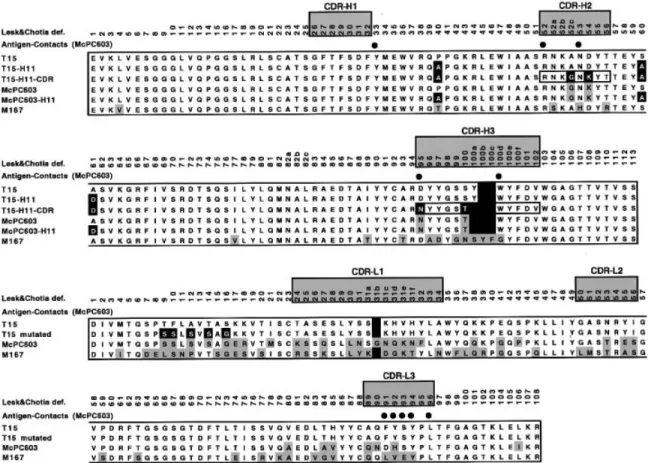
(2) J.G.Wall and A.Plu¨ckthun. streptomycin (25 µg/ml) and ampicillin (100 µg/ml). A 2–5 ml volume of this culture, depending on the OD550, was pelleted by centrifugation and resuspended in 0.5 ml LB. This cell washing was repeated once in order to reduce carry-over of β-lactamase that might break down ampicillin and facilitate the growth of plasmid-free cells. The resuspended cells were then added to 19.5 ml LB containing ampicillin and streptomycin as above and shaken in a water bath at 24°C until an OD of 0.5 was reached. Induction of antibody fragment production was achieved by the addition of 1 mM IPTG (final concentration). At the time of induction and for every hour thereafter, 200 µl samples of culture were removed; of this, 100 µl was used to determine the OD550 of the culture and the remainder to measure the β-lactamase activity in the culture supernatant, according to O’Callaghan and co-workers (1972). As the exact shape of each growth curve can be affected by many environmental parameters, each growth experiment was carried out three times on different days, with all mutants of interest assessed in parallel. Protein production and analysis Following harvesting of induced cultures, soluble and insoluble protein fractions were separated as previously described (Knappik and Plu¨ckthun, 1995). SDS–PAGE and immunoblotting were carried out using standard procedures and bacterially produced antibody fragments were detected using the commercially available M1 antibody (Prickett et al., 1989; Knappik and Plu¨ckthun, 1994), directed against the 4-amino acid FLAG peptide encoded at the N-terminal end of the fragments. Mutagenesis of the antibody fragments Introduction of the previously-described framework H40, H60 and H61 mutations (Kabat numbering), collectively termed the ‘H11 mutations’ (Knappik and Plu¨ckthun, 1995), into the T15 fragments was carried out according to Kunkel and coworkers (1987). Clones into which the mutations had been introduced were confirmed by Sanger dideoxy sequencing. Mutated clones were sequenced between convenient restriction sites in the vector and the mutated DNA was cloned back into the original expression vector for protein production and analysis. Exchange of CDRs between the three antibodies was carried out by restriction digestion cloning. Site-directed mutagenesis of the VL domain was achieved using the splicing by overlap extension method (SOE) of Ho and co-workers (1989). Affinity chromatography Protein production for affinity chromatography was carried out essentially as described by Knappik and Plu¨ckthun (1995). An overnight culture in 10 ml SB medium (20 g/l tryptone, 10 g/l yeast extract, 5 g/l NaCl, 2.5 g/l K2HPO4, 1 g/l Mg2SO4.7H2O) containing streptomycin (25 µg/ml) and ampicillin (100 µg/ml) was used to inoculate 2 l of the same medium in a 5 l culture flask. Cultures were shaken at 24°C until an OD550 of 0.5 was reached and were then induced by the addition of IPTG to a concentration of 1 mM. Induction was allowed to continue until shortly before extensive periplasmic leakiness began to occur, as determined from the smaller scale growth experiments described above. In the case of most clones studied, this involved a 3 h induction period. Cells were then pelleted by centrifugation at 4000 r.p.m. for 20 min at 4°C and resuspended in 30 ml BBS buffer (0.2 M boric acid, 0.16 M NaCl, pH 8.0) on ice. The cells were ruptured 606. by passage through a French pressure cell three times and centrifuged at 20 000 r.p.m. for 15 min. The supernatant was then passed through a 0.22 µM filter and the filtrate was loaded onto a PC-Sepharose 4B column. The column was run in BBS buffer with a flow rate of 1 ml/min and elution was carried out with 10 ml BBS buffer containing 5 mM PC. Aliquots of all fractions were stored at –20°C for analysis by SDS–PAGE and immunoblotting as described above. Screening for antigen binding in solution This method was modelled on that of Kazemier and co-workers (1996) and the affinity chromatography described above. Bacterial cultures were induced for protein production exactly as described above. Following pelleting of cells and resuspension in BBS buffer at a volume of 0.013OD550 at the time of harvesting, the cells were subjected to head-over-head rotation for 1 h at 4°C. After centrifugation at 20 000 r.p.m. for 15 min and passage of the supernatant through a 0.22 µM filter, 0.4 ml of the cell preparation was added to 0.6 ml PC-Sepharose in BBS in a 2 ml Eppendorf tube. Incubation was at 4°C for 1 h with head-over-head rotation. The PC-Sepharose resin was then washed by brief centrifugation in a microfuge, removal of supernatant, and re-addition of 0.4 ml BBS buffer. After five such washes, 0.4 ml BBS buffer containing 12.5 mM PC was added and the resin was incubated at 4°C for 30 min with head-over-head rotation. To check the specificity of the binding, this procedure was carried out in duplicate, with one of the cell preparations incubated with 10 mM PC for 30 min prior to incubation with the PC-Sepharose resin. The supernatant fractions from the washing and elution steps were analysed by SDS–PAGE and Coomassie blue staining or immunoblotting. Results The alignment of the heavy and light chain variable regions of the T15 antibody with those of the closely-related PCbinding McPC603 is shown in Figure 1. In a former study to identify factors which affect the efficiency of folding of antibodies in E.coli, three framework mutations, which greatly increased the folding efficiency of the antibody McPC603, were found (Knappik and Plu¨ckthun, 1995). These mutations are P40A, S60A and A61D (Kabat numbering), corresponding to P40A, S63A and A64D (sequential numbering in PDB file 2MCP) and have collectively been called the ‘H11’ set (Figure 2). The mutations were also found to reduce the level of lysis in bacterial cells producing the antibody fragments. While according to the definition of Kabat and co-workers (Kabat et al., 1991), which is based exclusively on sequence, two of these three mutations would be part of the CDR, a structural superposition makes it clear that all three are framework residues. We also show in Figure 1 the Chothia definition of CDRs (Chothia and Lesk, 1987) which, in common with the similarly more structurally based IMGT definition of CDRs (Barre et al., 1994; Giudicelli et al., 1997), emphasizes that these are not antigen contacting residues, nor are they structurally variable, although they vary in sequence. Following introduction of the H11 framework mutations into the T15 Fv and scFv fragments, E.coli cells were transformed with the relevant plasmids and induced to produce the mutated antibody fragments. The resulting effects on cell growth and leakiness are shown in Figure 3. Cells producing the mutated fragments attained a higher cell density before lysis began to occur than those producing the corresponding wild-type fragments (Figure 3a). Leakiness of the outer mem-.
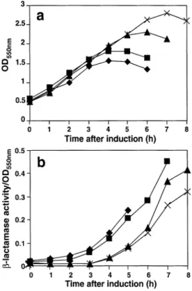
(3) Mutations influencing the folding of antibody domains in Escherichia coli. Fig. 1. Sequence alignments of the VH and VL domains of McPC603 and T15. The hypervariable regions, as defined by Chothia and Lesk (1987), are shaded and labelled. This definition, in common with the more recent IMGT definition (Barre et al., 1994; Giudicelli et al., 1997), is more structurally based than the sequence variability definition of Kabat (1991), leading to the classification of the VH mutations S60A and A61D as framework rather than CDR changes. Residues that differ from wild-type (T15 or McPC603) in the corresponding mutated antibody genes are indicated in black boxes. All other residues in McPC603, McPC603-H11 and M167 that differ from wild-type T15 are represented by shaded boxes. Filled circles in the table indicate ligand contact residues in McPC603, defined as those residues whose solvent-accessible area is smaller in the presence of ligand than in its absence.. Fig. 2. Model of the T15 Fv fragment. VH and VL domains are indicated, as are CDR loops in the two domains. Highlighted residues in heavy and light chains are those which were altered from wild type in VH and VL mutageneses.. brane of the cells, leading to release of the contents of the periplasm, was also reduced in the case of bacteria producing the mutated fragments (Figure 3b). Each of these growth experiments was carried out three times in independent experiments, since the exact shape of the growth curves can vary with minor changes in environmental conditions. The relative differences between the mutants always remained the same.. The effects of the framework mutations on the in vivo folding of the antibody fragments was also analysed. It was found that fragments containing the H11 mutations appeared to fold more efficiently in E.coli, as judged by the proportion of the total antibody protein that was produced in soluble form in the cells (Figure 4). In the case of the T15 antibody, it could be seen that the ratio of soluble to insoluble protein increased upon introduction of the H11 mutations, due somewhat to an increase in the soluble protein, but primarily to a reduction in the amount of protein produced in insoluble form (Figure 4). The levels of improvement during in vivo protein folding and E.coli physiology upon mutagenesis of the T15 fragments were not, however, as dramatic as those seen upon manipulation of the McPC603 fragment (Knappik and Plu¨ckthun, 1995). We therefore looked for additional determinants of poor folding and/or host cell lysis in the T15 amino acid sequence. On the basis of the rather extensive sequence differences between the VLs of the two antibodies (Figure 1), we first investigated the influence of this domain. The T15 VH gene was therefore expressed, in Fv fragments, in combination with its original VL and with the VLs of McPC603, M167 and hu4D5 Mab. The latter antibody has been shown to have excellent in vivo folding properties (Carter et al., 1992; Knappik and Plu¨ckthun, 1995). The growth of the host cells was optimal and cell leakiness at its lowest when the T15 VH was expressed in combination with its ‘natural’ VL (not shown), suggesting that factors still restricting the folding efficiency and growth 607.
(4) J.G.Wall and A.Plu¨ckthun. Fig. 3. Comparison of growth and leakiness of E.coli cells expressing various T15 antibody fragments. (a) Growth curves of cells expressing T15 wild-type Fv (r), H11 Fv (j), wild-type scFv (m) and H11 scFv (3) fragments. (b) Corresponding leakiness data for the same cells, expressed as β-lactamase activity per OD550 of the cultures; symbols are the same as in (a).. Fig. 4. Solubility analysis of T15 wild-type Fv, T15 (H11) Fv and T15 (H11-VH CDR3-grafted) Fv fragments. Western blots of preparations of soluble and insoluble protein are shown. ‘C’ represents control Fv; w.t. is the wild-type T15 Fv fragment; ‘H11’ is the framework-manipulated T15 wild-type Fv fragment; and ‘H11-CDR39 is the H11 Fv fragment containing the grafted McPC603 VHCDR3. Detection of the heavy chain is more sensitive than that of the light chain due to differences in the FLAG epitope between the two chains (Knappik and Plu¨ckthun, 1994). The lower band(s) visible in some lanes are products of degradation and the upper band in each sample lane was used for densitometry. A dilution series was incorporated into each experiment to overcome, for densitometric purposes, the non-linearity of the film. Because the most intense bands saturate the film, these strong bands appear to hardly give a reduction in signal upon dilution, while the weaker bands appear to disappear upon dilution.. characteristics of the framework-mutated fragments appeared to reside in the VH. Furthermore, these results suggest that the other combinations may form unstable VH/VL heterodimers, thereby not rescuing the poorly folding VH into a soluble Fv fragment. Sequence alignment of the VH genes of the two antiPC antibodies reveals that the only differences between the McPC603 and T15 VH genes occur in the CDR2 and CDR3 regions (Figure 1). We therefore replaced VH CDR2 (according to the structural definition, see above and Figure 1) or VH CDR3 608. from the T15 wild-type and framework-mutated antibodies with the corresponding CDR(s) from the antibody McPC603. Those residues differing from wild type in the grafted antibodies are highlighted in Figure 2. These newly-mutated fragments were then analysed as before: to determine the physiology of the host bacterial cells producing them and their in vivo solubility, used as a measure of folding efficiency. The results of these experiments are represented in Figure 5. It was found that the introduction of CDR-H3 from McPC603 into the wild-type Fv fragment had little effect on the host cells (Figures 5a and b). Introduction of the same CDR into the H11 mutant version of the same fragment, however, led to a somewhat improved growth physiology compared to cells expressing the H11 fragments. This improvement upon introduction of McPC603 VH CDR3 was also seen in the M167 (H11) Fv fragment, at a much more pronounced level (not shown); no improvement was seen when the McPC603 VH CDR3 was introduced into the M167 wild-type fragment. Analysis of the folding of the improved fragments, carrying the CDR-H3 from McPC603, (T15 Fv fragments shown in Figure 4) revealed that the proportion of soluble protein had increased significantly over that seen with the T15-H11 proteins. In contrast, the introduction of McPC603 VH CDR3 to the wild-type T15 Fv fragment resulted in soluble and insoluble protein levels the same as in the case of the wild-type fragment (not shown). It follows that the H11 mutations are required to see the beneficial effects of CDR-H3. Replacement of the T15 CDR-H2 with that from McPC603—i.e. introducing the two somatic mutations of McPC603 into the T15 sequence—yielded very different results from those observed with the CDR-H3. This manipulation was found to have no apparent effect on some of the proteins investigated [e.g. T15 (H11) Fv fragment—Figures 5c and d], but a detrimental effect on the cell physiology in the case of others (e.g. T15 wild-type Fv fragment—Figures 5a and b). This suggests that the T15 wild-type fragment cannot tolerate the additional problems located in the somatically-mutated version. Sequence alignment of the VL domains of McPC603 and T15 (Figure 1), carried out in a further search for determinants of poor folding in the T15 antibody fragments, led to 5 amino acid residues in the FW1 of T15 VL, which are different to the corresponding region in VL of McPC603 sequence (T9S, F10S, A12S, T14S and S16G; Figure 2) being exchanged together. To examine the effect of this mutagenesis on functional binding, a miniaturized functionality assay was developed. Since the affinity is too weak for a monovalent fragment to survive the washing steps of an ELISA, PC-resin, as used for affinity chromatography, was incubated with the bacterial supernatant and then centrifuged and washed. In this resin, the ligand concentration is millimolar and thus, the monovalent fragment remains in the resin phase upon washing. To eliminate non-specific binding as a possible source of errors, controls were incubated in 10 mM PC before adding the PC-resin. The bound fragment was analysed by SDS–PAGE. The VL FRI-mutations were first introduced in a T15–H11 VH background, resulting in the detection of this scFv by the PC-resin procedure outlined. The same protein was produced in a 2 l E.coli culture and purified by traditional affinity chromatography, the result of which is shown in Figure 6. This protein was quantified as approximately 200 µg. The mutant light chain was then cloned into vectors encoding the T15 wild-type and H11-McPC603 CDR3 heavy chain.
(5) Mutations influencing the folding of antibody domains in Escherichia coli. Fig. 5. Comparison of growth and leakiness of E.coli cells expressing various T15 antibody fragments. (a) Growth curves of cells expressing T15 wild-type Fv (r), wild-type 1 McPC603 VHCDR2 Fv (j) and wild-type 1 McPC603 VHCDR3 Fv (m) fragments. (b) Corresponding leakiness data for the same cultures, expressed as β-lactamase activity per OD550 of the cultures; the symbols are the same as in (a). (c) Growth curves of cells expressing T15 H11 Fv (r), T15 H11 1 McPC603 VHCDR2 Fv (u), and H11 1 McPC603 VHCDR3 Fv (m) fragments. (d) Corresponding leakiness data, with corresponding symbols, for the cells shown in (c).. Discussion. Fig. 6. Affinity chromatographic purification of T15 H11 scFv-mut. VL. The VL-mutated T15 scFv was produced in E.coli and purified by affinity chromatography as described. All fractions were electrophoresed in a SDS– polyacrylamide gel, followed by Coomassie staining. Lane 1, control scFv; lane 2, molecular weight marker; lane 3, column flow-through; lanes 4–8, washes; lane 9, eluted fraction.. constructs. These proteins were expressed and assayed for PC binding as before. Both were found to be produced in functional form and each could be purified at 30 µg/2 l culture. None of the corresponding fragments with the original T15 VL could be shown to bind antigen or to be purifiable using either this procedure or traditional affinity chromatography. This shows that these VL mutations are a prerequisite to purify any functional T15 fragment, and to observe any other beneficial mutations in VH at the level of purifiable protein.. The results we have outlined demonstrate for the first time that there is a hierarchy of mutations which influences the folding of antibody fragments in E.coli. They further provide a model for understanding the highly complex processes of protein folding and aggregation in the bacterium. T15 antibody fragments, previously found in our laboratory to be produced in a non-functional form and to be extremely susceptible to protease-degradation in E.coli, were first mutagenized to introduce the ‘H11’ triple mutation of the VH framework (Knappik and Plu¨ckthun, 1995). This mutation had been shown to improve the physiology of E.coli cells producing fragments of the related anti-PC antibody, McPC603, and the folding efficiency of the fragments themselves. This manipulation was found to significantly improve the folding of T15 Fv and scFv fragments, with a greater proportion of the mutated proteins being produced in soluble form than in the case of the wild-type proteins (Figure 4). The apparent toxicity of the mutated fragments to E.coli was also reduced, as judged by the lower level of periplasmic leakiness and delayed lysis of the cells producing the fragments (Figure 3); nevertheless, the levels of mutated McPC603 antibody fragments were not reached in either category. Since the VL genes are quite dissimilar and are derived from different Vκ genes (Perlmutter et al., 1984; Malipiero et al., 1987; Figure 1), the VH gene of T15 was combined with the VL gene from each of the T15, M167, McPC603, and the extremely well-folding huMab4D5 antibody (Plu¨ckthun and Pfitzinger, 1991; Carter et al., 1992). As the toxicity of the fragments to the host cell was lowest when the T15 VH was 609.
(6) J.G.Wall and A.Plu¨ckthun. expressed in combination with its original VL however (data not shown), it appeared that the other VL domains form only weak complexes with the T15 VH and do not rescue the VH into a soluble Fv fragment. The subsequent CDR exchange experiments, in which we replaced VH CDR2 (according to the structural definition) or VH CDR3 in the T15 fragments with the corresponding CDR(s) from the better-folding McPC603 fragment, demonstrated that mutations in the T15 fragments do not simply have additive or synergistic effects, but rather, there is a hierarchy of their effects. Introduction of the McPC603 VH CDR3 into the H11 mutant of the T15 antibody led to a further improvement in the solubility of the antibody fragments (Figure 4) and, to a lesser extent, in the growth of the host E.coli cells (Figure 5). However, this improved folding and solubility was seen in T15 fragments (and in M167 fragments) only upon prior introduction of the H11 framework mutations. In the absence of the framework mutations, therefore, the predominant pathway of periplasmic folding leads to aggregation and the composition of CDR-H3 only begins to have an effect when this diversion to aggregation has previously been overcome. This observed hierarchical effect of the manipulations also starts to explain the context dependence phenomenon of the effects of mutations in antibody fragments such as these. Introduction of the McPC603 CDR-H2 into T15 can be seen to have no effect on host cell physiology in the H11 version and to have a negative effect in the wild-type fragment. This suggests that the wild-type fragment cannot tolerate additional problems associated with the imposition of the McPC603 CDR, whereas the H11 mutant, while not suffering any further disadvantage due to introduction of the new VH CDR2, gains no advantage from it. It is likely that the mutations A52G and D54K facilitate the aggregation involving positions 60 and 61 even further. Finally, while the level of soluble protein being produced was greatly increased by this rational mutagenesis of T15 VH and the host bacterial cells showed a greatly stabilized physiology, none of the engineered fragments could be purified by affinity chromatography or using their peptide tag. This was thought to be due to their being produced in the bacteria in a soluble but non-functional form, the exact nature of which is unclear and is the subject of an ongoing study to characterize it precisely. Because the T15 VH domain has been converted entirely to VH of McPC603-H11 in this investigation, and because the Fv fragment McPC603-H11 (with its original VL domain) is easily purifiable by affinity chromatography, we chose to re-visit the VL domain in an attempt to restore the functionality of the T15 fragments. The mutagenesis of the VL FW1 region was designed with a view to identifying key residue(s) in the T15 VL gene that were responsible for the lack of functionality of the Fv and scFv fragments, by exchanging them for the corresponding residues in the antibody McPC603. The T15 VH FW1 sequence TFLAVTAS (Figures 1, 2) was, therefore, mutated to SSLSVSAG. Affinity chromatographic screening of the H11 heavy chain mutant containing this altered light chain led to the purification of a protein band of the expected size. It is important to note that no improvement had been observed upon exchange of the whole VL domain of McPC603 (see above), since several VL residues need to contact the antigen and to make crucial interdomain hydrogen bonds (Satow et al., 1986). Of the five residues changed in our analysis, modelling work indicated that the original Phe10 occurs in an exposed 610. location at the bottom of the T15 VL domain (Figure 2), where its replacement by a serine might be expected to disfavour aggregation. We propose that this residue in the T15 VL domain had previously been limiting the functionality of antibody fragments produced in E.coli and that it was only upon overcoming this problem that functional, affinity-purifiable fragments of the antibody could be produced for the first time. While the major effect of the mutation in this case was to restore functionality, the same mutation has also been observed by other workers to result in increased production in E.coli of an already functional Fab fragment (Forsberg et al., 1997). While the introduction of the FR1 mutations into VL by itself led to the production of a low level of functional protein, the H11 mutations in the VH, in the presence of the manipulated VL, then increased the level of affinity purifiable scFv produced by a factor of 7. In the case of the H11-McPC603 CDR3 scFv, the reduced level of purifiable protein is probably due to a perturbation in the binding pocket upon grafting of the McPC603 VH CDR3 to T15, resulting in a reduction in the affinity of the fragment. We conclude that this work demonstrates that there is a hierarchy of factors influencing the folding of recombinant antibody fragments produced in E.coli. The effectiveness of the structured approach we have employed is such that even previously non-functional antibodies can be engineered into well folding molecules. Furthermore, this analysis of the hierarchy of events should be informative in delineating the molecular mechanisms of aggregation. Acknowledgements We would like to thank Barbara Klinger for technical assistance and Annemarie Honegger, Hendrick Bothmann and Achim Knappik for useful discussions, and A.H. for help with the figures. Funding was provided by grants from the Schweizerischer Nationalfonds grant 3100–046624.96/1 (A.P.) and Enterprise Ireland Science and Innovation Development Agency grant IC/97/090 (G.W.).. References Bardwell,J.C. (1994) Mol. Microbiol., 14, 199–205. Barre,S., Greenberg,A.S., Flajnik,M.F. and Chothia,C. (1994) Nature Struct. Biol., 1, 915–920. Carter,P. et al. (1992) Biotechnology, 10, 163–167. Chothia,C. and Lesk,A.M. (1987) J. Mol. Biol., 196, 901–917. Derman,A.I., Prinz,W.A., Belin,D. and Beckwith,J. (1993) Science, 262, 1744–1747. Forsberg,G., Forsgren,M., Jaki,M., Norin,M., Sterky,C., Enhorning,A., Larsson,K., Ericsson,M. and Bjork,P. (1997) J. Biol. Chem., 272, 12430– 12436. Freund,C., Ross,A., Plu¨ckthun,A. and Holak,T.A. (1994) Biochemistry, 33, 3296–3303. Ge,L., Knappik,A., Pack,P., Freund,C. and Plu¨ckthun,A. (1995) In Borrebaeck,C.A.K. (ed.), Antibody Engineering, 2nd edn. Oxford University Press, Oxford, pp. 229–266. Giudicelli,V., Chaume,D., Bodmer,J., Muller,W., Busin,C., Marsh,S., Bontrop,R., Marc,L., Malik,A. and Lefranc,M.P. (1997) Nucleic Acids Res., 25, 206–211. Ho,S.N., Hunt,H.D., Horton,R.M., Pullen,J.K. and Pease,L.R. (1989) Gene, 77, 51–59. Jung,S. and Plu¨ckthun,A. (1997) Protein Engng, 10, 959–966. Kabat,E.A., Wu,T.T., Perry,H.M., Gottesman,K.S. and Foeller,C. (1991) Sequences of Proteins of Immunological Interest, 5th edn. National Institutes of Health, Bethesda. Kazemier,B., de Haard,H., Boender,P., van Gemen,B. and Hoogenboom,H. (1996) J. Immunol. Methods, 194, 201–201. Knappik,A. and Plu¨ckthun,A. (1994) BioTechniques, 17, 754–761. Knappik,A. and Plu¨ckthun,A. (1995) Protein Engng, 8, 81–89. Kunkel,T.A., Roberts,J.D. and Zakour,R.A. (1987) Methods Enzymol., 154, 367–382. Malipiero,U.V., Levy,N.S. and Gearhart,P.J. (1987) Immunol. Rev., 96, 59–74..
(7) Mutations influencing the folding of antibody domains in Escherichia coli Nieba,L., Honegger,A., Krebber,C. and Plu¨ckthun,A. (1997) Protein Engng, 10, 435–444. O’Callaghan,C.H., Morris,A., Kirby,S.M. and Shingler,A.H. (1972) Antimicrob. Agents Chemother., 1, 283–288. Perlmutter,R.M., Crews,S.T., Douglas,R., Sorensen,G., Johnson,N., Gearhart,P.J. and Hood,L. (1984) Adv. Immunol., 35, 1–37. Plu¨ckthun,A. (1993) Bioorg. Chem. Frontiers, 3, 25–66. Plu¨ckthun,A. and Pfitzinger,I. (1991) Annals New York Acad. Sci., 646, 115–124. Plu¨ckthun,A., Krebber,A., Krebber,C., Horn,U., Knu¨pfer,U., Wenderroth,R., Nieba,L., Proba,K. and Riesenberg,D. (1996) In McCafferty,J. (ed.), Antibody Engineering: A Practical Approach. IRL Press, Oxford, pp. 203–252. Prickett,K.S., Amberg,D.C. and Hopp,T.P. (1989) BioTechniques, 7, 580–589. Proba,K., Ge,L. and Plu¨ckthun,A. (1995) Gene, 159, 203–207. Satow,Y., Cohen,G.H., Padlan,E.A. and Davies,D.R. (1986) J. Mol. Biol., 190, 593–604. Steipe,B., Plu¨ckthun,A. and Huber,R. (1992) J. Mol. Biol., 225, 739–753. Ullrich,H.D., Patten,P.A., Yang,P.L., Romesberg,F.E. and Schultz,P.G. (1995) Proc. Natl Acad. Sci. USA, 92, 11907–11911. Yanisch-Perron,C., Vieira,J. and Messing,J. (1985) Gene, 33, 103–119. Received July 23, 1998; revised January 25, 1999; accepted March 22, 1999. 611.
(8)
Figure

Documents relatifs
In this paper, we seek to extend our understanding of the adoption of IT innovations, by developing an integrated model and identifying the factors that influence
Using an ion-neutral chemical model we calculate temporal density variations for var- ious atmospheric species in the relaxing phase after occurrence of a sprite streamer at
Ainsi, si la demande globale de bien « énergie » E constitue une contrainte pour ces dernières, la répartition de E entre bien dangereux et bien non dangereux est
Effect of cobalt fraction on the onset temperature of dehydration T i (upper-left), the width of the decomposition peak (upper-right), the enthalpy of dehydration.. (lower-left) and
Figure 6 a–d presents the rheological behaviors including complex viscosity (η*), storage modulus (G’), loss modulus (G”), and tan(delta) of PP, PP/polyphenol blends (PPT and PPL),
ًخجٳحى
Under a neutral divergence with gene flow model, morphological differences should not be evident among urban and non-urban sites, genetic diversity should be reduced in urban sites,
مﻠﻌﺗﻟا طﻣﻧ.. 16 دﯾﻬﻣﺗ ﻣ ﻲﻓ تﯾرﺟأ ﻲﺗﻟا ثوﺣﺑﻟا تدﻛأ طﺎﻣﻧﻷا لﺎﺟﻣ ﻲﻓ ﻲﺳﻔﻧﻟا زﯾﺎﻣﺗﻟاو ﺔﯾدرﻔﻟا قورﻔﻟا لﺎﺟ ﺎﻬﯾﻓ نورظﻧﯾ ﻲﺗﻟا ﺔﻘﯾرطﻟا ﻲﻓو ﺔﻠﺿﻔﻣﻟا
