Measurement of the Optical Properties of Tissues by a
Minimally-Invasive Probe
Bevilacqua F., Piguet D., Marquet R, Depeursinge Ch.
Institut dOptique Appliquee, Ecole Polytechnique F derale de Lausanne, CH-1015 Lausanne, Switzerland.
ABSTRACTA small probe (0 = 2 mm) was designed to measure locally the optical properties of biological tissues. The sensitivity of the measured Signal was assessed by experi-ments on microsphere suspensions and Monte Carlo simu-lations. Relative measurements of scattering and absorption coefficients should give local metabolic and tissue structure informations.
INTRODUCTION
Measuring the optical properties of biological tissues is a useful task for several medical applications. For example accurate optical coefficients are necessary for near infra-red imaging or for dosimetry in photodynamic therapy. Moreover, the optical properties could also help, in partic-ular cases, to differentiate directly normal and maJignant tissues [1]. In such cases, probing locally the optical prop-erties is a promising tool for mini-invasive surgery.
Photon migration in tissues is govemed by scattering and absoφtion. Therefore the important optical parameters of tissues are the absorption coefficient μ^ the scattering coefficient ^ and the phase function p(cos9). The phase function is the density probability function for the scatter-ing angle Θ. The first moment of p(cos9), denoted g, is the mean cosine of Θ. Generally, g varies in tissues between 0.7 to 0.99, which means that light is mainly scattered in the forward direction. It is also useful to define the reduced scattering coefficient \i^= mO-g), s explained later.
The scattering properties depend on the tissue structure: cell density and size, organelles density and size, etc. Therefore different types of tissues can be characterized by their scattering properties (u,', p(cos9)).
The absorption coefficient in tissues is related to the con-centration of chromophores such s haemoglobin or cy to-chrom. Since the absorption spectra of haemoglobin changes with its oxygenation state, measurements of j^ at different wavelengths allow the determination of the tis-sue blood content and haemoglobin oxygen Saturation, which are important metabolic indicators.
The probe described here allows local measurements of the optical properties in vivo. The method is based on spa-tially resolved measurements of the reflectance. Similar methods have been reported previously [2], but using measurement of the reflectance over large distances (typi-cally >1 cm) compared to the transport mean free path nupMlV+Ha)"1· In our case we measure the refiectance close to the light source (< 2mm) in order to use a rela-tively small diameter probe (2.2 mm) and to perform local
measurements.
MATERIAL AND METHOD
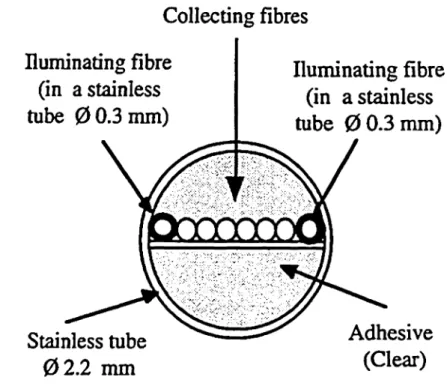
The probe, sketched in Fig. l, is a a linear array of optical fibres (core diameter of 200 μτη, Ν.Α.=0.37). Two fibres, coupled to laser diodes emitting at 670 nm and 820 nm, can be altematively used to illuminate the tissue. Six other fibres are used to collect the scattered light, and are imaged on a linear Charge-Coupled-Device (CCD). The illuminating are slid in small stainless tubes in order to avoid direct coupling with the collecting fibres. The b n-dle is set in a stainless tube of 2 mm diameter and 20 cm long. The probe is rigid, which allows an easier handling by the physician. However a flexible probe is also feasible for endoscopic application. The whole probe can be steri-lized. Collecting fibres Huminating fibre (in a stainless tube 00.3mm) Huminating fibre (in a stainless tube 00.3mm) Stainless tube 02.2 mm
Fig. L Measuring probe
Adhesive (Clear)
THEORY
A model of photon migration in tissues is necessary to explicit the relationship between the measured reflectance and the optical properties. We performed Monte Carlo simulations [3], which give indirectly exact Solutions of the transport theory. Analytical Solutions from the difru-sion equation are not appropriate in our case because we are interested in the reflectance close to the source, at dis-tance comparable to the transport mean free path mfp'. Our simulations take into account the exact diameter of the illuminating and collecting fibres, s well s their numerical apertures.
The reflectance depends on μ^ μ;, and p(cos9). Far from the source (d» mfp') only the first moment g of the phase function plays a role, and the reflectance depends only on two parameters: u^ and μ,'. These two parameters are
0.15
=1 ο.ι
ο ο 0.05ο
Mie phase function Henyey-Greenstein phase function
0.2 0.4 0.6 0.8 l 1.2 1.4 1.6
radial distance r [mm]
Fig. 2. Reflectance from simulations. The dots repre-sent the intensity collected by the sixfibres. £=0.92, μα=0.06 mnr't μ5'=1 mnr1
ally used to characterize a given tissue. Close to the source (d ~ mfjp') higher moments of the phase f ncdon also play an important role. This is illustrated in Fig. 2, where a Simulation of a probe reflectance measurement is plotted s a function of the radial distance r, using two different phase functions, with the same first moment g=0.92. The first one is the Henyey-Greenstein phase function, very often used in tissue modelling. The second one is the phase function obtained from Mie theory, corresponding to the microspheres used in the experiments described below. The important difference shown in Fig.2 between the two curves for radial distance < 0.5 mm, is explained by the role of the second and higher Order moments, dif-ferent for each phase function.
A complete analysis of light propagation close to the source, taking into account the importance of the phase function is under investigation. It will yield an optimized way to analyse the curve measured, in order to minimize the artefact due to the imprecisely known phase function of the investigated tissue.
PRELIMINARY RESULTS AND DISCUSSION
The sensitivity of the probe has been experimentally tested using polystyrene microsphere suspensions (0=1 μm). Mie theory allows to compute p(cos9) and m of the Suspension. The absorption is assumed to be equal to the water absorption. Absorption can be increased when ink is added. The ink absorption coefficient of the ink has been previously calibrated s a function of the concentra-tion.
In Fig. 3 and 4, measured Signals are shown for suspen-sions of different microsphere concentrations and ink con-centrations. The corresponding u,' and \L, are in the r nge of Standard optical properties of biological tissues. Fig. 3 and 4 clearly show that, s expected, the average meas-ured intensity increases if μ,' increases or u., decreases. The slope of the logarithm of the reflectance increases if μ,* increases. However the slope is only slightly affected by μ^ This observation is only valid close to the source. This shows that absolute intensity measurements is then
necessary to extract μ., from the measured curve. Signifi-cant systematic errors could then appear if the role of the phase function is not properly taken into account, s dis-cussed before. Nevertheless a relative measurement of μ., is still possible. In particular a spectroscopic analysis of the measured signal could give important informations about the metabolism of the investigated tissues.
μ,1 = 0.46 [mm-»] ' = 0.76 [mm-1] .07 [mm-1] .64 [mm-1] o υ υ α> Gν αί 0.1
-!·
= 1. 0.2 0.4 0.6 0.8 l 1.2 1.4 radial distance r [mm] 1.6Fig.3. Reflectance measured on microsphere suspen-sions. g=0.92, μα=0.0005 mnr't λ=670 nm. u, = 0.014 [mm-1] u, = 0.051 [mm·1] u, = 0.091 [mm-1] 185 [mm-1] 3 .cj. ou o t> c: l 0.8 0.6 0.4 0.2 0.2 0.4 0.6 0.8 l 1.2 1.4 1.6 radial distance r [mm]
Fig.4. Reflectance measured on microsphere suspen-sions (+ink). g=0.92, μ/=0.5 mnr1. λ=670 nm.
CONCLUSION
This preliminary study shows that the probe gives impor-tant local informations about the scattering and absorption properties of biological tissues. Clinical assessment of the probe is planned.
This work was supported by the Swiss National Priority Program in Optics (PPO/Med).
REFERENCES
[1] J. R. Mourant et al.; Lasers. Surg. Med. 17, 350, (1995).
[2] See T. J. Farrell et al.; Med. Phys. 19, 879 (1992) and the references therein.
[3] P. Marquet et al.; Opt. Eng. 34,2055, (1995).