HAL Id: inserm-00640066
https://www.hal.inserm.fr/inserm-00640066
Submitted on 3 Oct 2014HAL is a multi-disciplinary open access L’archive ouverte pluridisciplinaire HAL, est
RP1 and autosomal dominant rod-cone dystrophy: novel
mutations, a review of published variants, and
genotype-phenotype correlation.
Isabelle Audo, Saddek Mohand-Saïd, Claire-Marie Dhaenens, Aurore
Germain, Elise Orhan, Aline Antonio, Christian Hamel, José-Alain Sahel,
Shomi Bhattacharya, Christina Zeitz
To cite this version:
Isabelle Audo, Saddek Mohand-Saïd, Claire-Marie Dhaenens, Aurore Germain, Elise Orhan, et al.. RP1 and autosomal dominant rod-cone dystrophy: novel mutations, a review of published variants, and genotype-phenotype correlation.: RP1mutations in French adRP patients. Human Mutation, Wiley, 2012, 33 (1), pp.73-80. �10.1002/humu.21640�. �inserm-00640066�
RP1 and autosomal dominant rod-cone dystrophy: novel mutations, a review of published variants, and genotype-phenotype correlation
Isabelle Audo 1,2,3,4,5, Saddek Mohand-Saïd1,2,3,4, Claire-Marie Dhaenens6, Aurore
Germain1,2,3, Elise Orhan1,2,3, Aline Antonio1,2,3,4, Christian Hamel7,8, José-Alain
Sahel1,2,3,4,5,9, Shomi S. Bhattacharya1,2,3,5,10 and Christina Zeitz1,2,3.
1
INSERM, UMRS968, Paris, F-75012, France; 2 UPMC Univ Paris 06, UMR_S 968,
Institut de la Vision, Paris, F-75012, France; 3 CNRS, UMR_7210, Paris, F-75012,
France; 4 Centre Hospitalier National d’Ophtalmologie des Quinze-Vingts,
INSERM-DHOS CIC 503, Paris, F-75012, France; 5UCL-Institute of Ophthalmology, Bath Street,
London, UK ; 6Centre Hospitalier Régional Universitaire de Lille, UF Génopathies,
Laboratoire de Biochimie et Biologie Moléculaire - Université Lille Nord de France, Lille, France ; 7Centre de Référence Maladies Sensorielles Génétiques, Hôpital Guy de
Chauliac, Montpelier, France; 8U583, INSERM, Institute for Neurosciences de
Montpellier, Montpellier, France; 9Fondation Ophtalmologique Adolphe de Rothschild,
Paris, France; 10Department of Celular Therapy and Regenerative Medicine, Andalusian
Molecular Biology and Regenerative Medicine Centre (CABIMER), Seville, Spain. Running title: RP1mutations in French adRP patients
Key words: adRP, RP1 mutations, prevalence study Corresponding author: Isabelle Audo and Christina Zeitz Institut de la Vision, Department of Genetics
17, Rue Moreau 75012 Paris France
Financial support:
The project was financially supported by the Department of Paris, Foundation Fighting Blindness (I.A. FFB Grant N°: CD-CL-0808-0466-CHNO and the CIC503 recognized as an FFB center, FFB Grant No: C-CMM-0907-0428-INSERM04), ANR (S.S.B). NIHR Biomedical Research Centre for Ophthalmology and The Special Trustees of Moorfields Eye Hospital London, Foundation Voir et Entendre (C.Z) and French Ministry of Health (C.H. PHRC # 2008-A01238-47).
ABSTRACT
Rod-cone dystrophies (RP) are a clinically and genetically heterogeneous group of inherited retinal disorders characterized by photoreceptor degeneration. RP1 is a major gene underlying autosomal dominant (ad) RP though prevalence numbers vary depending on the origin of the cases from 0%-10% cases. Some mutations in RP1 also lead to autosomal recessive RP. Herein we review all previously reported and several novel RP1 mutations in relation to the associated phenotype in patients from a French adRP cohort. Prevalence studies from this cohort show that 5.3% of the cases have RP1 mutations. This is in accordance with other studies reported from UK and USA. The majority of mutations represent truncating mutations which are located in a hot spot region. Similarly, we identified in total four novel deletions and nonsense mutations, of which two may represent recurrent mutations in a French population. In addition a novel missense mutation of uncertain pathogenicity was identified. With our findings, to date 43 RP1 mutations are known to cause adRP. Variable penetrance of the disease was observed in our and other cohorts. Most patients with RP1 mutations show classical signs of RP with relatively preserved central vision and visual field.
Background
Rod-cone dystrophies, also known as retinitis pigmentosa (RP), are a clinically and genetically heterogeneous group of progressive inherited retinal disorders, which often starts with night blindness and leads to visual field constriction and secondary macular involvement, which can eventually result in loss of central vision and complete blindness (Hartong et al. 2006). RP occurs in 1 of 4000 births and affects more than 1 million individuals worldwide. The mode of inheritance can be x-linked (xl), autosomal dominant (ad) or autosomal recessive (ar). In addition, many patients represent isolated cases, due to the absence of family history of RP. To date, mutations in 23 different genes are associated with autosomal dominant RP (adRP) (http://www.sph.uth.tmc.edu/Retnet/)
and the majority of prevalence studies reveals rhodopsin (RHO: MIM#180380) being the most frequently mutated gene in adRP (Sullivan et al. 2006; Audo et al. 2010b). In addition, PRPF31(MIM#606419), PRPH2 (MIM#179605) and RP1 (MIM#603937) were proposed to represent major genes underlying this form of RP (Sullivan et al. 2006; Audo et al. 2010a). In some rare cases RP1 mutations were also found in patients with arRP and unilateral RP (Khaliq et al. 2005; Riazuddin et al. 2005; Singh et al. 2009; Chen et al. 2010; Mukhopadhyay et al. 2011). RP1 is located on chromosome 8q12.1, encompasses 4 exons of which 3 are coding. This work aims to review known mutations and to document novel mutations and genotype-phenotype correlations on a French cohort consisting of 114 cases.
Mutations in the RP1 gene
So far 39 disease causing mutations in RP1 have been identified of which most were found in the last exon, leading to a premature stop codon and are predicted to form a truncated protein (Table 1). The p.Arg677X has been described as the most commonly reported mutation (Bowne et al. 1999; Guillonneau et al. 1999; Pierce et al. 1999; Sullivan et al. 1999; Jacobson et al. 2000; Payne et al. 2000; Baum et al. 2001; Berson et al. 2001; Sohocki et al. 2001; Xiaoli et al. 2002; Schwartz et al. 2003; Ziviello et al. 2005; Chiang et al. 2006; Gamundi et al. 2006; Roberts et al. 2006; Sullivan et al. 2006).. Detailed phenotype-genotype correlations of patients with this mutation revealed incomplete penetrance and high variability of the disease expression in adRP suggesting modifiers to be involved (Jacobson et al. 2000). Similarly, incomplete penetrance was also noted for other mutations (Table 1). Mutation analysis in our cohort (85 of 114 adRP index patients screened for RP1) added two different deletions and two different nonsense mutations identified in 6 families to the previously described mutations, however none of the patients revealed the recurrent p.Arg677X mutation (Table 1, Supp. Table S1).With our findings, to date 43 RP1 mutations are known to cause adRP. The prevalence of RP1 mutations with 5,3% in our cohort is similar to studies from UK and USA, although the prevalence of RP1 mutations varied from 0%-10% from different adRP cohorts (Bowne et al. 1999; Pierce et al. 1999; Payne et al. 2000; Baum et al. 2001; Berson et al. 2001; Sohocki et al. 2001; Kawamura et al. 2004; Ziviello et al. 2005; Gamundi et al. 2006; Roberts et al. 2006; Sullivan et al. 2006; Zhang et al. 2010)
Different pathogenic mechanisms of RP1 truncating mutations have been proposed due to
in vitro and in vivo studies leading to ar or adRP. Chen and co-workers proposed four
classes of RP1 mutations depending on their site and underlying pathogenicity (Chen et al. 2010). Class 1 mutations are rare, thought to be nonsense-mediated decay (NMD)-sensitive and locate in exons 2-3 (amino acids 1-236). Haploinsufficiency is not believed to be the disease causing mechanism since heterozygous carriers of those mutations are not affected. Thus it is more likely that the mutation creates a null allele due to NMD. The RP1 protein made by the unaffected copy of the allele is completely functional and only if both alleles are affected, the patient will suffer from RP. Examples are p.Ser2ArgfsX16 (Chen et al. 2010) and p.Pro229GlnfsX35 (Pierce et al. 2010) (Table 1). Class II mutations occur frequently and are reported to be NMD-insensitive truncations, which are located approximately between amino acids 500 and 1053 in exon 4. The mutant proteins are expected to impose distinct dominant negative effect on photoreceptors, resulting in cell death and adRP (Chen et al. 2010) (Table 1). Similarly, the deletions and nonsense mutations identified in the French adRP cohort studied herein could also be classified as class II mutations. Two of them (p.Lys657AsnfsX7 and p.Ile725TyrfsX13) may represent re-current mutations in French adRP patients due to the appearance in two independent families (Table 1, Supp. Figure S1, Supp. Table S1). To our knowledge only one recessive truncating mutation lies in this region and leads to arRP (p.Asn949LysfsX32) (Table 1). Class III truncating mutations are thought to affect amino acids 264 to 499 and 1054 to 1751, which lead to loss-of-function in patients with arRP. Examples are p.Pro1648SerX13 (Chen et al. 2010) or p.Asn1751IlefsX4
(Riazuddin et al. 2005) (Table 1). Class IV truncating mutations are NMD-insensitive and thought to be located after amino acid 1816. The resulting truncated proteins are expected to be functional and thus not disease causing (Chen et al. 2010). A few missense mutations reported lead to ar or adRP (Table 1) (Khaliq et al. 2005; Chiang et al. 2006; Zhang et al. 2010). However for most of the missense mutations the pathogenic character remains to be elucidated or was considered to be benign (Supp. Tables S2 and S3). Similarly, we identified a missense mutation (c.4193C>G, p.Ser1398Cys, Supp. Figure S1, F745,) of which co-segregation analysis can not clearly predict if this variant lead to adRP (Supp. Table S4). Furthermore other variants in RP1 have been identified, which are presumably due to predictions or co-segregation analysis not pathogenic (Supp. Table S5). All mutations identified by our extensive literature review and described herein, will be submitted to the existing Locus Specific Databses (LSDB) on RP1, which before the submission of our review cites only 4 pathogenic mutations.
Genotype-Phenotype correlation
Variable penetrance of the disease was observed in our and other cohorts. Most patients with RP1 mutations show classical signs of RP with relatively preserved central vision and visual field. The detailed description about the phenotypic characteristics of the 7 index patients (3 females and 4 males) carrying different RP1 mutations of the French adRP cohort can be used as key insights about RP1 associated genotype-phenotype relationships (Table 2 and 3 with an illustration of retinal changes presented in figure 1). In brief, mean age of patients of the patients herein is 54.4. Except for one patient,
CIC01234 (carries the missense mutation of uncertain pathogenicity) examined at an asymptomatic young age during a family investigation, the majority of subjects were diagnosed in their twenties with an average age at time of diagnosis of about 23 years. This is older than reported in adRP associated with RHO and PRPF31 mutations (Audo et al. 2010a; Audo et al. 2010b) and in accordance with previous reports for patients with
RP1 mutations (Berson et al. 2001). Family history for two families (F556 and F745)
suggested incomplete penetrance and three patients reported variable age of onset in the family with diagnosis made as late as 55 years. This is in keeping with variable penetrance and expressivity associated with RP1 mutations (Jacobson et al. 2000; Berson et al. 2001). None of the families in this study mentioned unilateral involvement so far (Mukhopadhyay et al. 2011). Symptoms at presentation were dominated by night vision disturbances. Visual acuity ranges from 20/25 to 20/800. All patients had some lens opacities or had undergone cataract surgery. In most cases, fundus changes were typical of RP with narrowed retinal vessels, pale discs and pigmentary changes in the periphery. All but one had some degree of atrophy with loss of autofluorescence in the perifoveal region and one patient also had cystoid macular oedema in one eye. Color vision abnormalities were correlated with worse visual acuities. All patients had severe constricted visual fields and generalized retinal dysfunction with four out of seven with some preservation of central macular function. Therefore, although phenotypic variability was suggested in certain families in this study, the index patients had a fairly homogeneous degree of severity within their retinal involvement.
Biological Relevance
RP1 consists of 2156 amino acids with a molecular weight of 240 kD, which is expressed specifically in photoreceptors in the retina (Bowne et al. 1999; Guillonneau et al. 1999; Pierce et al. 1999). The N-terminal 28-228 amino acids of RP1 contains a tandem repeat domain (Kim et al. 2003), which is known to interact with microtubules and was initially detected in the protein doublecortin (DCX). DCX is involved in directing neuronal migration during development of the central nervous system and mutations in this gene in patients lead to x-linked lissencephaly and double cortex syndrome (des Portes et al. 1998; Gleeson et al. 1998; Francis et al. 1999). Immunofluorescence analyses detected the human and mouse RP1 protein in the connecting cilium and microtubule-based axoneme of rod and cone photoreceptors. In addition, the amino acid sequences between codons p.486 and p.635 shares homology with the Drosophila melangonaster protein BIF, which is essential for normal photoreceptor morphogenesis (Pierce et al. 1999). Due to the specific localization of RP1 and the existence of the typical DCX and BIF domain, it was suggested that RP1 might participate in transport of proteins between the inner and outer segments of photoreceptors or in maintenance of the ciliary structure (Liu et al. 2002). Different mouse lines lacking Rp1 have been created to better understand the function and pathogenic mechanism of RP1 protein and mutations: One deletes the conserved N-terminal Dcx domain (Gao et al. 2002) and produces an abnormal Rp1 product that is lacking exon 2 and 3, while the other mimics the frequently identified mutation in RP patients: p.Arg677X, which contains the N-terminal Dcx domain and produces the shorter Rp1 protein (Liu et al. 2003). Both homozygous models experience
rapid retinal degeneration and show outer segment dysplasia and misalignment, suggesting a general role of RP1 in outer segment organization. Initially, only heterozygous mice lacking the Dcx domain and expressing about 50% of normal Rp1 protein revealed some retinal dysfunction despite normal-appearing retinal morphology (Gao et al. 2002), while heterozygous mice mimicking the human mutation p.Arg677X did not show any phenotype (Liu et al. 2003). However, recently it was shown that the severity of the retinal degeneration is dependent on the genetic background and only heterozygous mice for the A.129S(B6) background indeed developed RP suggesting that specific modifiers present in one or the other mouse strain are responsible for the severity of the phenotype in some heterozygous mouse lines (Liu et al. 2009). Furthermore the photoreceptor degeneration of a knock-in mouse line carrying the p.Gln662X mutation, analogous to the human p.Arg677X mutation, could be delayed or prevented by adding wild-type Rp1 protein (Pierce et al. 2010). Another mouse model, which was chemically induced (N-ethyl-N-nitrosourea = ENU) carries a p.Arg533X mutation and again only homozygous mice were affected by photoreceptor degeneration (Won et al. 2011). Since the retinal phenotype in knock-in mice carrying mutations in this region could be delayed or prevented by adding the wild-type gene, the underlying pathogenic mechanism was suggested to be dominant-negative and not gain-of-function.
Clinical and Diagnostic Relevance
In the study presented here we review all reported disease causing RP1 mutations including 4 novel truncating mutations and 1 novel nucleotide change of uncertain
pathogenicity in 7 cases with a homogenous RP phenotype (Table 1, 2 and 3, Supp. Tables S1 and S2). Our study confirms that for adRP European cohorts, RP1 represents a major gene defect. We propose that most of the RP1 mutations leading to adRP represent truncation mutations and are located in a hot spot region (~nucleotides c.1490-c.3216). Therefore, to perform a time and cost-efficient mutations screening for adRP patients this region could be preferentially targeted. These truncating mutations can be classified as Class II mutations, which lead to a dominant negative effect of RP1 resulting in photoreceptor cell degeneration. In addition, incomplete penetrance was observed for some of the mutations in this class (Dietrich et al. 2002; Roberts et al. 2006) (Table 1). The exact mechanism of this incomplete penetrance is not known. Another form of adRP associated with mutation in splicing factors is also associated with incomplete penetrance (Evans et al. 1995; Al-Maghtheh et al. 1996; McGee et al. 1997; Vaclavik et al. 2010). In this case, haploinsufficiency is the admitted disease mechanism and an allelic imbalance with overexpression of the wild-type allele, compensating for the non-functional allele in asymptomatic carriers has been advocated (Vithana et al. 2003; Rivolta et al. 2006). In case of incomplete penetrance associated with RP1 mutation, such mechanisms with the influence of the trans allele has been suggested in asymptomatic female carriers by Daiger and colleagues but warrants further study (Daiger et al. 2006). Influence of mouse strains of Rp1 mutation upon severity in retinal degeneration advocate for modifying factors (Liu et al. 2009). A better understanding of the mechanisms underlying phenotypic variability should facilitate developing novel therapeutic approaches.
Clinically and genetically well-characterized patients such as those included here are critical in this regard.
ACKNOWLEDGEMENT:
The authors are grateful to patients and family members described in this study, to Thierry Léveillard, Dominique Santiard-Baron, Christine Chaumeil and clinical staff for their help in clinical data and DNA collection. The project was financially supported by the Department of Paris, Foundation Fighting Blindness (I.A. FFB Grant N°: CD-CL-0808-0466-CHNO and the CIC503 recognized as an FFB center, FFB Grant No: C-CMM-0907-0428-INSERM04), ANR (S.S.B). NIHR Biomedical Research Centre for Ophthalmology and The Special Trustees of Moorfields Eye Hospital London, Foundation Voir et Entendre (C.Z) and French Ministry of Health (C.H. PHRC # 2008-A01238-47).
REFERENCES:
Al-Maghtheh M, Vithana E, Tarttelin E, Jay M, Evans K, Moore T, Bhattacharya S, Inglehearn CF. 1996. Evidence for a major retinitis pigmentosa locus on 19q13.4 (RP11) and association with a unique bimodal expressivity phenotype. Am J Hum Genet 59(4):864-71.
Audo I, Bujakowska K, Mohand-Said S, Lancelot ME, Moskova-Doumanova V, Waseem NH, Antonio A, Sahel JA, Bhattacharya SS, Zeitz C. 2010a. Prevalence and novelty of PRPF31 mutations in French autosomal dominant rod-cone dystrophy patients and a review of published reports. BMC Med Genet 11:145. Audo I, Manes G, Mohand-Said S, Friedrich A, Lancelot ME, Antonio A,
Moskova-Doumanova V, Poch O, Zanlonghi X, Hamel CP, Sahel JA, Bhattacharya SS, Zeitz C. 2010b. Spectrum of rhodopsin mutations in French autosomal dominant rod-cone dystrophy patients. Invest Ophthalmol Vis Sci published online 17.02.2010.
Baum L, Chan WM, Yeung KY, Lam DS, Kwok AK, Pang CP. 2001. RP1 in Chinese: Eight novel variants and evidence that truncation of the extreme C-terminal does
Berson EL, Grimsby JL, Adams SM, McGee TL, Sweklo E, Pierce EA, Sandberg MA, Dryja TP. 2001. Clinical features and mutations in patients with dominant retinitis pigmentosa-1 (RP1). Invest Ophthalmol Vis Sci 42(10):2217-24.
Bowne SJ, Daiger SP, Hims MM, Sohocki MM, Malone KA, McKie AB, Heckenlively JR, Birch DG, Inglehearn CF, Bhattacharya SS, Bird A, Sullivan LS. 1999. Mutations in the RP1 gene causing autosomal dominant retinitis pigmentosa. Hum Mol Genet 8(11):2121-8.
Chen LJ, Lai TY, Tam PO, Chiang SW, Zhang X, Lam S, Lai RY, Lam DS, Pang CP. 2010. Compound heterozygosity of two novel truncation mutations in RP1 causing autosomal recessive retinitis pigmentosa. Invest Ophthalmol Vis Sci 51(4):2236-42.
Chiang SW, Wang DY, Chan WM, Tam PO, Chong KK, Lam DS, Pang CP. 2006. A novel missense RP1 mutation in retinitis pigmentosa. Eye (Lond) 20(5):602-5. Daiger SP, Shankar SP, Schindler AB, Sullivan LS, Bowne SJ, King TM, Daw EW,
Stone EM, Heckenlively JR. 2006. Genetic factors modifying clinical expression of autosomal dominant RP. Adv Exp Med Biol 572:3-8.
des Portes V, Pinard JM, Billuart P, Vinet MC, Koulakoff A, Carrie A, Gelot A, Dupuis E, Motte J, Berwald-Netter Y, Catala M, Kahn A, Beldjord C, Chelly J. 1998. A novel CNS gene required for neuronal migration and involved in X-linked subcortical laminar heterotopia and lissencephaly syndrome. Cell 92(1):51-61. Dietrich K, Jacobi FK, Tippmann S, Schmid R, Zrenner E, Wissinger B, Apfelstedt-Sylla
E. 2002. A novel mutation of the RP1 gene (Lys778ter) associated with autosomal dominant retinitis pigmentosa. Br J Ophthalmol 86(3):328-32.
Evans K, al-Maghtheh M, Fitzke FW, Moore AT, Jay M, Inglehearn CF, Arden GB, Bird AC. 1995. Bimodal expressivity in dominant retinitis pigmentosa genetically linked to chromosome 19q. Br J Ophthalmol 79(9):841-6.
Francis F, Koulakoff A, Boucher D, Chafey P, Schaar B, Vinet MC, Friocourt G, McDonnell N, Reiner O, Kahn A, McConnell SK, Berwald-Netter Y, Denoulet P, Chelly J. 1999. Doublecortin is a developmentally regulated, microtubule-associated protein expressed in migrating and differentiating neurons. Neuron 23(2):247-56.
Gamundi MJ, Hernan I, Martinez-Gimeno M, Maseras M, Garcia-Sandoval B, Ayuso C, Antinolo G, Baiget M, Carballo M. 2006. Three novel and the common Arg677Ter RP1 protein truncating mutations causing autosomal dominant retinitis pigmentosa in a Spanish population. BMC Med Genet 7:35.
Gao J, Cheon K, Nusinowitz S, Liu Q, Bei D, Atkins K, Azimi A, Daiger SP, Farber DB, Heckenlively JR, Pierce EA, Sullivan LS, Zuo J. 2002. Progressive photoreceptor degeneration, outer segment dysplasia, and rhodopsin mislocalization in mice with targeted disruption of the retinitis pigmentosa-1 (Rp1) gene. Proc Natl Acad Sci U S A 99(8):5698-703.
Gleeson JG, Allen KM, Fox JW, Lamperti ED, Berkovic S, Scheffer I, Cooper EC, Dobyns WB, Minnerath SR, Ross ME, Walsh CA. 1998. Doublecortin, a brain-specific gene mutated in human X-linked lissencephaly and double cortex syndrome, encodes a putative signaling protein. Cell 92(1):63-72.
Guillonneau X, Piriev NI, Danciger M, Kozak CA, Cideciyan AV, Jacobson SG, Farber DB. 1999. A nonsense mutation in a novel gene is associated with retinitis pigmentosa in a family linked to the RP1 locus. Hum Mol Genet 8(8):1541-6. Hartong DT, Berson EL, Dryja TP. 2006. Retinitis pigmentosa. Lancet
368(9549):1795-809.
Jacobson SG, Cideciyan AV, Iannaccone A, Weleber RG, Fishman GA, Maguire AM, Affatigato LM, Bennett J, Pierce EA, Danciger M, Farber DB, Stone EM. 2000. Disease expression of RP1 mutations causing autosomal dominant retinitis pigmentosa. Invest Ophthalmol Vis Sci 41(7):1898-908.
Kawamura M, Wada Y, Noda Y, Itabashi T, Ogawa S, Sato H, Tanaka K, Ishibashi T, Tamai M. 2004. Novel 2336-2337delCT mutation in RP1 gene in a Japanese family with autosomal dominant retinitis pigmentosa. Am J Ophthalmol 137(6):1137-9.
Khaliq S, Abid A, Ismail M, Hameed A, Mohyuddin A, Lall P, Aziz A, Anwar K, Mehdi SQ. 2005. Novel association of RP1 gene mutations with autosomal recessive retinitis pigmentosa. J Med Genet 42(5):436-8.
Kim MH, Derewenda U, Devedjiev Y, Dauter Z, Derewenda ZS. 2003. Purification and crystallization of the N-terminal domain from the human doublecortin-like kinase. Acta Crystallogr D Biol Crystallogr 59(Pt 3):502-5.
Liu Q, Zhou J, Daiger SP, Farber DB, Heckenlively JR, Smith JE, Sullivan LS, Zuo J, Milam AH, Pierce EA. 2002. Identification and subcellular localization of the RP1 protein in human and mouse photoreceptors. Invest Ophthalmol Vis Sci 43(1):22-32.
Liu Q, Lyubarsky A, Skalet JH, Pugh EN, Jr., Pierce EA. 2003. RP1 is required for the correct stacking of outer segment discs. Invest Ophthalmol Vis Sci 44(10):4171-83.
Liu Q, Saveliev A, Pierce EA. 2009. The severity of retinal degeneration in Rp1h gene-targeted mice is dependent on genetic background. Invest Ophthalmol Vis Sci 50(4):1566-74.
McGee TL, Devoto M, Ott J, Berson EL, Dryja TP. 1997. Evidence that the penetrance of mutations at the RP11 locus causing dominant retinitis pigmentosa is influenced by a gene linked to the homologous RP11 allele. Am J Hum Genet 61(5):1059-66.
Mukhopadhyay R, Holder GE, Moore AT, Webster AR. 2011. Unilateral Retinitis Pigmentosa Occurring in an Individual With a Germline Mutation in the RP1 Gene. Arch Ophthalmol 129(7):954-6.
Payne A, Vithana E, Khaliq S, Hameed A, Deller J, Abu-Safieh L, Kermani S, Leroy BP, Mehdi SQ, Moore AT, Bird AC, Bhattacharya SS. 2000. RP1 protein truncating mutations predominate at the RP1 adRP locus. Invest Ophthalmol Vis Sci 41(13):4069-73.
Pierce EA, Quinn T, Meehan T, McGee TL, Berson EL, Dryja TP. 1999. Mutations in a gene encoding a new oxygen-regulated photoreceptor protein cause dominant retinitis pigmentosa. Nat Genet 22(3):248-54.
Pierce EA, Saveliev A, Collin RWJ, van den Born I, Liu Q. Mutations in RP1 cause dominant retinitis pigmentosa via a dominant-negative mechanism.; 2010; Washington, USA.
Riazuddin SA, Zulfiqar F, Zhang Q, Sergeev YV, Qazi ZA, Husnain T, Caruso R, Riazuddin S, Sieving PA, Hejtmancik JF. 2005. Autosomal recessive retinitis pigmentosa is associated with mutations in RP1 in three consanguineous Pakistani families. Invest Ophthalmol Vis Sci 46(7):2264-70.
Rivolta C, McGee TL, Rio Frio T, Jensen RV, Berson EL, Dryja TP. 2006. Variation in retinitis pigmentosa-11 (PRPF31 or RP11) gene expression between symptomatic and asymptomatic patients with dominant RP11 mutations. Hum Mutat 27(7):644-53.
Roberts L, Bartmann L, Ramesar R, Greenberg J. 2006. Novel variants in the hotspot region of RP1 in South African patients with retinitis pigmentosa. Mol Vis 12:177-83.
Schwartz SB, Aleman TS, Cideciyan AV, Swaroop A, Jacobson SG, Stone EM. 2003. De novo mutation in the RP1 gene (Arg677ter) associated with retinitis pigmentosa. Invest Ophthalmol Vis Sci 44(8):3593-7.
Singh HP, Jalali S, Narayanan R, Kannabiran C. 2009. Genetic analysis of Indian families with autosomal recessive retinitis pigmentosa by homozygosity screening. Invest Ophthalmol Vis Sci 50(9):4065-71.
Sohocki MM, Daiger SP, Bowne SJ, Rodriquez JA, Northrup H, Heckenlively JR, Birch DG, Mintz-Hittner H, Ruiz RS, Lewis RA, Saperstein DA, Sullivan LS. 2001. Prevalence of mutations causing retinitis pigmentosa and other inherited retinopathies. Hum Mutat 17(1):42-51.
Sullivan LS, Heckenlively JR, Bowne SJ, Zuo J, Hide WA, Gal A, Denton M, Inglehearn CF, Blanton SH, Daiger SP. 1999. Mutations in a novel retina-specific gene cause autosomal dominant retinitis pigmentosa. Nat Genet 22(3):255-9.
Sullivan LS, Bowne SJ, Birch DG, Hughbanks-Wheaton D, Heckenlively JR, Lewis RA, Garcia CA, Ruiz RS, Blanton SH, Northrup H, Gire AI, Seaman R, Duzkale H, Spellicy CJ, Zhu J, Shankar SP, Daiger SP. 2006. Prevalence of disease-causing mutations in families with autosomal dominant retinitis pigmentosa: a screen of known genes in 200 families. Invest Ophthalmol Vis Sci 47(7):3052-64.
Vaclavik V, Gaillard MC, Tiab L, Schorderet DF, Munier FL. 2010. Variable phenotypic expressivity in a Swiss family with autosomal dominant retinitis pigmentosa due to a T494M mutation in the PRPF3 gene. Mol Vis 16:467-75.
Vithana EN, Abu-Safieh L, Pelosini L, Winchester E, Hornan D, Bird AC, Hunt DM, Bustin SA, Bhattacharya SS. 2003. Expression of PRPF31 mRNA in patients with autosomal dominant retinitis pigmentosa: a molecular clue for incomplete penetrance? Invest Ophthalmol Vis Sci 44(10):4204-9.
Won J, Shi LY, Hicks W, Wang J, Hurd R, Naggert JK, Chang B, Nishina PM. 2011. Mouse model resources for vision research. J Ophthalmol 2011:391384.
Xiaoli Z, Weiling F, Pang CP, Yeung KY. 2002. Screening for mutations in a novel retinal-specific gene among Chinese patients with retinitis pigmentosa. Chin Med Sci J 17(4):225-30.
Zhang X, Chen LJ, Law JP, Lai TY, Chiang SW, Tam PO, Chu KY, Wang N, Zhang M, Pang CP. 2010. Differential pattern of RP1 mutations in retinitis pigmentosa. Mol Vis 16:1353-60.
Ziviello C, Simonelli F, Testa F, Anastasi M, Marzoli SB, Falsini B, Ghiglione D, Macaluso C, Manitto MP, Garre C, Ciccodicola A, Rinaldi E, Banfi S. 2005. Molecular genetics of autosomal dominant retinitis pigmentosa (ADRP): a comprehensive study of 43 Italian families. J Med Genet 42(7):e47.
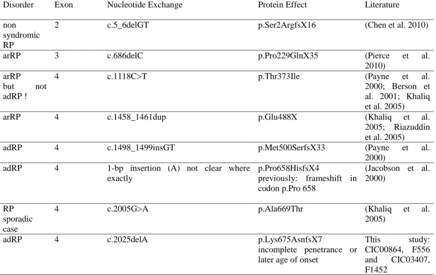
Table 1: Previously described and novel RP1 mutations leading to ar or adRP.
Disorder Exon Nucleotide Exchange Protein Effect Literature
non syndromic RP
2 c.5_6delGT p.Ser2ArgfsX16 (Chen et al. 2010)
arRP 3 c.686delC p.Pro229GlnX35 (Pierce et al.
2010) arRP
but not adRP !
4 c.1118C>T p.Thr373Ile (Payne et al.
2000; Berson et al. 2001; Khaliq et al. 2005)
arRP 4 c.1458_1461dup p.Glu488X (Khaliq et al.
2005; Riazuddin et al. 2005)
adRP 4 c.1498_1499insGT p.Met500SerfsX33 (Payne et al.
2000) adRP 4 1-bp insertion (A) not clear where
exactly p.Pro658HisfsX4 previously: frameshift in codon p.Pro 658 (Jacobson et al. 2000) RP sporadic case
4 c.2005G>A p.Ala669Thr (Khaliq et al.
2005)
adRP 4 c.2025delA p.Lys675AsnfsX7
incomplete penetrance or later age of onset
This study: CIC00864, F556 and CIC03407, F1452
adRP sporadic case de novo 4 c.2029C>T p.Arg677X incomplete penetrance (Bowne et al. 1999; Guillonneau et al. 1999; Pierce et al. 1999; Sullivan et al. 1999; Jacobson et al. 2000; Payne et al. 2000; Baum et al. 2001; Berson et al. 2001; Sohocki et al. 2001; Xiaoli et al. 2002; Schwartz et al. 2003; Ziviello et al. 2005; Chiang et al. 2006; Gamundi et al. 2006; Roberts et al. 2006; Sullivan et al. 2006)
adRP 4 c.2029delC p.Arg677AspfsX5 (Bowne et al.
1999)
adRP 4 c.2035C>T
in cis with c.5377C>T
p.Gln679X
in cis with p.Pro1793Ser
(Sullivan et al. 1999; Berson et al. 2001)
adRP 4 c.2056C>T p.Gln686X (Gamundi et al.
adRP 4 c.2098G>T p.Glu700X (Bowne et al. 1999; Payne et al. 2000)
adRP4 4 c.2115delA p.Gly706ValsfsX7
previously p.Lys705fsX712
(Gamundi et al. 2006)
adRP 4 c.2164_2165delinsG p.Lys722GlufsX16
previously p.Lys722fsX737
(Gamundi et al. 2006)
adRP 4 c.2167G>T p.Gly723X (Berson et al.
2001; Sohocki et al. 2001; Sullivan et al. 2006)
adRP 4 c.2168_2181del p.Ile725ArgfsX6 (Bowne et al.
1999; Payne et al. 2000)
adRP 4 c.2169delA p.Ile725TyrfsX13 This study:
CIC0009, F8 and CIC02805, F1039
adRP 4 c.2172dupG
Previously c.2169_2170insG
p.Ile725AspfsX4 (Bowne et al. 1999; Sohocki et al. 2001)
adRP 4 c.2171_2186del p.Gly724GlufsX9 (Payne et al.
2000)
adRP 4 c.2185delG p. Glu729LysfsX9 (Berson et al.
2001)
adRP 4 c.2206_2207insT p.Thr736IlefsX4 (Payne et al.
2000)
adRP 4 c.2232T>A p.Cys744X (Bowne et al.
1999; Payne et al. 2000)
adRP 4 c.2239delA p.Ser747ValfsX16 (Jacobson et al. 2000)
adRP 4 c.2275A>T p.Arg759X This study:
CIC01239, F749
adRP 4 c.2284_2289del p.Leu762_Asn763del
previously mentioned frameshift with premature X?
(Payne et al. 2000)
adRP 4 c.2285_2289del
previously c.2280_2284del
Leu762TyrfsX17 (Bowne et al. 1999; Pierce et al. 1999; Jacobson et al. 2000; Payne et al. 2000; Berson et al. 2001; Sohocki et al. 2001; Sullivan et al. 2006)
adRP 4 c.2287_2290del p.Asn763LeufsX11 (Pierce et al.
1999; Berson et al. 2001)
adRP 4 c.2304delC
previously c.2303delC
p.Lys769ArgfsX6 (Bowne et al. 1999; Sohocki et al. 2001)
adRP 4 c.2332A>T p.Lys778X
incomplete penetrence
(Dietrich et al. 2002)
adRP 4 c.2336_2337delCT p.Ser779X (Kawamura et al.
2004)
adRP 4 c.2590_2599del p.Ile864LysfsX11
incomplete penetrence
(Roberts et al. 2006)
adRP 4 c.2594_2596del p.Thr865_Leu866delinsIle (Payne et al. 2000) adRP 4 c.2613dupA previously c.2608_2609insA p.Arg872ThrfsX2 (Payne et al. 2000)
adRP 4 c.2732C>A p.Ser911X
incomplete penetrence
(Roberts et al. 2006)
arRP 4 c.2847delT p.Asn949LysfsX32 (Singh et al.
2009)
adRP 4 c.2951A>G p.Asp984Gly
phenoytpe variations in one family
(Chiang et al. 2006)
adRP 4 c. 3157delT p.Tyr1053ThrfsX4 (Jacobson et al.
2000)
Simplex RP 4 c.4108A>G p.Lys1370Glu (Zhang et al.
2010) arRP 4 c.4555delA previously c.4703delA p.Arg1519GlufsX2 previously p.Arg1519fsX1521 (Riazuddin et al. 2005) non syndromic RP 4 c.4941dupT previously c.4941_4942insT
p.Pro1648SerfsX13 (Chen et al. 2010)
Simplex RP 4 c.4955G>T p.Arg1652Leu (Zhang et al.
2005) arRP 4 c. 5252delA previously c.5400delA p.Asn1751IlefsX4 previously p.Asn1751fsX1754 (Riazuddin et al. 2005)
Nucleotide numbering reflects cDNA numbering with +1 corresponding to the A of the ATG translation initiation codon in the reference sequence NM_006269.1, according to journal guidelines (www.hgvs.org/mutnomen). The initiation codon is codon 1.
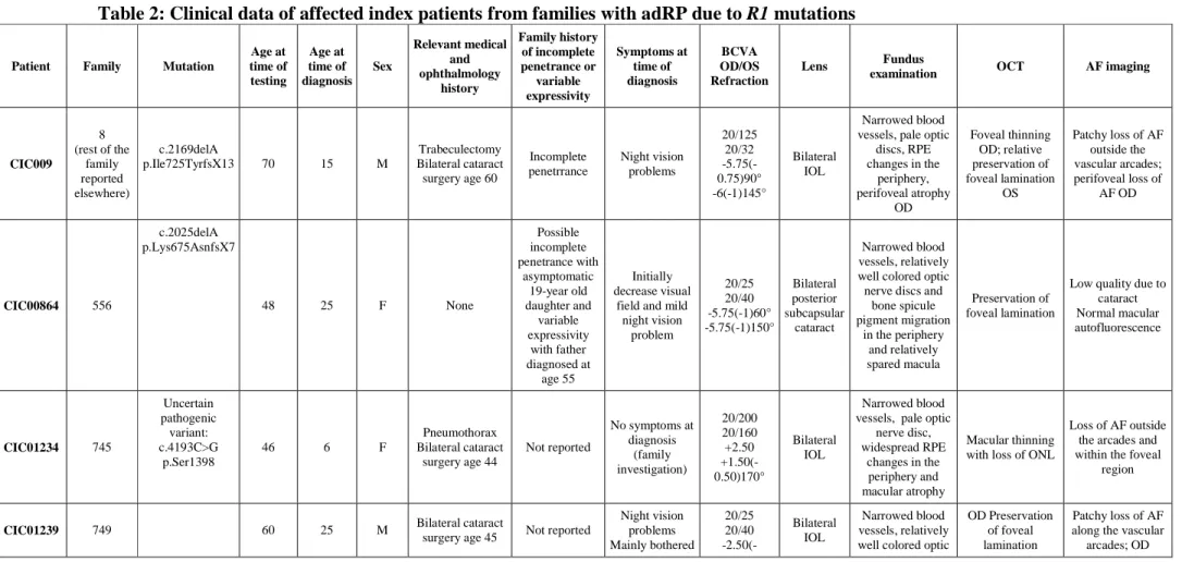
Table 2: Clinical data of affected index patients from families with adRP due to R1 mutations Patient Family Mutation
Age at time of testing Age at time of diagnosis Sex Relevant medical and ophthalmology history Family history of incomplete penetrance or variable expressivity Symptoms at time of diagnosis BCVA OD/OS Refraction Lens Fundus
examination OCT AF imaging
CIC009 8 (rest of the family reported elsewhere) c.2169delA p.Ile725TyrfsX13 70 15 M Trabeculectomy Bilateral cataract surgery age 60 Incomplete penetrrance Night vision problems 20/125 20/32 -5.75(-0.75)90° -6(-1)145° Bilateral IOL Narrowed blood vessels, pale optic
discs, RPE changes in the periphery, perifoveal atrophy OD Foveal thinning OD; relative preservation of foveal lamination OS Patchy loss of AF outside the vascular arcades; perifoveal loss of AF OD CIC00864 556 c.2025delA p.Lys675AsnfsX7 48 25 F None Possible incomplete penetrance with asymptomatic 19-year old daughter and variable expressivity with father diagnosed at age 55 Initially decrease visual
field and mild night vision problem 20/25 20/40 -5.75(-1)60° -5.75(-1)150° Bilateral posterior subcapsular cataract Narrowed blood vessels, relatively well colored optic nerve discs and
bone spicule pigment migration in the periphery and relatively spared macula Preservation of foveal lamination
Low quality due to cataract Normal macular autofluorescence CIC01234 745 Uncertain pathogenic variant: c.4193C>G p.Ser1398 46 6 F Pneumothorax Bilateral cataract surgery age 44 Not reported No symptoms at diagnosis (family investigation) 20/200 20/160 +2.50 +1.50(-0.50)170° Bilateral IOL Narrowed blood vessels, pale optic
nerve disc, widespread RPE changes in the periphery and macular atrophy Macular thinning with loss of ONL
Loss of AF outside the arcades and within the foveal
c.2275A>T p.Arg759X at age 55 by constricted visual field 0.50)95° -5(-0.50)90°
nerve discs, little pigment migration
in the periphery; OD relatively spared macula. OS
CME
OS CME Normal macular autofluorescence; OS perifoveol loss
of AF on the temporal side and abnormal foveal af related to the CMO
CIC02805 1039
c.2169delA p.Ile725TyrfsX13
58 30 M None Variable age of
onset Night vision problem Decreased vision 20/40 20/32 -2.25(-0.75)90° -2.75(-0.50)90) Bilateral posterior subcapsular cataract Narrowed blood vessels, pale optic
discs, little RPE changes in the periphery Relative conservation of foveal lamination Patchy loss of AF outside the vascular arcades and some perifoveal loss CIC01529 1332 c.2065C>T p.Gln689X 51 30 M None Later age of onset reported in other family members Night vision problem 20/32 20/25 -2.50(-1.25)20° -3(-1)140° Mild bilateral posterior subcapsular cataract Narrowed blood vessels, pale optic
nerve discs, RPE changes in the periphery and in perifoveal region Relative conservation of foveal lamination Patchy loss of AF outside the vascular arcades and in the perifoveal area CIC03407 1452 c.2025delA
p.Lys675AsnfsX7 48 27 F Deafness Not reported Night vision problem 20/200 20/800 Plano -0.25(-0.50)130° Significant bilateral posterior subcapsular cataract OD>OS Narrowed blood vessels, relatively well coloured optic nerve heads, little RPE changes in the periphery, macular atrophic
changes
Macular thinning with loss of ONL
Patchy loss of AF in periphery and
foveal region
BCVA: best corrected visual acuity; OD: Oculis dextra (right eye); OS: Oculis Sinistra (left eye); OCT: Optical Coherence Tomography; AF: autofluorescence; IOL: intra ocular lens; CME: cystoid macular edema; RPE: retinal pigment epithelium; ONL: Outer Nuclear Layer
Nucleotide numbering reflects cDNA numbering with +1 corresponding to the A of the ATG translation initiation codon in the reference sequence NM_006269.1, according to journal guidelines (www.hgvs.org/mutnomen). The initiation codon is codon 1.
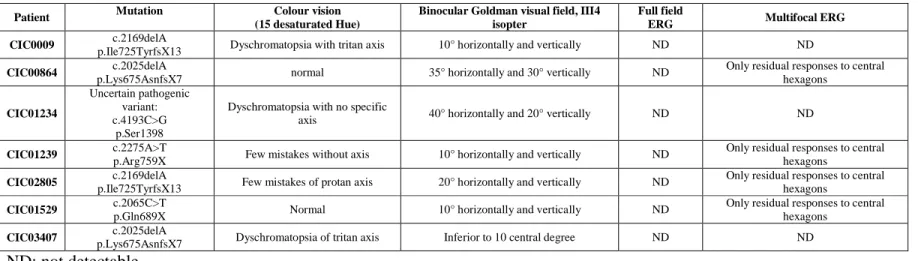
Table 3: Function data
Patient Mutation Colour vision (15 desaturated Hue)
Binocular Goldman visual field, III4 isopter
Full field
ERG Multifocal ERG CIC0009 c.2169delA
p.Ile725TyrfsX13 Dyschromatopsia with tritan axis 10° horizontally and vertically ND ND
CIC00864 c.2025delA
p.Lys675AsnfsX7 normal 35° horizontally and 30° vertically ND
Only residual responses to central hexagons CIC01234 Uncertain pathogenic variant: c.4193C>G p.Ser1398
Dyschromatopsia with no specific
axis 40° horizontally and 20° vertically ND ND
CIC01239 c.2275A>T
p.Arg759X Few mistakes without axis 10° horizontally and vertically ND
Only residual responses to central hexagons
CIC02805 p.Ile725TyrfsX13 c.2169delA Few mistakes of protan axis 20° horizontally and vertically ND Only residual responses to central hexagons
CIC01529 c.2065C>T
p.Gln689X Normal 10° horizontally and vertically ND
Only residual responses to central hexagons
CIC03407 p.Lys675AsnfsX7 c.2025delA Dyschromatopsia of tritan axis Inferior to 10 central degree ND ND
ND: not detectable
Nucleotide numbering reflects cDNA numbering with +1 corresponding to the A of the ATG translation initiation codon in the reference sequence NM_006269.1, according to journal guidelines (www.hgvs.org/mutnomen). The initiation codon is codon 1.
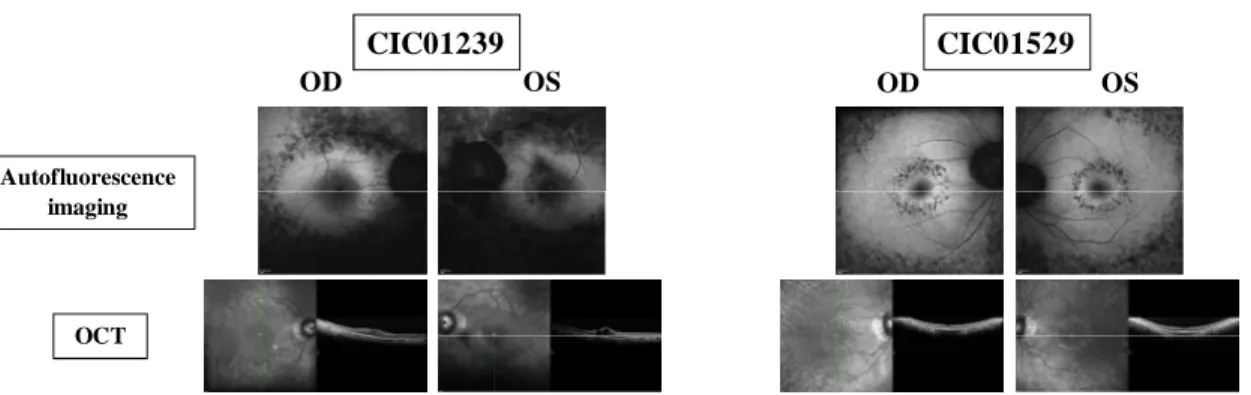
FIGURE LEGENDS
Figure 1: Fundus autofluorescence and optical coherence appearance in 2 patients from this French cohort with RP1 truncating mutations. CIC01239 Autofluorescence imaging OCT OD OS OD OS CIC01529
Supplementary data Methods
Clinical assessment
Patients with a provisional diagnosis of autosomal dominant rod-cone dystrophy, (adRP) were ascertained in the Clinical Investigating Centre of Quinze-Vingts Hospital. Informed consent was obtained from each patient and normal controls after explanation of the study and its potential outcome. The study protocol adhered to the tenets of the Declaration of Helsinki and was approved by the local ethics committee. Each patient underwent full ophthalmic examination with clinical assessment as described earlier (Audo et al., 2010, RHO mutations). For additional family members who could not come to our centre for examination, ophthalmic records were obtained from local ophthalmologists.
Mutation detection
Total genomic DNA was extracted from peripheral blood leucocytes according to manufacturer recommendation (Puregen Kit, Qiagen, Courtaboeuf, France). Subsequently, direct genomic sequencing of RP1 (Accession Number: NM_006269.1) was performed. All 4 exons of which exons 2-4 are coding, and flanking intronic regions of RP1 were PCR amplified in 20 fragments (RP1 RefSeq NM_006269.1) using a polymerase (HotFire, Solis Biodyne, Estonia) in the presence of 1.5 mM MgCl2 and at an annealing temperature of 58°C (Primers can be obtained by request). The PCR products were enzymatically
purified (ExoSAP-IT, USB Corporation, Cleveland, Ohio, USA purchased from GE Healthcare, Orsay, France) and sequenced with a commercially available sequencing mix (BigDyeTerm v1.1 CycleSeq kit, Applied Biosystems, Courtaboeuf, France). The sequenced products were purified on a presoaked Sephadex G-50 (GE Healthcare) 96-well multiscreen filter plate (Millipore, Molsheim, France), the purified product analyzed on an automated 48-capillary sequencer (ABI 3730 Genetic analyzer, Applied Biosystems) and the results interpreted by applying a software (SeqScape, Applied Biosystems). For the missense mutation 190 commercially available control samples were used to validate the pathogenicity (Human random control panel 1-3, Health Protection Agency Culture Collections, Salisbury, United Kingdom).
Supp. Figure 1: Pedigrees of families with different novel RP1 mutations and co-segregation in available family members. Filled symbols represent affected and unfilled unaffected persons. Squares indicate males, circles females. Arrows reflect the index patients. Equation symbols represent unaffected alleles.
Supplementary Table 1: Novel RP1 mutations in a French adRP cohort.
Index (families) Exon Nucleotide Exchange Protein Effect controls SIFT POLYPHEN CIC00864 (F556) (III.7) CIC00865 (IV.2) asymptomatic daughter CIC04903 (IV.8) unaffected brother CIC04950 (II.1) affected uncle CIC04968 (II.2) unaffected uncle CIC04969 (II.3) affected father CIC05003, (III.3) affected cousin CIC05043, (III.6) unaffected cousin 4 c.2025delA c.2025delA no c.2025delA no c.2025delA c.2025delA no p.Lys675AsnfsX7 p.Lys675AsnfsX7 no p.Lys675AsnfsX7 no p.Lys675AsnfsX7 p.Lys675AsnfsX7 no nd na na
CIC05051, (III.5) affected cousin CIC05053, (III.1) unaffected cousin CIC05093 (IV.2) asymptomatic daughter CIC05168 (III.2) affected cousin c.2025delA no c.2025delA c.2025delA p.Lys675AsnfsX7 no p.Lys675AsnfsX7 p.Lys675AsnfsX7 CIC03407 (F1452) (II.1) 4 c.2025delA p.Lys675AsnfsX7 nd na na CIC01529 (F1332) (IV.3) CIC05213 (III.1) affected father CIC05179 (IV.1) affected sister CIC05185 (IV.2) affected sister CIC05312 (IV.4) 4 c.2065C>T c.2065C>T c.2065C>T c.2065C>T no p.Gln689X p.Gln689X p.Gln689X p.Gln689X no nd na na
CIC05193 (IV.5) unaffected brother CIC05310 (V.3) unaffected nephew CIC05186 (V.4) unaffected niece CIC05253 (V.7) unaffected nephew CIC05311 (V.8) unaffected nephew CIC05192 (V.11) unaffected nephew no no no no no no no no no no no no CIC00009 (F8)* CIC00010 affected son 4 c.2169delA c.2169delA p.Ile725TyrfsX13 p.Ile725TyrfsX13 nd na na CIC02805 (F1039) (II.4) 4 c.2169delA p.Ile725TyrfsX13 nd na na CIC01239 (F749) (III.3) CIC04884 (II.3) affected mother 4 c.2275A>T c.2275A>T p.Arg759X p.Arg759X nd na na
*This family was also investigated by another French group (Christian Hamel, U583, INSERM, Institute for Neurosciences of Montpellier, France) and thus further details are given elsewhere.
Supplementary Table 2: Previously described RP1 variants with unclear pathogenicity
Disorder Exon Nucleotide Exchange Protein Effect Why
unclear/comment
Literture
adRP
3 c.652G>A p.Ala218Thr No other family
members available for co-segregation
(Berson et al. 2001)
adRP 4 c.1222A>C p.Ile408Leu PolyPhen and
SIFT benign but did not appear in 190 controls
(Zhang et al. 2010)
adRP 4 c.1437G>T p.Met479Ile Insufficient data
to conclude whether associated with RP (Baum et al. 2001; Chiang et al. 2006)
adRP? 4 c.1989G>T p.Lys663Asn no other family
members for testing, non-conserved residue in human and mouse (Bowne et al. 1999; Payne et al. 2000) adRP 4 c.2376A>C c.2393G>C p.Lys792Gln p.Arg798Asn no other family members, Lys conserved, Arg not (Payne et al. 2000)
adRP 4 c.2700A>C p.Lys900Thr conserved residue,
no other family members
(Payne et al. 2000)
adRP 4 c.2707T>A p.Pro903Leu 2 families, 1. 2
affected carried the mutation, while the
unaffected did not show the
mutation. 2. 1 affected and one at-risk individual incomplete
(Sheng et al. 2008)
penetrance RP 4 c.4955G>T p.Arg1652Leu Conserved residue, no other family member (Zhang et al. 2010)
adRP 4 c.5377C>T p.Pro1793Ser patient carried
also a nonsense mutation in cis, which was considered to be pathogenic (Payne et al. 2000; Berson et al. 2001)
adRP? 4 c.5423T>C p.Leu1808Pro no other family
members for testing
(Bowne et al. 1999)
adRP 4 c.6338C>A p.Thr2113Asn no other family
members for testing
(Payne et al. 2000; Sohocki et al. 2001)
Supplementary Table 3: Previously described rare RP1 variants which are unlikely to be pathogenic
Disorder Exon Nucleotide Exchange Protein Effect Why Literture
adRP 2 c.502C>G p.Arg168Gly did not
co-segregate
(Berson et al. 2001)
RP 3 c.746G>A p.Arg249His did not
co-segregate (Baum et al. 2001; Chiang et al. 2006) RP intron 3 c.787+34T>C - no evidence to be pathogenic (Zhang et al. 2010) RP 4 c.2116G>C p.Gly706Arg appeared in controls (Baum et al. 2001; Chiang et al. 2006; Zhang et al. 2010)
adRP 4 c.2255C>T p.Thr752Met did not
co-segregate, appeared in controls
(Roberts et al. 2006)
adRP 4 c.2953A>T p.Asn985Tyr Segregated in
one family but also
(Sullivan et al. 1999; Sohocki et al. 2001; Sheng
SNP rs2293869 and in specific cohorts appeared in controls
adRP 4 c.3215A>G p.Asp1072Gly did not
co-segregate (Berson et al. 2001) RP 4 c.3024G>A p.Gln1008Gln appeared in controls (Baum et al. 2001; Zhang et al. 2010)
RP 4 Wrong annotation, not clear
previously: c.3188G>A p.Gln1063Arg appeared in controls (Baum et al. 2001; Chiang et al. 2006)
adRP 4 c.4067T>C p.Leu1356Ser did not
co-segregate
(Berson et al. 2001)
adRP 4 c.4250T>C p.Leu1417Pro did not
co-segregate, 1/95 controls
(Berson et al. 2001)
adRP 4 c.4784G>A p.Arg1595Gln no other
family members now documented as SNP rs35084330 (Bowne et al. 1999; Payne et al. 2000; Sohocki et al. 2001)
controls 4 c.5797C>T p.Arg1933X appeared in
controls
(Baum et al. 2001; Chiang et al. 2006)
adRP 4 c.5805T>G p.Phe1935Leu did not
co-segregate
(Berson et al. 2001)
RP 4 c.6045A>G p.Leu2015Leu no evidence
to be pathogenic
(Zhang et al. 2010)
adRP 4 c.6196G>A p.Asp2066Asn did not
co-segregate, 1/91 controls
(Berson et al. 2001)
controls 4 c.6423A>G p.Ile2141Met appeared in
controls (Baum et al. 2001; Chiang et al. 2006) RP 4 c.6542C>T - appeared in controls (Baum et al. 2001; Zhang et
al. 2010)
Supplementary Table 4: Rare RP1 variants with uncertain pathogenicity in a French adRP cohort.
Index (families) Exon Nucleotide Exchange Protein Effect controls SIFT POLYPHEN
CIC01234 (F745) (III.4) CIC04861 (IV.8) unaffected niece CIC04983, (III.2) assymptomatic brother 4 c.4193C>G no c.4193C>G p.Ser1398Cys no p.Ser1398Cys 0/190 affect protein function possibly damaging
Supplementary Table 5: Rare RP1 variants in a French adRP cohort.
Index (families)
Exon Nucleotide Exchange Protein Effect controls SIFT POLYPHEN
CIC00869 (F558) Other CIC04860 affected daughter 1 1 c.1-51A>G no - nd na na CIC02802 (F933) CIC03164 affected son CIC03224 unaffected daughter CIC03420 unaffected son 4 4 4 4 c.6139G>A no no no p.Val12047Met no no no nd tolerated benign
affected cousin CIC01239 (F749)
4 c.5962A>G p.Ile1988Val nd tolerated benign
Nucleotide numbering reflects cDNA numbering with +1 corresponding to the A of the ATG translation initiation codon in the reference sequence NM_006269.1, according to journal guidelines (www.hgvs.org/mutnomen). The initiation codon is codon 1.