HAL Id: hal-01962025
https://hal.sorbonne-universite.fr/hal-01962025
Submitted on 20 Dec 2018HAL is a multi-disciplinary open access
archive for the deposit and dissemination of sci-entific research documents, whether they are pub-lished or not. The documents may come from teaching and research institutions in France or abroad, or from public or private research centers.
L’archive ouverte pluridisciplinaire HAL, est destinée au dépôt et à la diffusion de documents scientifiques de niveau recherche, publiés ou non, émanant des établissements d’enseignement et de recherche français ou étrangers, des laboratoires publics ou privés.
Synthesis of lipid-carbohydrate-peptidyl-RNA
conjugates to explore the limits imposed by the
substrate specificity of cell wall enzymes on the
acquisition of drug resistance
Matthieu Fonvielle, Ahmed Bouhss, Coralie Hoareau, Delphine Patin,
Dominique Mengin-Lecreulx, Laura Iannazzo, Nicolas Sakkas, Affaf El
Sagheer, Tom Brown, Mélanie Ethève-Quelquejeu, et al.
To cite this version:
Matthieu Fonvielle, Ahmed Bouhss, Coralie Hoareau, Delphine Patin, Dominique Mengin-Lecreulx, et al.. Synthesis of lipid-carbohydrate-peptidyl-RNA conjugates to explore the limits imposed by the substrate specificity of cell wall enzymes on the acquisition of drug resistance. Chemistry - A European Journal, Wiley-VCH Verlag, 2018, 24 (56), pp.14911-14915. �10.1002/chem.201802360�. �hal-01962025�
Synthesis of lipid-carbohydrate-peptidyl-RNA conjugates to explore the limits imposed
by the substrate specificity of cell wall enzymes on the acquisition of drug resistance
Matthieu Fonvielle[a]*, Ahmed Bouhss[b], Coralie Hoareau[a], Delphine Patin[b], Dominique
Mengin-Lecreulx[b], Laura Iannazzo[c], Nicolas Sakkas[c], Affaf el Sagheer, [d,e] Tom Brown[e], Mélanie
Ethève-Quelquejeu[c] *+ and Michel Arthur[a]*+
[a] Dr. Matthieu Fonvielle*, Dr. Michel Arthur*, Coralie Hoareau, INSERM UMRS 1138, Sorbonne Universités, UPMC Univ Paris 06; Sorbonne Paris Cité, Université Paris Descartes, Université Paris Diderot; Centre de Recherche des Cordeliers, 75006 Paris, France. E-mail: matthieu.fonvielle@crc.jussieu.fr michel.arthur@crc.jussieu.fr
[b] Dr. Ahmed Bouhss, Ms Delphine Patin, Dr. Dominique Mengin-Lecreulx, Institute for Integrative Biology of the Cell (I2BC), CEA, CNRS, Univ Paris-Sud, Université Paris-Saclay, 91198, Gif-sur-Yvette cedex, France. Present adress (AB): Laboratoire Structure-Activité des Biomolécules Normales et Pathologiques (SABNP), Univ Evry, INSERM U1204, Université Paris-Saclay, 91025 Evry, France.
[c] Dr. Laura Iannazzo, Nicolas Sakkas, Dr. Mélanie Ethève-Quelquejeu*, Laboratoire de Chimie et de Biochimie Pharmacologiques et Toxicologiques, Université Paris Descartes, UMR 8601, Paris, F-75005 France; CNRS UMR 8601, Paris, F-75006 France.
[d] Dr. Afaf H.El-Sagheer, Chemistry Branch, Dept. of Science and Mathematics, Faculty of Petroleum and Mining Engineering, Suez University, Suez, 43721 (Egypt)
[e] Dr. Tom Brown, Department of Chemistry, University of Oxford, 12 Mansfield Road, Oxford, OX1 3TA, UK
[*] Correspondence should be addressed to M.F. (email: matthieu.fonvielle@crc.jussieu.fr) or M.A. (email:
michel.arthur@crc.jussieu.fr)
[+] These authors contributed equally to this work.
Abstract: Conjugation of RNA with multiple partners to obtain mimics of complex biomolecules is limited
by the identification of orthogonal reactions. Here, lipid-carbohydrate-peptidyl-RNA conjugates were obtained by post-functionalization reactions, solid-phase synthesis, and enzymatic steps, to generate molecules mimicking the substrates of FmhB, an essential peptidoglycan synthesis enzyme of
Staphylococcus aureus. Mimics of Gly-tRNAGly and lipid intermediate II
(undecaprenyl-diphospho-disaccharide-pentapeptide) were combined in a single “bi-substrate” inhibitor (IC50 = 56 nM). The
synthetic route was exploited to generate substrates and inhibitors containing D-Lac instead of D-Ala at the C-terminus of the pentapeptide stem, a modification responsible for vancomycin resistance in the enterococci. The substitution impaired recognition of peptidoglycan precursors by FmhB. The associated fitness cost may account for limited dissemination of vancomycin resistance genes in S. aureus.
Various RNA conjugates have been synthesized to develop new therapeutic strategies and to investigate basic biological processes.[1] These molecules combine an oligonucleotide with
carbohydrates, peptides, small molecules, aptamers, or lipids.[1a] Carbohydrate-oligonucleotide and
small molecule-oligonucleotide conjugates have been mostly used to target specific receptors or to increase uptake in target organs.[2] Aptamer-RNAs were developed to improve the combined delivery of
drugs and siRNAs in multidrug-resistant cancer cells.[3] Peptidyl-RNAs have played pivotal roles in
mechanistic and structural studies of ribosomal and non-ribosomal peptide synthesis.[4] Peptidyl-RNA
conjugates were also developed to increase the cellular uptake of siRNA.[5] Lipid-RNA conjugates were
designed to increase the lipophilicity of negatively charged oligonucleotides and to leverage lipoprotein-mediated endocytosis.[2] All of these conjugates offer interesting properties and access to novel chemical
space.[1a] Various types of linkers have been used to conjugate RNAs to the various partners, including
amide, amine, oxyamine, oxyimine, carbamate, and triazoles, which are chemically and biologically stable.[6] Bioreversible linkers rely on disulfide bridges.[7] The conjugation is mostly performed at the 5’
and 3’ ends of the RNA, rather than internally, with the molecules attached to the nucleobase or to the ribose.
There are limited examples of the conjugation of a single RNA molecule with multiple partners. This type of adduct is required to investigate the biosynthesis of bacterial cell walls since its major component, the peptidoglycan, is assembled from a precursor combining a disaccharide [β-1,4-linked N-acetyl-glucosamine and N-acetyl-muramic acid, (GlcNAc-MurNAc)], a phospholipid carrier (undecaprenyl-diphosphate), and a branched pentapeptide, such as [L-Ala-D-iGlu-L-Lys(Gly5)-D-Ala-D-Ala] in
glycyl-tRNAs by amino acid transferases of the Fem family there is interest in developing lipid-carbohydrate-peptidyl-RNA conjugates (Figure 1c) to investigate the interaction of the enzymes with their substrates. The biological question addressed in our study concerns the acquisition of resistance to the glycopeptide antibiotic vancomycin in S. aureus by replacement of the terminal D-Ala residue by D-Lac. The substitution reduces the affinity of the drugs for peptidoglycan by 1,000 fold leading to high-level resistance (Figure 1d). Since the resistance mechanism is widespread in the enterococci but only sporadically detected in S. aureus in spite of easy horizontal gene transfer between these bacteria[8] our
specific aim is to evaluate whether the D-Ala to D-Lac substitution is tolerated by the S. aureus transferase FmhB responsible for incorporation of the first residue of the penta-glycine side chain.[9] We
focus on FmhB since this enzyme is essential presumably because the side-chain of peptidoglycan precursors directly participates in the peptidoglycan cross-linking reaction (Figure 1e).[9-10] To investigate
the specificity of FmhB, we have synthesized D-Ala- and D-Lac-containing substrates analogues (Figure 2) and lipid-carbohydrate-peptidyl-RNA conjugates acting as inhibitors (Figure 1c). This strategy was designed to assess the impact of the D-Ala to D-Lac substitution both on the catalytic efficacy of the transfer of Gly from Gly-tRNAGly to the peptidoglycan precursors and on the affinity of FmhB for inhibitors
that mimic both substrates of the enzyme (referred to as “bi-substrates”). The target inhibitor molecules (Figure 1c) comprise soluble analogues of the peptidoglycan precursors (Lipid II) covalently-linked by a triazole to the acceptor arm of tRNAGly. Our synthesis strategy enables to modulate the length of the
RNA and lipid moieties in order to obtain ligands with suitable size and solubility for investigating enzyme activity and affinity. It also enables to selectively modify the terminal residue of the peptide (D-Ala versus D-Lac). One of the major difficulty for obtaining the desired lipid-carbohydrate-peptidyl-RNA conjugates is the identification of orthogonal reactions compatible with the various functional groups specifically present in each component of the multi-RNA conjugates. In our synthetic strategy, we overcome these problems by developing post-functionalization of RNA and peptides moieties as well as a combination of enzymatic and chemical reactions (Figure 1c).
Figure 1. a) Assembly of the pentaglycine side-chain of peptidoglycan precursors by amino-acyl
transferases of the Fem family. (b) Reaction catalyzed by FmhB. (c) Target lipid-carbohydrate-peptidyl-RNA conjugates developed as bi-substrate inhibitors. (d) Hydrogen bonding interaction between vancomycin and the peptidyl-D-Ala-D-Ala extremity of peptidoglycan precursors. Substitution of the C-terminal D-Ala by D-Lac prevents formation of the hydrogen bond indicated in red. (e) Penicillin Binding Protein (PBP) mediated cross-linking for vancomycin-susceptible and -resistant strains of
MurG Bacteria Extraction (HCOOH 1.1 M) MraY Neryl-phosphate or heptaprenyl-phosphate GlcNAc, ATP Enterococcus faecalis UDP-MurNAc-pentapeptide ending in D-Ala4-D-Ala5
(X=NH)
Vancomycin-resistant S. aureus
UDP-MurNAc-pentadepsipeptide ending in D-Ala4-D-Lac5
(X=O) or O O O O AcHN OH O P O OH PO OH O HOHO NHAc OH O HN O HN CO2H O NH O HN O X CO2H H2N O HN O HN CO2H O NH O HN O X CO2H X = NH or O H2N O OH O O NHAc OH P OOH P HO O O N H N O O O O HO OH X = NH or O O n 2a: X=NH; n=6 2b: X=O; n=6 3a: X=NH; n=1 3b= X=O; n=1
Figure 2. Semi-synthesis of lipid II analogues. MraY,
Phospho-N-acetylmuramoyl-pentapeptide-transferase. MurG, N-acetylglucosamine- Phospho-N-acetylmuramoyl-pentapeptide-transferase.
To generate the D-Ala- and D-Lac-ending substrates of FmhB, the soluble nucleotide precursors were extracted from vancomycin-susceptible (D-Ala5) and vancomycin-resistant (D-Lac5) bacteria (Figure 2).
Transfer of the phospho-MurNAc-peptide moiety to the lipid and addition of GlcNAc were obtained enzymatically.[12] For these substrates, we used commercially available heptaprenyl-phosphate instead
of the natural undecaprenyl-phosphate lipid carrier (bactoprenyl). Transfer of [14C]Gly from [14
C]Gly-tRNAGly to the heptaprenyl-containing lipid II analogue was tested by a coupled assay involving acylation
of tRNAGly by purified glycyl-tRNA synthetase (GlyRS). The product was identified by thin layer
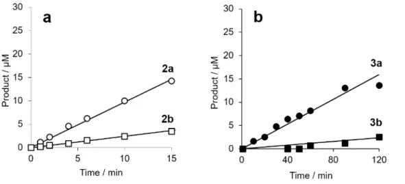
chromatography (Supplementary information, Figure S5). This analysis revealed that substitution of D-Ala by D-Lac at the C-terminus of the lipid II analogues reduces the catalytic efficacy of FmhB by 4.6 fold (Figure 3a).
There are several limitations resulting from the insolubility of the heptaprenyl-containing substrates including uncertainty regarding the concentrations of lipid II analogues actually accessible to FmhB and time consuming solvent extractions required for purification. In order to gain access to soluble lipid II analogues, we replaced heptaprenyl-phosphate by neryl-phosphate in the synthesis procedure. The
resulting neryl-containing lipid II analogues were effectively used as substrates by FmhB revealing again a reduced catalytic efficacy for D-Lac-ending substrates (7 fold; Figure 3b and Table 1).
Figure 3. Catalytic efficacy of FmhB for the transfer of Gly from Gly-tRNAGly to lipid II analogues ending
in D-Ala or D-Lac. (a) and (b), transfer of Gly from heptaprenyl- and neryl-containing substrates (30 µM) was tested with FmhB concentrations of 50 nM and 170 nM, respectively. Circles, D-Ala-ending substrate; squares, D-Lac-ending substrate.
Lipid-carbohydrate-peptidyl-RNA conjugates mimicking both the Gly-tRNAGly and lipid II
(lipid-carbohydrate-peptide) substrates were synthesized in order to explore the impact of the D-Ala to D-Lac substitution on the affinity of FmhB for peptidoglycan precursors (Figure 2). The presence of the carbohydrate and lipid parts have been shown to be essential for the activity of the FmhB transferase.[13] The synthesis of the Lipid II moiety started by four enzymatic reactions for sequential
addition of L-Ala, D-Glu, meso-cystine, and D-Ala-D-Ala or D-Ala-D-Lac to UDP-MurNAc by Mur synthetases (Figure 4). Meso-cystine is a structure analogue of meso-2,6-diaminopimelic acid (DAP), which is recognized by the enzyme MurE and enables post-functionalization of the peptide via formation of a dehydroalanine.[14] For this purpose, we have developed a new one-pot reaction based on reduction
of meso-cystine into cysteine by a soluble phosphine followed by its conversion to dehydroalanine by 2,5-dibromohexanediamide. By this approach, a dehydroalanine residue was introduced at the 3rd
position of the stem peptide. In the following step, 1,4-addition of 1-thio butynyl to the dehydroalanine afforded two stereoisomers containing an alkyne function at the extremity of the side-chain of the 3rd
residue. The (R) diastereoisomer was purified by rpHPLC as previously described.[15] MraY and MurG
were permissive for the transfer of the phospho-MurNAc-peptide moieties to heptaprenyl-phosphate and neryl-phosphate and the subsequent addition of GlcNAc, respectively, generating the corresponding alkyne-containing lipid II analogues.[12] For the RNA moiety, 2’-azido-5’-dimethoxytrityl-benzoyl
adenosine was coupled to a resin for solid phase synthesis of an 18-mer mimicking the acceptor arm of tRNAGly.[4a] During solid phase synthesis, a hexaethylene glycol linker was incorporated to stabilize the
hairpin duplex formed by the RNA strand. The alkyne-containing lipid II analogues were coupled to the azido-containing RNA by the CuI-catalyzed Huisgen-Sharpless cycloaddition reaction in the presence of
a metal ligand (THPTA).[15] The resulting triazole-containing lipid-carbohydrate-peptidyl-RNA conjugates
were purified by denaturing polyacrylamide gel electrophoresis and by rpHPLC for heptaprenyl- and neryl-containing molecules, respectively (Supplementary Information).
Our next objective was to assess the impact of the substitution of the C-terminal D-Ala by D-Lac on the inhibition of FmhB by lipid-carbohydrate-peptidyl-RNA conjugates (“bi-substrates”). The conjugates were tested as FmhB inhibitors in the GlyRS-FmhB coupled assay (Figure 5). Substitution of D-Ala by D-Lac at the C-terminus of the peptide stems of the conjugates led to a 15-fold increase in the IC50
values (from 56 ± 6 nM to 820 ± 50 nM for neryl-containing analogues; Table 1). This difference highlights the contribution of the D-Ala-D-Ala amide bond to the binding energy to the enzyme. IC50s of 56 ± 6 nM
and 5 ± 3 nM were determined for D-Ala-ending heptaprenyl- and neryl-PP-disaccharide-pentapeptide-RNA conjugates indicating that shortening of the lipid moiety (7 versus 2 prenyl groups) leads to a ca. 11-fold reduction in affinity (Supplementary Information and Table 1). This mirrors the 7- and 10-fold reduction observed with the substrates (Table 1).
O HO N A O P -O O O NH O HN CO2H O NH O HN O X CO2H N N C75 C74 U73 C72 G71 C70 C69 U68 U67 C66 A6 C2 G3 G4 S G5 G7 G1 NH O HN CO2H O NH O HN O X CO2H S S UDP-MurNAc H2N CO2H TCEP O NH2 O H2N Br Br NH O HN CO2H O NH O HN O X CO2H a: X = NH; n=7 b: X = O; n=2 c: X= NH; n=2 UDP-MurNAc 1a-b 2a-b SH NH O HN CO2H O NH O HN O X CO2H UDP-MurNAc 3a-b S MraY MurG NH O HN CO2H O NH O HN O X CO2H GlcNAc-MurNAc-PP-n-prenyl 4a-c S UDP-MurNAc MurE D-Ala-D-Ala or D-Ala-D-Lac MurF meso-cystine MurD D-Glu MurC L-Ala O O N3 N N N N NHBz DMTO O O N3 N N N N NHBz O O NH O O P O O OH O 6 DMT O O N3 N N N N NHBz O O OH N3 N N N N NH2 O O P O O 6 DMT N SPS SPS SPS C75 CU74 73 C72 GC71 70 C69 U68 U67 C66 C75 C74 U73 C72 G71 C70 C69 U68 U67 C66 C75 C74 U73 C72 G71 C70 C69 U68 U67 C66 A6 C2 G3 G4 G5 G7 G1 CuI THPTA GlcNAc-MurNAc-PP-n-prenyl 5 6a-c CN
Figure 4. Synthesis of the n-prenyl-carbohydrate-peptidyl-RNA conjugates. X = NH Ala) or X = O
(D-Lac); n = 2 (neryl); n = 7 (heptaprenyl). THPTA, tris(3-hydroxypropyltriazolylmethyl)amine. TCEP, tris(2-carboxyethyl)phosphine..
Total[10a,16] and enzymatic[12] synthesis of lipid II have been described previously and were mostly
used to incorporate fluorescent tags. Here, we report a hybrid strategy using dehydroalanine-containing lipid analogues for post-functionalization. This approach was used to generate lipid-carbohydrate-peptidyl-RNA conjugates mimicking the lipid II and Gly-tRNAGly substrates of the FmhB transferase from
S. aureus. Variability was introduced in the sequence of the peptide and in the number of isoprenyl
repeats in the lipid moiety. Solid-phase synthesis of the RNA moiety offers the possibility to modulate the size and the sequence of the RNA moiety. The 1,4 addition of the thiol to dehydroalanine also offers a versatile access to bi-substrates containing linkers with various lengths and structures. Our lipid-carbohydrate-peptidyl-RNA conjugates provide a versatile tool to study the tRNA-dependent amino acyltransferases involved in peptidoglycan synthesis.
In this study, we investigated the specificity of the FmhB aminoacyl-transferase by comparing substrates and inhibitors differing by the presence of D-Ala or D-Lac at the C-terminus of the stem peptide. Testing the substrates indicated that the D-Ala to D-Lac substitution impairs the activity of FmhB. Testing the inhibitors indicated that the substitution reduced the affinity of FmhB for the peptidoglycan
precursor analogues. Structural analyses of peptidoglycan purified from vancomycin-resistant S. aureus showed that a high portion of the stem peptides were devoid of the pentaglycine side-chain.[9,11] Our data
indicate that impaired activity of FmhB accounts for the presence of incomplete stem peptides lacking the pentaglycine side-chain. Such incomplete precursors are only used as acyl donors by the transpeptidases responsible for the formation of peptidoglycan cross-links (Figure 1e). Our data also indicate that the production of D-Lac-ending precursors may have a fitness cost by impairing the assembly of peptidoglycan precursors. Such a fitness cost could account for the lack of dissemination of vancomycin-resistance staphylococci in spite of the sporadic detection of this type of strain in patients treated with vancomycin. The origin of this fitness cost is not known. The presence of incomplete side-chain only in the “donor” stem of dimers suggests that impaired activity of the transpeptidases might be involved.
Figure 5. Inhibition of FmhB by lipid-carbohydrate-peptidyl-RNA conjugates. (a) Structure of the
inhibitors. X = NH (D-Ala) or X = O (D-Lac). (b and c) Separation of [14C]Gly from [14C]Gly-lipid II
analogues by rpHPLC coupled to a radioflow detector to assess the extent of inhibition of FmhB by increasing concentrations of inhibitors 8b and 8c ending in D-Ala and D-Lac, respectively. (d) % inhibition of FmhB by increasing concentrations of 8b and 8c (close and open circle, respectively). The IC50 values
(56 ± 6 nM and 820 ± 50 nM for 8b and 8c, respectively) were deduced by fitting the Morrison equation to the data.
This work was supported by the project SyntRNA from ANR program to MEQ and MA (ANR-17-CE07-0041-01). NS was funded by a DGA-MRIS scholarship.
Keywords: Fem • RNA conjugates • Staphylococcus aureus • VanA • vancomycin
[1] a) E. Valeur, S. M. Gueret, H. Adihou, R. Gopalakrishnan, M. Lemurell, H. Waldmann, T. N. Grossmann, A. T. Plowright, Angew. Chem. Int. Ed. Engl. 2017, 56, 10294-10323; b) J. Winkler, Therapeutic delivery 2013, 4, 791-809.
[2] a) M. Manoharan, Antisense Nucleic Acid Drug Dev. 2002, 12, 103-128; b) N. S. Kruglova, M. I. Meschaninova, A. G. Venyaminova, M. A. Zenkova, V. V. Vlassov, E. L. Chernolovskaya, Molecular Biology 2010, 44, 254-261; c) J. Baillet, V. Desvergnes, A. Hamoud, L. Latxague, P. Barthélémy,
2018, Advanced Materials, 1705078.
[3] a) G. Zhu, G. Niu, X. Chen, Bioconjug. Chem. 2015, 26, 2186-2197; b) H. Jeong, S. H. Lee, Y. Hwang, H. Yoo, H. Jung, S. H. Kim, H. Mok, Macromolecular bioscience 2017, 17, 1600343
[4] a) M. Fonvielle, I. Li de La Sierra-Gallay, A. H. El-Sagheer, M. Lecerf, D. Patin, D. Mellal, C. Mayer, D. Blanot, N. Gale, T. Brown, H. van Tilbeurgh, M. Etheve-Quelquejeu, M. Arthur, Angew. Chem. Int. Ed. Engl. 2013, 52, 7278-7281; b) S. Terenzi, E. Biala, N. Q. Nguyen-Trung, P. Strazewski, Angew. Chem. Int. Ed. Engl. 2003, 42, 2909-2912; c) H. Moroder, J. Steger, D. Graber, K. Fauster, K. Trappl, V. Marquez, N. Polacek, D. N. Wilson, R. Micura, Angew. Chem. Int. Ed. Engl. 2009, 48, 4056-4060.
[5] a) M. R. Alam, X. Ming, M. Fisher, J. G. Lackey, K. G. Rajeev, M. Manoharan, R. L. Juliano, Bioconjug. Chem. 2011, 22, 1673-1681; b) M. R. Alam, V. Dixit, H. Kang, Z. B. Li, X. Chen, J. Trejo, M. Fisher, R. L. Juliano, Nucleic Acids Res. 2008, 36, 2764-2776.
[6] a) M. Arthur, M. Ethève-Quelquejeu, Chemistry of organo-hybrids: synthesis and characterization of functional nano-objects. (Eds.: E. Lacôte, B. Charleux, C. Coperet), Wiley-VCH, 2015; b) E. Paredes, M. Evans, S. R. Das, Methods 2011, 54, 251-259.
[7] A. Ardana, A. K. Whittaker, K. J. Thurecht, Macromolecular Research 2017, 25, 599-614. [8] P. C. Appelbaum, International journal of antimicrobial agents 2007, 30, 398-408.
[9] A. Severin, K. Tabei, F. Tenover, M. Chung, N. Clarke, A. Tomasz, J. Biol. Chem. 2004, 279, 3398-3407.
[10] a) J. Nakamura, H. Yamashiro, H. Miya, K. Nishiguchi, H. Maki, H. Arimoto, Chem. Eur. J. 2013, 19, 12104-12112; b) V. Srisuknimit, Y. Qiao, K. Schaefer, D. Kahne, S. Walker, J. Am. Chem. Soc.
2017, 139, 9791-9794.
[11] D. Panesso, P. J. Planet, L. Diaz, J.-E. Hugonnet, T. T. Tran, A. Narechania, J. M. Munita, S. Rincon, L. P. Carvajal, J. Reyes, A. Londoño, H. Smith, R. Sebra, G. Deikus, G. M. Weinstock, B. E. Murray, F. Rossi, M. Arthur, C. A. Arias, Emerging infectious diseases 2015, 21, 1844-1848.
[12] L. Y. Huang, S. H. Huang, Y. C. Chang, W. C. Cheng, T. J. Cheng, C. H. Wong, Angew. Chem. Int. Ed. Engl. 2014, 53, 8060-8065.
[13] T. Schneider, M. M. Senn, B. Berger-Bachi, A. Tossi, H. G. Sahl, I. Wiedemann, Mol. Microbiol.
2004, 53, 675-685.
[14] J. M. Chalker, S. B. Gunnoo, O. Boutureira, S. C. Gerstberger, M. Fernadez-Gonzalez, J. G. L. Bernardes, L. Griffin, A. Hailu, C. J. Schofield, B. G. Davis, Chem. Sci. 2011, 2, 1666-1676.
[15] M. Fonvielle, D. Mellal, D. Patin, M. Lecerf, D. Blanot, A. Bouhss, M. Santarem, D. Mengin-Lecreulx, M. Sollogoub, M. Arthur, M. Etheve-Quelquejeu, Chem. Eur. J. 2013, 19, 1357-1363.
[16] a) M. S. VanNieuwenhze, S. C. Mauldin, M. Zia-Ebrahimi, B. E. Winger, W. J. Hornback, S. L. Saha, J. A. Aikins, L. C. Blaszczak, J. Am. Chem. Soc. 2002, 124, 3656-3660; b) Y. Zhang, E. J. Fechter, T. S. Wang, D. Barrett, S. Walker, D. E. Kahne, J. Am. Chem. Soc. 2007, 129, 3080-3081.
1
Supplementary information
Synthesis of lipid-carbohydrate-peptidyl-RNA conjugates to explore the limits imposed by the substrate specificity of cell wall enzymes on the acquisition of drug resistance
Matthieu Fonvielle[a]*, Ahmed Bouhss[b], Coralie Hoareau[a], Delphine Patin[b], Dominique
Mengin-Lecreulx[b], Laura Iannazzo[c], Nicolas Sakkas[c], Affaf el Sagheer[d,e], Tom Brown[e], Mélanie
Ethève-Quelquejeu[c]* and Michel Arthur[a]*
[a] Dr. Matthieu Fonvielle*, Dr. Michel Arthur*, Coralie Hoareau, INSERM UMRS 1138, Sorbonne Universités, UPMC Univ Paris 06; Sorbonne Paris Cité, Université Paris Descartes, Université Paris Diderot; Centre de Recherche des Cordeliers, 75006 Paris, France. E-mail: matthieu.fonvielle@crc.jussieu.fr michel.arthur@crc.jussieu.fr
[b]
Dr. Ahmed Bouhss, Ms Delphine Patin, Dr. Dominique Mengin-Lecreulx, Institute for Integrative Biology of the Cell (I2BC), CEA, CNRS, Univ Paris-Sud, Université Paris-Saclay, 91198, Gif-sur-Yvette cedex, France.
[c]
Dr. Laura Iannazzo, Nicolas Sakkas, Dr. Mélanie Ethève-Quelquejeu*, Laboratoire de Chimie et de Biochimie Pharmacologiques et Toxicologiques, Université Paris Descartes, UMR 8601, Paris, F-75005 France; CNRS UMR 8601, Paris, F-75006 France.
[d] Dr. Afaf H.El-Sagheer, Chemistry Branch, Dept. of Science and Mathematics, Faculty of Petroleum and Mining Engineering, Suez University, Suez, 43721 (Egypt)
[e] Dr. Tom Brown, Department of Chemistry, University of Oxford, 12 Mansfield Road, Oxford, OX1 3TA, UK.
[*] Correspondence should be addressed to M.F. (email: matthieu.fonvielle@crc.jussieu.fr) or M.A. (email: michel.arthur@crc.jussieu.fr)
1. General information.……….. p2 2. Mass spectrometry analysis of RNA containing compound………..……… p2 3. Enzyme production and purification………..……… p2 4. tRNA production and purification………..………..……… p2 5. Semi-synthesis.……… p3 6. Enzymatic assays.……….………….……… p9 7- Mass spectrometry analyses of synthesized compounds.………...……… p12 8- rpHPLC analysis of synthesized compounds.………..….………. p24 9- References.……….……… p32
2
1. General information
Solvents were dried using standard methods and distilled before use. Unless otherwise specified, materials were purchased from commercial suppliers and used without further purification. Spectra were recorded on a Bruker AVANCE 400 spectrometer for 1H (250 MHz), 13C (63 MHz), in CDCl
3.
Chemical shifts (δ) are expressed in ppm relative to residual 7.24 for 1H and 77.23 for 13C, as internal
references. Mass spectroscopy (MS) spectra were carried out on a LCQ-Deca XP-Max spectrometer in positive or negative ionization mode. Heptaprenyl-phosphate was purchased from the Institute of Biochemistry and Biophysics of the Polish Academy of Sciences, Warsaw, Poland.
2. Mass spectrometry analysis of RNA-containing compounds
Mass spectrometry analyses of tRNAGly, azido-RNA, and lipid-carbohydrate-peptidyl-RNA conjugates
were performed by LC-MS. Liquid chromatography (LC) was performed by anionic rpHPLC using a DNAPac RP column (4 µm; 2.1 x 50 mm; Thermo Fisher Scientific) at a flow rate of 300 µl/min with a 0 to 50% MeOH linear gradient applied from 2 to 10 min in Solution A [Solution A, triethylamine (15 mM) and 1,1,1,3,3,3-hexafluoro-2-propanol (400 mM)]. For mass spectrometry (MS), liquid chromatography was coupled to a LCQ Deca XP-Max mass spectrometer operating in the negative mode.
3. Enzyme production and purification
The FmhB glycyl transferase from S. aureus strain Mu50 was produced in E. coli BL21 (DE3) pREP4GroEL as a fusion protein with a C-terminal hexa-histidine tag (GSHHHHHH). FmhB was purified from a clarified lysate by metal affinity (Ni Sepharose 6 Fast flow; General Electrics) and size exclusion chromatography (DEX 75 HR 26/60; General Electrics) in 100 mM phosphate buffer (pH 7.5) containing 500 mM KCl. FmhB was concentrated (10 mg mL-1; Amicon Ultra, cutoff 10 kDa; Millipore) and stored
at -20°C following addition of one volume of glycerol per volume of purified FmhB.
The glycyl-tRNA synthetase of S. aureus strain Mu50 was produced in E. coli BL21 (DE3) pREP4GroEL and purified in two steps by metal affinity (Ni Sepharose 6 Fast flow; General Electrics) and anion exchange chromatography (Resource Q; General Electrics), as previously described.[1]
The translocase MraY from Aquifex aeolicus (for the synthesis of the lipid intermediate I) and the MurG transferase from E. coli (for the subsequent addition of GlcNAc) were purified as previously described.[2]
The concentration of proteins was determined by the Bradford assay (Biorad) using BSA as a standard.
4. tRNA production and purification
The sequence of the tRNAGly used in this study
(5’-GCGGGAGUAGUUCAACUUUUAGAACACGUUCCUUCCCGGAACGAGGUAUAGGUGUAAAUCCUAUCUUC CGCUCCA-3’) corresponds to one of the five tRNAGly of S. aureus.[3] The matrix used for in vitro
transcription was obtained by PCR using three overlapping oligonucleotides that cover the entire sequence of the tRNA and enable the introduction of the T7 promoter (underlined) (TTTAATACGACTCACTATAGCGGGAGTAGTTCAACTTTTAGAACA-3’, ATAGGATTTACACCTATACCTCGTTCCGGGAAGGAACGTGTTCTAAAAGTTGAACTA-3’, and 5’-TGGAGCGGAAGATAGGATTTACACCTATA-3’). In vitro transcription of the resulting PCR product (100 µl) was performed at 37°C for 4 h in a total volume of 450 µl containing NTPs (4 mM each; JenaBioscience), GMP (20 mM), T7 RNA polymerase (45 µg), Triton X-100 (0.05% v/v), MgCl2 (15 mM), spermidine (2
mM), dithiothreitol (10 mM), and Tris-HCl (40 mM, pH 7.5). The reaction mixture was incubated for 20 min at 65°C, DNAse was added (30 U; RQ1 RNase-Free DNAse, Promega), and incubation was continued for 1 h at 37°C. RNA was extracted with one volume of phenol (pH 4.5), followed by one volume of phenol/chloroform (1/1 v/v), and precipitated with 5 volumes of ethanol. RNA was
3
centrifuged at 4°C, dissolved in 100 µl of buffer A (25 mM Tris-HCl, pH 7.5; 300 mM NaCl; 5 mM MgCl2),
incubated for 1 min at 85°C and cooled to room temperature for 1 h. The RNA was purified by size-exclusion chromatography (Superdex® 75 HR 10/30, General Electrics) in buffer A. The tRNA was further purified by anion exchange chromatography (DNAPAc PA100; Dionex) with a linear gradient (25 mM to 2.5 M) of ammonium acetate (pH 8.0) containing 0.5% acetonitrile (v/v). The tRNA was lyophilized twice and dissolved in RNAse-free water (Sigma). The purity was assessed by denaturing (7 M urea) polyacrylamide gel electrophoresis (12 %) and by anion exchange chromatography as described above. The concentration of tRNA was determined by amino-acylation as described below (p10).
5. Semi-synthesis.
Synthesis of RNAs by solid phase synthesis. The azido-RNA helix used in this study, corresponding to
the acceptor arm of S. aureus tRNAGly described above, was obtained by solid phase synthesis as
previously described.[4] The helix consisted of a 7-mer (5’-GCGGGAG-3’) covalently bound to an 11-mer
(5’-CUUCCGCUCCAZ-3’) by a hexapolyethylene glycol linker. The ribose at the 3’-terminus of the
11-mer contained a 2’-azido moiety. Production of the resin substituted by the 2’-azido nucleotide, the coupling reactions, and the final deprotection step were performed as previously described.[4]
Synthesis of D-alanyl-D-lactate (Scheme S1). Synthesis of the depsipeptide D-Ala-D-Lac was performed
in two steps. In the first step, Boc-D-Ala-OH (3.8 mmol, 720 mg) was dissolved in dry THF at 0°C.
Carbonyldiimidazole (3.8 mmol, 617 mg) was added and the reaction was allowed to proceed for 30 min. D-Lac-OtBu (3.4 mmol, 500 mg) was dissolved in dry DCM (10 mL) and slowly added to the Boc-D
-Ala acyl imidazole solution at 0°C. The mixture was incubated for 24 h at room temperature. The reaction was quenched with 10 mL of 2 M acid acetic. The organic phase was washed with 20 mL of 1 M HCl, 20 mL of 1 M NaHCO3, and 20 mL of brine. The organic extract was dried over anhydrous
Na2SO4, filtered, and solvents were removed under reduced pressure. The Boc-D-Ala-D-LacOt-Bu
product was recovered as a clear oil (1 g, 92% yield) and was used without further purification. In the second step, crude Boc-D-Ala-D-LacOt-Bu (3.14 mmol, 1 g) was dissolved in 10 mL of DCM, cooled at
0°C, 10 mL of TFA were slowly added, and the solution was stirred at room temperature for 1 h. DCM and TFA were removed under reduced pressure and D-Ala-D-Lac was recovered as a clear oil (329 mg,
65% yield). 1H NMR (
250 MHz, D2O): δ 1.58 (d, 3H, 7 Hz); 1.52 (d, 3H, 7 Hz); 4.25 (q, 1H, 7 Hz); 5.19 (q, 1H, 7 Hz). MS (ESI+)
calculated for C6H12NO4+ 162.1, found 162.1. BocHN H3C OH O BocHN H3C O O COOt-Bu H2N H3C O O COOH i ii 92% 65 %
Scheme S1. (i) CDI; D-LacOt-Bu; (ii) TFA/DCM (1/1 v/v)
Synthesis of alkyne-containing UDP-MurNAc-peptide analogues ending in D-Ala or D-Lac (Scheme S2,
6a and 6b).
Semi-synthesis of the analogues involved five steps, as previously described.[5] (i) One pot addition of
L-Ala, D-Glu, meso-cystine to uridine di-phospho-N-acetyl-muramic acid (UDP-MurNAc) by synthetases
MurC, MurD, and MurE, respectively. (ii) Addition of D-Ala-D-Ala or D-Ala-D-Lac to
tripeptide by synthetase MurF. (iii) Reduction of the cystinyl residue of the resulting UDP-MurNAc-pentapeptide and -depsiUDP-MurNAc-pentapeptide analogues by DTT (20 mM) in Tris-HCl (50 mM; pH 8.0) at RT for 2 h. Yields: 90% for both D-Ala- and D-Lac-ending anlogues. (iv) Oxidation-elimination of the sulfhydryl
4
group of L-Cys with O-mesitylenesulfonylhydroxylamine (MSH) (20 mM) in DMF-phosphate buffer (50
mM; pH 8.0) (4/6, v/v) for 2 hours at RT. Yield: 28% for 5a (v) Addition of 3-butyne-1-thiol (50 mM) to the resulting dehydroalanyl residue (0.5 mM) in DMF-phosphate buffer (100 mM; pH 8.0) (1/19, v/v) for 2.5 h at 37°C. After addition of 1 volume of buffer A (50 mM ammonium acetate, pH 5.0), the desired isomer (depicted in Scheme 2) was purified by rpHPLC (EC 250/4.6 Nucleosil 100-5 C18;
Macherey Nagel) with a linear gradient of acetonitrile (0 to 5%) in ammonium acetate (50 mM; pH 5.0) applied between 10 and 40 min at a flow rate of 1 mL min-1. The S and R isomers eluted at 34.7 and
36.9 min for the analogues ending in D-Ala (Figure S1). The S and R isomers eluted at 35.5 and 39.7 min
for the D-Lac-ending analogues. The final products, 6a and 6b, were lyophilized twice and dissolved in RNAse free water (Sigma-Aldrich). Purity of the two diastereoisomers was assessed by mass spectrometry and analytical rpHPLC with a linear gradient of acetonitrile (0 to 15%) in ammonium acetate (50 mM; pH 5.0) applied between 10 and 40 min (260 nm, 1 mL min-1; RT = 24.4 min and 26.8
min for the (R) diastereoisomers ending in D-Ala (6a) and D-Lac (6b), respectively. Concentrations were determined by spectrophotometry (ε260nm = 10,000 M-1 cm-1). Yields: 19.3% and 19.6% for 6a and 6b,
respectively. N NH O O P O O -O P O -O O O HN HO HO O O HO O O OH OH O i UDP-MurNAc HN NH O HOOC HN OH O S S HOOC NH2 O UDP-MurNAc ii UDP-MurNAc HN NH O HOOC HN NH O S S HOOC NH2 O iii X O O HO UDP-MurNAc HN NH O HOOC HN NH O SH O X O O HO iv UDP-MurNAc HN NH O HOOC HN NH O O X O O HO UDP-MurNAc HN NH O HOOC HN NH O O X O O HO S v 4a: X=NH 4b: X=O 5a: X=NH 5b: X=O 6a: X=NH 6b: X=O 28% 25% (6a) 90%
Scheme S2. (i) 1. L-Ala, MurC; 2. D-Glu, MurD; 3. meso-cystine, MurE; (ii) D-Ala-D-Ala or D-Ala-D-Lac, MurF; (iii) DTT, Tris-HCl buffer, pH 8.0, 2 h, RT; (iv) MSH, DMF, 2 h, RT; (v) 3-butyne-1-thiol, DMF, 2 h, 37°C.
Alternative one pot procedure for conversion of meso-cystine to dehydroalanine in nucleotide precursors (Scheme S3, 5a and 5b). The preceding procedure requires purification of the intermediate
obtained by reduction of meso-cystine since DTT is not compatible with MSH-mediated oxidation-elimination. A less effort-consuming method was therefore developed based on the one pot reaction depicted in Scheme 3. The method is based on disulfide reduction by a water soluble tris(2-carboxyethyl)phosphine (TCEP) coupled to 2,5-dibromohexanediamide. Briefly, the mixture comprised
meso-cystine-containing precursors ending in D-Ala or D-Lac (0.5 M), TCEP (20 mM),
tris-(2,5-dibromohexanediamide) (100 mM), potassium phosphate (100 mM, pH 8.0), and 20% DMF. The solution of tris-(2,5-dibromohexanediamide) was freshly prepared in DMF. Incubation was performed for 1 h at 37°C. The products of the reaction were purified in two steps. (i) Size-exclusion chromatography (SuperDexpeptide 10/300 GL, general Electrics) in water (260 nm, 1 mL min-1; RT =
8.1 min). (ii) rpHPLC (EC 250/22 Nucleosil 100-5 C18; Macherey Nagel) with a linear gradient of
acetonitrile (0 to 15%) in ammonium acetate (50 mM; pH 5.0) applied between 10 and 40 min (260 nm, 10 mL min-1). RTs = 16.8 min and 18.6 min for 5a and 5b, respectively. Yields: 30% and 22%
5 H2N NH2 O Br Br O UDP-MurNAc HN NH O HOOC HN NH O SH O X O O HO H2N NH2 O Br O UDP-MurNAc HN NH O HOOC HN NH O S O X O O HO UDP-MurNAc HN NH O HOOC HN NH O O X O O HO H2N NH2 O O S UDP-MurNAc HN NH O HOOC HN NH O O X O O HO UDP-MurNAc HN NH O HOOC HN NH O S S HOOC NH2 O X O O HO TCEP 4a: X=NH 4b: X=O 5a: X=NH 5b: X=O 30%(6a) 22%(6b)
Scheme S3. Mechanism for one pot conversion of meso-cystine to dehydroalanine
0 10 20 30 40 50 60 0 10 20 30 40 50 60 Abs or ba nc e / m AU Volume / mL 0 5 10 15 20 25 30 35 40 0 10 20 30 40 50 60 Abs or ba nc e / m AU Volume / mL NH O HOOC HN NH O O HN O O HO O OH O O NHAc OH O P OOH P HO NH O O N H N O O O O HO OH S UDP-MurNAc-pentapeptide-th NH O HOOC HN NH O O O O O HO O OH O O NHAc OH O P OOH P HO NH O O N H N O O O O HO OH S UDP-MurNAc-pentadepsipeptide-thio
6
Figure S1. rpHPLC resolution of diastereoisomers generated by 1,4-addition of 3-butyne-1-thiol to the
dehydroalanine of UDP-MurNAc-peptide analogues.
Purification of nucleotide precursors from bacterial extracts (1a and 1b). U
DP-MurNAc-pentapeptide (ending in D-Ala-D-Ala, 1a) or UDP-MurNAc-pentadepsipeptide (ending in D-Ala-D-Lac, 1b) were purified from vancomycin-susceptible E. faecalis[6] and vancomycin-resistant S. aureus,[7] as
previously described.[8] Briefly, bacteria were grown at 37°C in 2 L of brain heart infusion (BHI) broth,
containing 50 µg mL-1 of vancomycin to induce production of UDP-MurNAc-pentadepsipeptide 1b by
the S. aureus strain. Bacteria were grown to an optical density of 0.7 at 600 nm and further incubated in the presence of bacitracin (200 µg mL-1) for 30 min at 37 °C. Bacteria were collected by centrifugation
and ice-cold formic acid (30 mL, 1.1 M) was rapidly added to the pellet. Peptidoglycan precursors were extracted for 30 min at 4°C under gentle agitation. After centrifugation (7,000 x g; 15 min) the supernatant was filtrated, desalted on a G-25 column, and lyophilized. Peptidoglycan precursors were purified by rpHPLC with a preparative C18 column (5 µm; 22 x 250 mm; Nucleosil, Macherey-Nagel) equilibrated with ammonium formate buffer (50 mM; pH 5.0) at a flow rate of 10 mL min-1. A linear
gradient of methanol was applied from 0 to 20 % between 10 and 40 min. Fractions containing the precursors were identified by mass spectrometry and lyophilized. The precursors were dissolved in water and quantified by UV absorption at 260 nm (ε260nm = 10,000 M-1.cm-1). Yield: 500 nmol per litter
of bacterial culture.
Synthesis of heptaprenyl-containing lipid II analogues (Scheme S4, 2a, 2b, and 7a). Synthesis of the
lipid II analogues containing a heptaprenyl moiety, instead of the undecaprenyl moiety present in the peptidoglycan precursors, was performed in a one pot enzymatic reaction containing MraY from A.
aeolicus, MurG from E. coli, nucleotide precursors, and heptaprenyl-phosphate, as previously
described [9] with minor modifications. Briefly, the synthesis of the lipid II analogues was performed
for 18 h at 37°C in a volume of 40 µl containing Tris-HCl (50 mM; pH 8.0), heptaprenyl-phosphate ammonium salt (15 mM), UDP-MurNAc-pentapeptide (12.5 mM), UDP-MurNAc-pentadepsipeptide or the alkyne containing analogue of the former nucleotide (12.5 mM), uridine 5′-diphospho-N-acetylglucosamine (UDP-GlcNAc) sodium salt (15 mM; Sigma-Aldrich), MgCl2 (10 mM), NaCl (50 mM),
N,N-dimethyldodecylamine N-oxide (LDAO; 10 mM; Sigma-Aldrich), dimethyl sulfoxide (DMSO; 2% v/v;
Sigma-Aldrich), MraY (1 mg mL-1), and MurG (0.5 mg mL-1). The reagents were added to lyophilized
heptaprenyl-phosphate. Cefoxitine (40 µg mL-1) and meropenem (40 µg mL-1) were also present in the
reaction mixture to inhibit any contaminating D,D-carboxypeptidase activity that might have been
present in the MraY or MurG preparations. After 18 h of incubation, the reaction mixture was centrifuged (12,000 x g for 10 min at room temperature), 80 µl of buffer A (20 mM ammonium carbonate containing 50% methanol (v/v) were added to the supernatant, and the lipid II analogues were purified by rpHPLC (EC 250/4.6 Nucleosil 100-5 C8; Macherey Nagel) with a linear gradient (0 to
100%) of buffer B (80% methanol, 20% isopropanol; v/v) applied between 5 min and 35 min at a flow rate of 1 mL min-1. The lipid II analogues were detected by the absorbance at 214 nm and analyzed by
mass spectrometry in the negative mode (LCQ Deca XP-Max; Thermofisher). The concentration of lipid II analogues was determined by amino acid analysis after acid hydrolysis and injection into a Hitachi L8800 amino acid analyzer equipped with a 2620 MSC-PS column.
7 HN O COOH NH HN O O X O O OH Y O O O O AcHN OH O PO OH PO OH O HOHO NHAc OH HN O O MurG HN O COOH NH HN O O X O O OH Y O HO O O AcHN OH O PO OH P OH HN O O N H N O O O O OH HO MraY ii) n-prenyl-phosphate
iii) GlcNAc, ATP
NH2 S Y = n 2a: X= NH; n=6 2b: X=O; n=6 3a: X= NH; n=1 3b: X=O; n=1 7a: X= NH; n=6 7b: X= NH; n=1 7c: X=O; n=1
Scheme S4. One pot enzymatic synthesis of lipid II analogues. heptaprenyl-phosphate, n = 6;
neryl-phosphate n = 1.
Synthesis of neryl-containing lipid II analogues used as substrates of FmhB (Scheme S4, 3a, 3b, 7b,
and 7c). Soluble lipid II analogues containing a neryl group were obtained in a one pot enzymatic reaction containing MraY from A. aeolicus, MurG from E. coli, nucleotide precursors ending in D-Ala5
or D-Lac5, and neryl-phosphate (Z,w) (Sigma-Aldrich), as previously described,[9] with minor
modifications. Briefly, the synthesis of lipid II analogues was performed in a volume of 40 µl containing Tris-HCl (50 mM; pH 8.0), neryl-phosphate litium salt (15 mM), nucleotide precursors (12.5 mM), uridine 5′-diphospho-N-acetylglucosamine (UDP-GlcNAc) sodium salt (15 mM; Sigma-Aldrich), MgCl2
(10 mM), NaCl (50 mM), Tween20 (5 mM), MraY (1 mg mL-1), MurG (0.5 mg mL-1). The reagents were
added to lyophilized neryl-phosphate lithium salt. Cefoxitine (40 µg mL-1) and meropenem (40 µg mL -1) were also present in the reaction mixture to inhibit any contaminating D,D-carboxypeptidase activity
that might have been present in the MraY or MurG preparations.
After 18 h of incubation at 37°C, the reaction mixture was centrifuged (12,000 x g for 10 min at room temperature), 80 µl of buffer A (20 mM ammonium carbonate) were added to the supernatant, and the lipid II analogues were purified by rpHPLC (EC 250/4.6 Nucleosil 100-5 C8; Macherey Nagel)
with a linear gradient of methanol (0 to 50%) in buffer A applied between 6.5 min and 36.5 min at a flow rate of 1 mL min-1. The lipid II analogues, detected by the absorbance at 214 nm and identified by
mass spectrometry in the negative mode (LCQ Deca XP-Max; Thermofisher), were lyophilized and dissolved in RNAse free water (Sigma-Aldrich). The concentration of lipid II analogues was determined by amino acid analysis after acid hydrolysis and injection into a Hitachi L8800 amino acid analyzer equipped with a 2620 MSC-PS column. The purity of the lipid II analogues was assessed by rpHPLC (EC 250/4.6 Nucleosil 100-5 C18; Macherey Nagel) with a linear gradient of acetonitrile (0 to 50%) in
ammonium formate (10 mM; pH 5.0) applied between 6.5 and 36.5 min at a flow rate of 1 mL min-1.
Synthesis of PP-disaccharide-peptide-RNA (8a, Scheme S5). For the
heptaprenyl-containing analogue, the Huisgen-Sharpless cycloaddition reaction was performed in a volume of 20 µl containing the lipid II analogue (110 µM), the azido-RNA helix (62 µM; figure S2b), tris(3-hydroxypropyltriazolylmethyl)amine (THPTA) (3.5 mM), copper sulfate (0.5 mM), sodium ascorbate (10 mM), and sodium phosphate (100 mM; pH 8.0). The reaction was allowed to proceed at 37°C for 18 h. The peptidyl-RNA was purified by denaturing polyacrylamide gel electrophoresis. Briefly, the gel (20 x 20 x 0.1 cm), [acrylamide (12%, w/v), bis-acrylamide (4.4%, w/v), urea (8 M), tetramethylethylenediamine (TEMED, 0.04% v/v), ammonium persulfate (0.08% w/v), and TBE buffer (89 mM Tris, 89 mM boric acid, 2 mM EDTA, pH 8.2)] was loaded (lane width 6 mm) with the crude cycloaddition reaction (20 µl), which was lyophilized, dissolved in 2 µl of DMF, and supplemented with 5 µl of loading buffer [50% glycerol v/v containing 0.25% (w/v) bromophenol blue]. Electrophoresis was performed for 100 min at 600 V. The gel was stained with ethidium bromide (0.5 mg mL-1) and
8
imaged with a Herolab E.A.S.Y 429K camera. RNA-containing band was cut off from gel with a sterile scalpel (Figure S2a) and electroeluted in TBE buffer for 2 h at 100 V in MINI GeBaFlex-tube, 8 kDa cutoff; Gene Bio-Application, L.T.D.). The potential was inverted for 30 s and the lipid analogue was dialyzed for 30 min against RNAse-free water (Sigma). The concentration of peptidyl-RNA adduct was determined by spectrophotometry (ε260 nm = 180,000 M-1 cm-1; 232 pmol; yield: 18%) and analyzed by
LC-MS.
Synthesis of neryl-PP-disaccharide-peptide-RNA (8b and 8c, Scheme S5). For neryl-containing
analogues, the Huisgen-Sharpless cycloaddition reaction was performed in a volume of 10 µl containing lipid II analogues (1 mM), the azido-RNA helix (0.5 mM), tris(3-hydroxypropyltriazolylmethyl)amine (THPTA) (3.5 mM), copper sulfate (0.5 mM), sodium ascorbate (10 mM), sodium phosphate (100 mM; pH 8.0). The lipid II analogues and the azido-RNA helix were lyophilized together prior to the addition of the remaining components. Incubation was performed in a PCR thermocycler at 37°C for 2 h. The peptidyl-RNAs were purified by anion exchange chromatography (DNAPAc PA100; Dionex) with a linear gradient (25 mM to 2.5 M) of ammonium acetate (pH 8.0) containing 0.5% acetonitrile (v/v), which was applied between 8.5 and 33.5 min (Figure S2 c and d). The peptidyl-RNA adducts were lyophilized twice and dissolved in RNAse-free water (Sigma). The purity of peptidyl-RNA adducts was assessed by denaturing (7 M urea) polyacrylamide gel electrophoresis (12 %), by anion exchange chromatography, and by LC-MS. The concentration of peptidyl-RNA adducts was determined by spectrophotometry (ε260 nm = 180,000 M-1 cm-1).
HN O COOH NH HN O O X O O OH S N N N O OH O CC75 74 UC73 72 G71 C70 C69 U68 U67 C66 A6 C2 G3 G4 G1 G5 G7 O O O O AcHN OH O POHO PO OH O HOHO NHAc OH HN O O HN O COOH NH HN O O X O O OH S O O O O AcHN OH O PO OH PO OH O HOHO NHAc OH HN O O
CuSO4 /Ascorbic acid / THPTA
N3 O OH O CC75 74 UC73 72 G71 C70 C69 U68 U67 C66 A6 C2 G3 G4 G1 G5 G7 + n n 7a-c 8a-c a: X=NH; n=6 b: X=NH; n=1 c: X=O; n=1 N N N N NH2 N N N N NH2 Buffer pH 8.0 / 37°C / 2h Azido-RNA
Scheme S5. Coupling of lipid II analogues to RNA by click chemistry. Phosphate buffer, 2 h, 37°C; 2'-N3
9
Figure S2. (a) Upper panels: purification of the heptaprenyl-PP-disaccharide-peptidyl-RNA conjugate
(8a) by denaturing polyacrylamide gel electrophoresis. Lane 1, reaction; lane 2, purified azido-RNA (160 pmoles). The red box indicates the portion of the gel containing the band of interest prior (left panel) and after (right panel) excision. Lower panels: Analyses of rpHPLC purifications of the cycloaddition of azido-RNA with alkyne-containing neryl-PP-disaccharide-peptide ending in D-Ala or D
-Lac. lane 1, purified azido-RNA (250 pmoles), lanes 2 to 4, rpHPLC fractions from panel b and c. (b)
rpHPLC analysis of purified azido-RNA by anionic rpHPLC (RT = 20.6 min). (c and d) Purification of
neryl-PP-disaccharide-peptidyl-RNA conjugates ending in D-Ala (8b) or D-Lac (8c). Anionic rpHPLC was
performed on an analytical DNAPac PA-100 column (Thermofisher Scientific, 4.6 x 250 mm) at a flow rate of 1 mL min-1 with a linear gradient (0 to 100% B) applied between 8 and 33 min. Buffer A, 25 mM
ammonium acetate (pH 8.0) containing 0.5% ACN (v/v). Buffer B, 2.5 M ammonium acetate (pH 8.0) containing 0.5% ACN (v/v). Absorbance was monitored at 260 nm.
6. Enzymatic assays
Aminoacylation of tRNAGly by the GlyRS amino-acyl-tRNA synthetase. The acceptor capacity of
tRNAGly obtained by in vitro transcription was evaluated in a volume of 15 µl containing RNA (ca. 3 µM,
as determined by spectrophotometry, ε260 nm = 708,000 M-1 cm-1), GlyRS (0 to 800 nM), [14C]Gly (3.74
GBq/mmol, 20 µM), ATP (2.5 mM), Tris-HCl (25 mM; pH 7.5), MgCl2 (7.5 mM), KCl (12.5 mM), DTT (2
mM), and BSA (0.1 mg mL-1). The reaction mixture was incubated for 8 min at 30°C and the acylated
[14C]Gly-tRNAGly was precipitated by the addition of 1 mL of cold trichloroacetic acid (7%) containing
0.5% casaminoacid (w/v). The precipitate was filtrated on a GF/C Whattmann filter previously washed three times with 1 mL of the TCA-casamino acids solution. The filters were washed three times with 1 mL of the TCA-casamino acids solution and dried at room temperature for 30 min. [14C]Gly-tRNAGly was
determined with a Beckmann Coulter LS6500 liquid scintillation counter (Figure S3). 0 20 40 60 80 100 120 0 5 10 15 20 25 30 35 40 Abs or ba nc e / m AU Volume / mL 0 200 400 600 800 1000 1200 0 5 10 15 20 25 30 35 40 Abs or ba nc e / m AU Volume / mL 20.5 min Azido-RNA 21.1 min (8b) 0 100 200 300 400 500 600 700 800 900 1000 0 5 10 15 20 25 30 35 40 Abs or ba nc e / m AU Volume / mL 21.2 min (8c) 20.6 min Azido-RNA DAla DLac 1 2 3 4 1 2 3 4
10
Figure S3. Enzymatic determination of tRNAGly. The reaction mixtures contained 45 pmoles of tRNAGly
based on spectrophotometry (ε260 nm = 708,000 M-1 cm-1). The radioactive assay indicates that 32
pmoles (71%) of tRNAGly, as determined by spectrophotometry, were acylated by GlyRS.
FmhB-GlyRS coupled assay. The FmhB transferase catalyzes the transfer of a glycyl residue from
Gly-tRNAGly to the lipid II peptidoglycan precursor (Figure S4). In this study, Gly-tRNAGly was generated in
the reaction mixture by a high concentration of GlyRS that maintains full acylation of tRNAGly during
the entire time course of the reaction (Figure S3, above).[1] The reaction mixture (25 µL) contained
GlyRS (0.8 µM), heptaprenyl- or neryl-containing lipid II analogues (30 µM), [14C]Gly (50 µM; 3.74
GBq/mmol), tRNAGly (0.4 µM), Tris-HCl (50 mM; pH 7.5), ATP (7.5 mM), MgCl2 (12.5 mM) and DTT (2
mM). The reaction was initiated by the addition of FmhB (0.05 µM and 0.17 µM for heptaprenyl- and neryl-containing lipid II, respectively). The heptaprenyl-containing lipid II analogues were extemporaneously lyophilized prior to dissolution in a 10% TritonTM X-100 solution. The final
concentration of TritonTM X-100 in the assay was 1%. The inhibitors were used at concentrations
ranging from 0 to 2.5 µM. The reactions were allowed to proceed at 30°C and aliquots (2 µl) were withdrawn at 1, 10, 20, 30, 40, 50, 60, 90, and 120 min. The reactions were stopped at 80°C for 2 min, cooled in ice for 1 min, and centrifuged at 12,000 x g for 5 min. The supernatants were spotted on chromatography paper (Whatman 4MM, Elancourt, France) or on pre-coated cellulose TLC-sheet (ALUGRAM CEL 300; Macherey-Nagel) for heptaprenyl- and neryl-containing lipid II, respectively. [14C]Gly and the heptaprenyl- or neryl [14C]Gly-lipid II analogues were separated by descending paper
chromatography or ascending thin-layer chromatography, respectively (Fig. S5 for representative results). Isobutyric acid/1 M ammonia (5:3 v/v) was used as the mobile phase in both methods. The radioactive spots were identified by autoradiography, cut off, and counted by liquid scintillation. HN O COOH NH HN O O X O O OH H2N O O O O AcHN OH O PO OH PO OH O HOHO NHAc OH HN O O OH O OH + n X = NH or O O O OH Gly, ATP GlyRS O NH2 FmhB HN (S) O COOH NH HN O O X O O OH HN O O O O AcHN OH O PO OH PO OH O HOHO NHAc OH HN O O + n O H2N OH O OH N N N N NH2 N N N N NH2 N N N N NH2 tRNAGly
Gly-tRNAGly
Lipid II
tRNAGly
Gly-Lipid II
Figure S4. Reactions catalyzed by GlyRS and FmhB. The glycyl-tRNA synthetase (GlyRS) acylates tRNAGly
(depicted as a clover leaf). FmhB transfers Gly from Gly-tRNAGly to the lipid II analogues.
Heptaprenyl-phosphate, n = 6; neryl-Heptaprenyl-phosphate, n = 1. 0 5 10 15 20 25 30 35 0 200 400 600 800 1000 GlyRS (nM)
11
Figure S5. (a) and (b) Kinetics of transfer of [14C]Gly from Gly-tRNAGly to heptaprenyl- and
neryl-containing lipid II analogues, respectively. The reactions products were analyzed by descending paper chromatography and ascending thin-layer chromatography, respectively.
Figure S6. Inhibition of FmhB by D-Ala- and heptaprenyl-containing disaccharide-peptidyl-RNA
conjugate (8a). The IC50 value (5± 3 nM) was deduced by fitting the Morrison equation to the data of
three independent experiment.
0 1 2 4 6 10 15 30 60 160 min 0 1 2 4 6 10 15 30 60 160 min D-Lac5 [14C]Gly [14C]Gly-LipidII D-Ala5 [14C]Gly [14C]Gly-LipidII 30 40 50 60 90 120 m 0 40 50 60 90 120 m se CCM chromatograp Inhibitor (nM) 0 200 400 600 800 1000 R e s idu al ac tiv it y (% ) 0 20 40 60 80 100
12
7- Mass spectrometry analyses of synthesized compounds
dha, dehydroalanine; UM, UDP-MurNAc; GM, GlcNAc-MurNAc.
Compound N° Formula Calculated Observed
UM-pentapeptide 1a C40H64N9O26P2- [M-H]- = 1,148.34 1,148.33
UM-pentadepsipeptide 1b C40H63N8O27P2- [M-H]- = 1,149.33 1,149.27
D-Ala5 containing hepta-prenyl-lipid II 2a C74H124N8O26P2- [M-H]- = 1,601.81 1,601.84
D-Lac5 containing hepta-prenyl-lipid II 2b C74H123N7O27P2- [M-H]- = 1,602.79 1,602.87
D-Ala5 containing neryl-lipid II 3a C49H83N8O26P2- [M-H]- = 1,261.49 1,261.47
D-Lac5 containing neryl-lipid II 3b C49H82N7O27P2- [M-H]- = 1,262.47 1,262.33
UM-L-Ala-D-iGlu-cystine-D-Ala-D-Ala 4a C40H64N9O28P2S2+ [M+H]+ = 1,244.26 1,244.27
UM-L-Ala-D-iGlu-cystine-D-Ala-D-Lac 4b C40H63N8O29P2S2+ [M+H]+ = 1,245.25 1,245.27
UM-L-Ala-D-iGlu-dha-D-Ala-D-Ala 5a C37H55N8O26P2- [M-H]- = 1,089.27 1,089.33
UM-L-Ala-D-iGlu-dha-D-Ala-D-Lac 5b C37H54N7O27P2- [M-H]- = 1,090.25 1,090.33
UM-L-Ala-D-iGlu-(R)-thio-butynyl-D-Ala-D-Ala 6a C41H61N8O26P2S- [M-H]- = 1,175.33 1,175.29
UM-L-Ala-D-iGlu-(R)-thio-butynyl-D-Ala-D-Lac 6b C41H60N7O27P2S- [M-H]- = 1,176.20 1,176.27
GM-heptaprenyl-L-Ala-D-iGlu-(R)-thio-butynyl-D-Ala-D-Ala 7a C75H120N7O26P2S- [M-H]- = 1,628.75 1,628.80
GM-neryl-L-Ala-D-iGlu-(R)-thio-butynyl-D-Ala-D-Ala 7b C50H80N7O26P2S- [M-H]- = 1,288.44 1,288.53
GM-neryl-L-Ala-D-iGlu-(R)-thio-butynyl-D-Ala-D-Lac 7c C50H79N6O27P2S- [M-H]- = 1,289.42 1,289.47
tRNAGly - C710H881N274O529P75 [MW] = 24,040 24,043
Azido-RNA - C182H238N70O133P18 [MW] = 6,091.8 6,092.0
Heptaprenyl-PP-GM-pentapeptide-RNA (D-Ala) 8a C257H359N77O159P20S [MW] = 7,722.6 7,723.0
Neryl-PP-GM-pentapeptide-RNA (D-Ala) 8b C232H319N77O159P20S [MW] = 7,382.0 7,381.9
13 UDP-MurNAc-pentapeptide (1a) UDP-MurNAc-pentadepsipeptide (1b) p y g y , , , , , T:- p ESI Full ms [100,00-1500,00] 200 300 400 500 600 700 800 900 1000 1100 1200 1300 1400 1500 m/z 0 5 10 15 20 25 30 35 40 45 50 55 60 65 70 75 80 85 90 95 100 R el at iv e A bundanc e 1148,27 573,60 1170,20 1192,20 1214,20 584,67 402,73 1417,60 1267,33 842,07 592,73 744,20 1006,00 559,20 212,73 325,73 159,13 447,13 882,73 1056,07 1477,07 T:- p ESI Full ms [200,00-1500,00] 300 400 500 600 700 800 900 1000 1100 1200 1300 1400 1500 m/z 0 5 10 15 20 25 30 35 40 45 50 55 60 65 70 75 80 85 90 95 100 R el at iv e A bundanc e 1149,27 574,13 226,80 1171,33 896,93 538,20 644,33 842,93 945,40 1097,00 787,87 1421,80 529,47 403,00 673,00 1033,87 1293,07 239,93 364,93 463,00 [M-2H] 2-[M-H] -+Na +2Na +3Na +Na [M-2H] 2-[M-H]
-14
D-Ala-and heptaprenyl-containing Lipid II (2a)
D-Lac- and heptaprenyl-containing Lipid II (2b)
p g , , , T:- p ESI Full ms [440,00-2000,00] 500 600 700 800 900 1000 1100 1200 1300 1400 1500 1600 1700 1800 1900 2000 m/z 0 5 10 15 20 25 30 35 40 45 50 55 60 65 70 75 80 85 90 95 100 R el at iv e A bundanc e 1601,84 800,38 1623,46 811,36 555,16 947,61 1398,40 1639,90 1856,98 509,32 635,53 766,19 900,04 1055,78 1156,971245,19 1310,52 1566,32 1784,86 1978,92 _ , , , T:- p ESI Full ms [500,00-2000,00] 600 700 800 900 1000 1100 1200 1300 1400 1500 1600 1700 1800 1900 2000 m/z 0 5 10 15 20 25 30 35 40 45 50 55 60 65 70 75 80 85 90 95 100 R el at iv e A bundanc e 1602,87 1625,73 801,00 1647,87 1669,73 1726,60 812,33 1744,27 764,93 1112,60 1354,80 1532,53 1382,67 1836,00 1928,87 876,60 964,27 1187,67 1307,47 756,00 596,93 1080,87 [M-2H] 2-[M-H] -[M-2H] 2-[M-H] -+Na +2Na +3Na
15
D-Ala- and neryl-containing lipid II (3a)
D-Lac- and neryl-containing lipid II (3b) T:- p ESI Full ms [345,00-2000,00] 400 500 600 700 800 900 1000 1100 1200 1300 1400 1500 1600 1700 1800 1900 2000 m/z 0 5 10 15 20 25 30 35 40 45 50 55 60 65 70 75 80 85 90 95 100 R el at iv e A bundanc e 1261,42 630,27 1283,44 1058,44 412,73 641,31 474,60 617,70 757,41 826,59 947,41 1003,82 1136,88 1238,87 1299,14 1423,08 1570,18 1716,34 1861,04 1931,82 T:- p ESI Full ms [1000,00-1500,00] 1050 1100 1150 1200 1250 1300 1350 1400 1450 1500 m/z 0 5 10 15 20 25 30 35 40 45 50 55 60 65 70 75 80 85 90 95 100 R el at iv e A bundanc e 1262,33 1284,33 1300,20 1306,20 1344,33 1059,20 1149,27 1366,07 1028,20 1082,07 1125,20 1172,47 1212,13 1251,60 1391,87 1443,93 1482,93 [M-2H] 2-[M-H] -[M-H] -+Na +2Na
16
UDP-MurNAc-L-Ala-D-iGlu-cystinyl-D-Ala-D-Ala (4a)
UDP-MurNAc-L-Ala-D-iGlu-cystinyl-D-Ala-D-Lac (4b)
, , , , , T:+ p ESI Full ms [250,00-2000,00] 300 400 500 600 700 800 900 1000 1100 1200 1300 1400 1500 1600 1700 1800 1900 2000 m/z 0 5 10 15 20 25 30 35 40 45 50 55 60 65 70 75 80 85 90 95 100 R el at iv e A bundanc e 1244,27 1266,33 1282,20 393,13 404,00 1306,27 840,40 1328,13 376,40 622,27 1951,40 773,53 599,80 1368,33 1758,33 1932,07 500,27 692,13 897,67972,33 1080,07 1186,13 1527,13 1697,67 268,07 T:+ p ESI Full ms [250,00-2000,00] 300 400 500 600 700 800 900 1000 1100 1200 1300 1400 1500 1600 1700 1800 1900 2000 m/z 0 5 10 15 20 25 30 35 40 45 50 55 60 65 70 75 80 85 90 95 100 R el at iv e A bundanc e 1245,27 1283,27 1299,20 404,07 841,33 1868,07 1399,27 796,40 376,20 523,20 642,20 863,27 985,93 1039,73 1213,20 1559,27 1678,80 1774,47 1936,67 [M-H] -+Na +2Na [M-H]
-17
UDP-MurNAc-L-Ala-D-iGlu-dha-D-Ala-D-Ala (5a)
UDP-MurNAc-L-Ala-D-iGlu-dha-D-Ala-D-Lac (5b)
T:- p ESI Full ms [500,00-1500,00] 600 700 800 900 1000 1100 1200 1300 1400 1500 m/z 0 5 10 15 20 25 30 35 40 45 50 55 60 65 70 75 80 85 90 95 100 R el at iv e A bundanc e 1089,27 1111,20 544,20 1127,07 1263,00 1149,20 1171,40 632,00 692,13 787,27 1281,93 1411,67 595,73 748,20 828,53 882,53 934,00 992,53 1032,20 1345,13 1462,00 T:- p ESI Full ms [500,00-1500,00] 600 700 800 900 1000 1100 1200 1300 1400 1500 m/z 0 5 10 15 20 25 30 35 40 45 50 55 60 65 70 75 80 85 90 95 100 R el at iv e A bundanc e 1090,27 544,73 1112,20 1128,13 1149,93 1265,07 766,20 555,67 625,00 703,27 845,93 931,47 1001,20 1078,40 1302,33 1387,27 1446,93 [M-H] -+Na [M-H] -[M-2H] 2-[M-2H]
2-18
UDP-MurNAc-L-Ala-D-iGlu-(R)-thio-butynyl-D-Ala-D-Ala (6a)
UDP-MurNAc-L-Ala-D-iGlu-(R)-thio-butynyl-D-Ala-D-Lac (6b)
T:- p ESI Full ms [1000,00-1500,00] 1050 1100 1150 1200 1250 1300 1350 1400 1450 1500 m/z 0 5 10 15 20 25 30 35 40 45 50 55 60 65 70 75 80 85 90 95 100 R el at iv e A bundanc e 1197,27 1175,27 1219,13 1241,20 1361,27 1496,27 1263,13 1338,87 1287,20 1157,33 1373,67 1113,20 1091,20 1309,27 1044,73 1123,20 1011,00 1070,67 1383,20 1406,271439,33 1471,67 y p j p , , , T:- p ESI Full ms [1000,00-1500,00] 1050 1100 1150 1200 1250 1300 1350 1400 1450 1500 m/z 0 5 10 15 20 25 30 35 40 45 50 55 60 65 70 75 80 85 90 95 100 R el at iv e A bundanc e 1176,27 1198,20 1242,27 1220,27 1266,13 1170,20 1362,07 1104,33 1126,33 1289,00 1340,33 1384,13 1411,53 1086,40 1067,33 1156,40 1429,47 1463,00 1024,47 1493,73 [M-H]- +Na +2Na +3Na +4Na [M-H] -+Na +2Na +3Na
19
GlcNAc-MurNAc[L-Ala-D-iGlu-(R)-thio-butynyl-D-Ala-D-Ala]-PP-hepta-prenyl (7a)
GlcNAc-MurNAc[L-Ala-D-iGlu-(R)-thio-butynyl-D-Ala-D-Ala]-PP-neryl (7b)
y y p p , , , , T:- p ESI Full ms [500,00-2000,00] 600 700 800 900 1000 1100 1200 1300 1400 1500 1600 1700 1800 1900 2000 m/z 0 5 10 15 20 25 30 35 40 45 50 55 60 65 70 75 80 85 90 95 100 R el at iv e A bundanc e 814,07 1650,80 1628,80 1672,80 1695,80 825,13 1754,67 835,87 974,20 1537,80 586,33 1081,07 1259,801315,93 1403,73 1781,73 1971,47 635,60 794,13 930,40 1487,60 1915,13 T:- p ESI Full ms [800,00-1550,00] 850 900 950 1000 1050 1100 1150 1200 1250 1300 1350 1400 1450 1500 1550 m/z 0 5 10 15 20 25 30 35 40 45 50 55 60 65 70 75 80 85 90 95 100 R el at iv e A bundanc e 1288,53 1310,47 1332,27 1403,47 1354,33 974,60 1217,33 898,93918,73 1003,13 1054,80 1103,47 1147,53 1256,80 1445,47 1489,201529,33 832,00 [M-H] -+Na +2Na +3Na [M-2H] 2-+Na [M-H] -+Na +2Na
20
GlcNAc-MurNAc[L-Ala-D-iGlu-(R)-thio-butynyl-D-Ala-D-Lac]-PP-neryl (7c)
tRNAGly T:- p ESI Full ms [800,00-1550,00] 850 900 950 1000 1050 1100 1150 1200 1250 1300 1350 1400 1450 1500 1550 m/z 0 5 10 15 20 25 30 35 40 45 50 55 60 65 70 75 80 85 90 95 100 R el at iv e A bundanc e 1289,47 1311,47 1333,33 1040,93 846,67 886,60 1355,40 1091,67 1405,73 942,73 836,67 853,07 997,13 1135,73 1198,60 1260,33 1431,00 1490,73 1525,07 T:- p ESI Full ms [800,00-1550,00] 1200 1250 1300 1350 1400 1450 1500 m/z 0 5 10 15 20 25 30 35 40 45 50 55 60 65 70 75 80 85 90 95 100 R el at iv e A bundanc e 1334,87 1413,27 1264,53 1501,40 1202,47 1298,40 1236,00 1444,20 1273,60 1218,53 1294,13 1245,40 1306,93 1354,60 1473,40 1327,40 1393,40 1433,73 1462,53 1535,93 1491,60 1386,27 1529,40
Z=18
Z
Z=19
Z=20
[M-H] -+Na +2Na +3Na21 Azido-RNA Z=5 Z=4 Z=6 Z=7
Z=4
Z=5
Z=6
Z=7
22
D-Ala- and heptaprenyl-containing disaccharide-peptidyl-RNA conjugate (8a)
D-Ala- and neryl-containing disaccharide-peptidyl-RNA conjugate (8b)
p , , , T:- p ESI Full ms [500,00-2000,00] 600 700 800 900 1000 1100 1200 1300 1400 1500 1600 1700 1800 1900 2000 m/z 0 5 10 15 20 25 30 35 40 45 50 55 60 65 70 75 80 85 90 95 100 R el at iv e A bundanc e 701,20 1543,80 1929,93 771,53 964,40 857,27 1467,47 1766,00 1239,00 1102,33 1043,47 909,00 664,33 981,73 1400,67 1593,60 1181,73 1721,60 1627,60 1816,00 598,20
Z=6
Z=8
Z=7
Z=9
Z=10
Z=11
T:- p ESI Full ms [800,00-1550,00] 850 900 950 1000 1050 1100 1150 1200 1250 1300 1350 1400 1450 1500 1550 m/z 0 5 10 15 20 25 30 35 40 45 50 55 60 65 70 75 80 85 90 95 100 R el at iv e A bundanc e 1053,53 1229,13 1475,13 1257,27 1328,87 933,87 1077,00 1276,27 941,73 1119,40 1377,87 1507,93 921,73 1385,40 959,33 1135,40 819,27 1360,60 968,73 1084,27 1017,07 1287,67 984,20 1436,53 839,40 913,67 1518,73 1192,00 864,80 1531,73 898,47 1407,33Z=7
Z=8
23
D-Lac- and neryl-containing disaccharide-peptidyl-RNA conjugate (8c)
p , , , T:- p ESI Full ms [800,00-1550,00] 850 900 950 1000 1050 1100 1150 1200 1250 1300 1350 1400 1450 1500 1550 m/z 0 5 10 15 20 25 30 35 40 45 50 55 60 65 70 75 80 85 90 95 100 R el at iv e A bundanc e 1475,73 1229,33 1053,87 1511,80 1077,53 1259,53 921,87 1111,40 983,00 936,27 1209,27 1270,00 1311,13 836,80 857,87 953,60 1531,33 1350,87 820,93 1027,00 1127,47 1460,87 913,33 1425,73 1182,87 1385,93
Z=6
Z=7
24
8- rpHPLC analysis of synthesized compounds tRNAGly
Anionic rpHPLC on semi-preparative DNAPac PA-100 (Thermofisher Scientific, 10 x 250 mm) at 3.5 mL min-1 with a 30 min gradient from 25 mM to 2.5 M ammonium acetate pH 8.0 containing 0.5% ACN
(absorbance measured at 260 nm).
Azido-RNA
Anionic rpHPLC on analytical DNAPac PA-100 (Thermofisher Scientific, 4.6 x 250 mm) at 1 mL min-1 with
a 25 min gradient applied between 8 and 33 min from 25 mM to 2.5 M ammonium acetate pH 8.0 containing 0.5% ACN (absorbance measured at 260 nm).
0 50 100 150 200 250 300 350 400 0 20 40 60 80 100 120 140 Abs or ba nc e / m AU Volume / mL A G G A U A U A U G C U U U U C C U C C U A U U U C A C C C U A G C G U C U G C A U A U U A A C C C CA C U U U C A A A A A A C G G G G G G G G G G G OH 68.3 mL 0 500 1000 1500 2000 2500 3000 0 5 10 15 20 25 30 35 40 Abs or ba nc e / m AU Volume / mL G G A UU C C U C A C C C G C G G G N3 20.3 mL
25
UDP-MurNAc-pentapeptide (1a)
Anionic rpHPLC on analytical DNAPac PA-100 (Thermofisher Scientific, 4.6 x 250 mm) at 1 mL min-1
with a 22 min gradient from 25 mM to 375 mM ammonium acetate pH 8.0 containing 0.5% ACN between 1 and 23 min (absorbance measured at 260 nm).
UDP-MurNAc-pentadepsipeptide (1b)
Anionic rpHPLC on analytical DNAPac PA-100 (Thermofisher Scientific, 4.6 x 250 mm) at 1 mL min-1
with a 22 min gradient from 25 mM to 375 mM ammonium acetate pH 8.0 containing 0.5% ACN between 1 and 23 min (absorbance measured at 260 nm).
UDP-MurNAc-pentapeptide NH O HOOC HN NH O O HN O O HO NH2 O OH O O NHAc OH O P OOH P HO NH O O N H N O O O O HO OH 0 50 100 150 200 250 300 350 400 450 500 0 5 10 15 20 25 Abs or ba nc e / m AU Volume / mL 8.6 min 0 50 100 150 200 250 300 350 400 450 500 0 5 10 15 20 25 Abs or ba nc e / m AU Volume / mL NH O HOOC HN NH O O O O O HO NH2 O OH O O NHAc OH O P OOH P HO NH O O N H N O O O O HO OH UDP-MurNAc-pentadepsipeptide 9.9
26
D-Ala- and neryl-containing lipid II (3a)
rpHPLC on analytical 100-5 C18 Nucleosil column (4.6 x 250 mm; Macherey-Nagel), 10 mM ammonium
formate pH 5.0; 30 min linear gradient from 0 to 50% ACN applied between 6.5 and 36.5 min (absorbance measured at 214 nm).
D-Lac- and neryl-containing lipid II (3b)
rpHPLC 100-5 C18 Nucleosil (4.6 x 250 mm; Macherey-Nagel), 10mM ammonium formate pH 5.0; 30
min linear gradient from 0 to 50% ACN applied between 6.5 and 36.5 min (absorbance measured at 214 nm). 0 10 20 30 40 50 60 70 80 90 100 0 5 10 15 20 25 30 35 m AU mL HN O COOH NH HN O O NH O O OH O O O O NHAc OH O PO OH PO OH HN O O O HO AcHN OH HO H2N Di-prenyl-LipidII 0 50 100 150 200 250 300 350 0 5 10 15 20 25 30 35 m AU mL HN O COOH NH HN O O O O O OH O O O O NHAc OH O PO OH PO OH HN O O O HO AcHN OH HO H2N Di-prenyl-depsi-Lip


![Figure S5. (a) and (b) Kinetics of transfer of [ 14 C]Gly from Gly-tRNA Gly to heptaprenyl- and neryl- neryl-containing lipid II analogues, respectively](https://thumb-eu.123doks.com/thumbv2/123doknet/13328439.400729/23.892.111.781.110.499/figure-kinetics-transfer-heptaprenyl-neryl-containing-analogues-respectively.webp)
