Cell Cycle Regulation During Gametogenesis in
Budding Yeast
By
Michelle Andrea Attner B.A. Biology, University of Pennsylvania SUBMITTED TO
ARCHNES
MA SSACHUSETTS INSTrTOTE OF4ECHNOLOGYMAY 3
13
LIBRARIES
THE DEPARTMENT OF BIOLOGY IN PARTIAL FULFILLMENT OF THE REQUIREMENTS FOR THE DEGREE OF
DOCTOR OF PHILOSOPHY IN BIOLOGY AT THE
MASSACHUSETTS INSTITUTE OF TECHNOLOGY JUNE 2013
© Michelle A. Attner. All rights reserved. The author hereby grants to MIT permission to reproduce
and to distribute publically paper and electronic copies of this thesis document in whole or in part
in any medium now known or hereafter created.
Signature of Author: Department of Biology April 29, 2013 Certified by: Accepted by Angelika Amon Professor of Biology Thesis Supervisor Stephen P. Bell Professor of Biology Chair, Committee for Graduate Students
Cell Cycle Regulation During Gametogenesis in Budding Yeast
By
Michelle Andrea Attner
Submitted to the Department of Biology on May 24, 2013 in Partial Fulfillment of the
Requirements for the Degree of Doctor of Philosophy in Biology
ABSTRACT
Sexual reproduction depends on meiosis, the specialized cell division that gives rise to gametes. During meiosis, two consecutive rounds of chromosome segregation follow one round of DNA replication to yield four haploid gametes from one diploid
progenitor. In meiosis I, homologous chromosomes segregate and in meiosis 11, sister chromatids split. Much of the same cell cycle machinery controls mitosis and meiosis.
However, segregation of homologous chromosomes in meiosis I and progression into meiosis 11 directly after meiosis I necessitate several modifications to the basic cell cycle machinery. In this thesis, I have investigated how cell cycle regulators function during gametogenesis. First, I show that the mitotic exit network, which is a signaling pathway essential for mitotic exit, is dispensable for the meiotic divisions, and in fact signals via a mechanism distinct from mitosis. Second, I present data that the Polo kinase Cdc5, which activates mitotic exit in budding yeast, has gained additional roles during meiosis
1. I show that CDC5 is required for the removal of cohesin from chromosome arms in meiosis I, which is a prerequisite for meiosis I segregation. Despite the central role of
CDC5 in regulating meiosis I, CDC5 is dispensable during meiosis 11. In sum,
understanding how cell cycle regulators control the specialized meiotic divisions has improved our understanding of how different cell division types are established.
Thesis supervisor: Angelika Amon Title: Professor of Biology
Acknowledgments
First and foremost, I would like to thank my advisor Angelika Amon. Angelika: your brilliance and enthusiasm is inspiring. I have learned so much from you, and it has truly been a privilege to work in your lab. The work in this thesis would not have been possible without my fantastic colleagues in the Amon lab, past and present. Every member of the lab has challenged me, supported me, inspired me, and contributed greatly to my graduate work. I am particularly grateful for scientific input from Gloria, Elgin, Dr. Bret, Matt, Leon, Jeremy, Luke, Sarah, Becky, and Ana. I have made lifelong friends during long hours in the lab. I am thankful for the wonderful friendships I have
formed with Brett, Elgin, my big brothers Matt and Jeremy, Ana, and Sarah.
Teaching biology has greatly informed my research. I would like to thank all of the undergraduate students I have worked with over the past few years for their insights and passion for learning biology. I had the pleasure of working as a TA for lain
Cheeseman and Terry Orr-Weaver, who both taught me how to communicate cell biology to undergraduates. I am also thankful to Anu, Frank, and Michelle M. for their insights about science education. I would like to acknowledge two of my teachers, Ted Abel and Marcelo Wood, who mentored me during my undergraduate years and
encouraged me to pursue research.
A huge thank you to my thesis committee members Frank Solomon and lain Cheeseman for their input and support. I would particularly like to especially thank Frank for always being available to talk about my project and my goals. Thank you to Monica Colaiacovo for participating in my thesis defense.
I would like to thank many friends for their keen intellects and constant support throughout the years. I would like to especially thank several members of Biograd2006: Claire, Heather, Matt, Jeremy, Sylvain, Jason, Noah, and Peggy. I cannot thank my former roommates Claire and Nozomi enough for the years of love, support, and laughter. I can't imagine ever living in a happier home. To Susannah: I love you with affection unspeakable! To my guitar teacher Janet: thank you for your positive energy and for encouraging me to be creative.
Finally, I would like to thank my incredible family. I love you all very much, and am eternally grateful for your support. To Granny: thank you for always taking an interest in my research. To my brother-in-law David and sister Rachel: thank you for your unwavering support. You both inspire me with your kindness, generosity, intellect, and humor. To Mish: thank you for always encouraging me. To Mom and Dad: I could never fully express my gratitude for all you have done for me, and all you continue to do.
Table of contents Abstract 3 Dedication 4 Acknowledgments 5 Table of contents 7 Chapter 1: Introduction 11
A comparison between mitosis and meiosis 13
Mitotic cell cycle 15
CDK activity drives progression through the cell cycle 16 Overview of cell cycle events leading to mitosis 17
Overview of prophase and metaphase events 20
Function of Cdc14 in mitotic exit 23
The FEAR network 24
The Mitotic Exit Network (MEN) 26
Control of the MEN by spatial and temporal signals 29
NDR-kinase signaling in higher eukaryotes 33
Meiosis: A specialized cell division required for sexual reproduction 37
Entry into gametogenesis 38
Pre-meiotic DNA replication 40
Meiotic prophase and homologous recombination 41
Regulation by CDK, Polo kinase and the APC/C 44
Specializations of Meiosis 1 47
Co-orientation of sister chromatids 47
Stepwise loss of cohesion 50
The meiosis l-meiosis 11 transition 53
Meiosis is coupled to a developmental program 55
Concluding remarks 56
References 58
Chapter 2: Control of the Mitotic Exit Network During Meiosis 75
Abstract 77
Introduction 78
Results 82
The Mitotic Exit Network is required for the timely
exit from meiosis II 82
MEN activity is restricted to meiosis 11 90 MEN components are not detected on SPBs in meiosis 96
NUDI is not required for Dbf20 kinase activity in meiosis 11 100 Dbf2 and Dbf20 are differentially regulated 105 The Dbf20-Mob1 interaction peaks at exit from meiosis 11
Discussion 114
MEN functions in meiosis 114
Signaling through the MEN differs between mitosis and
meiosis in multiple ways 116
Parallels in other organisms 119
Materials and Methods 121
References 125
Strain Table 130
Chapter 3: The Polo Kinase Cdc5 Is A Central Regulator of Meiosis I 135
Abstract 137
Introduction 138
Results 142
Phosphorylation of Rec8 residues S136 and S179 is
CDC5-dependent 142
Phosphorylation of S136 and S179 contributes to cohesin removal 147
CDC5 is required for Rec8 cleavage 152
Meiosis I is suppressed in cells overexpressing CDC5 155
CDC5 regulates the stability of Spol 3 160
CDC5 is dispensable during meiosis II 165
Many Cdc5 substrates are only phosphorylated during meiosis 11 168 Rec8 phosphorylation is dispensable for anaphase 11 entry 173
Discussion 176
CDC5's multiple roles in cohesin removal 176
CDC5's role in meiosis 11 179
Cdc5 - a versatile protein kinase 181
Materials and Methods 182
References 187
Strain table 192
Chapter 4: Discussion and Future Directions 199
Summary of key conclusions 201
The mechanism of MEN signaling in meiosis 202
MEN signaling is controlled in an SPB-independent manner 203
Relevance to higher eukaryotes 208
The role of Cdc14 in exit from meiosis 11 208
The control of Cdc14 release in meiosis 211
The roles of polo kinase in meiosis 213
Cdc5 gains additional functions in meiosis 1 216
CDC5 is required for securin degradation in meiosis 1 216
CDC5 controls sister kinetochore co-orientation 218
CDC5 is dispensable during meiosis Il 219
Regulation of the stepwise loss of cohesion 221
CDC5 regulates the stepwise loss of cohesion 221 Towards a model for the stepwise loss of cohesion in meiosis 223
References
Appendix: The Function of the Mitotic Exit Network (MEN) During Meiosis 237
Introduction 239
Results 242
The MEN is not required for cyclin degradation at exit
from meiosis II 242
Overexpressing the cyclin Clb2 does not enhance the requirement
for the MEN in meiosis 245
Deletion of BUB2 does not hyperactivate MEN signaling in meiosis 247 Overexpression of a CDC15 truncation in meiosis does not
hyperactivate Dbf2O kinase 250
Tethering Tem1 to the SPB does not promote MEN signaling
in meiosis 254
Discussion 256
The function of the MEN in meiosis 11 257
The MEN cannot be hyperactivated in meiosis 1 257
Materials and Methods 259
References 262
Strain table 265
Chapter 1:
A comparison between mitosis and meiosis
Accurate chromosome segregation lies at the heart of all cellular reproduction. Asexual reproduction and the development of a multicellular organism from a single cell are just two processes requiring faithful cell division. The cell division cycle is achieved
by alternating DNA replication (S phase) with chromosome segregation (mitosis).
During mitosis, chromosomes are segregated such that two cells genetically identical to the progenitor cell are formed. In sexually reproducing organisms, a specialized cell division called meiosis occurs in cells destined to become gametes. During meiosis, two rounds of chromosome segregation follow one round of DNA replication to produce four cells with half the genomic content of the progenitor cell. In diploid organisms, the fusion of two haploid gametes creates a zygote with the proper ploidy in the next generation.
Mitosis and meiosis have many similarities (Figure 1). Cyclin-dependent kinase (CDK) activity and regulated proteolysis drive progression through both cell division types. Both mitosis and meiosis require sister chromatids to be attached to a
microtubule-based spindle and to be linked to each other by a protein complex called cohesin. Upon dissolution of cohesion, chromosomes segregate. Despite these general similarities, mitosis and meiosis have several very important distinctions. First, at exit from mitosis, CDKs are inactivated. The rapid resetting of the cell to a low CDK state creates conditions that allow cytokinesis and entry into the next cell cycle. In contrast, it is thought that CDKs are partially inactivated during meiosis I exit in order to ensure that cells disassemble the meiosis I spindle but immediately undergo a second division without exiting the meiotic program. Second, during mitosis sister chromatids segregate. However, during meiosis I, homologous chromosomes rather than sister chromatids
segregate. Understanding how signaling pathways and key mitotic events are
modulated during meiosis has provided critical insights into how different cell division types are established. In this chapter, I will provide a broad overview of the mitotic cell cycle and review how one such cell cycle regulatory pathway, the mitotic exit network (MEN), functions in mitosis. I will then describe the meiotic cell divisions in more detail, focusing on the parallels between mitosis and meiosis and the specializations required for homologous chromosomes to segregate during meiosis I.
MITOSIS G1 Homologs SPB(*) Cohesin () MEIOSIS Gi Homologs SPB (#) Cohesin (2) S/G2 Sister chromatids
II
Sister chromatid cohesion S/G2 Sister chromatids crossover Sister chromatid cohesion Recombination Mitosis G Sister chromatid segregation Melosis I Homolog segregationMelosis i1 End of melosis
Sister chromatid segregation . . . .I
Figure 1. Comparison between mitosis and meiosis
(Top panel) During mitosis, two cells genetically identical to the original cell are formed. This is achieved through alternating rounds of DNA replication and chromosome
segregation. During S phase, DNA replicates and sister chromatids are held together by cohesin complexes. In mitosis, chromosomes attach to the mitotic spindle, and sister chromatids are separated. (Bottom panel) During meiosis, the genomic complement of the original cell is halved. In most cases, a diploid progenitor produces four haploid gametes. In meiosis, much of the fundamental cell cycle machinery is the same as in mitosis. However, during meiosis, two rounds of chromosome segregation follow one round of DNA replication. Meiosis 11 is a mitosis-like division in which sister chromatids split. However, meiosis I is unique in that homologous chromosomes segregate. Figure used with permission from Matt Miller and Elgin
Onal.
Mitotic cell cycle
Mitosis is the cell cycle stage when chromosomes are segregated. Chromosome segregation must be accurate; chromosome missegregation yields aneuploid cells with an incorrect amount of DNA. Remarkably, countless mitoses occur in a developing human embryo from fertilization until birth, and the resulting cells in the newborn have the correct genomic complement. Furthermore, chromosome missegregation is a hallmark of cancer. Understanding the fundamental principles underlying correct and aberrant cell division is therefore of important medical relevance. The core components required for mitosis are conserved across species; therefore, the genetically tractable, simple eukaryote Saccharomyces cerevisiae (budding yeast) has provided important insights into how chromosome segregation occurs in all organisms.
CDK activity drives progression through the cell cycle
Progression through the cell cycle is driven by cyclin-dependent kinase (CDK) activity, initially discovered through seminal genetic screens identifying mutants that arrest in specific cell cycle stages and biochemical experiments that identified the existence of a cell-cycle oscillator and purified CDK activity (Evans et al., 1983; Hara et al., 1980; Hartwell et al., 1970; Lohka et al., 1988; Masui and Markert, 1971; Nurse,
1975). CDKs are composed of two subunits: a catalytic kinase subunit and a regulatory cyclin subunit. In budding yeast, there is one CDK subunit, encoded by CDC28, which is expressed throughout the cell cycle. Budding yeast contain nine cyclins: three
G1-cyclins CLN1-3, and six B-type G1-cyclins, encoded by CLBI-6 (Mendenhall and Hodge, 1998). CLB5-6 promote S-phase, and CLBI-4 predominantly control mitotic events (Fitch et al., 1992; Schwob and Nasmyth, 1993). CDK activity of the various cyclin-CDK complexes in budding yeast is illustrated in Figure 2. Changes in CDK activity lead to changes in substrate phosphorylation, and thus substrate function. CDKs phosphorylate a wide range of substrates to control cell cycle processes. I will discuss the regulation and role of CDKs as it becomes relevant.
Sic1
0
GI S Mitosis GI
Figure 2. CDK activity oscillations throughout the budding yeast cell cycle. G1-CDK activity is high in G1, and serves the important role of inactivating the B-type-CDK inhibitor Sici. Sic1 degradation triggers the rise of S-B-type-CDK activity. M-B-type-CDK activity peaks during mitosis. M-CDK activity declines precipitously at mitotic exit when B-type cyclin degradation is triggered by the E3 ubiquitin ligase APC/C. Exit from mitosis also establishes conditions necessary to begin accumulating the next round of G1 cyclins.
Overview of cell cycle events leading to mitosis
During G1, cells prepare to replicate DNA by assembling pre-replicative complexes (pre-RCs) onto origins of replication. Pre-RCs consist of an origin
recognition complex (ORC) bound to origin DNA, Cdc6, and Cdtl. The major role of the pre-RC is to load the replicative helicase Mcm2-7 (Bell and Dutta, 2002). S-CDKs
(Clb5/6-CDK) promote DNA replication and are activated in part by Cln-CDK-dependent
degradation of the Clb-CDK inhibitor Sic1 (Schwob et al., 1994; Tyers, 1996). S-CDKs promote DNA replication through two major functions. First, S phase-CDKs inhibit pre-RC components, thus ensuring that pre-pre-RCs are not reassembled during subsequent
control the formation of the preinitiation complex and helicase activation (Bousset and Diffley, 1998; Donaldson et al., 1998). Two critical substrates of S-CDK in budding yeast are Sld2 and Sld3, two essential components of the replication pre-initiation complex. CDK-dependent phosphorylation of Sld2/3 promote their association with other factors in the pre-initiation complex, and is required for DNA replication to occur (Masumoto et al., 2002; Tanaka et al., 2007; Zegerman and Diffley, 2007).
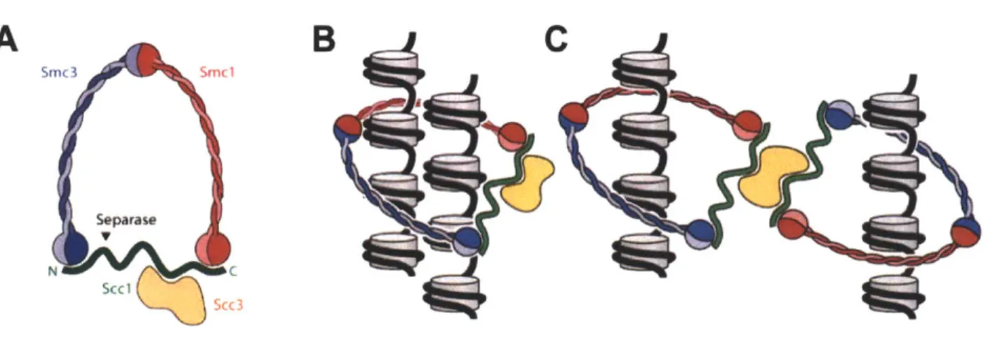
During S phase, deposition of cohesin molecules, in addition to DNA replication, occurs (Hirano, 2000; Uhlmann and Nasmyth, 1998). Cohesin is the protein complex that provides linkages between sister chromatids, and cohesion between sister chromatids is critical for the execution of mitosis (Guacci et al., 1997; Losada et al., 1998; Michaelis et al., 1997). Cohesin is a ring-shaped, evolutionarily conserved protein complex made up of four subunits: two SMC (structural maintenance of chromosomes) proteins Smcl and Smc3, the cohesin subunit Scc3, and the a-kleisin Sccl/Mcdl (Figure 3). Sccl/Mcdl (henceforth referred to as Sccl) is the a-kleisin subunit of cohesin that connects the SMC dimer. How cohesin grasps sister chromatids is under debate. One model posits that the cohesin ring encircles the two sister chromatids, and a second model posits that cohesin rings oligomerize to tether sister chromatids
(Nasmyth and Haering, 2009; Onn et al., 2008; Figure 3).
Cohesin binds chromatin during the G1/S transition in budding yeast at
pericentromeric regions and on chromosome arms. Cohesin binds at a lower density around chromosome arms and is concentrated at cohesin-associated regions (CARs) which occur at approximately 15kb intervals (Blat and Kleckner, 1999; Glynn et al., 2004; Laloraya et al., 2000). Through a poorly defined mechanism, cohesin associates
with chromatin in a process dependent on SCC2 and SCC4 (Ciosk et al., 2000). Cohesin loading onto chromosomes does not automatically create cohesion between sister chromatids. Cohesion is established by the evolutionarily conserved protein Ecol/Ctf17 (Skibbens et al., 1999; Toth et al., 1999). Ecol acetylates the Smc3 subunit of cohesin to promote the cohesive state of cohesin (Unal et al., 2008). Ecol is not needed for the maintanence of cohesion; once cohesion is generated, Ecol is dispensable (Skibbens et al., 1999; Toth et al., 1999). After S phase is complete,
mammalian cells enter a second gap phase, G2. In contrast, G2 in budding yeast is not clearly defined.
A
B
C
Smc3 Smc1 Separase N C Scc 1K..Iu Scc 3Figure 3. Structure of the cohesin complex.
(A) Structure of the cohesin complex. Cohesin is a large ring-shaped structure. Smcl and Smc3 contain long coiled-coil domains. Sccl (also called Mcdl and Rad2l) links the two head regions of the Smc proteins. Sccl is the subunit of cohesin that is cleaved by separase. During meiosis, Sccl is replaced by the meiosis-specific a-kleisin subunit Rec8. (B-C) Models for how cohesin links sister chromatids. Cohesin might encircle both chromatids (B) or cohesin molecules might associate (C). It is also possible that cohesin molecules catenate (not shown). Adapted from Haering and Nasmyth 2009.
Overview of prophase and metaphase events
Ultimately, in all eukaryotes analyzed, mitotic cyclin dependent kinase (CDK) activity promotes entry into mitosis (Morgan, 1997). Activation of M-CDKs in most
organisms (other than budding yeast) depends on the dephosphorylation of an inhibitory tyrosine on the catalytic CDK subunit by Cdc25-family phosphatases and an ensuing positive feedback loop (Nurse, 1990). M-CDKs trigger the events of mitosis:
chromosome condensation, centrosome splitting, nuclear envelope breakdown, spindle formation, metaphase I-anaphase I transition and spindle elongation (Miele, 2004; Nigg, 2001; Rahal and Amon, 2008). M-CDKs govern mitosis in all eukaryotes, but the
mechanics of mitosis vary slightly between organisms. Notably, budding yeast
undergoes mitosis with an intact nucleus and with the centrosomes, called spindle pole bodies (SPBs), embedded in the nuclear envelope. Differences in cyclins also exist. Interestingly, in yeast, different B-type cyclins (Clbs) have different abilities to promote mitotic events. Only Clb2 is able to sustain mitotic progression on its own (Fitch et al.,
1992).
In addition to CDK activity, an additional conserved kinase coordinates multiple aspects of mitosis. This family of kinases, polo kinases, is defined by a kinase domain at its N-terminus and a polo-box domain at its C-terminus. The polo-box domain can
recognize phosphorylated serines and threonines in certain contexts, thus targeting the polo kinase to proteins that have already been phosphorylated (Elia et al., 2003). In most eukaryotes, Polo kinases play a critical role in the full activation of M-CDKs, cohesin removal, centrosome separation, spindle assembly, exit from mitosis, and
cytokinesis (Archambault and Glover, 2009). In most organisms examined, polo kinase mutants build abnormal mitotic spindles (Lane and Nigg, 1996; Sunkel and Glover, 1988). In contrast, temperature-sensitive mutants in the sole budding yeast polo kinase
CDC5 arrest in anaphase (Kitada et al., 1993). Therefore, the essential role for polo kinase in budding yeast is in exit from mitosis.
In prophase, chromosomes condense, a process mediated by condensin complexes, in order to transform chromosomes into discrete, movable units (Hirano, 2005). In addition, the majority of arm cohesion is lost during prophase in vertebrates. The mammalian polo-like kinase, PIk1, mediates cohesion loss via the prophase pathway (Losada et al., 2002; Sumara et al., 2002). The vertebrate homolog of Scc3, SA2, is the critical target of PIk1. An allele of SA2 containing the PIk1 phosphorylation sites mutated to alanine blocks prophase removal. Furthermore, the cohesin removed from chromosomes during prophase contains phosphorylated SA2 (Hauf et al., 2005). In addition, prophase removal of cohesion depends on another factor called Wapl, as cells depleted for WapI contain phosphorylated SA2 on chromosome arms in
metaphase (Gandhi et al., 2006; Kueng et al., 2006). Cohesion is protected around centromeric regions by a shugoshin/MEI-S332 family member (Kerrebrock et al., 1995; Watanabe and Kitajima, 2005). Indeed, in human cells depleted for Sgol, cohesin and cohesion are lost from the entire chromosome prematurely (McGuinness et al., 2005; Salic et al., 2004). In contrast to mammalian cells, budding yeast lose all cohesion at the metaphase - anaphase transition. Although the budding yeast polo kinase Cdc5 does not promote prophase removal of cohesin, Cdc5 does phosphorylate Sccl to facilitate its cleavage (Alexandru et al., 2001; Hornig and Uhlmann, 2004).
During metaphase, chromosomes attach to the mitotic spindle. Attachment occurs via the kinetochore, a large protein complex that links the chromosomes to spindle microtubules. The inner kinetochore binds centromeric regions, and the outer kinetochore captures microtubules (Cheeseman and Desai, 2008). In yeast, where only one microtubule binds to a kinetochore, it is thought that the outer kinetochore
component Dam1 forms a collar around the microtubule and links to the outer
kinetochore by binding to the Ndc80 complex (Cheeseman et al., 2001; Westermann et al., 2006). The conserved Ndc80 complex contains microtubule binding activity as well, thus linking the outer kinetochore to spindle microtubules (Cheeseman et al., 2006; DeLuca et al., 2006; Wei et al., 2007). The metaphase - anaphase transition is not triggered until sister chromatids are properly attached to microtubules emanating from opposite spindles poles. Tension on the mitotic spindle can be achieved because cohesion between the sister chromatids resists the pulling force of the microtubules. The spindle assembly checkpoint (SAC) senses the proper biorientation of sister
chromatids on the mitotic spindle, and inhibits the metaphase - anaphase transition until all chromosomes are bioriented. Unattached kinetochores send a "wait anaphase
signal" that inhibits the E3 ubiquitin ligase called the anaphase promoting
complex/cyclosome (APC/C), which is required for the metaphase-anaphase transition, until all chromosomes are properly bioriented on the spindle (Musacchio and Hardwick, 2002; Musacchio and Salmon, 2007). If chromosomes are not bioriented, and thus not
under tension, the Aurora B kinase phosphorylates kinetochore components including Dam1, causing destabilization of the microtubule-kinetochore interaction (Cheeseman et al., 2002).
The dissolution of sister chromatid cohesion triggers the metaphase - anaphase transition, and is achieved by the proteolytic cleavage of the Sccl subunit of the cohesin complex by the protease separase (Uhlmann et al., 1999). When the SAC is satisfied, the APC/C in association with its activating subunit Cdc20 targets the separase inhibitor securin for destruction. Securin degradation frees separase to cleave Sccl, thus
triggering anaphase, the specific stage of mitosis when chromosomes segregate (Ciosk et al., 1998; Cohen-Fix et al., 1996). After chromosomes segregate, cells exit from mitosis. During this stage of mitosis, the mitotic spindle is disassembled, chromosomes decondense, and mitotic CDKs are inactivated. Exit from mitosis establishes conditions necessary for cells to undergo cytokinesis, enter G1 of the next cell cycle, and license origins for the next round of DNA replication (Stegmeier and Amon, 2004).
Function of Cdc14 in mitotic exit
In higher eukaryotes, most mitotic CDKs are inactivated at the metaphase-anaphase transition (Peters, 2002). However, in budding yeast a significant pool of mitotic CDKs remains after chromosome segregation (Wasch and Cross, 2002). CDK inactivation occurs through the activity of the essential phosphatase Cdc14. Cdc14 reverses CDK-dependent phosphorylation, and has several key targets that promote the inactivation of CDKs (Visintin et al., 1998). First, Cdc14 de-phosphorylates the APC/C activating subunit Cdhl (Jaspersen et al., 1999; Visintin et al., 1998).
De-phosphorylation of Cdhl promotes its association with the APC/C, and thus the
destruction of mitotic cyclins and polo kinase. Cdc14 also de-phosphorylates (and thus activates) the Clb inhibitor Sic1 and the S/C1 transcription factor, Swi5, to further ensure
that Clbs are inhibited (Toyn et al., 1997; Visintin et al., 1998). Cdc14 activity is tightly controlled; it is bound by its inhibitor Cfi1/Netl in the nucleolus for most of the cell cycle, and is released into the nucleus and cytoplasm during anaphase (Shou et al., 1999; Visintin et al., 1999). It is thought that dissociation of the Cdcl4-Cfi1/Netl complex is promoted by the phosphorylation of both Cdc14 and Cfi1/Netl (Shou et al., 2002; Shou et al., 1999; Visintin et al., 2003; Yoshida and Toh-e, 2002). Two signaling pathways control the dissociation of the Cdc14-Cfi/Net1 complex, thus releasing Cdc14 from the nucleolus: the non-essential FEAR (Cdc-fourteen early anaphase release) network, and the essential mitotic exit network (MEN).
The FEAR network
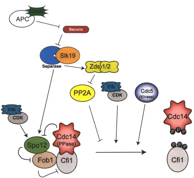
The FEAR network is a non-essential pathway that promotes a transient burst of Cdc14 release during early anaphase (Pereira et al., 2002; Stegmeier et al., 2002; Yoshida et al., 2002). FEAR network components include separase, the separase-binding protein Slk19, the replication fork block protein Fob1, a protein of unknown function Spo12, the polo kinase Cdc5, and the phosphatase PP2ACdC5 5. The FEAR network controls Cdcl4-Cfil/Netl dissociation through a poorly defined signaling mechanism (Figure 4). First, separase links the metaphase-anaphase transition to exit from mitosis. Indeed, mitotic exit is inhibited when securin is stabilized, as Clb2 is not degraded in these cells (Cohen-Fix and Koshland, 1999). Separase not only causes dissolution of sister chromatid cohesion, but also acts in conjunction with SIk1 9 to mediate the early release of Cdc14, possibly by downregulating PP2A and allowing CDKs to phosphorylate Cfil/Netl (Azzam et al., 2004; Queralt et al., 2006). Activation
of the proteins Zdsl and Zds2 may also contribute to PP2A downregulation (Queralt and Uhlmann, 2008). Second, the ESPI-SLK19 branch of the FEAR network acts upstream and parallel to SPO12. Spo12, a protein activated by CDK-dependent phosphorylation in anaphase and dephosphorylated by Cdc14, acts positively in the FEAR network, likely by inhibiting Fob1, a nucleolar protein that stabilizes the Cdc14-Cfi/Netl interaction (Stegmeier et al., 2004; Tomson et al., 2009; Visintin et al., 2003). Third, the polo kinase Cdc5 promotes phosphorylation of both Cdc14 and Cfil/Net1
(Shou et al., 2002; Visintin et al., 2003; Yoshida and Toh-e, 2002).
Although non-essential, the FEAR network serves important roles in coordinating anaphase. Proper segregation of the rDNA repeats, which encode the rRNA and define the nucleolus, is dependent upon the FEAR network (Rock and Amon, 2009). Cdc14
dephosphorylates several microtubule-binding proteins, resulting in the stabilization of the mitotic spindle (Higuchi and Uhlmann, 2005; Woodbury and Morgan, 2007). Finally,
FEAR-released Cdc14 plays an important role in activating the MEN (Jaspersen and Morgan, 2000; Stegmeier et al., 2002). Importantly, in contrast to the 10-20 minute mitotic exit delay that FEAR mutants display, the FEAR network plays an essential role in regulating the meiosis I-meiosis Il transition (Buonomo et al., 2003; Marston et al.,
--
Y
rn
Zs 1I
Zdsi1i2
na
(I
Figure 4. Schematic of the FEAR network
The FEAR network links the metaphase - anaphase transition to mitotic exit. Upon APC/Ccdc2o- dependent degradation of securin, separase promotes FEAR network
activity. Separase-SIkl9 inhibits the PP2A phosphatase and acts upstream of Spo12. Spol 2 inhibits the Fob1 inhibitor during anaphase. Together with CDK activity and Cdc5 activity, Cdc14 is released from the nucleolus early in anaphase.
The Mitotic Exit Network (MEN)
The MEN is essential for mitotic exit in budding yeast. MEN mutants arrest in late anaphase with high CDK activity and Cdc14 sequestered in the nucleolus (Jaspersen et
(:AP7
al., 1998; Shou et al., 1999; Visintin et al., 1999). The MEN is a conserved GTPase signaling cascade, consisting of a small GTPase, a two-component GAP, a sterile 20-like kinase, an NDR kinase, a Mob1-family protein (activating subunit for NDR kinase), and a scaffold (Stegmeier and Amon, 2004). The small GTPase central to MEN
signaling is encoded by TEMI (Shirayama et al., 1994) and is negatively regulated by Bub2-Bfal, a two-component GAP (Bardin et al., 2000; Fesquet et al., 1999; Geymonat et al., 2002; Pereira et al., 2000; Wang et al., 2000). During anaphase, Tem1 is
activated and propagates a signal to the sterile 20-like kinase Cdcl 5, which then activates the NDR-kinase complex Dbf2-Mobl (Asakawa et al., 2001; Mah et al., 2001; Visintin and Amon, 2001). Nud1, a component of the spindle pole body (SPB), acts as a scaffold for MEN signaling at the SPB (Adams and Kilmartin, 1999; Bardin et al., 2000; Geymonat et al., 2002; Gruneberg et al., 2000; Visintin and Amon, 2001). Finally, although not a core component of the MEN, the polo kinase Cdc5 activates the MEN at multiple points (Hu et al., 2001; Rock and Amon, 2011). A schematic diagram of the MEN is shown in Figure 5.
MEN components are organized into signaling modules at the SPB. NUDI, which encodes the scaffold at the SPB, is required for the localization of MEN components to the SPB, and NUDI-temperature sensitive mutants arrest in anaphase (Adams and Kilmartin, 1999; Gruneberg et al., 2000; Visintin and Amon, 2001). Tem1 localizes to SPBs from metaphase until late anaphase, and SPB localization is essential for Tem1 function (Bardin et al., 2000; Pereira et al., 2000; Valerio-Santiago and Monje-Casas, 2011). Upon Tem1 activation in anaphase, Temi-GTP is thought to recruit Cdcl 5 to SPBs (Asakawa et al., 2001). Although Tem1 is enriched on the daughter-bound
spindle pole, Cdc15 is recruited to both SPBs, which in turn recruits Dbf2-Mobl to SPBs (Cenamor et al., 1999; Luca et al., 2001; Visintin and Amon, 2001; Xu et al., 2000; Yoshida and Toh-e, 2001). Cdcl 5 then promotes Dbf2-Mobl activation through two
mechanisms. First, Cdcl 5 has been shown to phosphorylate Dbf2 directly in vitro, and this phosphorylation likely contributes to Dbf2-Mobl activation in vivo (Mah et al., 2001).
Second, Cdc 5 recruits Dbf2-Mobl to SPBs by phosphorylating the scaffold Nud1,
which creates a binding site for Mobl, the activating subunit for the Dbf2 kinase (Rock et al., personal communication). How the MEN causes sustained Cdcl4 release is not completely clear, but occurs at least partially through Dbf2-dependent phosphorylation near the nuclear localization signal of Cdc14 (Mohl et al., 2009).
Mitotic Exit Network (MEN)
FEAR network
Figure 5. Diagram of the Mitotic Exit Network
The MEN is a GTPase signaling cascade anchored at the SPB. The FEAR network and Cdc5 promote an early burst of Cdc14 activity that contributes to the activation of the MEN. The MEN is required for sustained release of Cdc14 from its inhibitor in the nucleolus and mitotic exit.
Control of the MEN by spatial and temporal signals
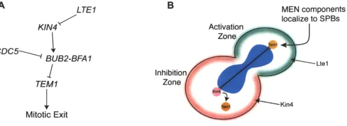
The MEN integrates multiple spatial and temporal cellular signals, thus ensuring that MEN activation only occurs during anaphase. Much progress has been made in understanding the spatial control of MEN activation by spindle position. Because budding yeast divide asymmetrically, an additional challenge to proper chromosome segregation is present: one complement of the genome must be segregated into the bud. The bud site is selected in G1, so the axis of cell division is determined prior to mitosis. Therefore, budding yeast must only activate the MEN once the spindle is properly positioned along the mother-bud axis. Teml, the GTPase central to MEN signaling which is enriched on the daughter-bound SPB, acts as a sensor moving between two zones: an inhibitory zone in the mother cell and an activating zone in the daughter cell (Bardin et al., 2000; Pereira et al., 2000). A diagram of the zone model is shown in Figure 6.
The inhibitory zone is created by the protein kinase Kin4, which localizes to the mother cell cortex throughout the cell cycle and to the SPB of the mother cell during anaphase (Maekawa et al., 2007). Kin4 was initially identified as a central component of the spindle position checkpoint (SPOC). In response to a mispositioned spindle, Kin4
inhibits MEN activation until the spindle is properly positioned. Indeed in kin4A mutants, cells with mispositioned spindles will exit from mitosis with both nuclei contained in the mother cell, whereas wild-type cells will delay mitotic exit until the spindle has been properly positioned (D'Aquino et al., 2005; Pereira and Schiebel, 2005). In cells in which an anaphase spindle has elongated within the mother cell, Kin4 localizes to both SPBs, and Tem1 is no longer localized to SPBs (D'Aquino et al., 2005). Interestingly, tethering of Tem1 to SPBs suppresses the anaphase arrest caused by overexpression of KIN4, suggesting that a critical function for the SPOC is keeping Tem1 off SPBs
(Valerio-Santiago and Monje-Casas, 2011). Kin4 also promotes phosphorylation of Bfal in a manner that protects it from inhibitory phosphorylation by Cdc5. (Maekawa et al., 2007). It is tempting to speculate that through activating Bfal, Kin4 promotes Tem1 in a GDP-bound state, which in turn promotes Tem1 loss from SPBs. Finally, Kin4 itself is
regulated; it functions in its dephosphorylated form in anaphase and is regulated by the kinase Elm1 and the phosphatase PP2A bound to its specificity subunit Rtsl (Caydasi et al., 2010; Chan and Amon, 2009; Moore et al., 2010).
The activating zone is created in the bud by the protein Ltel, which primarily localizes to the bud cortex (Seshan et al., 2002). Ltel contains putative GEF domains, and was long thought to positively regulate MEN signaling by acting as a GEF for Tem1, despite the observations that Ltel was not seen on SPBs and does not exhibit GEF activity in vitro (Geymonat et al., 2009). Recently, it has become clear that Ltel acts positively within the MEN by inhibiting Kin4. Two parallel mechanisms ensure that Kin4 does not inhibit the MEN in the daughter cell: restriction of Kin4 to the mother cell cortex and inhibition of Kin4 loading onto the dSPB by LTE1. In the absence of LTE1, Kin4
loads onto the dSPB in a fraction of cells (Falk et al., 2011). Loading of Kin4 onto the dSPB is enhanced in the absence of LTEI when a mutant version of Kin4 that localizes to the mother and bud cortex is expressed (Chan and Amon, 2010; Falk et al., 2011).
Furthermore, expression of a symmetric mutant of LTEI that localizes to both the mother and daughter cell inhibits Kin4 localization to the mother SPB (Bertazzi et al., 2011; Falk et al., 2011). An alternative to the zone model posits that interactions
between cytoplasmic microtubules in the bud neck control the SPOC. In support of this model, laser ablation of cytoplasmic microtubules leads to mitotic exit in cells with
mispositioned spindles. Further, the Elm1 kinase that regulates Kin4 localizes to the bud neck (Caydasi et al., 2010; Moore et al., 2010). However, the observation that mutants lacking cytoplasmic microtubules only exhibit a minor defect in SPOC function in cells that misposition spindles suggests that this is not the major mechanism cells use to coordinate spindle position with mitotic exit (Adames et al., 2001; Moore et al., 2009). The data therefore support the zone model: in every cell cycle only when a Tem1-containing SPB escapes the inhibitory zone created by Kin4 in the mother and enters the activating zone created by Ltel in the bud can mitotic exit be triggered.
LTE1 MEN components KlN4 Activation localize to SPBs Zone CDC5- BUB2-BFAI Inhibition TEM1 Zone 4Kin4 Mitotic Exit
Figure 6. Control of MEN activation by spindle position
(A) Diagram of the genetic interactions between components controlling the activation of
Tem1, and thus MEN signaling. (B) Diagram of the zone model. Kin4 (pink) resides in the mother cell and establishes an inhibitory zone. Ltel (green) resides in the daughter cell and establishes an activating zone. Tem1 (orange) acts as a sensor moving
between the zones, and only when Tem1 contacts the daughter cell can mitotic exit be triggered. The nucleus is shown in blue, and the spindle microtubules are indicated by the black line.
Spatial control is not necessary for mitotic exit in cells with properly positioned spindles, as ItelA kin4A double mutants still activate MEN signaling in anaphase (Rock and Amon, 2011). Therefore, temporal control of MEN activation exists, and the Tem1 GTPase as well as the polo-like kinase Cdc5 have been implicated in this temporal control. Evidence that TEMI mediates temporal control is seen in the observation that cells lacking TEMI, but that are kept alive by overexpression of CDC15, prematurely activate Dbf2-Mobl in metaphase (Rock and Amon, 2011). Moreover, tethering Tem1 to SPBs results in premature localization of Cdcl5 to SPBs, further suggesting that Tem1
2011). Furthermore, CDC5 is required for the recruitment of Cdcl5 to SPBs in anaphase (Rock and Amon, 2011). In addition, CDC5 promotes MEN activation in several other ways. Cdc5 phosphorylates Bfal to reduce its GAP activity, and thus promote MEN activation (Hu et al., 2001; Lee et al., 2001). Also, as part of the FEAR network, CDC5 controls the early release of Cdc14, which leads to the
dephosphorylation of Cdcl 5 and further promotes MEN activation (Jaspersen and Morgan, 2000; Stegmeier et al., 2002). Taken together, the polo kinase Cdc5 acts at multiple points to activate MEN signaling.
After mitotic exit has been achieved, MEN and FEAR signaling must be reversed. Spol 2 and Cdc5 are both degraded by the APC/Ccdhl at exit from mitosis. However, FEAR signaling is inactivated prior to exit from mitosis. Spol2 phosphorylation is an indication of FEAR activity, and is antagonized by Cdc14 (Tomson et al., 2009).
Therefore FEAR-released Cdc14 may contribute to the inactivation of FEAR signaling. MEN signaling is inactivated at least in part by APC/CCdhi dependent degradation of Cdc5 as well as Cdc14 activity, which both promote the return of Cdc14 to the nucleolus (Manzoni et al., 2010; Visintin et al., 2008).
NDR-kinase signaling in higher eukaryotes
The MEN has served as an important paradigm for many problems in cell
biology. First, studying the MEN has elucidated important concepts in how the cell cycle is reset after chromosome segregation. Second, studying the MEN has shed light on how asymmetric cell division is achieved. Asymmetric cell division is not unique to budding yeast. For example, a major way that cells in a developing embryo become
different from each other is through asymmetric cell division. Third, the MEN is an important paradigm for understanding how homologous signaling pathways function. How NDR-kinase signaling pathways function in different cellular contexts will be addressed in Chapter 2, and insights from other organisms will be described briefly below.
The core signaling components of the MEN are conserved (Figure 7). This pathway is known as the septation initiation network (SIN) in fission yeast, where it controls cytokinesis. Like the MEN, the SIN pathway is a GTPase signaling cascade that utilizes spindle pole bodies to assemble signaling modules (Krapp et al., 2004b). Similarly, polo kinase activity positively regulates the SIN (Krapp et al., 2004a; Ohkura et al., 1995). In contrast, the fission yeast Cdc14 homolog CIp1/FIp1 is not required for cyclin degradation, and its release from the nucleolus is not dependent on the SIN (Cueille et al., 2001; Trautmann et al., 2001).
Homologous signaling pathways also exist in higher eurkaryotes, although the pathway has evolved to control organ growth. Hippo signaling in Drosophila and NDR-kinase signaling in mammals use the same core signaling module as the MEN. The Cdcl5-Dbf2-Mobl module of the MEN is conserved in the hippo/NDR kinase signaling pathway as Hippo-Warts-Mats in Drosophila and Mst1/2-Lats1/2-Mob1A/B in mouse. The Hippo pathway effector is not Cdc14, but a transcriptional co-activator Yorkie (YAP/TAZ in mammals). Yorkie promotes transcription of pro-growth genes, and Hippo pathway activity leads to phosphorylation and nuclear exclusion of Yorkie (Halder and Johnson, 2011; Hergovich et al., 2006). Therefore, Hippo/NDR kinase signaling
results in the binding of Yorkie to 14-3-3 proteins and cytoplasmic retention (Basu et al., 2003; Zhao et al., 2007). The Hippo pathway also contains a scaffold, Salvador/WW45, which appears to be regulated by phosphorylation, similarly to Nud1. It is becoming clear that Sav/WW45 can interact with both Hpo/Mst and with Warts/Lats, thus enabling Hpo/Mst to phosphorylate and activate the Warts/Lats kinase (Callus et al., 2006; Guo et al., 2007).
The upstream signals regulating the Hippo kinase are beginning to emerge, and, analogous to yeast, involve the integration of information from other signaling pathways and positional cues (Yu and Guan, 2013). Upstream signals restrain Hippo signaling when division is needed, and activate Hippo signaling when cells are contact-inhibited. Upstream signals regulating the Hippo pathway are most clear in Drosophila, and many signals exist. Signals transmitted from the atypical cadherin Fat and the transmembrane apical-basal polarity factor Crumbs, activate Hippo signaling through a poorly defined mechanism mediated in part by the Expanded/Merlin/Kibra complex. (Baumgartner et al., 2010; Hamaratoglu et al., 2006; Ling et al., 2010; Reddy and Irvine, 2008). Ajuba
proteins, proteins that localize to adherens junctions, inhibit Hippo signaling (Das Thakur et al., 2010; Marie et al., 2003) when a cell is not contact-inhibited. Finally, crosstalk between signaling pathways influences Hippo signaling. Mst proteins have been shown to interact with RASSF proteins, which may allow crosstalk between the Ras and Hippo pathways (Guo et al., 2007; Matallanas et al., 2007). Taken together, although many inputs for Hippo/NDR kinase signaling have been identified, much remains to be learned about the mechanisms controlling activation or inhibition of the Hippo pathway. Moreover, the NDR kinase pathway appears to be regulated differently
in different cell types and contexts, with much remaining to discover (Halder and Johnson, 2011). A CK Dase)0
C
D
Warts
(Kinase)S. cerevisiae
.melanogaster B DFigure 7. Conservation of the core signaling components in NDR-kinase signaling.
The mitotic exit network is conserved in higher eukaryotes. Although the upstream signals and downstream effectors vary between species, the core components of the
pathway are conserved, shown for budding yeast (A), fission yeast (B), fly (C), and vertebrates (D). The GTPase is shown in blue, the sterile-20-like kinase in green, scaffold in gray, NDR kinase in yellow, and NDR-kinase activating subunit in orange.
S. pombe
Vertebrate
Lats
Meiosis: A specialized cell division required for sexual reproduction
All sexual reproduction depends on meiosis, the specialized cell division that yields four meiotic products with half of the amount of genetic material. In diploid
organisms, meiosis produces four haploid cells from one diploid progenitor. In yeast the products of meiosis, or gametes, are spores. It is essential that gametes with the correct number of chromosomes are produced; chromosome mis-segregation during meiosis causes miscarriages and Down's syndrome in humans (Hassold and Hunt, 2001).
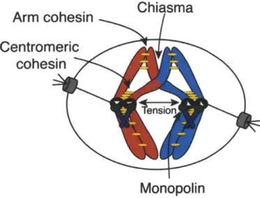
During meiosis, two rounds of chromosome segregation follow one round of DNA replication. In meiosis I, homologous chromosomes separate and in meiosis 11, sister chromatids split. While meiosis 11 segregation resembles mitosis, meiosis I segregation is unique in that homologous chromosomes segregate. Several modifications are required for the meiosis I chromosome segregation pattern. These specializations -creation of physical linkages between homologous chromosomes, stepwise loss of cohesion between sister chromatids, and attachment of sister kinetochores to microtubules from the same pole of the meiotic spindle - establish the meiosis I
chromosome segregation pattern (Figure 8). Additionally, cells must exit from meiosis I without establishing conditions permissive for DNA re-replication (Marston and Amon, 2004). In the following section of this chapter I will describe the stages of meiosis in greater detail. I will highlight similarities between the cell cycle machinery in mitosis and meiosis and focus on the specializations required for homologous chromosomes to segregate in meiosis 1.
Arm cohesin
Centromeric
c oei
Ml specializations:Crossover formation
Co-orientation of sister kinetochores Stepwise loss of cohesin
Monopolin
Figure 8. Meiosis I Specializations
Several modifications occur to establish conditions that allow homologous
chromosomes, rather than sister chromatids to split in meiosis 1. (1) Recombination links homologous chromosomes. Chiasmata are the physical manifestations of crossover events. (2) Cohesion links sister chromatids. Arm cohesion is removed in meiosis I, but centromeric cohesion is retained, thus allowing sister chromatids to be held together until meiosis 11. (3) The monopolin complex links sister kinetochores so that they can be
attached to microtubules from the same spindle pole. Together, these modifications allow tension to be achieved on the meiosis I spindle.
Entry into gametogenesis
In budding yeast, sporulation is a starvation response. If diploid cells are starved, they undergo meiosis coupled with the developmental program of spore formation to produce stress-resistant, haploid gametes (spores) that can return to growth when nutrients are restored. Therefore, entry into sporulation requires the integration of nutritional signals with mating-type information: only nutrient-starved (lack of a nitrogen source and a fermentable carbon source) cells of an a/a mating type will enter the
sporulation program. IME1, a transcription factor, is the master regulator of entry into gametogenesis in budding yeast. Cells lacking IMEI do not sporulate, and cells
overexpressing IMEI will undergo meiosis even in nutrient rich conditions or in the a or a mating type (Kassir et al., 1988; Smith and Mitchell, 1989). The promoter of IME1 integrates information about mating type (which is a read-out for ploidy) and nutritional state. The IMEI promoter is complex; it is approximately 2kb long and contains binding sites for proteins whose presence or absence at the promoter is reflective of mating type or nutritional state (van Werven and Amon, 2011). In addition, two non-coding RNAs present in the IMEI promoter mediate mating-type control of IMEI expression (van Werven et al., 2012).
Once expressed, Imel initiates a wave of transcription of early meiotic genes. These early meiotic genes encode proteins required for pre-meiotic DNA replication, chromosome condensation, and homologous recombination. One critical target of Imel is IME2, which encodes a protein kinase (Dirick et al., 1998; Mitchell et al., 1990; Yoshida et al., 1990). Indirectly, Imel promotes expression of NDT80, which encodes another transcription factor. Ndt80 promotes the expression of middle meiotic genes including polo kinase, cyclins, and other factors necessary for the meiotic divisions. Finally, in a late wave of transcription, genes that encode factors essential for building spore walls, are expressed (Chu and Herskowitz, 1998; Pak and Segall, 2002; Primig et al., 2000).
Entry into gametogenesis is quite different in mammals and other metazoans, but may require a master regulator. In multicellular organisms, germ cells are specified early in development. After specification, primordial germ cells (PGCs) migrate to the
gonad where they become meiosis-competent. It is likely that a master regulator of meiosis exists in mammals; the putative transcription factor Stra8 is required for meiosis in both males and females (Baltus et al., 2006; Tedesco et al., 2009). Stra8 also
integrates multiple signals. Stra8 requires the RNA-binding protein Dazi for its
expression, and Dazl is expressed only in PGCs in the gonad (Lin et al., 2008). Stra8 also responds to an extracellular signal, the presence of retinoic acid (Bowles et al., 2006). Upon integration of intracellular and extracellular signals, Stra8 then promotes entry into the gametogenesis program.
Pre-meliotic DNA replication
After cells have entered the gametogenesis program, DNA is replicated. In both pre-meiotic and pre-mitotic S phase, CIb5/6-CDK and Dbf4-dependent kinase (DDK) are required. Additionally, an important target of Imel, the kinase Ime2, is required for entry into pre-meiotic S; degradation of the Clb-CDK inhibitor Sic1 depends on ime2
(Benjamin et al., 2003; Dirick et al., 1998). Although similar origins of replication are used, meiotic S phase proceeds more slowly than pre-mitotic S phase, as a
consequence of delayed initiation (Blitzblau et al., 2012). As in the vegetative cell cycle, during pre-meiotic S phase, cohesin molecules are deposited. Cohesin complexes are similar between mitosis and meiosis with one important difference: the a-kleisin subunit of cohesin (the Sccl subunit that is cleaved by separase) is replaced by Rec8 in
meiosis. Rec8 is utilized across species including mammals in meiotic cohesin
complexes. The replacement of Sccl by Rec8 has important consequences in prophase 1. Rec8 plays multiple roles in pairing of homologous chromosomes, homologous
recombination, and synaptonemal complex assembly (Brar et al., 2009; Klein et al., 1999) that cannot be supported by Sccl.
Meiotic Prophase and homologous recombination
Once pre-meiotic S phase is completed, cells undergo an elongated prophase I (sometimes referred to as meiotic G2), in which homologous chromosomes pair and recombine. I will briefly discuss the relevance of homologous recombination to
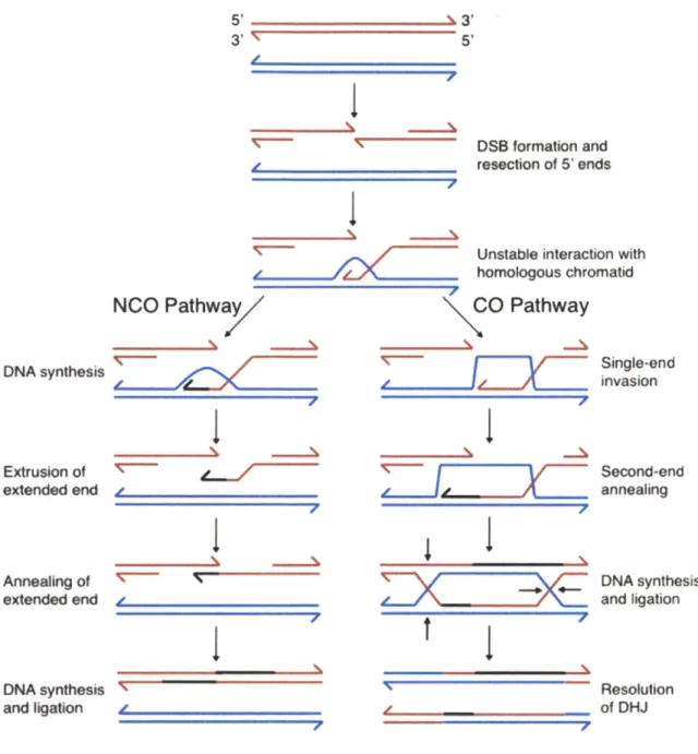
chromosome segregation. Recombination is an amazing and risky process in which a large amount of DNA damage is introduced into chromosomes and correctly repaired (Figure 9). The conserved endonuclease Spol1 is responsible for introducing these programmed double strand breaks (DSBs; (Keeney et al., 1997)). After DSBs are produced, the 5' ends of the DNA are resected, and in a process dependent on DMCI, a homology search biased toward the homolog begins (Bishop et al., 1992; Schwacha and Kleckner, 1997; Shinohara and Shinohara, 2004). DSBs can be processed as either crossovers (COs) or noncrossovers (NCOs), and this decision is made early, soon after strand invasion (Allers and Lichten, 2001). Therefore, double Holliday junctions are repaired into crossovers. A surveillance mechanism exists to ensure that cells do not progress into meiosis before all DNA damage is repaired. This checkpoint is conserved since mouse spermatocytes and oocytes failing to repair DNA damage do not undergo meiosis. In budding yeast, unrepaired damage leads to the inhibition of Clb-CDKs and Ndt80, the transcription factor required for expression of B-type cyclins, polo kinase, and progression into the meiotic divisions (Hochwagen and Amon, 2006).
5'-3
I
53' 5' 4, DSB formation and resection of 5' endsUnstable interaction with
_ _ _homologous chromatid
NCO Pathwa CO Pathway
DNA synthesis Extrusion of extended end Annealing of extended end y DNA synthesis and ligation
I
'c-7
I77~Z
.1
7
I
DQIIPxE
tj,
I,I
I
Single-end invasion Second-end annealing DNA synthesis and ligation Resolution of DHJFigure 9. Schematic of homologous recombination.
During prophase, programmed double-strand breaks are created throughout the genome. DNA is resected, and single strands undergo a homology search. If the extended end is ejected, the break will be repaired as a non-crossover (NCO). If the extended end anneals, double Holliday junctions (DHJs) will form and ultimately resolve into a crossover. Adapted from Bishop and Zickler, 2004.
In addition to homologous recombination, the synaptonemal complex (SC), a proteinaceous scaffold, forms between the homologs during meiotic prophase to enable efficient homologous chromosome pairing and recombination (Page and Hawley, 2004). SC formation depends on cohesin, condensin, and DSBs (in budding yeast). The SC forms a zipper-like structure that synapses homologous chromosomes together during prophase. The lateral elements are composed of Red and Hop1 and are thought to promote inter-homolog repair (Hollingsworth et al., 1990; Smith and Roeder, 1997). In late prophase, transverse filaments containing Zip1 form and are required for crossover maturation (Borner et al., 2004). SC function varies in different organisms. In budding yeast, SC formation requires the initiation of recombination, whereas in C. elegans, the SC can form in the absence of recombination initiation, but is important for the
completion of recombination (Colaiacovo et al., 2003; Dernburg et al., 1998; Roeder, 1997). In contrast, Drosophila males and fission yeast do not form SC at all (Page and Hawley, 2004).
Crucially for chromosome segregation, at least one crossover must be present on each pair of homologous chromosomes. The chiasmata produced by repair via crossovers provide the linkages between homologous chromosomes. Together with the cohesin molecules that link sister chromatids distal to the crossover, tension can be generated on the meiosis I spindle. Although recombination exists to physically link homologous chromosomes, it has the important evolutionary consequence of generating gametes that are not genetically identical to either parent. Due to independent assortment and the swapping of genetic material between parental
chromosomes, new combinations of alleles are contributed to the next generation, leading to diversity within the population.
Regulation of meiotic chromosome segregation by CDK, polo kinase, and the APC/C
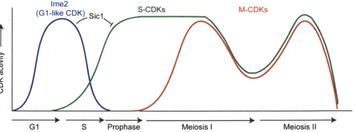
Among the targets of Ndt80 essential for progression through the meiotic divisions are the genes encoding the B-type cyclins CLBI, CLB3, and CLB4, and the polo kinase CDC5. Analogously to mitosis, CDK activity is required for progression through the meiotic divisions (Figure 10). However, the regulation of meiotic CDK
activity exhibits important differences from the regulation of M-CDKs. First, the mitotic cyclin of central importance to mitosis, Clb2, is absent during meiosis (Grandin and
Reed, 1993). Second, Clb1 kinase activity, but not protein, is restricted to meiosis I (Carlile and Amon, 2008). Third, Clb3 is translationally controlled, and produced only during meiosis II (Carlile and Amon, 2008). The intricate regulation of cyclins in meiosis
is critical for proper chromosome segregation. Over-expression of CLB3 or CLBI, but curiously not CLB4, prior to the first meiotic division leads to the separation of sister chromatids rather than homologs during meiosis I (Carlile and Amon, 2008; Miller et al., 2012). Unraveling in greater detail how B-type cyclins are regulated and how cyclin specificity contributes to meiotic progression are important questions for the future, as CDK activity drives meiotic progression across species.
Ime2
Af
G1 S Prophase Meiosis I Meiosis i
Figure 10. CDK activity in budding yeast meiosis
CDK activity drives progression through meiosis. Although G1-CDKs are absent in meiosis, the kinase Ime2 acts as a G1-like CDK, fulfilling the important role of triggering Sic1 degradation. S-CDKs drive progression through S phase and M-CDKs drive progression through the meiotic divisions. It is thought that CDK activity dips between the two meiotic divisions. Finally, how CDKs are inactivated at the end of meiosis is unclear.
The Ndt80 target, the polo kinase Cdc5, is also a central regulator of meiotic progression in all species analyzed. In budding yeast, cells lacking Cdc5 in meiosis arrest in metaphase I (Clyne et al., 2003; Lee and Amon, 2003). CDC5, unlike in mitosis, is an essential coordinator of nearly all aspects of the meiosis I division; it is required for chiasmata resolution, securin degradation, sister kinetochore co-orientation, cohesion removal, and Cdc14 release in anaphase I (Clyne et al., 2003; Lee and Amon, 2003). Precisely how Cdc5 functions during the meiotic divisions is less clear, and experiments addressing this question will be described in Chapter 3. The role of polo kinase in coordinating the meiotic divisions is conserved. For example, in Drosophila,
fact, a polo inhibitor called matrimony is present during prophase to ensure that polo is not activated prematurely (Xiang et al., 2007). Furthermore, in Drosophila, polo also functions in the regulation of cohesion (Clarke et al., 2005).
In addition to the conserved role for CDK activity and polo kinase activity in mitosis and meiosis, regulated proteolysis is also a conserved theme. Just as in mitosis, securin is degraded at the metaphase I-anaphase I and metaphase II-anaphase 11 transitions (Salah and Nasmyth, 2000). However, APC/C activity differs in several key ways. First, the APC/Ccdhl does not appear to play a key role in the meiosis 1-meiosis II transition, highlighted by the observation that Clb1, which is targeted by the APC/CCdhl,
is not degraded at the meiosis l-meiosis 11 transition (Carlile and Amon, 2008). In contrast with mitosis, an additional meiosis-specific APC/C activator exists in meiosis called AMA 1. The APC/CAmal primarily functions to degrade mitotic regulators during
prophase I in order to ensure that mitotic regulators are repressed during prophase I (Okaz et al., 2012). An APC inhibitor called Mnd2 also exists in meiosis. Mnd2 ensures that securin is not degraded prematurely during the elongated meiotic prophase
(Oelschlaegel et al., 2005; Penkner et al., 2005). Meiosis-specific APC/C activators are not specific to budding yeast. In Drosophila, a female-specific activator of the APC/C called cortex is required for proper meiosis, as it targets cyclin A for degradation prior to the metaphase I arrest in oocytes (Pesin and Orr-Weaver, 2007).
Specializations of Meiosis I
Co-orientation of sister chromatids
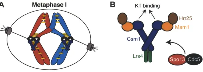
In order for homologous chromosomes to segregate during meiosis I, they must not only be linked to each other, but also linked properly to the meiosis I spindle. To achieve co-orientation, kinetochores from sister chromatids must be attached to
microtubules emanating from the same spindle pole. Sister kinetochore co-orientation is best understood in budding yeast where it is achieved by the monopolin complex
(Figure 11). Monopolin consists of Mam1, Csml, Lrs4, and the casein kinase Hrr25. Mam1 is a meiosis I specific protein, whereas Csm1 and Lrs4 localize to the nucleolus during interphase. Late in prophase I, Csm1 and Lrs4 are released from the nucleolus. They associate, along with Mam1 and Hrr25, with kinetochores until metaphase I (Petronczki et al., 2006; Rabitsch et al., 2003; Toth et al., 2000), and MAMI is required for robust localization of Csm1 and Lrs4 to kinetochores (Rabitsch et al., 2003). The localization of Mam1 depends on SPO13, a gene encoding a meiosis I-specific protein of unknown function (Katis et al., 2004b; Lee et al., 2004). In addition, Cdc5 and DDK also control monopolin function as Mam1 localization and Lrs4 phosphorylation and nucleolar release depend on both CDC5 and CDC7 (Clyne et al., 2003; Lee and Amon, 2003; Matos et al., 2008). Cdc5 may act through Spol3, as the two proteins physically interact (Matos et al., 2008).
Finally, kinetochore co-orientation is regulated by temporal regulation of microtubule-kinetochore interactions. NDC80, an outer kinetochore component, is transcriptionally and translationally controlled such that Ndc80 protein is undetectable