Supporting Information
Size specific copper nanoparticle cytotoxicity varies between human cell lines
Ina Na, and David C. Kennedy*
Metrology, National Research Council Canada, 1200 Montreal Road, Ottawa K1A 0R6, Canada;
Figure S1. TEM images for copper nanoparticles in media supplemented with fetal bovine
serum
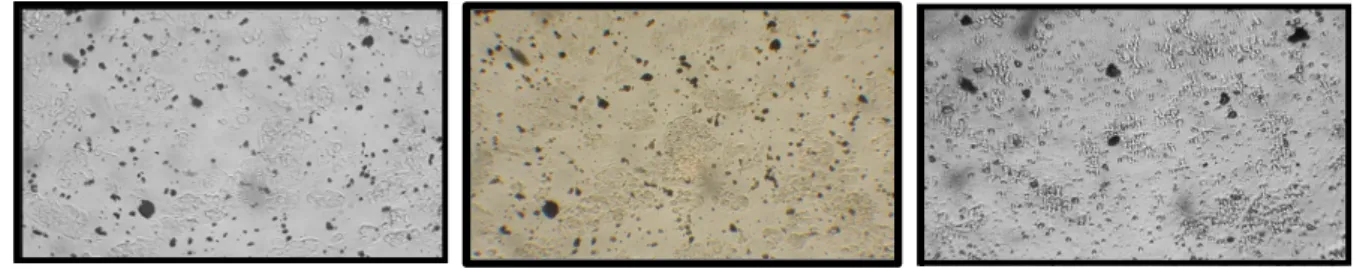
Figure S2. Brightfield images of A549 (left), HepG2 (middle) and SH-SY5Y (right) cells
exposed to 200 μg/mL of 25 nm CuNPs immediately after exposure. Large black CuNP agglomerates are easily observed but are not present after 24 h exposure.