HAL Id: tel-01726953
https://tel.archives-ouvertes.fr/tel-01726953
Submitted on 8 Mar 2018
HAL is a multi-disciplinary open access archive for the deposit and dissemination of sci-entific research documents, whether they are pub-lished or not. The documents may come from teaching and research institutions in France or abroad, or from public or private research centers.
L’archive ouverte pluridisciplinaire HAL, est destinée au dépôt et à la diffusion de documents scientifiques de niveau recherche, publiés ou non, émanant des établissements d’enseignement et de recherche français ou étrangers, des laboratoires publics ou privés.
islet function
Mohamad Kassem
To cite this version:
Mohamad Kassem. In vivo and in vitro study of pancreatic islets aging : impact of endothelial senes-cence and microparticles on islet function. Endocrinology and metabolism. Université de Strasbourg, 2017. English. �NNT : 2017STRAJ007�. �tel-01726953�
UNIVERSITÉ DE STRASBOURG
ECOLE DOCTORALE 414-Sciences de la vie et de la santé
EA7293 : Stress vasculaire et tissulaire en transplantation :
Microparticules et environnement
THÈSE
Présentée par :
Mohamad KASSEM
Soutenue le : 20 Janvier 2017
Pour obtenir le grade de :
Docteur de l’Université de Strasbourg
Discipline/ Spécialité
: Aspects Moléculaires et Cellulaires de la Biologie
Etude in vivo et in vitro du vieillissement des îlots
pancréatiques : Impact de la senescence endothéliale et
des Microparticules sur la fonction des îlots
THÈSE dirigées par :
Mme Laurence KESSLER Professeur, Université de Strasbourg
Mme Florence TOTI Professeur, Université de Strasbourg
Rapporteurs externes :
M. Domenico BOSCO Professeur, Université de Genève
Mme. Angela TESSE Professeur, Université de Nantes
Examinateur interne :
Page i
This thesis is dedicated to my
beloved
Father and Mother,
For sacrifices, endless love, encouragement and
Patience make me able to get such success and honor
Brothers
Who never allowed me to give up on the pursuit of my dream
Nour
My lovely wife for her love, patience and understanding
Jamal
My little man who was my big gift that give to my life another
meaning and another hope
Page ii
Acknowledgments
This might be my only opportunity to thank the talented individuals in writing. For that, forgive me if I am a bit more verbose in my thanks than necessary. I wish to express my warmest gratitude to all the people whose comments, questions, criticism, support and encouragement, personal and academic, have left a mark on this work. Regrettably, but inevitably, the following list of names will be incomplete. Plenty of people have helped me along the way. For those who I failed to mention, accept my sincere appreciation on your influence on this work.
First and foremost, I offer my deepest gratitude to my thesis director, Pr. Laurence KESSLER, for her continuous support and mentoring throughout all my PhD study and research, for your patience, motivation, enthusiasm and immense knowledge. Her wise supervision, lead me to be independent and has motivated me to be a better researcher. Above all Pr. KESSLER, you are a women of your word. You supported me when support was needed, helped me in the time of need, and your pure honesty showed me the value of having principles in science. It has been both a pleasure and an honor to be your student.
My sincere thanks and appreciation goes to Pr. Florence TOTI, co-director, for the continuous follow up, and for critically reading and refining both my dissertation and all publications. I am deeply grateful to her for the long discussions that helped me sort out the technical details of my work. Pr. Florence has been always there to listen, gave me priceless advice whenever needed in both aspects of my professional and even personal life. Florence, it was your patience, understanding, support and guidance that made me a better researcher and I have learned so much thanks to you.
With your busy schedules, and coming from faraway place in Switzerland & France. I am thankful and honored to have such esteemed, recognized researchers as my thesis committee. Professor Domenico BOSCO, Professor Angela TESSE, Professor Olivier HUCK, I would like to extend my sincere thanks to you for letting my defense be an enjoyable moment, and for your brilliant comments and suggestions, thanks to you.
I am also very thankful to Professor Valerie SCHINI-KERTH and her research lab, Laboratoire de Biophotonique - UMR7213 CNRS/UDS- Faculté de Pharmacie, for the generous non-stop support in achieving my professional goals. I was fortunate really to have had the chance to work alongside you and to have you as collaborators. Many special thanks go to Cyril, Antonio,
Page iii
I express my warm thanks to Pr. Nelly BOEHM and to Dr. Brigitte SAMAMA for their patience and guidance. Pr. Nelly, as you always told me “Mohamad learning immunohistochemistry is a career by itself “, so thank you very much.
I would especially like to thank my fellow colleagues, Ali, Guillaume, Lamia, Malak, Fatiha, Blandine, Andrei, Céline, Philippe, Sophie, Anne, Justine, Sarah, Hassan, Benjamin, Amissi, Zahid, Sonia, Raed, Abdul Wahid, Akmal, Hera, Lee, SinHee, Faraj, Fathi, Sherzad, Hassouna and to all other scientific colleagues which have been alongside me all this way. Thank you for keeping me motivated throughout the hardships of my thesis. It is a real privilege to have such a big and loving family. I truly appreciate all the encouragement and guidance from everybody. I especially acknowledge my friends, Malak ABBAS, Hafez EL SAYYED, Mohamed ATTIA, Ali EL HABHAB, Sonia KHEMAIS and Lamia AMOURA for their permanent conduct, for love and fun, they were real friends. I hope that our friendship will continue forever.
The reason I am here, the reason I grew up to be who I am today, the love and happiness I possess, are the outcome of my great parents’ sacrifices. In addition, none of this would have been possible without the love and the support of my brothers ‘’Bilal, Youssef, Ibrahim & Ahmad’’. My gratitude to them is beyond words.
To my wonderful wife Nour, if I am to count my blessing, you would be the most precious. Being married to a researcher is not an easy life. Nevertheless, despite practically living in the lab, you never complained and supported me. No matter how far I will get in life, no prizes, nor recognition, tempt me for you are the prize of my life and I will cherish you as long as I live. I had a dream, even before I started my PhD, that my son or daughter would be there the day of my PhD defense. Jamal, my sweet lovely son, I could not be happier to have you by my side in this day that I dreamt about for years. Life is not easy, but just by the mere look of you, I find the strength to do the impossible. I hope that you will be proud of your father one day and that I can be the father, you would like me to be. You are the definition of joy and for that I dedicate my thesis, my life, and my being to serve your innocent smile all my life.
Thank you Mohamad KASSEM
Page iv ACKNOWLEDGMENTS ... II RESUME EN FRANÇAIS ... 1 LIST OF ABBREVIATIONS ... 8 LIST OF FIGURES ... 13 LIST OF TABLES ... 15
LIST OF ARTICLES IN ANNEXES ... 16
I THE PHYSIOLOGY OF PANCREATIC ISLETS ... 18
I.1 BRIEF DESCRIPTION OF THE PANCREATIC FUNCTION AND ITS VASCULARIZATION ... 18
I.2 THE ISLETS STRUCTURE AND FUNCTION ... 18
I.3 INTERACTION BETWEEN THE ISLETS AND THE EXOCRINE PANCREAS ... 20
II PANCREATIC ISLET DYSFUNCTION: DIABETES ... 25
II.1 DIABETES ... 25
II.2 DIFFERENT CAUSES OF DIABETES AND SIMILAR OUTCOME ... 25
II.3 TYPE 1DIABETES: AN AUTO-IMMUNE DISEASES THAT DESTROY ISLETS ... 26
II.3.1 Epidemiology of T1D ... 26
II.3.2 Molecular and cellular causes of TID ... 27
II.4 T1D TREATMENT ... 29
II.4.1 Stabilization and prevention of T1D ... 29
II.4.2 Replacement of defective β-cells by Stem cell: an alternative therapy ... 30
III PANCREATIC ISLET TRANSPLANTATION: A CELLULAR TREATMENT OF T1D ... 32
III.1 ISLET GRAFT ... 32
III.2 PANCREATIC ISLET TRANSPLANTATION PROTOCOL ... 32
III.2.1 Islet transplantation steps ... 32
III.2.2 Donor selection ... 34
III.2.3 Pancreas dissociation and islet isolation ... 34
III.2.4 Assessment of isolated islets ... 35
III.2.5 Islet culture... 35
III.3 MONITORING OF GRAFTED ISLETS ... 36
III.4 FAVORABLE SITES FOR ISLET TRANSPLANTATION ... 37
III.5 ADVANTAGES OF ISLETS TRANSPLANTATION ... 38
IV DIFFICULTIES AND PROSPECTS FOR BETTER ISLET GRAFT OUTCOME ... 41
IV.1 NON VASCULAR DIFFICULTIES ... 41
IV.1.1 Immunosuppressive treatment: a double-edged weapon ... 41
Page v
IV.1.3 Ischemia reperfusion ... 46
IV.1.4 Efficiency of the islet isolation ... 47
IV.2 VASCULAR DIFFICULTIES: ISLET REVASCULARIZATION... 47
V ISLET ENDOTHELIUM: AN IMPORTANT STRUCTURE FOR ISLET SURVIVAL AND FUNCTION ... 52
V.1 GENERAL STRUCTURE AND DEFINITION OF THE ENDOTHELIUM ... 52
V.2 ISLET ENDOTHELIUM STRUCTURE AND FUNCTION ... 54
V.2.1 Morphology and characteristics of islets vasculature ... 54
V.2.2 The structures of islet microvessels ... 56
V.2.3 Islet endothelial cell specialized functions and crosstalk with islet endocrine cells ... 56
V.3 ENDOTHELIUM FUNCTION IN LARGER VESSELS: ... 57
V.3.1 Endothelium-derived vasorelaxing factors ... 57
V.3.2 Endothelium-derived vasocontracting factors (EDCF) ... 62
V.3.3 The endothelium: sites of coagulation ... 67
V.4 ENDOTHELIUM DYSFUNCTION: ROLE IN ISLET TRANSPLANTATION OUTCOME ... 68
V.4.1 Endothelium dysfunction in diabetes ... 68
V.4.2 Progressive endothelium dysfunction with age and premature senescence ... 69
VI THE IMPACT OF SENESCENCE ON ISLET Β-CELLS AND TRANSPLANTATION ... 73
VI.1 DEFINITION AND PHYSIOPATHOLOGICAL IMPACT OF CELLULAR SENESCENCE ... 73
VI.2 PHYSIOPATHOLOGICAL IMPACT AT CELLULAR LEVEL OF ENDOTHELIAL SENESCENCE ... 74
VI.3 THE DIFFERENT SENESCENCE TYPES ... 76
VI.3.1 Replicative senescence ... 76
VI.3.2 Pre-mature cellular senescence ... 77
VI.4 SENESCENCE FEATURES AND BIOMARKERS ... 77
VI.4.1 Morphological transformation ... 77
VI.4.2 Growth arrest ... 79
VI.4.3 Induction of senescence associated -beta-galactosaidase activity (SA-βgal) ... 79
VI.4.4 Senescence-associated heterochromatic foci (SAHF) ... 79
VI.4.5 Secreted factors in senescence ... 81
VI.4.6 Reactive oxygen species (ROS) ... 82
VI.4.7 Autophagy ... 82
VI.5 MOLECULAR MECHANISMS OF CELLULAR SENESCENCE ... 82
VI.6 P53/P21 PATHWAYS ... 83
VI.7 IMPACT OF SENESCENCE ON PANCREATIC Β-CELL FUNCTION AND IN DIABETES ... 85
VI.8 SENESCENCE AND TRANSPLANTATION OUTCOMES ... 88
VI.8.1 Impact of donor age ... 88
VI.8.2 Immunosenescence of recipient and organ transplantation ... 89
Page vi
VII MICROPARTICLES: IMPORTANT BIOMARKERS & EFFECTORS IN GRAFT ... 95
VII.1 MICROPARTICLES: STRESS MARKERS AND CELLULAR EFFECTORS ... 95
VII.2 MP FORMATION MECHANISMS ... 98
VII.2.1 Plasma membrane: a central player in vesiculation ... 98
VII.2.2 Membrane remodeling and vesiculation ... 99
VII.3 MPS CLEARANCE ... 105
VII.4 MICROPARTICLES ISOLATION AND MEASUREMENT ... 107
VII.5 MICROPARTICLES IN DIABETES AND ITS RELATED VASCULAR DISEASES ... 109
VII.5.1 Pathogenic effects of MPs in some vascular complications related to diabetes ... 112
VII.6 MICROPARTICLES AS A BIOMARKERS IN TRANSPLANTATION ... 113
VII.6.1 Pancreatic islets transplantation ... 114
VII.6.2 Kidney transplantation ... 114
VII.6.3 Heart transplantation ... 115
VII.6.4 Lung Transplantation ... 115
VII.6.5 Hematopoietic Stem Cell Transplantation ... 117
VII.7 MICROPARTICLES ROLE IN SENESCENCE AND ISLET SURVIVAL ... 117
ARTICLE I ... 123
ARTICLE II ... 136
GENERAL DISCUSSION ... 158
CONCLUSIONS & PERSPECTIVES ... 168
COMMUNICATIONS & PUBLICATIONS ... 185
REFERENCES ... 188
Page 1
Résumé en Français
L’organisation mondiale de la santé (OMS) fait actuellement état de 630 millions de personnes diabétiques dans le monde. L’OMS prévoit qu’en 2030 le diabète sera la 7ème cause de décès dans le monde. En France entre 2,9 et 3 millions de personnes sont diabétiques selon les données de l’assurance maladie avec une augmentation de plus de 6 % du nombre de patients atteints par le diabète entre 2000 et 2009.
On distingue 3 principales formes de diabète : le diabète de type 1, de type 2 et le diabète gestationnel. Le diabète de type 1 (DT1) est un trouble du métabolisme glucidique lié à une carence absolue en insuline du fait de la destruction des cellules β (cellules productrices d’insuline) par un phénomène auto immun. Le diabète de type 1 instable se traduit par des hypoglycémies fréquentes, sévères, malgré un traitement insulinique optimal. Il expose à un risque vital à court terme et à une altération de la qualité de vie en raison d’un risque hypoglycémique permanent.
Le traitement du DT1 consiste en un apport exogène et définitif d’insuline par injections sous cutanées et surveillance de la glycémie capillaire pluri quotidiennes. La greffe d’îlots pancréatiques est une thérapie cellulaire proposée depuis une dizaine d’année à des patients avec un DT1 instable mettant en jeu leur pronostic vital. En effet l’apport de cellules α et β fonctionnelles améliorent de manière spectaculaire l’équilibre glycémique en réduisant notamment les importantes variations glycémiques observées auparavant. La sensibilité aux hypoglycémies est également restaurée, les patients ressentant à nouveau les signes d’appels d’hypoglycémies. Malgré la nécessité d’un traitement immunosuppresseur, le bénéfice pour ces patients dans leur vie quotidienne est important.
Les îlots pancréatiques constituent la part endocrine du pancréas soit environ 1-2% du parenchyme total et sont formés d’un amas de cellules de 5 types principaux : cellules α sécrétant le glucagon, cellules β sécrétant l’insuline, cellules δ sécrétant la somatostatine, cellules ε sécrétant la ghreline et cellules PP sécrétant le polypeptide pancréatique. L’îlot se présente sous une forme arrondie de 200 μm à 500 μm de diamètre en moyenne et est séparé des cellules exocrines par une fine capsule de collagène. La structure des îlots pancréatiques humains est complexe, adoptant une architecture dite "tri-laminaire" avec une couche de cellules β inclue entre deux couches de cellules α.
La greffe d’îlots pancréatiques consiste en l’injection par voie intra portale d’une suspension cellulaire constituée d’environ 50% de cellules endocrines, qui va s’installer dans les micro
Page 2 vaisseaux hépatiques. Les îlots sont isolés par traitement enzymatique (collagénase) et mécanique du pancréas du donneur. En général l’injection successive de deux à trois préparations issues de donneurs différents est nécessaire pour espérer obtenir l’insulino-indépendance.
On estime que 60% des îlots sont détruits après la greffe du fait d’une réaction inflammatoire nommée IBMIR (Instant Blood-Mediated Inflammatory Reaction). L’IBMIR provoquerait la perte précoce de la fonction du greffon et survient immédiatement après l’injection. Elle est caractérisée par l’activation de la coagulation et du complément conduisant à l’activation et l’agrégation rapide des plaquettes à la surface des îlots pancréatiques suivi du recrutement de leucocytes qui vont infiltrer les îlots. Un des facteurs d’initiation de l’IBMIR est la présence de facteur tissulaire (FT) à la surface des îlots pancréatiques. L’ensemble de ces phénomènes conduit à une perte d’intégrité de l’îlot. De plus, le micro environnement hépatique expose les îlots à des signaux inflammatoires qui vont altérer leur fonction et leur survie ainsi que leur revascularisation.
Malgré l’amélioration des protocoles d’immunosuppression on observe une perte progressive de la fonction des îlots marquant une dysfonction chronique du greffon. Ainsi, à trois ans post-greffe, 50% des patients reprennent un traitement insulinique.
Actuellement les pancréas des donneurs les plus jeunes sont préférentiellement attribués à la greffe pancréatique et ceux des donneurs les plus âgés à la greffe d’îlots. Alors que les techniques d’isolement d’îlots provoquent une ischémie insulaire par la rupture de la vascularisation. Par ailleurs, le vieillissement s’accompagne d’une perte des capacités vasoprotectrices de l’endothélium due à une sénescence endothéliale liée à l’âge, autre cause probable de la dysfonction des îlots.
L’endothélium est la monocouche cellulaire tapissant l’intérieur des vaisseaux sanguins. Il constitue le plus grand compartiment cellulaire en contact direct avec le sang. Il est directement impliqué dans la régulation du tonus vasculaire et le maintien de l’hémostase.
La sénescence endothéliale est définie par l’arrêt définitif et irréversible du cycle cellulaire de la cellule endothéliale s’accompagnant de modifications géniques et chromosomiques et est associée au stress oxydant et à une réponse inflammatoire par recrutement de cellules leucocytaires et libération de cytokines. La sénescence cellulaire s’etablit avec le nombre de réplications cellulaires, en réponse à de forts signaux mitogéniques, la réduction de taille des télomères, les dommages de l’ADN et le stress oxydant notamment. L’entrée en sénescence
Page 3 implique différents acteurs de la régulation du cycle cellulaire dont p53, p21 et p16 qui appartiennent à des voies suppresseurs de tumeurs. Les capacités anti-oxydantes du pancréas sont faibles, ce qui suggère un organe particulièrement sensible à la dysfonction endothéliale. La revascularisation des îlots greffés s’effectue en partie à partir des cellules endothéliales intra-insulaires du donneur, des cellules endothéliales du receveur et des progéniteurs endothéliaux sous l’action de facteurs pro-angiogéniques secrétés par les cellules endothéliales du donneur et du receveur. Les microparticules (MPs) sont des vésicules de membrane plasmique de diamètre 50 à 1000 nm libérées par les cellules dans les fluides biologiques et dans l’espace péricellulaire en réponse à une stimulation ou à un stress. Dans le sang, elles ont été décrites par le laboratoire et d’autres équipes comme des biomarqueurs du devenir du greffon. Par ailleurs, les MPs sont des effecteurs cellulaires in vitro ou dans les modèles animaux. Récemment, la possibilité d’un effet pro-angiogénique des MPs émises par les progéniteurs endothéliaux a été rapportée dans un modèle d’îlot reconstitué. En greffe d’îlots pancréatiques, les travaux récentes du laboratoire ont montré une augmentation des taux de MPs totales circulantes contemporaine d’une augmentation des besoins en insuline témoignant d’une perte de fonction des îlots pancréatiques, le plus souvent liée à un processus de rejet.
Hypothèse et Objectifs :
Nous faisons l’hypothèse que la sénescence endothéliale des îlots liée au vieillissement physiologique ou induite par les troubles de la vascularisation initiale du greffon, est présente au sein de l’endothélium de l’îlot et participe à sa dysfonction post-greffe. L’IBMIR induit un stress endothélial pouvant entrainer également une sénescence précoce à l’origine d’une libération de MPs aux effets potentiellement délétères sur la fonction de l’îlot pancréatique greffé et sur sa survie.
Le présent travail de thèse vise à étudier l’impact de la sénescence endothéliale sur le devenir de l’îlot pancréatique afin de proposer de nouvelles stratégies susceptibles de préserver la fonction des îlots au cours de la greffe d’îlots pancréatiques en clinique.
L’objectif du projet est de déterminer :
l’impact du vieillissement du pancréas sur la morphologie, le devenir et la fonction de l’îlot pancréatique par analyse comparative entre pancréas de rats jeunes et d’âge moyen.
le rôle des MPs endothéliales pro-sénescentes dans la sénescence prématurée des îlots pancréatiques isolés.
Page 4
Travaux :
Première partie : Etude morphologique et fonctionnelle du pancréas de rats d’âge moyen avec fonction vasculaire normale
Le vieillissement s’accompagne d’une perte des capacités vasoprotectrices de l’endothélium due à une sénescence endothéliale liée à l’âge. Les capacités anti-oxydantes du pancréas sont réduites, ce qui le prédisposerait à la dysfonction endothéliale. Nous avons étudié l’effet de l'âge sur la qualité des îlots et sur le dommage vasculaire et endothélial chez le rat.
Des pancréas de rats jeunes de 12 semaines (n=8) et d’âge moyen de 52 semaines (n=8) ont été prélevés. L’analyse histologique a permis de déterminer la taille des îlots, leur infiltration en collagène et les marqueurs de la fonction insulaire en immunofluorescence (insuline, glucagon). La sénescence du pancréas a été évaluée par l’expression en western blot des protéines p53, p21 et p16, le stress oxydant par l’expression d’eNOS. L’expression des ROS (espèce réactive de l’oxygène) a été étudiée en immunofluorescence par DHE (dihydroéthidium). Des prélèvements sanguins ont été réalisés au sacrifice des rats jeunes et âgés pour dosage de la glycémie, de l’insulinémie, des MPs et du profile lipidique et en même temps le tonus vasculaire a été étudié dans des anneaux d’artère mésentérique secondaire utilisé pour la mesure de la réactivité vasculaire.
Une proportion augmentée d’îlots de petite taille (diamètre = 20-150 µm) est retrouvée chez les rats d’âge moyen vs rats jeunes : 92,7 ± 1,9 % VS 86,8 ± 1,4 % (p<0,05), alors que le nombre d’îlots de diamètre supérieur chez les rats jeunes est doublé. Le ratio surface glucagon / surface des îlots des rats d’âge moyen dans l’analyse histologique des coupes pancréatiques augmente de 6,04 ± 0,1 % à 10,40 ± 0,01 % (p<0,05) alors que le ratio surface insuline / surface îlot diminue de 93,96 ± 0,01 % à 89,60 ± 0,01 % (p<0,05).
Une augmentation significative de l’expression de marqueurs de senescences p53, p21 et p16 (2, 7 et 3 fois respectivement), du facteur tissulaire TF (4 fois) et une diminution de 30 % de l’eNOS sont observées chez les rats d’âge moyen comparativement aux jeunes. L’accumulation d’espèces réactives de l’oxygène (ROS) est doublée chez les rats âgés. Les sources principales de ROS déterminées par des inhibiteurs pharmacologiques sont le NADPH oxydase et l’eNOS découplée. La glycémie est comparable dans les 2 groupes des rats alors que l’insulinémie augmente significativement chez les rats d’âge moyen de 5,69 ± 0,47 à 7,05 ± 0,35 ng/ml (p<0,05), ainsi que le taux de cholestérol de 1,59 ± 0,04 à 2,02 ± 0,19 mM (p<0,05) et LDL-CHOL 0,15 ± 0,01 à 0,21 ± 0,02 mM (p<0,05). Les MPs totales, d’origine leucocytaire,
Page 5 plaquettaire et endothéliale sont comparables dans les 2 groupes. Lors de l’étude de réactivité vasculaire, l’artère mésentérique secondaire montre une contraction et une relaxation similaire entre les rats jeunes et d’âge moyen.
L’ensemble de ces résultats montrent que :
i. Les altérations morphologiques et fonctionnelles des îlots pancréatiques sont des indicateurs précoces de l’altération du pancréas détectables chez le rat d’âge moyen. ii. Le vieillissement macro vasculaire est indétectable ni par l’étude de la fonction ni par
dosage de MPs chez les rats d'âge moyen.
iii. La sénescence est associée au stress oxydant et au dysfonctionnement du pancréas chez le rat d’âge moyen suggérant un rôle déterminant de l'âge du donneur pour un fonctionnement optimal de la greffe d’îlots pancréatiques.
Deuxième partie : rôle des MPs de cellules endothéliales pro-sénescentes sur le devenir de l’îlot pancréatique
Les cellules endothéliales sénescentes libèrent des MPs capables d’induire la sénescence endothéliale prématurée de cellules jeunes et la revascularisation des îlots post-greffe est un facteur limitant majeur. Cette revascularisation a lieu essentiellement à partir des cellules endothéliales du receveur mais également des cellules endothéliales intra insulaires (du donneur) pour 7% à 40% des nouveaux vaisseaux néoformés d’où l’importance de notre étude sur l’effet des MPs de cellules endothéliales pro-sénescentes sur l’îlot pancréatique greffé. Des îlots pancréatiques de rats été isolés de pancréas prélevés sur des rats mâles de type WISTAR pesant 250g environ et mis en culture. Une partie des îlots a été traitée pour obtenir une suspension totalement dissociée de cellules insulaires. Pour pallier aux difficultés d’isolement de quantités suffisantes de cellules endothéliales de l’îlot, les MPs endothéliales pro-sénescentes ont été isolées à partir de cellules endothéliales primaires de porc, cultivées selon un modèle de sénescence réplicative disponible au laboratoire (UMR 7213). Les MPs seront récoltées dans le surnageant de culture de cellules endothéliales sénescentes puis isolées par centrifugation différentielle et ont été quantifiées par dosage enzymatique prothrombinase. Après sélection manuelle et mise au repos pendant 24h, les îlots pancréatiques sont stimulés par trois agents : les microparticules P1 (isolées de cellules jeunes), les microparticules P3 (sénescentes récoltées au troisième passage) et le peroxyde d’hydrogène (H2O2 de concentration
100 µM) en tant que contrôle positif. Le contrôle négatif correspond à l’ajout d’un volume équivalent de milieu. La concentration des microparticules utilisée a été définie par une courbe
Page 6 dose réponse. Pour cela des MPs P1 et P3 ont été appliquées sur 200 îlots pancréatiques pendant 24h à la dose de 5, 10 et 20 nM éq PhtdSer. La concentration de MPs retenue pour toutes les expériences ultérieures retenue est de 5 nM éq PhtdSer. Le temps d’application a été fixé à 24h d’après des résultats préliminaires du laboratoire. La réponse aux MPs a été évaluée par l’expression de protéines de la sénescence (p16, p21, p53) par Western-Blot. L’expression de p53, p21 et p16 doublait significativement après 24h de traitement par les MPs P3 par rapport au contrôle négatif.
Concernant la viabilité, le traitement par les MPs P3 ou l’H2O2 entraîne une diminution non
significative de la viabilité (89±1,7% pour P3 ; 87±2% pour H2O2 ; p>0,05 respectivement par
rapport au contrôle).
La fonctionnalité des îlots pancréatiques a été évaluée par le rapport entre la quantité d’insuline sécrétée après incubation dans un milieu hyperglucidique (25 mM de glucose) et la quantité d’insuline sécrétée après incubation dans un milieu hypoglucidique (2,5 mM de glucose), définissant l’index de stimulation qui est la mesure de la capacité des îlots à secréter d’insuline en réponse au glucose. Une diminution significative de l’index de stimulation est observée après traitement par les MPs pro-sénescentes (1,7±0,2 pour les MPs P3 ; 2,7±0,2 pour le contrôle ; p<0,05).
Une quantification de l’apoptose par cytométrie en flux (double marquage IP/Annexine-V) a également été réalisée sur des suspensions cellulaires issues de la dissociation complète des îlots pancréatiques traités. Le traitement par MPs P3 augmente l’apoptose mais de manière non significative (37,5±3,5% pour les MPs P3 ; 24±3,5% pour le contrôle ; p>0,05) signant un potentiel pro-apoptotique nettement inférieur à celui du contrôle positif H2O2 . Le traitement
par H2O2 qui augmente l’apoptose cellulaire jusqu’à 42±5% (p<0,05 contre le contrôle).
Le marquage des MPs par la sonde lipidique PKH26 permet d’affirmer une interaction entre MPs et îlots pancréatiques. L’observation des îlots après 24h de traitement par 5 nM éq Phtd-Ser de MPs (P1 et P3) marquées met en évidence l’apparition d’une fluorescence des îlots pancréatiques (56% pour P1 et 27% pour P3).
Cette étude a permis de mettre en évidence que le traitement d’îlots pancréatiques de rats par des microparticules endothéliales porcines pro-sénescentes entraînait :
(i) L’intégration de microparticules endothéliales pro-sénescentes, indiquant la possibilité d’un transfert d’information
Page 7 (iii) La réduction de la capacité de sécrétion d’insuline en réponse aux conditions hyper-et hypo-glucidiques des îlots pancréatiques hyper-et cela sans altération importante de la viabilité.
Conclusion :
En conclusion, l’ensemble de nos données suggère que le pancréas est un organe précocement sensible aux dysfonctions vasculaires associées à la sénescence endothéliale et plus particulièrement au cours de la greffe d’îlots pancréatiques, et la présence d’un effet délétère des microparticules endothéliales porcines pro-sénescentes sur la fonctionnalité de l’îlot pancréatique de rat. Cet effet délétère est observé conjointement à une augmentation de la sénescence cellulaire possiblement liée à un effet paracrine des microparticules endothéliales sénescentes. Ces données sont en faveur de l’existence d’un cross-talk délétère entre les cellules endothéliales et les îlots pancréatiques par l’intermédiaire de microparticules endothéliales. Ces résultats suggèrent l’importance de chercher de modulateurs pharmacologiques susceptibles de moduler la réponse des îlots pancréatiques aux microparticules ce qui serait du plus grand intérêt dans le contexte clinique de la transplantation.
Page 8
List of Abbreviations
ACE Angiotensin Converting Enzyme AgII Angiotensin II
AM Adrenomedullin
AMPK AMP Activated Protein Kinase Ang II Angiotensin II
APCS Antigen Presentig Cells
AT1R Ang II type 1 Receptors AT2R Ang II type 2 Receptors
BL B Lymphocytes
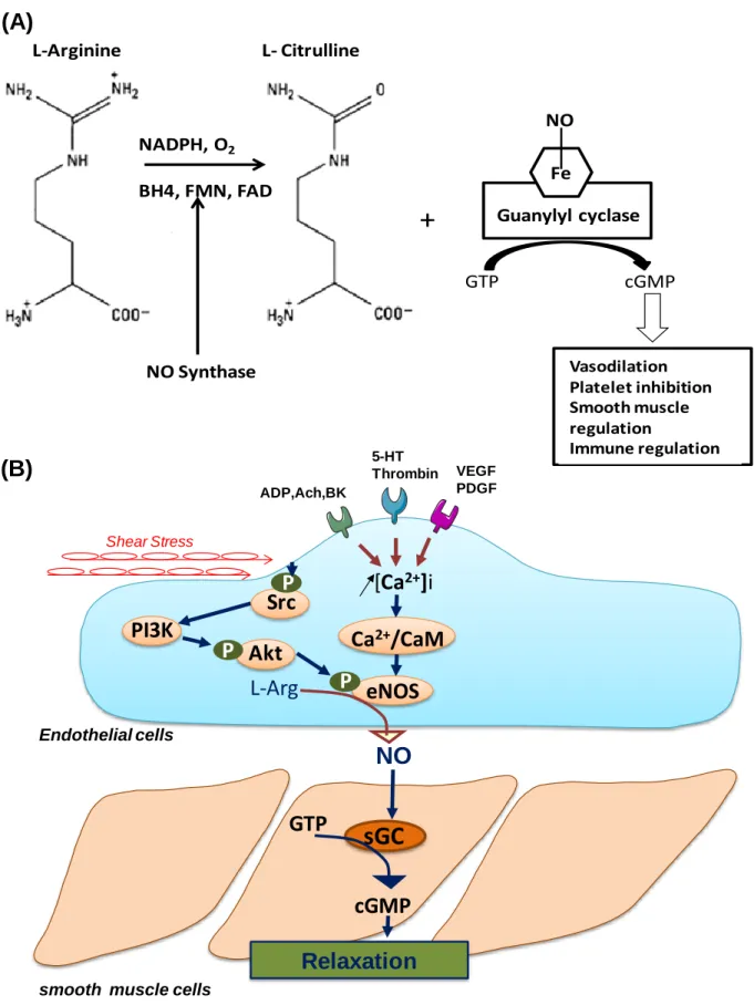
BH4 Tetrahydrobiopterin
BMI Body Mass Index
BM-MSCs Bone Marrow – Mesenchymal Stem Cells
CDK Cyclin dependent kinase
CMRL Connaught Medical Research Laboratory
CNP Natriuretic Peptide
COX Cyclooxygenase
COX-2 Cyclooxygenase-2
CTL Cytotoxic T Cell
CYP Cytochrome P450
DDR DNA Damage Response
Del-1 Developmental Endothelial Locus -1
DHE Dihydroethidium
DM Diabetes Mellitus
EC Endothelial Cell
EDCF Endothelium derived vasocontracting factors
EDHF Endothelium derived hyperpolarizing factor
EEI Exocrine Endocrine Interface
EETs Epoxyeicosatrienoic acids
EGF-8 Epidermal Growth Factor -8
eNOS endothelial Nitric oxide synthase
ER Endoplasmic Reticulum
ERK Extracellular signal-regulated kinases
Page 9
ET Endothelin
FAD Flavin Adenine Dinucleotide
FDA Fluorescein Diacetate
FGF Fibroblast Growth Factor
FMN Flavin Mononucleotide
GAD Glutamic Acid Decarboxylase
GC Guanylate Cyclase
GLP-1 Glucagon-like peptide-1
GPI Glycosylphosphatidylinositol
H2O2 Hydrogen Peroxide
H3K9Me Hypo-acetylated histones , Methylated histones
Hcd20 Anti-Human CD20
hESCs Human Embryonic Stem Cells
HGF Hepatocyte Growth Factor
HGP Hereditary / Genetic Pancreatitis
HLA-DR Human Leukocyte Antigens DR
HLA-DQ2 Human Leukocyte Antigens DQ class II
HMGB1 High-mobility group box-1
HP1 Heterochromatin Protein 1
HTK Histidine-Tryptophan-Ketoglutarate
IA-2 Islet Antigen-2
IBMIR Instant Blood-Mediated Inflammatory Reaction
IE Islet Equivalent
IEI Islet-Exocrine Interface
IL Interleukin
IL-1 Interleukin 1
IL-6 Interleukin 6
IL-8 Interleukin 8
iNOS Inducible nitric oxide synthase
IPCs Insulin Producing Cells
LMWDS Low-Molecular-Weight Dextran Sulfate
LPS Lipopolysaccharide
MCP-1 Monocyte Chemoattractant Protein-1
Page 10
MHC Major Histocompatibility Complex
MMPS Matrix Metalloproteases
MPs Microparticles
MRI Magnetic Resonance Imaging
mTOR mammalian Target Of Rapamycin
NADPH Nicotinamide-Adenine-Dinucleotide Phosphate
NF1 Neurofibromin 1
NO Nitric Oxyde
NOD Non-Obese Diabetic
NOS Nitric oxide synthase
O2.- Superoxide
OIS Oncogene-Induced Senescence
ONOO- Peroxynitrite
PAI-1 Plasmingon Activor Inhibitor 1
Pc Pericytes
PECAM-1 Platelets Endothelial Cell Adhesion Molecule
PET Positron Emission Tomography
PG12 Prostanoid Prostacyclin
PGI Prostacyclin
PGIS Prostacyclin synthase
PhChol Phosphatidycholine
PhEth Phosphotidylethanolamine
PhtdSer Phosphatidylserine
PI Propidium Iodide
PKA Protein Kinase A
PLA2 Phospholipase A2
POT1 Protection of Telomere 1
pRb Retinoblastoma Protein
PSGL-1 P-Selectin Glycoprotein Ligand-1
PTEN Phosphatase and Tensin homolog
Rap1 Repressor activator protein 1
RAS Renin-Angiotensin System
RB Retinoblastoma
Page 11
ROCK-1 Rho-Kinase-1
ROS Reactive Oxygen Species
S1177 serine 1177
SAHF Senescence-Associated Heterochromatic Foci
SASP Senescence-Associated Secretory Phenotype
SA-βGAL Senescence Associated-Beta-Galactosaidase
SIPS Stress Induced Premature Senescence
SKCa Small conductance Calcium – dependent potassium Channels
IKCa Intermediate conductance Calcium – dependent potassium Channels
SM Sphingomyelin
–SNO S- nitrosothiol
T regs Regulatory T cells
T1D Type 1 Diabetes T2D Type 2 Diabetes T494 Threonine 494 TAC Tacrolimus TL T Lymphocytes TF Tissue Factor
TGFβ Transforming Growth Factor β
Th1 T helper 1
Th2 T helper 2
Th17 T helper 17
TIN2 TRF1 Interacting Nuclear Protein 2
TM Thrombomodulin
TP Thromboxane Prostanoid
TPP1 Tripeptidyl-Peptidase 1
TRF1 Telomere Repeat-binding Factor 1
TRF2 Telomere Repeat-binding Factor 2
TxA2 Thomboxane A2
VEC Vascular Endothelial Cells
VE-cadherin Vascular Endothelial cadherin VEGF Vascular Endothelial Growth Factor
VEGF R2 Vascular Endothelial Growth Factors Receptor 2
Page 12
VHL Von Hippel Lindau
VSMCs Vascular Smooth Muscle cells
vWF von Willebrand Factor XRP8 Xk-related family Protein 8
Page 13
List of Figures
Figure 1 : Pancreas anatomy ... 19
Figure 2 : Human islets organization ... 21
Figure 3 : Islet exocrine interface - IEI in rat model ... 23
Figure 4 : Immunological factor implicated in the autoimmune destruction of β-cells in T1D ... 28
Figure 5 : Islet isolation and transplantation ... 33
Figure 6 : IBMIR (Instant Blood Mediated Inflammatory Reaction) ... 43
Figure 7 : Sources of endothelial cells in islets revascularization ... 49
Figure 8 : Structure of Blood Vessel ... 53
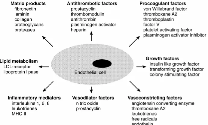
Figure 9: Different mediators that show metabolic and synthetic functions of the endothelial cells ... 55
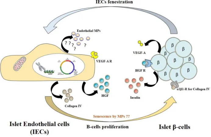
Figure 10 : Cross-talk relationship between islet endothelial cells (IECs) and β-cells ... 58
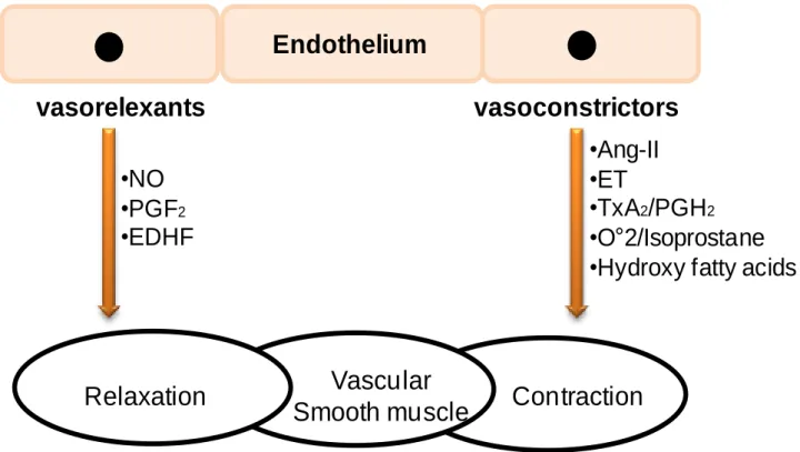
Figure 11: Biochemical mediators of vasocontracting and vasorelaxing released by the endothelium ... 59
Figure 12: NO generation from L-Arginine and its functional properties in the endothelial cell and its actions in the vascular smooth muscle cell ... 61
Figure 13: Prostacyclin (PGI2) receptor signaling ... 63
Figure 14: EDHF-mediated responses ... 64
Figure 15: RAS (renin-angiotensin system) ... 66
Figure 16 : Progression of endothelial dysfunction in relation to the progression of insulin resistance (Hsueh et al., Am J Med, 2004) ... 70
Figure 17: Biological causes and consequences of cellular senescence ... 75
Figure 18: Pathways of senescence leading to either replicative senescence or premature ... 78
Figure 19: Biochemical and Morphological characteristics of Senescent Cells ... 80
Figure 20: pRb in cell cycle regulation ... 84
Figure 21: Senescences and diabetes ... 87
Figure 22 : Immunosenescence and accompanying changes of all innate and adaptive immune cells ... 90
Figure 23: Impact of old donor and recipient on transplantation outcome ... 91
Figure 24: Membrane remodeling and MP generation ... 96
Figure 25: MPs a circulant pool of bioactif effectors ... 97
Page 14
Figure 27: Involvement of scramblase activity in XRP8 PhSer exposure following an apoptotic stress ... 101 Figure 28: Membrane mechanisms responsible for the release of MPs ... 103 Figure 29 : Reorganization of the cytoskeleton during apoptosis ... 104 Figure 30: Lipid rafts compositions ... 106 Figure 31: Mechanisms involved in clearance of circulating MPs ... 108 Figure 32 : Isolation of Exosomes & MPs ... 110 Figure 33: Variations of the C-peptide, the need for insulin and circulating microparticles during islet
transplantation ... 116 Figure 34: Possible relation between IBMIR, Senescence & MPs shedding. ... 121 Figure 35 : Endothelium damage occurs at the end of the hepatic microvessels and within the islets ... 164
Page 15
List of Tables
Page 16
List of articles in annexes
Annex 1. β cell membrane remodeling and procoagulant events occur in inflammation-driven
insulin impairment: A GLP-1 receptor dependent and independent control.
Annex 2. Differential influence of tacrolimus and sirolimus on mitochondrial-dependent signaling for apoptosis in pancreatic cells.
Annex 3. Chronic intake of EPA:DHA 6:1, a superior omega-3 PUFA formulation, prevents
the angiotensin II-induced hypertension and endothelial dysfunction in rats.
Annex 4. Endothelial Microparticles release after activated Protein C protect Beta cells through
Page 17
Chapter I
Page 18
I The physiology of pancreatic islets
I.1 Brief description of the pancreatic function and its vascularization
The pancreas, an exocrine and endocrine gland, has a head, body and tail. It is about 15 cm long, tapered organ located in the abdominal cavity behind the stomach [1] (Figure 1). Pancreas serves as two glands in one: a digestive exocrine gland and a hormone-producing endocrine gland.
The endocrine gland constitutes 1-4% of total pancreas volume where islets of Langerhans is the main unit with about 3 million islets [2].
The exocrine pancreas constitutes 96-99% of the total pancreas volume. It is mostly composed of arranged clusters of cells called exocrine acinar cells that secrete the pancreatic juice containing digestive enzymes like trypsin and chymotrypsin. These enzymes are secreted in the inactive form called proenzymes. Once they are secreted in the duodenum by enteropeptidase enzyme, which activate a cascade of enzymatic reaction that leads to proenzymes cleavage. It is initiated by the cleavage activation of proenzyme trypsinogen breakdown to the active form trypsinogen. The activated form of trypsinogen have the ability to activate other reactions including conversion of chymotrysinogen to chymotrypsin. Furthermore, besides their role in helping to digest and absorb nutrients in the small intestine. They are involved in protein, carbohydrates, lipids and nucleic acids digestion [3]. The pancreas vascularization is adequately based on blood flow coming by branches of both the coeliac artery and the superior mesenteric artery. The splenic artery run over the top border of the pancreas and supplied the body and pancreatic tail by its branches, the pancreatic artery. On the other hand, the head of the pancreas is supplied by the superior and inferior pancreatic duodenal arteries [1].
Pancreas dysfunction is defined as the incapability of the pancreas to properly perform its function. It can be caused by many diseases such as pancreatitis, cystic fibrosis, cysts, pancreatic cancer and the most importantly by diabetes.
I.2 The Islets structure and function
The pancreas is derived from two buds (dorsal and ventral endoderm), at 6 weeks of gestation, both bud fused together. This fusion occurs by turning of the ventral bud and then reaching towards the dorsal bud followed by subsequent fusion. In the fetal period, islet cell groups form by differentiating from the pancreatic bud endoderm and the pancreatic islets [4].The first evidence of islets formation in this period can be detected at 9 to 11 weeks of gestation. The
Page 19
Figure 1 : Pancreas anatomy (OpenStax College, Anatomy and Physiology) Head
Body
Page 20 pancreas growth is controlled by a composite cascade of transcription factors [5], where Ngn3 is essential to initiate endocrine differentiation [6]. The main units of the endocrine pancreas are the islets of Langerhans. It is formed as the result of agglomeration of endocrine cells. This crucial unit is scattered between the acini and ductal structures all along the exocrine parenchyma. It is an essential unit for body survival, which controls blood glucose level through insulin secretion. In the human body, islets are made up of distinct cell types [7] including: - α cells (20–40%), which produce the glucagon hormone that increases blood glucose in response to low blood glucose level.
- β cells (50–70%), which produce the insulin hormone that decreases blood glucose in response to elevated blood glucose level.
- δ cells (<10%), which produce the somatostatin hormone that inhibits the release of both glucagon and insulin.
- ε cells (<1%) which produce the Ghrelin hormone that inhibits glucose-stimulated insulin secretion from beta cells in the pancreatic islets [8].
- PP (<5%) which produces the pancreatic polypeptide hormone that plays a role in appetite and in the regulation of pancreatic exocrine and endocrine secretions [9].
In humans, 70% of the islets are in the size range of 50-250 μm in diameter with an average range of 100-150 μm. It has a regular spherical shape and it is separated from exocrine compartment by a thin collagen capsule [10]. Islet cells are organized into a trilaminar plate where one layer of β-cells is sandwiched between two α-cell-enriched layers. Moreover, this structure of islets has a folded pattern where the vessels circulate along both of its sides [11] (Figure 2).
I.3 Interaction between the islets and the exocrine pancreas
The pancreas has a dual function depends on the intercellular communications between the exocrine and endocrine parts. This islet-exocrine interface (IEI or exocrine-endocrine interface EEI) is a very important anatomical and functional region for the cell-cell communication between the endocrine islets and exocrine acinar cells of the pancreas. In IEI region, a capillary network of islets connect with the surrounding capillaries of acinar cells. Together, they form the insulo-acinar portal system that is defined as the anatomical substrate by which the endocrine pancreas may influence the exocrine pancreas. In addition, the islets have an important structure characterized by its fenestrated capillaries which constitute 8%–10% of
Page 21
Figure 2 :Human islets organization (Bosco D et al. Diabetes; 2010)
A: α-Cells (green) and β-cells (red) are organized into a thick folded plate boarded with vessels on both sides (blue). α-Cells are found on the outer surface of islets in direct contact with vessels. β-Cells, on the other hand, constitute the core part of the islets. Despite being in the core, β-Cells achieve endocrine secretion by developing cytoplasmic extensions that run between α-cells to reach the surface of vessels. B: Islet form by folding of the plate with adjacent vessels.
Page 22 the islet volume. It is likely that the islets capillaries fenestrations are 10 times wider as compared to exocrine pancreatic capillaries [12].
Most of the studies reveal the inference of oxidative stress in diabetes pathogenesis. Diabetes and oxidative stress can contribute to the loss of cell interface [13] (Figure 3). Moreover, recent studies showed that circulating microparticles (MPs), the small vesicles released from the cell surface upon activation, were significantly elevated in both diabetic patients and diabetic rats. MPs were devised as a distinguishable way to study endocrine-exocrine interactions in many diseases like cystic fibrosis where the mechanisms of these interactions are still unclear [14].
Page 23
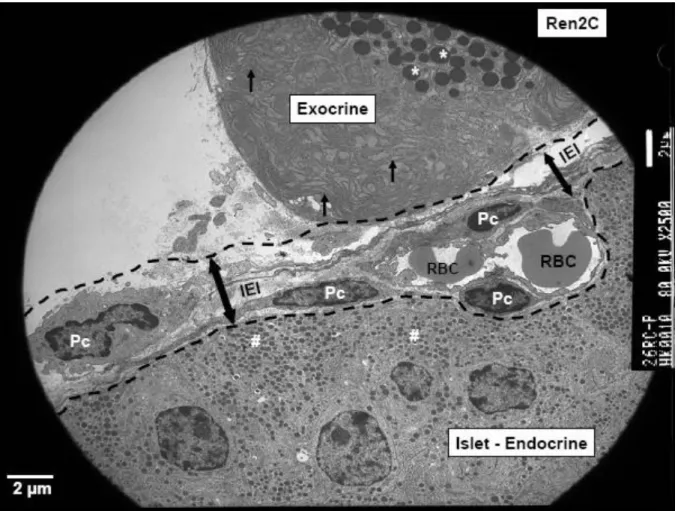
Figure 3 :Islet exocrine interface - IEI in rat model (Hayden MR, JOP 2007)
Electron microgram of the tail sections of the pancreatic tissue demonstrating the special anatomical area: The islet (endocrine)-exocrine interface (IEI) (double arrows) and is highlighted by bold black dashed lines. This islet-exocrine interface contains an anuclear endothelial capillary containing two red blood cells (RBC) with four adjacent pericytes (Pc) (magnification: 2,500x; bar: 2 μm).
Page 24
Chapter II
Page 25
II Pancreatic islet dysfunction: Diabetes
II.1 Diabetes
Diabetes nowadays is a serious public health problem. It is described as a metabolic disorder characterized by chronic hyperglycemia (increase in blood glucose level) either because insulin production is inadequate (insulin deficinecey), or the body’s cells don’t respond properly to insulin in target tissues especially liver, muscle and adipose tissue. The latter is mainly due to defective insulin receptors in target tissues.
Recently, the number of diabetic patients is constantly increasing. Moreover, diabetes causes and aggravates health complications with time. Over time, diabetes can cause serious complications and leading to damage of many organs such as heart, blood vessels, eyes, kidneys and nerves. The prevelance of diabetes is increasing among all ages. The world health organization (WHO) studies showed that number of diabetic people has abruptly increased from 108 million to 422 million between 1980 and 2014. Noteworthy, there was an increase in diabetes cases from 4.7% to 8.5 % among adults over 18 years old with higher increasing rate in middle and low-income countries. WHO estimated that 1.5 million deaths were directly caused by diabetes in 2012, 2.2 million deaths, were also linked to high blood glucose [15]. Other study by WHO showed that by the year 2030, diabetes will be the 7th cause of death in the world [16]. In France, there was more than 3.3 million according to the statistical data of medical assurance with a 6 % increase in the number of patients with diabetes between 2000 and 2009.
The prevalence of diabetes in adults in France is 4.8 to 7.4 %. Among diabetic patients, two million, seven hundred thousand subjects are diagnosed with Type 2 Diabetes mellitus (T2DM) and one hundred sixty thousand, have type 1 diabetes (T1D).
II.2 Different causes of diabetes and similar outcome
T1D is an autoimmune disease characterized by the destruction of the beta-cells (β-cells) of the pancreas due to aberrant “self” antibodies production that wrongly bind to β-cells , initiating immune-mediated reactions lead to their destruction and lack of insulin.
T1D, is more prevalent in young adults and children, hence the name juvenile diabetes [17]. The T1D autoimmunity progresses for 5 to 10 years or more after onset. Normally, hyperglycemia occurs when patients have less than 10% of functional β-cells. Chronic hyperglycemia plays a major role in the initiation of diabetic vascular damage. On the long
Page 26 term, it causes severe complications, mainly affecting the vascular endothelium. As a result, diabetes is strongly associated with microvascular and macrovascular complications, including retinopathy, nephropathy and neuropathy [18]. T1D can be diagnosed by systemic detection of auto-antibodies to insulin, islet antigen-2 (IA-2), glutamic acid decarboxylase (GAD 65) and ZnT8A [19, 20].
T2DM, also called insulin-independent diabetes is majorly caused by insulin resistance. It manifests when cells within the muscles, liver and fat tissue are unable to respond correctly to normal levels of insulin [21]. This mainly leads to hyperglycemia induction as the result of the decrease in muscular glucose utilization and increased hepatic glucose production. T2DM is a multi-factorial disease resulting from a combination of several susceptibility genes with differential expression depending on environmental factors such as obesity, sedentary lifestyle, stress and weight of birth [22]. Type 2 diabetes, accounts for ∼90–95% of all diabetes cases [23]. It is a complex disease that combines insufficient insulin production from β-cells that usually precedes 10 to 20 years hyperglycemia and peripheral insulin resistance most often in the context of obesity or overweight.
However, the causes of these two types of diabetes are different, but the consequences are almost the same. Diabetic patients of different types suffer chronically and equally from the unstable blood glucose level which obliges them to be extremely prudent in their life style and stay on treatment to avoid any further complications from progressing.
II.3 Type 1 Diabetes: an auto-immune diseases that destroy islets
II.3.1 Epidemiology of T1DTypes 1 diabetes incidence increase worldwide, it is actually estimated to account for up to 5-10 % of all cases of diabetes [24]. It mainly affects children younger than 15 years. Its incidence has increased from 0.6% in 1989 to 9.3% in 2003. Based on EURODIAB study, the number of new cases of diabetes in Europe will increase from 15,000 in 2005 to 24,400 in 2020. In addition, the prevalence of T1D in children younger than 15 years will increase from 94,000 in 2005 to 160,000 in 2020 [25]. T1D has been found recently to have a higher tendency of earlier onset. It has been speculated that T1D prevalence increases and will be doubled among children less than 5 years of age by 2020 [26]. The prevalence of T1D is growing globally, so cure cannot be done with the current knowledge, but with better understanding of the pathophysiological mechanisms in both the treatment and the management of T1D.
Page 27
II.3.2 Molecular and cellular causes of T1D
T1D is an autoimmune disease characterized by the destruction of insulin secreting cells (β-Cells) which leads to insulin insufficiency and impaired glucose homeostasis. Multiple causative genes have been identified that are dispersed loci situated in up to 41 distinct genomic locations. The first identified set of genes associated with T1D was found on chromosome 6 and it is called the Human Leukocyte Antigens DR and DQ class II (HLA-DR and HLA-DQ2) [26-28]. In addition to genetic predisposition as aforementioned, T1D can also be inflicted by environmental predispositions that are involved in the pathogenesis of the disease by a T cell-mediated autoimmune attack against β-cells [27].
Many studies suggest that T-lymphocytes (TL), the key players in cellular immunity, play also a pivotal role in the autoimmune attack of β cells [29]. The onset of T1D requires two types of T lymphocytes: CD4+ and CD8+. They were identified after prevention of diabetes by elimination of CD4+ and CD8+ T cells. The activation of TL is debuted by the β-cell antigens recruitment by dendritic cells, members of the family of antigen presenting cells (APCs). Then two possible pancreatic β-cells destruction pathways are possible: i) via direct cell-to-cell contact, by a cytotoxic process, or ii) indirectly the secretion of pro-inflammatory cytokines, perforin, or granzyme B [20].
The CD4+ T cells are primarily involved in the primary immune response by stimulating the activation and proliferation of both CD8+ T cellsand B-lymphocytes. Human CD4+ T cells from the pancreatic lymph nodes of subjects with T1D respond to the first 15 amino acids of the insulin A-chain [30]. CD4+ T cells can be divided into two categories: T helper (Th1, Th2 and Th17) and regulatory T cells (Tregs). Both type are differently involved in T1D pathogenesis depending on different cytokines secretion (Figure 4).
The cytotoxic T cell (CTL) are the result of the activation of CD8+ T cells, which recognize
pathogen-derived peptides presented by major histocompatibility complex (MHC) class I molecules. Cytotoxicity is expressed by secreting proteins such as Fas and cytokines such as TNF-α and IFN-γ [20]. B cells (B Lymphocytes, BL) are not directly linked to islet-β cells destruction but rather to the progress of the disease by promoting autoimmunity. B-cells promote differentiation of CD8+ T-cells to CTL and provide survival signals for CD8+ T-cells to maintain high levels of aggressive CTLs [31]. Indeed, they stimulate the production of auto-antibodies whose presentation generates self-reaction of TL and the maintenance of CD4+ T cell memory [32]. However, they also play a role in the initial phase of the disease by being
Page 28
Figure 4 : Immunological factor implicated in the autoimmune destruction of β-cells in T1D. (Min Li et al. J Cell Mol Med, 2014)
β-cells are damaged by various factors and the released autoantigens are presented by antigen-presenting cells. Then CD4+ T, CD8+T andNK cells are activated and CD4+ helper T lymphocytes differentiate into Th1, Th2, Th17
and Tregs. Th1 cells can destroy the β-isletcells and accelerate the course of T1DM via production of IL-2 and IFN-γ. IL-2 has been shown to prevent diabetes, while it can activate CD8+ T cells and Tregs. In addition, IFN-γ
plays a dual role in β-cells destruction. Via the signal transducer and activator of transcription-1 (STAT-1) pathway and in protection via the IRF-1 pathway. Th2 cells mainly produce IL-4 and IL-10, which are responsible for strong antibody production, have been ascribed with a protective role. Th17 can destroy the β-islet cells by secreting IL-17. Whether Tregs play a preventive role in the pathogenesis of T1DM remains a question. In addition, NK cells are involved in direct killing of β-cells through the interaction of NK cell markers, such as NKp46 and KIRs. Furthermore, CD8+ T cells contribute to the development of T1DM by secreting proteins such as Fas and
Page 29 essential antigen presenting cells of the islet but to a less extent compared to dendritic cells [20].
II.4 T1D treatment
T1D is inevitable, so the main target of T1D treatment is to maintain blood glucose levels at normal range, improving functional residual β-cell mass, preventing acute and chronic complications of the disease and to ensure good quality life for the patient. Some researchers postulated that it might be possible to prevent or delay T1D at the latent autoimmune stage and before pancreatic beta cells destruction [33]. To reach these target two types of treatments were developed:
1- Treatments to maintain normal blood glucose level and to prevent the destruction of the β-cells by many techniques like immunotherapy.
2- Treatments to replace the insulin-producing cells with stem cells, pancreas or pancreatic islets transplantation.
II.4.1 Stabilization and prevention of T1D
T1D disease stabilization can be maintained by several day-to-day practices. Daily-based insulin injections, several times per day, are the common and effective way to maintain normoglycaemia. Glucose level is then constantly monitored by a blood glucose monitor that helps adjusting insulin doses. Insulin treatment should also be accompanied by adopting a healthier life style with a low-carbohydrate diet [34], physical exercise and quitting both smoking and alcohol intake.
Immuno-suppressors have been also used to treat T1D such as Cyclosporine, the first successful immunosuppressive agent used. Unfortunately, despite retaining β-cell function, Cyclosporine showed high kidney toxicity along with other side effects of long-term treatment. Altogether, forced the termination of the cyclosporine therapy [33].
In autoimmune diabetes, studies in NOD mice using anti-CD20 antibody
have shown contrasting effects of B-cell depletion on reversal or prevention of T1D. Recently, researchers have also shown that the disease progression was totally stopped by the removal of the antibodies at a preinsulitis stage in NOD mice [35]. B cells depletion in NOD mice using anti-CD20 mAb showed effective in reversing hyperglycemia at onset [36]. The combined treatment of anti-human CD20 (hCD20) and oral anti-CD3 have also been shown to reverse diabetes in more than 60% of NOD mice diagnosed with diabetes. Additional studies proved
Page 30 that the suppressive function of regulatory T cells was synergistically improved by the addition of oral anti-CD3 to the B-cell depletion therapy [37]. Most significantly, T1D can be reversed by using a small number of antigen-specific Tregs, which propose a novel approach to cellular
immunotherapy for autoimmunity [38].
Several therapeutic approaches were studied to prevent the onset of T1D symptoms by improving immune tolerance for β-cells and maintaining its function. However, despite the promising results, few treatments stood out. Moreover, most treatments have been found either toxic on the long term or failed to maintain sustainable pancreatic β-cell function [39].
II.4.2 Replacement of defective β-cells by Stem cell: an alternative therapy
Scientist have tried to replace the defective β-cells with a bio-mechanical pump. The method is based on the development of an artificial pancreas combining continuous analysis of blood glucose system coupled with an automatic insulin delivery system. Nevertheless, the injection of exogenous insulin didn’t succeed to stop the development of T1D and may have caused serious complication such as kidney failure, vascular diseases and retinopathies [40].
As part of a predominantly later management of T1D, the replacement of defective β-cells is as an essential therapeutic strategy. Therefore, the next step in research was the replacement of defective β-cells by Stem-cell therapy or pancreatic islet transplantation.
β-cells replacement approaches aim to generate stem cell derived insulin-producing cells (IPCs), which could make diabetes cellular therapy available for millions diabetic patients. Human embryonic stem cells (hESCs), a cell line with unlimited proliferative capacity and a high differentiating potential, was the main source to generate IPCs [41]. The principal characteristic of hESCs was the potential to differentiate into specialized cells of all three primary germ-layers, including pancreatic IPCs. When transplanted into the epididymal fat pads of SCID/NOD mice, the hESC-derived pancreatic IPCs were able to reverse hyperglycemia for ≥8 weeks [42]. The principal challenge to scientists now is to have safe and efficient transplanted cells without the risk of immune rejection or tumorigenesis, not to mention, the big concern regarding the ethical issue [40].
Despite the huge advancement in studies to obtain an unlimited source of functional and immunocompetent β-cells, their application to humans is limited by the need to graft a sufficient amount of cells with complete cell functionality. For this purpose, the grafting of the functional units of insulin secretion directly has been used to quickly help re-establish the normal blood glucose level.
Page 31
Chapter III
Pancreatic islet transplantation:
Page 32
III Pancreatic islet transplantation: A cellular treatment of T1D
III.1 Islet graft
The development of islet cell transplantation aims at decreasing surgical complications and to limit or eliminate immunosuppression by protection of grafted islets. Islet transplantation started in 1972 by Ballinger et al. in rodents, which proved that diabetes mellitus could be reversed by pancreatic islets transplantation [43]. Then in 1990, a 1 month insulin independence was reported in T1D patient by Scharp et al. [44]. In the year 2000, Shapiro et al. were successful in maintaining insulin independence for 1 year in seven T1D patients using a protocol called Edmonton. It had an overwhelming success that lead to enormous development in clinical islet transplantation [45]. This technique was normally used in patients with unstable T1D and aged > 50 years. Islet auto-transplantation after total pancreatectomy represents a big hope for patients with chronic pancreatitis due to hereditary/genetic pancreatitis (HGP). It offers long-term pain relief (90%) and β-cell function preservation. Islet auto-transplantation done prior to pancreatic inflammation results in a higher degree of pancreatic fibrosis risk, pancreatic cancer and islet cell function loss [46]. Cystic fibrosis patients develop hyperglycemia after lung transplantation. This aggravates if patients are diabetic causing complication that might lead to mortality [47]. Combination of lung and islet transplantation from the same donor can minimize and even ablate complications. This combination has been shown to lead to optimal blood glucose level with no sign of hypoglycemic and 50 % reduction of used insulin [48]. A 5-year follow-up, in a large cohort showed that islet transplantation was safe and efficient in patients with T1D [49].
III.2 Pancreatic islet transplantation protocol
III.2.1 Islet transplantation stepsIslet transplantation proceeded in two steps. The first step started by the isolation of the islets. A pancreas of a brain-dead patient is harvested then processed to isolate only islets. The pancreas is digested by collagenase treatment. Then by using density gradient, islet cells are separated from exocrine cells, purified and then used for culture. The second step occurred in the hospital by the surgical intervention performed in diabetic patients, usually under local anesthesia. The procedure is summarized by the infusion of islets into the patient’s through the portal venous circulation under ultrasound guidance (Figure 5). To overcome the severe shortage of available pancreas islet transplantation and experimental status of other sources of
Page 33
Page 34 β-cells, optimization rate, viability, integrity and functionality of the islets at each stage of the procedure appears to be crucial in the long-term success of the islets.
III.2.2 Donor selection
The main source of donors is from brain-dead patients but actual studies have shown a comparable results from non-heart beating donor [50]. The establishment of criteria, which determine the best donor selection, is very important to improve the success of islet transplantation due to the high cost procedure and the complications that may occur. Previous studies review several selection criteria that should be taken into careful consideration, for which they have been proven effectively in multiple transplantation surgeries. These include, the donor’s age, high body mass index (BMI), cold ischemia time and blood glucose level [4]. The donor’s age is one of the most essential factor, where a pancreas isolated from donors older than 50 years old with BMI greater than 30 is undesirable for a whole organ transplant, but is favored for islet transplantation [51]. This is because the higher the BMI the higher the quantity of isolated islets [52]. Putative donors with HbA1c levels higher than 7% are not selected as islet
transplantation donors. HbA1c is correlated with Diabetes onset. Evidently, T2DM patients are
considered unsuitable to be islets donors [53].
III.2.3 Pancreas dissociation and islet isolation
After pancreas isolation, it is preserved prior to islet isolation in a UW or histidine–tryptophan– ketoglutarate (HTK) solution. Recent studies have shown a similar preservation characteristics of these 2 solutions especially in terms of islets yield and functionality [54]. The principal protein structure in the exocrine-islet interface is collagen [55]. As a result, after preservation, collagenases dissociate the strong collagen interface structure [56].This is an essential part that will affect whole procedure and downstream steps. Especially, it affects both the yield and the viability of islets.
In 1990s, Roche introduced the liberase, which is the first blend of enzymes made up of collagenase type (I & II) and thermolysin (a derivative of Bacillus thermoproteolyticus as a non-collagenase impurity that is needed to enhance pancreas dissociation). This new product helped reducing the variability of enzyme effectiveness [57]. Another collagenase cocktail called collagenase NB1 that is supplemented with neutral protease NB, showed more improvements in islet morphology, viability and in vitro function that makes it a promising product for human-islets isolation to avoid the inter-batch variability [58]. Camillo Ricordi has