HAL Id: tel-01164987
https://tel.archives-ouvertes.fr/tel-01164987
Submitted on 18 Jun 2015HAL is a multi-disciplinary open access archive for the deposit and dissemination of sci-entific research documents, whether they are pub-lished or not. The documents may come from teaching and research institutions in France or abroad, or from public or private research centers.
L’archive ouverte pluridisciplinaire HAL, est destinée au dépôt et à la diffusion de documents scientifiques de niveau recherche, publiés ou non, émanant des établissements d’enseignement et de recherche français ou étrangers, des laboratoires publics ou privés.
peptides : from amyloid precursor protein (APP) to
amyloid β-oligomers and γ-secretase modulators
Anna Itkin
To cite this version:
Anna Itkin. Multidisniplinary study of Alzheimer’s disease-related peptides : from amyloid precursor protein (APP) to amyloid β-oligomers and γ-secretase modulators. Agricultural sciences. Université de Strasbourg, 2012. English. �NNT : 2012STRAF051�. �tel-01164987�
ÉCOLE DOCTORALE DES SCIENCES CHIMIQUES
[ UMR 7177 ]
EN CO-TUTELLE AVEC
UNIVERSITÉ LIBRE DE BRUXELLES
FACULTÉ DES SCIENCES ULB
THÈSE
présentée par
[ Anna ITKIN ]
soutenue le : 14 Mai 2012
pour obtenir le grade de
Docteur de l’université de Strasbourg
Discipline / Spécialité : Résonance magnétique et biophysique des membranes
Multidisciplinary study of Alzheimer’s
disease-related peptides: from Amyloid
precursor protein (APP) to Amyloid
β-oligomers and
-secretase modulators
THÈSE dirigée par :
M. BECHINGER Burkhard Prof., Université de Strasbourg M. RAUSSENS Vincent Prof., Université Libre de Bruxelles
M. RUYSSCHAERT Jean-Marie Prof. Honoraire, Université Libre de Bruxelles
RAPPORTEURS :
M. HUSTER Daniel Prof., University of Leipzig M. WARSCHAWSKI Dror H.d.R., CNRS-UMR 7099
MEMBRES DU JURY :
Mme. HELLWIG Petra Prof., Université de Strasbourg M. GOORMAGHTIGH Erik Prof., Université Libre de Bruxelles
i n t r o d u c t i o n
La maladie d’Alzheimer (AD) est une maladie neuro-dégénérative affectant presque 2% de la population des pays industrialisés. L’AD est la forme la plus commune de démence caractérisée par la destruction des cellules du cerveau, une perte de mémoire et une détérioration cognitive et des processus comportementaux suffisamment sérieuse pour affecter l’activité professionnelle, la vie sociale et les loisirs des personnes atteintes. Cette maladie mortelle s’aggrave au fil du temps. L’un des marqueurs histopathologiques caractéristiques de l’AD est la présence de dépôts protéiques et de plaques amyloïdes dans le cerveau. Ces plaques se composent de peptides bêta-amyloïdes (Ab) de 40 et 42 résidus de longueur, et sont les produits du clivage par des protéases de la protéine précurseur de l’amyloïde (APP).
L’APP est une protéine membranaire intégrale avec un seul domaine transmembranaire (TM) exprimée dans de nombreux tissus et concen-trée dans les synapses des neurones. L’APP est plus connue et plus communément étudiée en tant que molécule précurseur dont la pro-téolyse génère des peptides Ab, des peptides de 38 à 42 acides aminés de longueur, et dont la forme fibrillaire est le principal composant des plaques amyloïdes retrouvées dans le cerveau des patients atteints de l’AD.
On a récemment découvert qu’un mécanisme de clivage protéoly-tique séquentiel de libération d’Ab était à l’origine des multiples clivages par γ-sécrétase, générant des peptides Aβ de différentes longueurs. Il a été prouvé que la protéolyse intramembranaire par γ-sécrétase nécessitait un effilochage local de la structure hélicoïdale secondaire du domaine TM. En effet, certaines fonctions inhabituelles de la séquence APP_TM pourraient déstabiliser sa structure hélicoïdale dans la membrane. Premièrement, on constate une densité d’acides aminés bêta-branchés (Valine, Thréonine, Isoleucine) élevée autour des sites de clivage Ab40- et Ab42- susceptible de déstabiliser la structure secondaire des hélices TM.
Deuxièmement, plusieurs des glycines du domaine de l’APP_TM se produisent dans des motifs GxxxG, Gly625, Gly629, and Gly633, et on a constaté qu’elles jouaient un rôle de médiateur dans l’homodimérisation de l’hélice. On constate que l’hélice TM se déstabilise au point de transition proche du site de clivage-ε et les données de RMN solide indiquent que ces conformations hélicoïdales coexistent avec des con-formations non hélicoïdales dans le domaine de l’APP_TM. Suggérant
rôle dans le traitement de l’APP et donc dans la production d’Aβ. La caractéristique pathologique de l’AD est attribuée à l’accumulation constante de fibrilles d’Aβ dans des plaques et à la formation de sites contenant une hyperphosphorylation de la protéine Tau, entraînant une perte neuronale. L’hypothèse de la cascade amyloïde (Hardy et Higgins, 1992) explique les effets toxiques des plaques amyloïdes sur les connexions synaptiques et les neurones. Cependant, les expéri-ences menées pour établir une relation de cause à effet directe entre les dépôts d’Aβ et la neurodégénérescence à l’origine de l’AD n’ont pas été concluantes. Selon les récentes théories, de petits assemblages oligomériques solubles ou protofibrillaires d’Aβ sembleraient toxiques pour les neurones et leurs interconnexions vitales, ce qui expliquerait cette relation de cause à effet. Bien que les peptides Aβ puissent avoir différentes longueurs d’acides aminés comprises entre 38 et 43, les alloformes que l’on retrouve le plus dans le cerveau sont l’Aβ (1-40) et l’Aβ (1-42). On a retrouvé ces deux peptides dans des plaques amyloïdes et on a constaté qu’ils formaient des oligomères et des protofibrilles. Des analyses post-mortem prati-quées sur des sujets humains ont démontré que l’Aβ (1-40) plutôt que l’Aβ (1-42), que ce soit sous forme soluble ou insoluble, tendait à s’accumuler davan-tage chez les patients atteints de l’AD. En plus des cas sporadiques d’AD, on a découvert et étudié plusieurs mutations de la maladie d’Alzheimer familiale (FAD). La mutation Arctique dans la séquence Aβ (Aβ(1-40)E22G) crée des protofibrilles bien plus rapidement et en plus grande quantité que le type sauvage Aβ et induit une neurotoxic-ité rapidement. Les caractéristiques cliniques et anatomopathologiques de la FAD sont similaires aux cas sporadiques, sauf que la maladie se déclare bien plus tôt.
La recherche n’a, pour l’instant, pas permis d’établir le mécanisme primaire unique à l’origine de l’agrégation d’Aβ responsable de la dégénérescence neuronale et du décès des patients atteints de l’AD. Il semblerait, au contraire, que l’agrégation d’Aβ et le développement de la maladie découlent de nombreux processus. Tandis que la source d’ions Ca2+ est soit extracellulaire soit intracellulaire, l’activation neuronale est plus généralement associée à une augmentation de la concentration de Ca2+intracellulaire ([Ca2+]i). Parmi les nombreuses
hypothèses mises en avant pour expliquer l’étiologie de l’AD, on s’accorde à dire que le dérèglement de l’homéostase du Ca2+joue un
rôle très important dans le développement de la maladie. L’idée qu’une homéostase du Ca2+altérée puisse servir de facteur déclenchant du développement de l’AD a été formulée pour la première fois en 1982 pour être plus tard revue par Khachaturian. Les nombreuses études sur l’AD ont déterminé une déficience du Ca2+ prouvant la relation
entre le signal du Ca2+et la voie amyloïdogénique.
la formation d’oligomères Aβ pathogènes, qui à leur tour peuvent intensifier la dyshoméostasie du Ca2+. Les espèces oligomériques
pourraient dont être tenues pour responsable de la médiation de la blessure neuronale et de l’inhibition de LTP de l’AD chez les sujets d’âge avancé, similaire à la situation chez les patients FAD porteurs de la mutation Arctique.
Malgré les progrès incroyables faits dans la compréhension de l’AD, nous ne sommes toujours pas parvenus à développer de nouveaux médicaments suffisamment efficaces pour guérir, ou mieux encore, prévenir la maladie. Les études épidémiologiques ont démontré une association étroite entre la diminution du risque d’AD et l’usage prolongé de médicaments anti-inflammatoires non stéroïdiens (AINS). On a découvert que certains d’entre eux modulaient le clivage de la γ-sécrétase. Dont la molécule carprofène–un inhibiteur du COX-2–on l’a isolée des autres agents pharmacologiques disponibles dans le commerce et constaté que sa N-substitution créait des modulateurs puissants de γ-sécrétase. Le carprofène sulfonyle et le carprofène benzyle ont eut un effet minime voire nul sur le clivage de la γ-sécrétase du site-ε (Narlawar et al., 2006 ; Narlawar et al., 2007) mais ont une spécificité élevée dans la modulation de l’activité de la γ-sécrétase (Schmidt et al., 2006 ; Narlawar et al., 2006).
o b j e c t i f s
L’objectif principal de cette thèse était de comprendre certains des processus clés à l’origine, du développement et du traitement de l’AD en suivant une approche multidisciplinaire.
Pour comprendre le mode d’action des protéines membranaires au niveau moléculaire, il est essentiel d’avoir des connaissances précises sur les propriétés structurelles de celles-ci, ainsi que sur la structure de base et les angles d’inclinaison des segments transmembranaires. Dans nos efforts de compréhension du rôle des variations structurelles de la séquence TM dans le traitement de l’APP, nous tentons de déterminer les détails structurels, les angles d’inclinaison d’hélice du peptide APP_TM dans son environnement naturel, la double couche lipidique de la membrane. La RMN solide s’est avérée être un outil biophysique très utile au sondage de la structure et des dynamiques parmi de nombreuses biomolécules inaccessibles par cristallographie ou par la RMN en phase liquide comme les protéines membranaires et les agrégats protéiniques liés à la maladie.
Dans le but de découvrir les mécanismes moléculaires responsables des risques de l’AD liés à l’âge, nous avons comparé l’agrégat d’Aβ (1-40) et d’Aβ (1-40)E22G dans le but d’étudier les similarités struc-turelles et morphologiques des espèces–oligomériques ou fibrillaires–
sur gel et le transfert de protéines, la fluorescence de la thioflavine T (ThT), la spectroscopie infrarouge à transformée de Fourier (FTIR) et la microscopie atomique (AFM) ont été utilisés.
Nous avons utilisé la RMN solide pour découvrir le mode d’action des deux puissants modulateurs de γ-sécrétase, le carprofène benzyle et le carprofène sulfonyle, dans les doubles couches lipidiques. La détermination de leur position et orientation dans les membranes lipidiques nous donnera un bon aperçu du mécanisme moléculaire du clivage protéolytique de la γ-sécrétase et donc de la production d’Aβ. r é s u ltat s e t c o n c l u s i o n s
Le dérèglement de l’homéostase du Ca2+ dans le cerveau de sujets
d’âge avancé et dans les maladies neurodégénératives joue un rôle essentiel dans de nombreux processus et contribue à la déficience des cellules et au décès. Nous avons émis l’hypothèse que le Ca2+pourrait
déclencher ou accélérer l’agrégation d’Aβ. Par conséquent, nous avons comparé les modèles d’agrégation de l’Aβ(1-40) et de l’Aβ(1-40)E22G avec ou sans Ca2+.
Des échantillons d’Aβ(1-40) et d’Aβ(1-40)E22G ont été préparés et ont incubé pendant 96 heures avec ou sans 2 mM de Ca2+. Après 0, 2, 4, 6, 24, 72, et 96 h, les échantillons ont été analysés par électrophorèse sur gel de polyacrylamide sans SDS (0,1% de SDS était présent dans le tampon de migration) et imagés par transfert de protéines. Le modèle d’agrégation de l’Aβ (1-40) a présenté des différences frappantes avec ou sans Ca2+ ajouté. À 72 h et à 96 h, l’Aβ (1-40) est constituée des monomères et des oligomères contenant du Ca2+dont les masses moléculaires sont comprises entre celles des dimères (environ 8 kDa) et des hexamères et de celles des oligomères ayant une masse moléculaire plus élevée. Sans Ca2+, principalement les oligomères, protofibrilles et fibrilles ayant une masse moléculaire élevée ont pu être détectés en raison de l’étendue de l’agrégation de l’Aβ(1-40). Contrairement à ceux d’Aβ(1-40), les échantillons d’Aβ(1-40)E22G n’ont présenté aucune différence dans leurs profils d’agrégation avec ou sans Ca2+
ajouté. Comparé aux résultats obtenus pour les deux peptides, nous avons clairement constaté qu’en présence de Ca2+, l’Aβ(1-40), tout comme l’Aβ(1-40)E22G, s’agrégeait pour produire des oligomères et des protofibrilles, alors que sans Ca2+, seul l’Aβ(1-40) formait des fibrilles.
La fluorescence de la ThT a été utilisée pour distinguer les oligomères et les fibrilles des peptides Aβ. Une étude a été menée, qui consistait en une incubation de 96 heures, avec ou sans Ca2+ajouté pour les deux
peptides. Après une incubation de 72 et de 96 heures de l’Aβ(1-40) sans Ca2+, l’intensité de la fluorescence de la ThT a grandement aug-menté, ce qui indique la formation de fibrilles. Après une incubation
d’oligomères et l’absence de fibrilles. Avec de l’Aβ(1-40)E22G, avec ou sans Ca2+, on a constaté une intensité de la fluorescence de la
ThT stable voire même relativement basse pendant les 96 heures, ce qui signifie que le processus d’agrégation de l’Aβ(1-40) est bien plus lent que celui de l’Aβ(1-40)E22G, ou que ce dernier a une tendance relativement élevée à former des oligomères plutôt que des fibrilles.
Les structures secondaires des agrégats d’Aβ sont réputées comme possédant une haute teneur en feuillet bêta. Nous avons récemment découvert, au moyen de l’ATR-FTIR, qu’une signature caractéristique d’oligomères solubles d’Aβ indiquait une conformation de feuillet bêta antiparallèle, tandis qu’une présence de fibrilles d’Aβ indiquait une conformation de feuillet bêta parallèle. Dans le but d’évaluer la présence d’oligomères ou de fibrilles, nous avons étudié les mod-èles d’agrégation de l’Aβ(1-40) et de l’Aβ(1-40)E22G sur un temps d’incubation donné et avons suivi l’évolution des pics à 1630 cm−1 et 1695cm−1.
Au cours des premières 48 heures, nous avons observé deux car-actéristiques particulières, et ce, pour toutes les conditions évaluées: deux pics, un à ~1695 cm−1et un autre à ~1630 cm−1. Les pics à ~1695
cm−1et à ~1630 cm−1 dans le spectre infrarouge sont caractéristiques de la conformation du feuillet bêta antiparallèle, indiquant des espèces structurellement différentes des fibrilles. On a observé une réduction importante à ~1695 cm−1 puis un passage à des nombres d’ondes inférieurs et pour finir à une seconde diminution à ~1630 cm−1 au bout de 48 heures d’incubation d’Aβ(1-40) sans Ca2+ ajouté. Ceci
illustre le processus de fibrillation dynamique du peptide Aβ(1-40) in vitro, ayant commencé avec des monomères et des dimères qui se sont transformés en fibrilles au fil du temps. On a remarqué, pour l’Aβ(1-40) avec du Ca2+, les deux pics caractéristiques à ~1695 cm−1 et à ~1630 cm−1 à tous les points d’observation. Malgré la légère diminution d’intensité observée à ~1695 cm−1, aucune réduction
im-portante n’a été observée à ~1630 cm−1. Il est également intéressant de noter que ce comportement spectral de l’Aβ(1-40) en présence de Ca2+ a les mêmes particularités que celui de l’Aβ(1-40)E22G avec ou
sans Ca2+. Dans les deux conditions et pendant 96 heures, les pics de l’Aβ(1-40)E22G à ~1695 cm−1 et à ~1630 cm−1 sont caractéristiques d’une conformation du feuillet bêta antiparallèle. Cette découverte confirme les résultats découlant de l’analyse PAGE et des expériences ThT, indiquant toutes–comme prévu, et conformément aux données publiées–la formation de nombreux oligomères et éventuellement de protofibrilles par l’Aβ(1-40)E22G.
L’imagerie AFM a révélé des similarités morphologiques entre les oligomères de l’Aβ(1-40) avec Ca2+et de l’Aβ(1-40)E22G, qui étaient
tous sphériques ou curvilignes. Nous ne savons toujours pas si la
de Ca aux oligomères de l’Aβ(1-40)E22G formés en l’absence de Ca2+ ajouté indique des effets toxiques similaires.
La similarité entre les modèles d’agrégation de l’Aβ(1-40) avec du Ca2+et de l’Aβ(1-40)E22G avec ou sans Ca2+, observée ici au moyen de trois techniques différentes, soulève une question fondamentale sur un changement éventuel dans le mécanisme de l’agrégation de l’Aβ(1-40) en présence de Ca2+. Les ions Ca2+semblent stimuler la formation préférentielle d’oligomères et de protofibrilles par l’Aβ, s’écartant ainsi de la voie de la fibrillogénèse préférable.
Les propriétés conformationnelles du segment TM de l’APP pourrait affecter son traitement protéolytique par la γ-sécrétase. Ces propriétés n’ont pas été définitivement établies. Afin de mieux comprendre le clivage protéolytique de l’APP à l’origine de la création de peptides Aβ, nous avons étudié les particularités structurelles de la séquence de l’APP TM qui pourrait s’avérer essentielles à son traitement par la γ-sécrétase. Nous avons réussi à synthétiser et à reconstituer chim-iquement le domaine transmembranaire de l’APP en doubles couches POPC sous forme de dimère présentant en grande partie une confor-mation alpha hélicoïdale. Une étude détaillée de la RMN solide des peptides APP_TM4K marqués avec les isotopes15N et2H à divers
points de la séquence TM, indique une importante hétérogénéité con-formationnelle et orientationnelle aux positions Ala-630, Ala-642 et Val-646.
La valeur chimique15N du Gly-637 était de 191 ppm indiquant une orientation transmembranaire pour un peptide hélicoïdal. Il a été déjà publiéque le Gly-637 participait à la formation d’un dimère stable. Le Gly-637, faisant partie du dernier motif GxxxG, est le dernier acide aminé ancrant l’orientation transmembranaire et stabilisant le dimère. Cette stabilité pourrait s’avérer nécessaire à l’effilochage de la structure hélicoïdale aux points de clivage. La théorie du contrôle du traitement de l’APP par la force de la dimérisation a été avancée.
Le même peptide était également marqué avec du 2H à la
posi-tion Ala-642, et n’a pas présenté de division du quadrupole claire, mais plutôt de nombreuses divisions d’une largeur de ~ 27 kHz. L’explication la plus plausible quant à la forme du spectre de décalage chimique15N de l’Ala-642 est que ce site possède de nombreuses ori-entations différentes, mais pas toutes celles d’un échantillon de poudre. Une réorientation lente (plus lente que la gamme en kHz) de ce site est probablement responsable de l’apparition relativement importante du "pic principal " dans le spectre. Cela signifie que la structure pourrait ne pas être hélicoïdale à cet emplacement. Afin de bien comprendre les diverses orientations observées, on a simulé le spectre du décalage chimique15N. Le résultat indique que pour le spectre expérimental
du 15N Ala-642 une gamme restreinte d’orientations " autorisées "
(ex. restreintes par les angles de liaison Cα–Cβ) existe, dans laquelle
parallèles à la membrane normale. La liaison NH de l’Ala-642 adopte clairement de nombreuses orientations différentes. Cette découverte conforte l’idée qui veut que l’Ala-642 du site de clivage-γ se trouve dans une conformation non hélicoïdale.
Le troisième site marqué 15N Val-646 était localisé à 209 ± 28
ppm présentant un large spectre de 26 kHz, indiquant une hélice transmembranaire possédant un nombre d’orientations correspondant au sens du champ magnétique. Nous avons découvert que pour une gamme complète d’angles d’attaque rotationnels (0° - 360°) éventuels, les angles d’inclinaison sont compris entre 0° à 50°.
Le Val-646 et l’Ala-642 se situent dans les sites de clivage ζ- et γ-, re-spectivement. Le première présente une orientation transmembranaire, tandis que le second présente une hétérogénéité conformationnelle ample et semble n’avoir aucune orientation définie. L’existence d’une conformation non hélicoïdale sur le site Ala-642 est proposée par Lu et al., 2011. Cependant, c’est la première fois qu’on observe une dis-tribution orientationnelle avec une approche structurelle. Une preuve de la variabilité conformationnelle autour des sites de clivage γ- et ζ-peut avoir des implications importantes pour le mécanisme de clivage et donc pour la production d’Aβ.
Le peptide portant le marqueur15N Val-646 a également été marqué sur le site 2H Ala-630. La largeur du spectre quadrupole 2H sur le
site Ala-630 était de 26 kHz indiquant une distribution hétérogène des alignements, qui ne sont pas un modèle de poudre (dans lequel toutes les orientations sont possibles). L’Ala-630 se trouve près de la limite de la membrane N-terminale et pourrait subir moins de con-traintes près des interactions de van der Waals avec des molécules lipidiques et donc il est raisonnable de penser que cet acide aminé soit plus mobile. Cependant, des publications récentes prouvent qu’il implique l’existence de mouvements sur le site Ala-630 qui pourraient être associés à l’échange entre les conformations hélicoïdales et non hélicoïdales. Il est intéressant de noter que les spectres au deutérium des deux alanines, Ala-630 et Ala-642, ont une forme relativement sem-blable. Les deux sont relativement les larges ~26 kHz et ~27 kHz et ces deux valeurs maximales de clivage ont des orientations de la liaison Cα–Cβde 42◦et 71◦, pour les valeurs positives et négatives,
respec-tivement. En gardant à l’esprit qu’il n’y a aucune structure du peptide APP_TM à aligner, le large spectre du décalage chimique du 15N pour l’interaction quadrupole du Val-646 et du2H pour l’Ala-630 n’a pas été en mesure de donner des contraintes orientationnelles claires pour cette paire de marqueurs. Les spectres sont, par conséquent, en grande partie déterminés par la distribution orientationnelle des molécules par rapport à la direction du champ magnétique. Cette grande hétérogénéité conformationnelle démontrée est plus
hélicoïdales du peptide sur ces sites. En fonction des résultats de ces travaux, nous avons proposé un modèle des orientations et confor-mations possibles du peptide APP_TM4K dans les doubles couches POPC (voir le texte complet).
On a découvert que le carprofène benzyle et le carprofène sulfonyle étaient des modulateurs efficaces de la γ-sécrétase. Ils affectent le clivage sur les sites γ38, γ40, et γ42 et bloquent, en particulier, la formation d’Aβ42 pour améliorer la formation d’Aβ38, présentant ainsi un profil similaire aux AINS efficaces. On en a trouvé sur la plupart des inhibiteurs puissants de l’Aβ42 (Narlawar et al., 2006). Les connaissances sur l’interaction de ces molécules avec la double couche lipidique sont essentielles à la compréhension de leur mode d’action. Nous avons déterminé, dans ces travaux, l’orientation des dérivés du carprofène dans les doubles couches lipidiques au moyen de la RMN en phase solide.
Des fragments spécifiques de molécules de carprofène sulfonyle et de carprofène benzyle ont été marqués avec l’isotope 2H. Les
molécules ont été incorporées dans des doubles couches lipidiques composées d’un mélange lipidique tertiaire (POPC, cholestérol et sphyngomyéline) et on a préparé des échantillons pour les mesures à la RMN solide. L’analyse des données de la RMN solide a entraîné des contraintes orientationnelles pour les molécules de carprofène sulfonyle et de carprofène benzyle. Nous avons observé une liai-son C–S du carprofène sulfonyle orientée à ± 27,4° par rapport à la direction du champ magnétique, ce qui correspondait aux deux orien-tations possibles (voir le texte complet). Quant au benzyle carprofène, l’orientation correspondant aux résultats expérimentaux est celle où l’angle d’attaque rotationnel (rotation autour de l’axe z) est de ~31◦ et où l’angle d’inclinaison rotationnel (rotation autour de l’axe y) est de ~137◦. Les expériences de diffraction des neutrons (effectuées par nos collaborateurs) ont montrées que les molécules allaient se placer à l’interface de la membrane lipidique.
Le site d’interaction particulier de ces composants doit encore être identifié. Nous supposons qu’il pourrait interagir avec le PS1, l’unité catalytique de la γ-sécrétase ou directement avec l’APP. Les résultats de ces travaux soutiennent l’hypothèse d’une interaction directe avec l’APP. Sachant que le site catalytique du PS1 se situe dans la double couche lipidique et que les dérivés du carprofène se situent près des groupements chargés des lipides, il est difficile d’imaginer comment ils atteindraient le site actif. De plus, une interaction directe des dérivés du carprofène avec le PS1 semble arrêter, plutôt que modifier, l’activité de sa γ-sécrétase. Il semble plus probable que les dérivés du carprofène interfèrent avec la formation des dimères de l’APP, nécessaires au clivage séquentiel par la γ-sécrétase. Lorsque la stabilité d’un dimère TM est gravement affectée, le modèle de Munter et al., 2007 dit que
production d’Aβ42.
Plus de 100 ans après sa découverte, l’AD est toujours un mys-tère. Malgré les nombreux efforts faits pour comprendre le processus d’agrégation de l’Aβ dans les plaques fibrillaires, qui était encore récemment considéré comme la cause de la maladie. Cependant, de nouvelles idées ont vu le jour, et les plaques amyloïdes ne sont peut être pas aussi néfastes qu’on le pensait. Il semblerait, au contraire, que les oligomères soient les espèces toxiques responsables de la plupart des dommages neuronaux. Toutefois, les questions sur le mécanisme de production de l’Aβ, la formation des oligomères Aβ et les traite-ments éventuels restent sans réponse. Nous pensons que les résultats de ces travaux permettront de mieux comprendre les processus de l’AD et nous éclaireront sur le mode d’action des médicaments poten-tiels.
. . .
Alzheimer’s disease (AD) is a progressive neurodegenerative disorder and the most common form of dementia. There is no cure and the disease is fatal. One of the characteristic histopathological markers of AD is the presence of proteinaceous deposits, amyloid plaques, in the brain. These plaques are formed by the amyloid b-peptides (Ab) 40- and 42-residue-long, which are protease cleavage products of the amyloid precursor protein (APP). Elucidation of some of the key processes in the cause and the development of AD is crucial for the development of new and efficient treatments.
Conformational properties of the transmembrane (TM) segment of APP may affect its proteolytic processing byg-secretase. These prop-erties have not been definitely established. In addressing the role of structural variations of the TM sequence in APP processing, structural details of the chemically synthesized APP_TM4K peptides within the membrane bilayers were studied using Attenuated total reflection Fourier transform spectroscopy (ATR-FTIR) and solid-state nuclear magnetic resonance (ssNMR) techniques. While the overall secondary structure of the APP_TM4K peptide is ana-helix, conformational and orientational heterogeneity was observed for theg-cleavage site and, to a smaller extent, for thez-cleavage site. Evidence for the conforma-tional variability aroundg- and z-cleavage sites may have important implications for the cleavage mechanism and hence for the Ab pro-duction. It was also found that the last glycine within the sequence of GxxxG motifs is in the transmembrane orientation, implying that dimerization via these motifs may act as an anchor, confining the TM dimer to the stable transmembrane orientation.
Amyloidb-peptide is directly linked to AD. Starting from its monomeric form, Ab aggregates into fibrils and / or oligomers, the latter being the most neurotoxic. Dysregulation of Ca2+
homeostasis in aging brains and in neurodegenerative disorders plays a crucial role in numer-ous processes and contributes to cell dysfunction and death. Here we postulated that calcium may enable or accelerate the aggregation of Ab. The aggregation pattern of Ab(1-40) and of Ab(1-40)E22G, an amyloid peptide carrying the Arctic mutation that causes early on-set of the disease, were compared. We found that in the presence of Ca2+
, Ab(1-40) preferentially formed oligomers similar to those formed by Ab(1-40)E22G with or without added Ca2+
, whereas in the absence of added Ca2+ the Ab(1-40) aggregated to form fibrils.
Morphological similarities of the oligomers were confirmed by con-tact mode atomic force microscopy (AFM) imaging. The distribution of oligomeric and fibrillar species in different samples was detected
the samples without Ca2+
, Fourier transform infrared spectroscopy revealed conversion of oligomers from an anti-parallelb-sheet to the parallelb-sheet conformation characteristic of fibrils. Overall, these results led us to conclude that calcium ions stimulate the formation of oligomers of Ab(1-40), that have been implicated in the pathogenesis of AD.
Despite the tremendous progress in understanding AD, there re-mains the challenge of the development of new and efficient drugs. In order to shed light onto the mechanism of action of two new po-tentg-secretase modulators – benzyl-carprofen and sulfonyl-carprofen within lipid bilayers, ssNMR technique was employed. Using neutron scattering experiments it was previously found that sulfonyl-carprofen and benzyl-carprofen partition into the headgroup region of the lipid bilayer. The orientational constraints derived from the ssNMR experi-ments refined their position into precise orientation. Combined, these results indicate that carprofen-derivatives can directly interact with the region of APP that mediates dimerization. Such interaction, would interfere with proper APP-dimer formation, which is necessary for the sequential cleavage by γ-secretase, diminishing or greatly reducing Ab42 production.
Results obtained during this work shed new light onto some of the key processes in AD: Ab production from APP, formation of Ab oligomers and insights into the mechanism of action of poten-tial therapeutics. We believe that these results will promote a better understanding of the disease and will help in future drug design.
. . .
Some ideas and figures have appeared previously in the following publications:
“Calcium ions promote formation of Amyloidb-peptide (1-40) oligomers causally implicated in neuronal toxicity of Alzheimer’s disease.” Anna Itkin, Vincent Dupres, Yves F. Dufrêne, Burkhard Bechinger, Jean-Marie Ruysschaert, Vincent Raussens. PLoS ONE 6(3): e18250. cf. Appendix.
POSTERS
“Effects of Ca2+
Ions on Aggregation of Synthetic Amyloidb-peptides Associated with Alzheimer’s disease“ (Anna Itkin), 22nd Symposium of the Protein Society “Machines of Life” in San-Diego, California, US, 2008
“Effects of Ca2+
Ions on Aggregation and Conformational Changes of Synthetic Amyloidb-peptides Associated with Alzheimer’s disease” (Anna Itkin, Burkhard Bechinger, Jean-Marie Ruysschaert, Vincent Raussens), 52nd Annual Meeting of the Biophysical Society & 16th International Biophysics Congress, Long Beach, California, US, 2008
Friedrich Nietzsche
Dedicated to my beloved grandmother Frida, without whom I would not have the opportunity to begin my academic pursuits many years
Friedrich Nietzsche
A C K N O W L E D G M E N T S
I would like to express my gratitude to my supervisors Prof. Jean-Marie Ruysschaert, Prof. Vincent Raussens, and Prof. Burkhard Bechinger for their guidance and support and for enabling me to develop an understanding of this fascinating topic and of the complex biophysical tools. I am truly thankful to Jean-Marie and Vincent for their continu-ous interest in my work, much needed support and encouragement, even when I was already at another University.
I would like to thank our collaborators without whom this work would not be complete: Dr. Vincent Dupres from the Université catholique de Louvain who performed Atomic Force Microscopy (AFM) experiments and Dr. Thomas Haußfrom the Helmholtz-Zentrum in Berlin who provided the carprofen derivatives.
This thesis would not have been possible had a number of great scientists not have agreed to share their knowledge and their vast experience to help me in my journey through the complex subject matter of the ssNMR field. I would like to express my gratitude to Dr. Christopher Aisenbrey for his tremendous help with the solid-phase peptide synthesis, the ssNMR experiments, his advice and many interesting and stimulating discussions. I am very sad that he will not be able to attend my thesis defense due to his health condition, but he will be in my thoughts. I owe my deepest gratitude to Dr. Evgeniy Salnikov, who selflessly helped me with the ssNMR experiments and data analysis along with many critical discussions and review of this manuscript. It is my pleasure to thank Dr. Jésus Raya for being a true and very gifted teacher who showed me with pens and pencils how magnetization is affected by radiofrequency pulses, what this has to do with a spectrometer and which experiments are used for which nuclei. I also received lessons in fixing probe heads, which is an inseparable part (as I learned) in engaging in ssNMR experiments. I would also like to thank Dr. Philippe Bertani and Dr. Jérôme Hirshinger for their help as ssNMR specialists who contributed to my knowledge and understanding in this field.
I am indebted to many of my colleagues for supporting me, provid-ing valuable insights and offerrprovid-ing their help in the laboratory. My special and warm thanks to Elise Glattard (who is also a Dr., but since she is my friend it is a bit strange to refer to her in this way) for being there to help and advise and support me at all times. To Barbara, who
ago. Barbara’s spirit never failed, hiking and laughing together was our way to combat the difficulty of being PhD students. And at the end, her help with the word-processing software LYX was a life-saver. I will miss you!
I would like to thank my colleagues from Université Libre de Brux-elles, especially Rabia Sarroukh for introducing me to FTIR experi-ments.
It is my fortune to thank Danny, my amazing husband, life partner and my friend for his love and support, his patience and his humor, his courage to engage with me in this overseas affair and to stand by me at all times. I want to mention our daughter, who challenged me from the moment she was born and I owe it to her, more than to anybody else, to finish my dissertation and finally fulfill my long-standing promise to her – to spend more time together. Her unconditional love is my Northern star.
I would like to express my love and eternal gratitude to my grand-mother. For her hard work and many sacrifices, which made it possible to start on this long academic journey in the first place. Few possess strength and humbleness like hers these days. And, of course, I am thankful to my parents for their love, support, advice, for listening to my complaints and for being themselves (I can always count on this). Many thanks to my wonderful parents-in-law for their belief in me, in us, and their support of all our adventures.
My special acknowledgments go to all my wonderful friends, who are with me for many years. You all were there to stand beside me and I deeply love you for that.
Accomplishment of this PhD thesis was made possible with the financial support from the European Community’s Sixth Framework Program through a Marie Curie Research Training Network ( Biocontrol MRTN-CT-2006-033439). I would also like to thank “la Fondation David et Alice Van Buuren” for the prize they awarded me.
i i n t r o d u c t i o n a n d m e t h o d s 1
1 i n t r o d u c t i o n 3
1.1 Amyloid precursor protein 3
1.2 Amyloidb-peptides 6
1.3 Carprofen derivatives 8
1.4 Goals 9
1.5 Techniques 11
1.5.1 Model lipid membranes 11
1.5.2 Theory of NMR spectroscopy 12
1.5.3 Attenuated total reflection Fourier transform in-frared spectroscopy 18
2 m e t h o d s 23
2.1 Solid-state NMR experiments 23
2.1.1 Solid-state NMR spectroscopy techniques: 2H,
15
N,31
P. 23
2.1.2 Hahn (spin) echo 27
2.1.3 Quadrupole (solid) echo 29
2.1.4 Cross-polarization 29
2.2 Solid-phase peptide synthesis using Fmoc chemistry 30
2.3 Linear dichroism using ATR-FTIR spectroscopy 34 ii r e s u lt s a n d d i s c u s s i o n 37
3 r e s u lt s: calcium ions promote formation of ab(1-40) oligomers 39
3.1 Preferential formation of oligomers of Ab(1-40) in the presence of Ca2+
. 39
3.1.1 Experimental 39
3.1.2 Results and discussion 40
3.2 Ca2+inhibits formation of ThT-positive Ab(1-40) species, but does not affect Ab(1-40)E22G. 41
3.2.1 Experimental 41
3.2.2 Results and discussion 43
3.3 Similar secondary structures for Ab(1-40) and Ab(1-40)E22G oligomers in the presence of Ca2+. 45
3.3.1 Experimental 45
3.3.2 Results and discussion 45
3.4 Morphological similarity between 40) and Ab(1-40)E22G species formed in the presence of Ca2+. 48
3.4.1 Experimental 48
3.4.2 Results and discussion 48
4 r e s u lt s: ssnmr and atr-ftir study of amyloid pre-c u r s o r p r o t e i n 55
4.1 ATR-FTIR measurements of APP_TM4K 55
4.1.1 Experimental 55
4.1.2 Results 57
4.2 Polyacrylamide gel electrophoresis (PAGE) analysis 58
4.2.1 Experimental 58
4.2.2 Results 59
4.3 15N and2H ssNMR investigation of APP_TM4K 60
4.3.1 Experimental 60
4.3.2 Results and discussion 62
5 r e s u lt s: ssnmr of 2h-labeled carprofen derivatives i n l i p i d b i l ay e r s 75
5.1 Experimental 75
5.1.1 Sample preparation 75
5.1.2 ssNMR measurements 76
5.1.3 Calculation of Orientational Constraints from Experimental Spectra 77
5.2 Results and discussion 80
5.2.1 Sulfonyl-carprofen 80 5.2.2 Benzyl-carprofen 82 iii c o n c l u s i o n s 93 6 c o n c l u s i o n s 95 b i b l i o g r a p h y 101 iv a p p e n d i x 123 a s u p p l e m e n ta r y e x p e r i m e n t s o n a m y l o i d β-peptides 125
a.0.3 Synthesis and purification 125
a.0.4 Aggregation studies: PAGE and fluorescence analyses 126
a.0.5 Oligomers separation trials using FPLC 136
a.0.6 Oligomers separation using spin filters 136
a.0.7 NMR experiments 139
a.0.8 SANS measurements (in collaboration with Dr. Preu Julia and Forschungszentrum Jülich) 145
a.0.9 2H NMR of lipids and Aβ peptides 149
Figure 1 Schematic diagram illustrating proteolytic cleav-age of the APP.a-secretase cleaves APP within the Ab sequence (non-amyloidogenic pathway) to liberate two peptides, including the neuropro-tective sAPPa. b- and g-secretases act sequen-tially to cleave APP within the N- and C-terminal parts of the Ab sequence, respectively producing Ab peptide (amyloidogenic pathway) and AICD.
4
Figure 2 Partial sequence of the APP where the TM do-main is enclosed by dashed lines. The red rectan-gle marks the region synthesized for this study. Underlined are isotopically-labeled amino acids that were incorporated for solid-state nuclear magnetic resonance (ssNMR) studies. The β-branched amino acids are marked in blue. The positions of the glycines in the 3 consecutive GxxxG are presented in bold and labeled. The cleavage sites of α-, β-, γ-secretase are indicated with scissors. 5
Figure 3 Amino acid sequence of Ab(1-40) and of AbE22G(1-40) peptides. The Glutamic acid at position 22 of the Ab(1-40) sequence is marked in bold, while the mutation to Glycine in AbE22G(1-40) se-quence is marked in red. 7
Figure 4 Graphical representation of deuterated sulfonyl-carprofen (left) and deuterated benzyl-sulfonyl-carprofen (right). 10
Figure 5 Schematic representation of the model mem-brane components used in this thesis. Images were taken from Avanti website. Transition tem-perature range of Sphingomyelin (Brain, Porcine) is taken fromShipley et al.[1974]. 12
Figure 6 Graphical representation of the chemical shift anisotropy (CSA) tensor (cf. text). The length of the principal axes of the tensor is represented by 1/svii, withsvii being the main tensor elements
sv11,sv22,sv33[Bechinger and Sizun,2003]. 15
Figure 7 The spin 1 deuterium nucleus is characterized by three energy levels and thereby exhibits two Zeeman transitions with ∆m = 1. The transitions are modulated by the quadrupolar interaction, therefore the two transitions exhibit different resonance frequencies that are separated by the deuterium quadrupole splitting (4νQ). 17
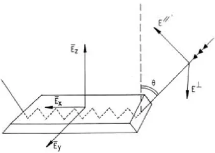
Figure 8 Schematic representation of the internal reflec-tion element (IRE) and of the light pathway. The Cartesian components of the electric field are shown along the x, y and z axes. Two possible planes of polarization of the incident light are indicated by Ek (polarization parallel to the
in-cidence plane) and E⊥(polarization
perpendic-ular to the incidence plane). The incident beam makes an angle θ with respect to the normal to the IRE surface. The edges of the IRE are beveled so that the incident beam penetrates the IRE through the surface that is perpendicular to its propagation. Adapted from [Goormaghtigh et al.,1999]. 19
Figure 9 Side view of the IRE with details of the elec-tric field of the electromagnetic radiation at one point of reflection. A standing wave exists within the IRE while the evanescent field decays expo-nentially outside the IRE. Z in µm. Adapted from [Goormaghtigh et al.,1999] 20
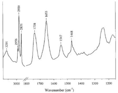
Figure 10 An example of FTIR spectrum of a membrane protein in lipids. The following peaks are ob-served: amide A at 3291 cm-1; asymmetric stretch of CH3 at 2956 cm-1; asymmetric stretch of CH2
at 2929 cm-1; symmetric stretch of CH3 at 2872
cm-1; symmetric stretch of CH2 at 2851 cm-1;
C=O stretch at 1738 cm-1; amide I region - a-helix at 1653 cm-1; amide II region -a-helix at 1549cm-1; γw(CH2) progression from 1200 cm-1 -1350cm-1; CH bend and COO-symmetric stretch 1350 cm-1 - 1500 cm-1; C-C, C-O stretch, PO2
symmetric stretch 1130 cm-1- 1180 cm-1. 22
Figure 11 2
H powder pattern spectrum arising from two transitions, eq.2.4. 24
Figure 12 15
N NMR of orienteda-helical peptides, showing the alignment of the15
N–2
H vector. The figure is a courtesy of E. Salnikov. 26
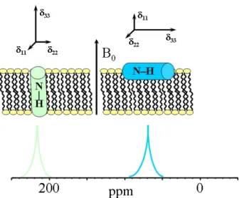
Figure 13 31
P chemical shift spectrum of pure POPC lipids in oriented sample at RT. The chemical shift around 30 ppm corresponds to lipids oriented parallel to the external magnetic field, while a very small spectral discontinuity around -18 ppm corresponds to lipids oriented perpendicu-lar to the external magnetic field. 27
Figure 14 The spin-echo pulse sequence. The behaviour of the transverse magnetization component is shown. Magnetization components dephase un-der the effects of chemical shift anisotropy or heteronuclear dipolar coupling during the first τperiod. The subsequent 180◦pulse rotates the magnetization components 180◦ about the pulse axis ( y in this case) so that the components refo-cus after a further τ period. The τ delay is chosen to be long enough to include the dead time of the probe. Then the FID can be completely recorded from the true echo maximum. 28
Figure 15 The quadrupole echo pulse sequence. 29
Figure 16 The cross polarization pulse sequence. The se-quence is designed to transfer magnetization from the abundant1
Hspins in the sample to the I spins via dipolar coupling between 1
H and I spins. Reproduced from [Duer,2004]. 30
Figure 17 Chemical structure of TentaGel resin. The figure adopted from “Rapp Polymere” website. 31
Figure 18 General scheme of SPPS. Reproduced from Nov-abiochem catalog (Merck Millipore). 33
Figure 19 Aggregation profiles of Ab(1-40) and Ab(1-40)E22G during 96 h of incubation in the presence or ab-sence of added Ca2+
were followed using West-ern blot analysis. Samples were separated using gel electrophoresis on a 12% bis-Tris gel. For each condition, samples were taken at t = 0, 2, 4, 6, 24, 72, and 96 h. Following the loading of 1 µg of protein sample into each lane, the membrane was probed with a mixture of mon-oclonal antibodies 6E10 and 4G8 that recognize residues 1-17 and 17-24, respectively. Panels A and B are representative Western blots of Ab(1-40) and Ab(1-40)E22G in phosphate buffer (“– Ca2+
condition”), respectively. Panels C and D are representative Western blots of Ab(1-40) and Ab(1-40)E22G in 2 mM Ca2+
(“+Ca2+
condition”), respectively. At least four separate experiments were carried out to confirm these results. All im-ages were taken from a single 96-h experimental procedure. 42
Figure 20 Oligomers and fibrils formation differentiated by ThT fluorescence. ThT fluorescence intensity was monitored to follow fibrillogenesis of Ab(1-40) and Ab(1-40)E22G in the presence and in the absence of 2 mM Ca2+. Black bars, Ab(1-40)
in phosphate buffer (“–Ca2+
condition”); light grey bars, Ab(1-40) in 2 mM CaCl2; dark grey
bars, Ab(1-40)E22G in phosphate buffer; light blue bars, Ab(1-40)E22G in CaCl2. Shown are
averages of values obtained in four independent experiments; error bars indicating the standard error of the average. 44
Figure 21 ATR-FTIR spectra of Ab(1-40) and Ab(1-40)E22G. FTIR spectra of Ab(1-40) and Ab(1-40)E22G were taken in the presence and in the absence of added Ca2+
, showing the amide I region of the spectra (1600–1700 cm-1). Aliquots of 2 µl were taken from each sample at t = 0, 2, 6, 24, 48, 72, and 96 h (shown in blue, green, red, cyan, pur-ple, mustard, and dark blue, respectively). The data shown here were collected in one continu-ous experiment and are representative of three independent trials. 46
Figure 22 Morphological comparison of Ab(1-40) and Ab(1-40)E22G. Contact mode AFM images (5 µm × 5 µm, Z scale 15 nm) of 40) and Ab(1-40)E22G peptides on mica, recorded either in phosphate buffer or in MOPS buffer with Ca2+
. Samples of Ab(1-40) and Ab(1-40)E22G in the presence and absence of added Ca2+
(marked as “+Ca2+
” or “ Ca2+
”, respectively) at t = 0, 6, or 72h. Closer views (1 µm × 1 µm, Z scale 15 nm) of oligomers, protofibrils and fibrils are shown as insets in the panel of t = 72 h (C, F, I, L). Im-ages A, D, G, J were taken at t = 0; imIm-ages B, E, H, K were taken at t = 6 h. Peptide concentration was the same in all samples. 49
Figure 23 IR spectra of APP_TM4K in POPC bilayers. The spectrum in red was obtained with 90◦polarized light. The spectrum in green was obtained with 0◦ polarized light. Spectrum in black is the dif-ference between the red and the green spectra.
58
Figure 24 Bis-Tris acrylamide gel electrophoresis. APP_TM in the presence of POPC lipids migrated with an apparent molecular weight of a dimer (~5.6 kDa) and APP_TM without lipids migrated with an apparent molecular weight of a monomer (~2.8 kDa). 59
Figure 25 Proton-decoupled15
N and31
P spectra of APP_TM4K labeled with15
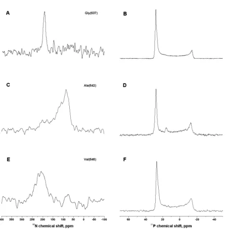
N at positions Gly637(A, B), Ala642
(C, D), Val646 (E, F) and reconstituted into
ori-ented POPC lipid membranes. Samples were measured with the membrane normal parallel to the magnetic field direction. 63
Figure 26 2
H NMR spectra of APP_TM4K peptides recon-stituted into oriented POPC lipid membranes and labeled at positions2
H-Ala-630 (A) and2
H-Ala-642 (B). Samples were measured with the membrane normal parallel to the magnetic field direction. Spectrum in panel A is a representa-tive spectrum of two peptides, APP_TM4K15
N-Ala-642 /2
H-Ala-630 and APP_TM4K15
N-Val-646/2H-Ala-630. 64
Figure 27 Contour plot that results from experimental mea-surements of the 15
N chemical shift (191 ± 7) ppm of Gly-637 in APP_TM4K peptide. x-Axis shows the rotational pitch angle around molecu-lar z-axis (along the helix long axis), while y-axis shows the rotational tilt angle, relative to the membrane normal. 65
Figure 28 Contour plot that results from experimental mea-surements of the2
H quadrupole interaction of
2
H-Ala-642 in APP_TM4K peptide. x-Axis shows the rotational pitch angle around molecular z-axis (along the helix long z-axis), while y-z-axis shows the rotational tilt angle, relative to the membrane normal. Dotted arc marks the angu-lar pairs taken for the simulation of15
N chemical shift values for Ala-642, cf. text for details. 67
Figure 29 Comparison between the experimental (A) and the simulated (B) spectra of15
N-Ala-642. 67
Figure 30 Contour plot that results from experimental mea-surements of the 15
N chemical shift (209 ± 28) ppm of Val-646 in APP_TM4K peptide. x-Axis shows the rotational pitch angle around molec-ular z-axis (along the helix long axis), while y-axis shows the rotational tilt angle, relative to the membrane normal. The grey area represents the space of possible pitch and tilt angles, while white areas are excluded from that space. 69
Figure 31 A possible model of the APP_TM4K peptide orientation and conformation in POPC bilayer, based on the results obtained in this work. 71
Figure 32 Schematic drawing of the [d7] - benzyl. 78
Figure 33 Illustration of the averaged2
H quadrupolar ten-sor due to 180º flips about the Cβ– Cγ bond.
Orientation of the averaged principal axes VxxF ,
VyyF , VzzF and the angles between them and either
of the C–D bonds are shown. Reproduced with modifications from [Schmidt-Rohr and Spiess,
Figure 34 Benzyl-carprofen molecules showing quadrupole tensor elements attached to each of the methy-lene deuterons: (A) for D1 atom the tensor
el-ements are labeled as x0, y0, and z0 (B) for D2
atom the tensor elements are labeled as x00, y00, and z00 . The molecular coordinate system is shown where z-axis is along the Cγ-Cβ bond,
x axis is out of the page plane and y axis is orthogonal to them. 79
Figure 35 Deuterium solid-state NMR spectra of sulfonyl-carprofen reconstituted into model lipid mem-branes measured at 288 K (A and B). Spectrum in panel A is of the sample oriented parallel to the magnetic field direction; spectrum in panel B is of the sample oriented perpendicular to the magnetic field direction. Panel C shows the proton-decoupled31
P chemical shift spectrum, indicating the orientation of the lipids mem-branes in the sample. 81
Figure 36 Alignments of sulfonyl-carprofen in oriented lipid membrane that agree with the experimen-tal data. Only the orientations corresponding to θ = ± 27.4◦ are shown. Orientations corre-sponding to θ = ± 152.6◦ are not represented as these angles are complementary to 180◦and are essentially the same. The structure of sulfonyl-carprofen was produced using ChemDraw3D software (CambridgeSoft, PerkinElmer Informat-ics) implementing MM2 energy minimization. It was then superimposed on the fluid phase POPC-only lipid bilayer generated by molecular dynamics simulation [Heller et al., 1993].
Pan-els A and B show the molecule oriented with different pitch angles. 82
Figure 37 Deuterium solid-state NMR spectra of benzyl-carprofen reconstituted into model lipid mem-branes recorded at 288 K (panels A and B). Spec-trum in panel A is of the sample oriented paral-lel to the magnetic field direction; spectrum in panel B is of the sample oriented perpendicular to the magnetic field direction. Panel C shows a proton-decoupled31
P ssNMR spectrum, indicat-ing the alignment of the lipid membranes in the sample. 83
Figure 38 Contour plot depicting all spatial orientations of benzyl-carprofen, represented by the tilt and the pitch angles. The red lines trace angular pairs that agree with the experimental2H
quadrupo-lar splitting of 46 ± 5 kHz, the dark blue lines are in agreement with the2H quadrupolar
split-ting of 66 ± 3 kHz, the green and the light blue lines are in agreement with the2H quadrupolar splitting of 20 ± 10 kHz. The circled cross section shows combinations of values that agree with the experimental results (where the constraints from all the segments meet in one point). 86
Figure 39 Simulated static 2H NMR spectra of
benzyl-carprofen deuterons in the tilted (90◦) sample. The red line shape corresponds to reduced ten-sor of four benzyl ring deuterons; the grey line shape corresponds to single deuteron in position ζ of the benzyl ring; the blue line shape corre-sponds to methylene deuterons. Simulation was done by Dr. Salnikov E. 88
Figure 40 Alignment of benzyl-carprofen in oriented lipid membrane, corresponding to the spatial orien-tation shown in the contour plot. The structure of benzyl-carprofen was produced using Chem-Draw3D software (CambridgeSoft, PerkinElmer Informatics) implementing MM2 energy mini-mization. It was then superimposed on the fluid phase POPC-only lipid bilayer generated by molec-ular dynamics simulation [Heller et al.,1993]. 89
Figure 41 Potential interplay between Ab oligomers, Ca2+
, and a target cell in the initial stages of Alzheimer’s disease. (1) Age-related increase in [Ca2+
]i
pro-motes oligomerization of intracellular Ab. (2) Disruption of Ca2+
homeostasis by oligomers, by either binding to or modulating the activity of a number of receptors such as ryanodine (Ry) and inositol triphosphate (IP3R) [Stutzmann,2005].
(3) Increase in [Ca2+
]i. These three steps might
form an inimical cycle leading to increases in both cytosolic calcium and Ab oligomer con-centrations. (4) Ab oligomers disrupt intracel-lular membranes, leading to apoptosis [ Kawa-hara et al.,2000, Arispe et al.,1993a,b]. (5)
Ex-tracellular calcium concentration ([Ca2+
]e)
pro-motes oligomerization of extracellular Ab. (6) Oligomers form nonspecific pores in the plasma membrane, disturbing cellular integrity and lead-ing to apoptosis [Bucciantini et al., 2004]. (7)
Ab oligomers can interact and impair calcium channels at the membrane surface, opening cal-cium importers and blocking calcal-cium exporters such as the voltage-dependent calcium chan-nel [Rovira et al., 2002]. Ab oligomers can
af-fect surface expression of N-methyl-D-aspartate receptors (NMDARs) [Dewachter et al., 2009],
may increase [Molnár et al., 2004] or decrease
the conductance [Shankar et al., 2007], and
fa-cilitate long-term synaptic depression by dis-rupting neuronal glutamate uptake [Li et al.,
2009]. 97
Figure 42 Specific isotopic labels on glutamic acid and ly-sine amino acids destined for peptide synthe-sis. 125
Figure 43 Mass spectrometric analysis of the crude syn-thetic unlabeled Aβ peptides. (a) Aβ(1-40) pep-tide with expected mass of 4329.8, (b) Aβ(1-40)E22G peptide with expected mass of 4257.8. 127
Figure 44 Mass spectrometric analysis of the crude labeled Aβ(1-40) peptides with expected mass of 4329.8. Here the apparent mass is 4399.3. 128
Figure 45 HPLC purification of Aβ peptides - elution pro-files: (a) Aβ(1-40) peptides eluted in two ma-jor peaks, (b) Aβ(1-40)E22G peptides eluted in one major peak and another smaller peak. For each peptide, two peaks were collected and ana-lyzed. 128
Figure 46 HPLC purification of labeled Aβ(1-40) peptides - elution profile. Two main peaks, each split into three smaller peaks. 129
Figure 47 Aggregation trial for Aβ(1-40) and Aβ(1-40)E22G in PB and MOPS buffers: 0h, 2h and 24h sam-ples: (a) Aβ(1-40) after 0h and 2h of incubation, (b) Aβ(1-40) and Aβ(1-40)E22G after 24h of in-cubation. 129
Figure 48 Aggregation trial for Aβ(1-40) crude preparation (peptide before purification), peak I and peak II at different temperatures in PB and MOPS buffers: representative 0h and 168h samples. (a) Aβ(1-40) at time 0h, crude peptide, peak I, peak II, (b) Aβ(1-40) crude peptide, peak I, peak II after incubation at 4◦C for 168h, (c) Aβ(1-40) crude peptide, peak I, peak II after incubation at 37◦C for 168h. 131
Figure 49 New buffer conditions for oligomers of Aβ(1-40) growth. The color coding is as follows: light blue is the MOPS buffer condition; dark blue is the salt condition - NaKCaMg. 132
Figure 50 Spin-filter separation trials of 40) and Aβ(1-40)E22G oligomers grown in high-salt condi-tions. 133
Figure 51 Effect of salts on aggregation of labeled and non-labeled Aβ(1-40) and Aβ(1-40)E22G peptides - in preparation for ssNMR experiments: (a) aggre-gation profiles of Aβ(1-40) and of Aβ(1-40)E22G at t = 0 h, (b) aggregation profiles of Aβ(1-40) and of Aβ(1-40)E22G at t = 72 h, (c) aggregation profiles of isotopically labeled Aβ(1-40) at t = 0 h, (d) aggregation profiles of isotopically labeled Aβ(1-40) at t = 72 h. 135
Figure 52 On-column oligomerization of Aβ(1-40). FPLC elution profile and its consequent PAGE analysis.
Figure 53 Oligomers separation using centrifugal concen-tration units, comparison of different brands. The order of the samples is as follows from right to left: Aβ(1-40) after elution from FPLC, con-centrate and a flow-though from “Centricon”, concentrate and flow-through from “Amicon ul-tra” 30 K, concentrate and flow-though from “Vivaspin”, and concentrate and flow-through from “Macrosep”. 138
Figure 54 NMR spectrum of Aβ(1-40) in phosphate buffer. (a) full spectrum, (b) closeup into the aromatic and amine regions. 140
Figure 55 NMR spectrum of Aβ(1-40) in phosphate buffer. (c) closeup into the backbone amide region, (d) closeup into the aliphatic region. 141
Figure 56 Aβ(1-40)13C natural abundance, MAS NMR at RT. 143
Figure 57 13C and 15N chemical sifts from CP
experi-ments of Aβ(1-40) oligomers grown in MOPS buffer. 144
Figure 58 13C and15N chemical shift CP spectra of pure
synthetic Aβ(1-40) peptide labeled at Glu22 and Lys28. 146
Figure 59 13C CP of Aβ(1-40) oligomers from PB (black
trace) and purified dry powder of Aβ(1-40) (red trace), measured at 244◦K. 147
Figure 60 Effect of 60 % D2O on aggregation profiles of
1mM Aβ40 and Aβ40E22G in MOPS or MOPS + 2mM Ca2+(a) sample at 0 h, (b) sample at 4 h, (c) sample at 24 h, (d) sample at 48 h, (e) sample at 72 h. 148
Figure 61 Results from SANS experiment arranged in a table. 150
Figure 62 Results from SANS experiment arranged in a table. Continue. 151
Figure 63 Results from SANS experiment arranged in a table. Continue. 152
Figure 64 Results from SANS experiment arranged in a table. Continue. 153
lipid vesicles mixture, which include H-labeled POPE: (a) effect of Aβ(1-40) on POPE lipids as observed through quadrupolar splittings of2H
POPE incorporated in lipid vesicles consistent of POPC, POPE, cholesterol; (b) effect of cholesterol on POPE lipids as observed through quadrupo-lar splittings of2H POPE incorporated in lipid vesicles consistent of POPC, POPE with 3 % Aβ(1-40); (c) effect of the Aβ(1-40) on lipids mea-sured via d31POPC or via d31POPE. 155
Figure 66 Temperature effect on the order parameter. POPC, deuterium labeled d31POPE and cholesterol vesi-cles with 1% or 3 % of Aβ(1-40) peptide or with-out were measured either at 298◦K or at 310◦K. The legend inside the figure specifies the color coding. 156
Figure 67 Comparison of the order parameter between different lipid vesicles compositions, monitor-ing quadrupolar splittmonitor-ings of either d31POPC or d31POPE. The color coding is as follows: black rectangles - d31POPC only; red stars - d31POPC with 3 % Aβ; black triangles - d31POPC, POPE; red snowflakes - d31POPC, POPE, 3 % Aβ(1-40); black circles POPC, d31POPE; red circles -d31POPE, POPC, 3 % Aβ(1-40); black and white circles - POPC, d31POPE, cholesterol; blue 4-x symbols - POPC, d31POPE, cholesterol, 3 % Aβ(1-40); black plus-in-a-circle symbols - d31POPC, POPE, cholesterol; empty grey squares - d31POPC, POPE, cholesterol, 3 % Aβ(1-40). 157
Figure 68 Effect of POPS and of Aβ (1-40) peptides in the presence of sphingomyelin and varying temper-atures on the order parameter of POPE. (a) the order parameter of POPC lipids in the presence of POPS and Aβ(1-40); (b) the effect of the tem-perature and the amount of Aβ (1-40) peptides on the order parameter in the presence of SM (all vesicles have the same composition). The leg-end inside the figure specifies the color coding.
158
N O M E N C L AT U R E
[Ca2+]i intracellular Ca2+ concentration
α − CT F alpha C-terminal fragments β − CT F beta C-terminal fragment Aβ amyloid beta-peptide ACN Acetonitril
AD Alzheimer’s disease
ADHH adiabatic passage through Hartman-Hahn condition AFM Atomic force microscopy
AICD APP intracellular domain Aph − 1 anterior pharynx-defective 1 APP Amyloid precursor protein APS Ammonium persulfate BSA Bovine serum albumin CaBPs calcium-binding proteins cmc critical micelle concentration
CODEX Center-band Only Detection of Exchange COX cyclooxygenase
CP Cross polarization DCM dichloromethane DMF dimethylformamide EDT Ethanedithiol
EDT A Ethylenediaminetetraacetic acid ER Endoplasmic reticulum
FAD Familial Alzheimer’s disease FID free induction decay
FT IR Fourier transform infrared GdnCl guanidine hydrochloride
HBT U Benzotriazole tetramethyl uronium hexafluoro phosphate HH Hartman-Hahn
HPLC high performance liquid chromatography IP3 Inositol trisphosphate
JM juxtamembrane
LMPG lyso-myristoylphosphatidylglycerol LT P Long-term potentiation
MALDI matrix assisted laser desorption or ionization MALDI matrix assisted laser desorption or ionization MD molecular dynamics
MOPS 3-(N-morpholino)propanesulfonic acid nAChR nicotinic acetylcholine receptor Nct Nicastrin
NMDA N-methyl-D-aspartate NMR nuclear magnetic resonance
NSAID nonsteroidal anti-inflammaroy drug PAGE Polyacrylamide gel electrophoresis Pen − 2 presenilin enhancer gene 2
POPC 1-palmitoyl-2-oleoyl-sn-glycero-3-phosphocholine PS1 Presenilin 1
PS2 Presenilin 2
REDOR Rotational Echo DOble Resonance RF radio frequency
Ry Ryanodine SAP serum amyloid P SDS Sodium dodecyl sulfate SPPS solid phase peptide synthesis
ssNMR solid-state nuclear magnetic resonance T EMED N, N, N’, N’-tetramethylethylenediamine T FA Trifluoroacetic acid T hT Thioflavine T T IS Triisopropylsilan T M transmembrane (domain) T OF time of flight
1
I N T R O D U C T I O N
1.1 a m y l o i d p r e c u r s o r p r o t e i n
Alzheimer’s disease (AD) is a progressive neurodegenerative disorder that affects nearly 2% of the population in industrialized countries. AD is the most common form of dementia and is characterized by brain cell destruction, memory loss, and deterioration of cognitive and behavioral processes severe enough to affect work, lifelong hobbies, and social life. Symptoms worsen over time and the disease is fatal. One of the characteristic histopathological markers of AD is the pres-ence of proteinaceous deposits, amyloid plaques, in the brain. These plaques are formed by amyloidb-peptides (Ab) 40- and 42-residue-long, which are protease cleavage products of the amyloid precursor protein (APP).
APP is an integral membrane protein with a single transmembrane (TM) domain expressed in many tissues and concentrated in the synapses of neurons [Lambert et al., 1998]. Its primary function is
not known, though it seems not to be required for the expression of a critical cell function but, may be involved in the modulation of neuronal functions at the cellular level. It has been shown that in brain a distinct percentage of APP is present on the cell surface as a membrane protein of type I (i.e. it has a single-pass transmembrane domain, with the portion of the polypeptide on the NH2-terminal
side of the TM domain exposed on the exterior side of the membrane and the COOH-terminal portion exposed on the cytoplasmic side). Involvement of APP in neuronal development, synaptogenesis, and synaptic plasticity indicated that some aspects of these processes are mediated by cell-associated APP [Nishimoto,1998,Perez et al.,1997, Weidemann et al., 1989]. APP is best known and most commonly
studied as the precursor molecule whose proteolysis generates Ab peptides, a 38- to 42-amino acid-long peptides whose amyloid fibrillar form is the primary component of amyloid plaques found in the brains of AD patients (described in more details in the next section).
APP undergoes sequential cleavage (Figure1on page4). First, the
bulk ectodomain is removed by membrane-bound a- or b-secretases, leading to secreted forms of APP, sAPPa or sAPPb respectively, and a membrane-bound a-C-terminal fragment (a-CTF) or b-C-terminal fragment (b-CTF), respectively [Annaert and De Strooper,2002,Vassar et al., 1999]. Regulated transmembrane proteolysis of the b-CTF by
g-secretase occurs after ectodomain shedding and results in Ab pro-duction [De Strooper and Annaert,2000].g-Secretase cleavage also
Figure 1: Schematic diagram illustrating proteolytic cleavage of the APP. α-secretase cleaves APP within the Aβ sequence (non-amyloidogenic pathway) to liberate two peptides, including the neuroprotective sAPPα. β- and γ-secretases act sequentially to cleave APP within the N- and C-terminal parts of the Aβ sequence, respectively producing Aβ peptide (amyloidogenic pathway) and AICD.
releases the APP intracellular domain (AICD), which could have a role in transcriptional regulation (Figure1on page4).g-Secretase is a
multiprotein complex composed of presenilin 1 (PS1) or presenilin 2 (PS2); nicastrin (Nct), a typeI transmembrane glycoprotein; anterior pharynx-defective 1 (Aph-1) and presenilin enhancer gene-2 (Pen-2), two multipass transmembrane proteins [Bergmans and De Strooper,
2010]. This complex is essential for the sequential intramembraneous
proteolysis of a variety of transmembrane proteins. The functions of the variousg-secretase proteins and their interactions in the complex are not yet fully defined, but it has been suggested that the ectodomain of nicastrin recognizes and binds to the amino-terminal stubs of previ-ously cleaved transmembrane proteins. Aph-1 aids the formation of a precomplex, which interacts with PS1 or PS2 while Pen-2 enters the complex [Edbauer et al.,2003,Go et al.,2004] to initiate the cleavage of
PS1 or PS2 to form an N-terminal 28-kDa fragment and a C-terminal 18-kDa fragment, both of which are critical to theg-secretase complex [Takasugi et al.,2003].
Recently, an explanation for the multiple cleavages byg-secretase was provided, indicating a sequential proteolytic cleavage mechanism to release Ab [Qi-Takahara et al.,2005,Zhao et al.,2004]. Accordingly,
the first cut occurs at the cytoplasmic edge of the TM at thee-site, that is, residue 49 or 48 ofb-CTF (Figure2on page5) [Sastre et al.,2001, Weidemann et al.,2002]. The products Ab49/Ab48 remain membrane
Figure 2: Partial sequence of the APP where the TM domain is enclosed by dashed lines. The red rectangle marks the region synthesized for this study. Underlined are isotopically-labeled amino acids that were incorporated for solid-state nuclear magnetic resonance (ss-NMR) studies. The β-branched amino acids are marked in blue. The positions of the glycines in the 3 consecutive GxxxG are pre-sented in bold and labeled. The cleavage sites of α-, β-, γ-secretase are indicated with scissors.
that is, the z-site [Zhao et al.,2004]. Ab46 is further processed into
Ab43 and finally Ab40, whereas Ab45 is the direct precursor of Ab42 [Zhao et al., 2004, Qi-Takahara et al., 2005]. There is evidence that
cellular APP exists as a homodimer, which promotes or attenuates the Ab production in the b-secretory pathway [Scheuermann et al.,2001].
Therefore, further cleavage that generates Ab38 and Ab37 will occur only if not inhibited by strong dimerization between two TM helices of APP [Munter et al.,2007,Miyashita et al.,2009,Munter et al.,2010].
There are evidence that suggest that intramembraneous proteoly-sis by g-secretase requires local unraveling of the helical secondary structure of the TM domain to expose backbone carbonyl carbons for nucleophilic attack by polarized water in the enzyme active site [Sato et al., 2009]. Indeed, there are some unusual features of the
APP_TM sequence that might destabilize its helical structure in the membrane. First, there is a high density ofb-branched amino acids (Va-line, Threonine, Isoleucine) surrounding the Ab40- and Ab42-cleavage sites (Figure 2on page 5). Sequentialb-branched residues can have a
destabilizing effect on the secondary structure of TM helices [Li and Deber,1992].
Second, several of the glycines in the APP_TM domain occur in GxxxG motifs, which in TM sequences were shown to mediate he-lix dimerization [Russ and Engelman,2000]. Mutational studies also
show the importance of glycines in GxxxG motifs for APP process-ing [Munter et al., 2007, Kienlen-Campard et al., 2008]. Molecular
dynamics and solid-state NMR (ssNMR) experiments point out that Gly625, Gly629, and Gly633 (Figure2on page5) within GxxxG motifs
mediate TM helix homodimerization, and that the TM helix breaks at the transition point near thee-cut site [Sato et al.,2009,Miyashita et al., 2009]. There is ssNMR data that indicates that helical conformations
coexist with non-helical conformations within the APP_TM domain [Lu et al.,2011]. These suggest that the conformational variability of
the TM segment might play a role in APP processing and therefore in Ab production [Antzutkin et al.,2003,Munter et al.,2007], for example