HAL Id: hal-03089154
https://hal.archives-ouvertes.fr/hal-03089154
Submitted on 28 Dec 2020
HAL is a multi-disciplinary open access
archive for the deposit and dissemination of
sci-entific research documents, whether they are
pub-lished or not. The documents may come from
teaching and research institutions in France or
abroad, or from public or private research centers.
L’archive ouverte pluridisciplinaire HAL, est
destinée au dépôt et à la diffusion de documents
scientifiques de niveau recherche, publiés ou non,
émanant des établissements d’enseignement et de
recherche français ou étrangers, des laboratoires
publics ou privés.
Picometer Resolution Structure of the Coordination
Sphere in the Metal-Binding Site in a Metalloprotein by
NMR
Andrea Bertarello, Ladislav Benda, Kevin Sanders, Andrew Pell, Michael
Knight, Vladimir Pelmenschikov, Leonardo Gonnelli, Isabella Felli, Martin
Kaupp, Lyndon Emsley, et al.
To cite this version:
Andrea Bertarello, Ladislav Benda, Kevin Sanders, Andrew Pell, Michael Knight, et al..
Picome-ter Resolution Structure of the Coordination Sphere in the Metal-Binding Site in a Metalloprotein
by NMR. Journal of the American Chemical Society, American Chemical Society, 2020, 142 (39),
pp.16757-16765. �10.1021/jacs.0c07339�. �hal-03089154�
1
Pico-meter resolution structure of the coordination sphere in the
metal-binding site in a metalloprotein by NMR
Andrea Bertarello,
1,4,‡
Ladislav Benda,
1,‡
Kevin J. Sanders,
1,$
Andrew J. Pell,
1,¥
Michael J. Knight,
1
Vladimir Pelmenschikov,
2
Leonardo Gonnelli,
3
Isabella C. Felli,
3
Martin Kaupp,
2
Lyndon Emsley,
4,*
Roberta Pierattelli,
3,*
Guido Pintacuda
1,*
1
Université de Lyon, Centre de RMN à Très Hauts Champs, FRE 2034 CNRS/Université Claude Bernard Lyon 1/ENS Lyon,
5 rue de la Doua, 69100 Villeurbanne, France ;
2Technische Universität Berlin, Institut für Chemie, Straße des 17 Juni 135,
10623 Berlin, Germany;
3University of Florence, Department of Chemistry and Magnetic Resonance Center (CERM), Via L.
Sacconi 6, 50019 Sesto Fiorentino, Italy;
4École Polytechnique Fédérale de Lausanne (EPFL), Institut des Sciences et Ingé-nierie Chimiques, CH-1015 Lausanne, Switzerland
Supporting Information Placeholder
ABSTRACT:
Most of our understanding of chemistry derives from atomic-level structures obtained with single crystal X-ray diffrac-tion. Metal centers in X-ray structures of small organometallic or coordination complexes are often extremely well defined, with
errors in the positions on the order of 10
-4-10
-5Å. Determining the metal coordination geometry to high accuracy is essential for
understanding metal center reactivity, as even small structural changes can dramatically alter the metal activity. In contrast, the
resolution of X-ray structures in proteins is limited typically to the order of 10
-1Å. This resolution is often not sufficient to develop
precise structure-activity relations for the metal sites in proteins, since the uncertainty in positions can cover all the known ranges
of bond-lengths and bond-angles for a given type of metal-complex. Here we introduce a new approach that enables determination
of a high-definition structure of the active site of a metalloprotein from a powder sample, by combining magic-angle spinning (MAS)
nuclear magnetic resonance (NMR) spectroscopy, tailored radio-frequency (RF) irradiation schemes, and computational
ap-proaches. This allows us to overcome the “blind sphere” in paramagnetic proteins, and to observe and assign
1H,
13C, and
15N reso-
nances for the ligands directly coordinating the metal center. We illustrate the method by determining the bond lengths in the struc-ture of the Co
IIcoordination sphere at the core of human superoxide dismutase 1 (SOD) with 0.7 pm precision. The coordination
geometry of the resulting structure explains the non-reactive nature of the Co
II/Zn
IIcenters in these proteins, that allows them to
play a purely structural role.
INTRODUCTION
Metal ions play an important role in many processes at the core
of modern chemistry, biochemistry, and materials research,
and the accurate description of their coordination environment
is essential for understanding the function of organometallic
catalysts and metalloenzymes.
1,2The capacity of single crystal
X-ray diffraction
3to determine atomic positions to within 10
-4-10
-5Å has enabled the development of today’s structure-activ-ity based approaches to chemistry.
4Indeed, high accuracy is es-sential for understanding metal center reactivity, as even small
structural changes can dramatically alter the metal activity.
5However, in proteins the resolution of crystal structures is lim-ited typically to the order of 10
-1Å,
2,6,7mostly by the need for
highly ordered crystalline samples, and this is often not suffi-cient to develop precise structure-activity relations for the
metal sites in proteins,
8since the uncertainty in positions can
cover all the known ranges of bond-lengths and bond-angles
for a given type of metal-complex. Metal-ligand coordination
distances can be refined with X-ray absorption techniques but
these approaches share many of the limitations of X-ray diffrac-tion.
9Nuclear Magnetic Resonance (NMR) spectroscopy is a direct
probe for the electronic structure and coordination geometry
of metal ion complexes in the presence of paramagnetism orig-inating from unpaired electrons, which is a common feature of
many transition metal ions of biological relevance in metallo-proteins. Paramagnetic metal ions induce peculiar effects on
the NMR signals of the surrounding nuclei, with a well-defined
dependence on the electronic configuration and coordination
environment of a metal center. This results in long-range per-turbations such as hyperfine shifts and NMR relaxation
en-hancements, which are routinely used as structural probes for
proteins in both solution
10,11and solid state.
12-14When it comes
to the proximity of a metal, however, these paramagnetic ef-
fects are the strongest, which has to date hindered the acquisi-tion of NMR signals in the “blind sphere” around the metal ion.
MAS solid-state NMR is particularly well suited to circumvent
the main barriers for the acquisition of paramagnetic signals in
solution, since Curie relaxation is absent in solids,
15and
nu-clear spin coherences can be transferred efficiently via strong
dipolar couplings. However, in paramagnetic solids, additional
effects broaden NMR signals and spread them over very large
spectral windows. Recently, we and others have shown that
these drawbacks can be significantly reduced with fast (60 kHz
and above) MAS and high-power radio-frequency pulses,
16,17opening the way to the detection of significantly
hyperfine-shifted and broadened resonances in large biomolecules.
18,192
Here, we show that it is possible to obtain data from the “blind
sphere” of a paramagnetic Co-containing metalloprotein by
combining magic-angle spinning (MAS) NMR at 100 kHz MAS
rate, with tailored radio-frequency (RF) irradiation schemes
and modern computational approaches. We determine the
high-resolution structure of the metal coordination sphere
(Fig. 1) in the core of the thermostable mutant of human super-oxide dismutase 1 in microcrystalline form containing a Co
IIion
(CoSOD), by measuring paramagnetic NMR effects for the
1H,
13C, and
15N nuclei of the Co
IIligands. This leads to pico-meter
resolution in the precision of the bond-lengths and ±1° resolu-tion of bond-angles in the metal center, and we show that the
coordination geometry of the resulting structure is precise
enough to explain the non-reactive nature of the Co
II/Zn
IIcen-ters in these proteins.
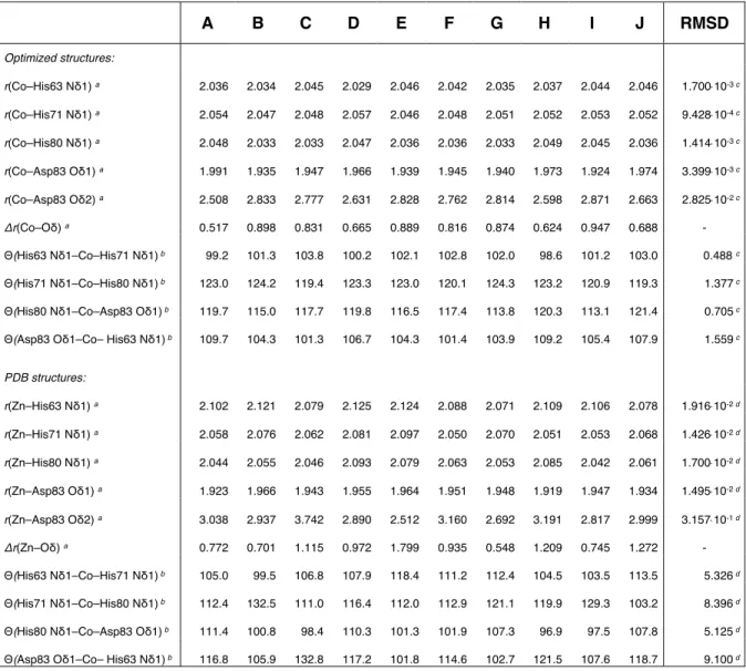
Figure 1. Structure determination of the Co
IIsite in CoSOD. (A)
Overlay of Zn
IIsite in ten protein chains of the single-crystal X-
ray structure of SOD 1 (PDB code 1SOS), illustrating the crys-tallographic uncertainty in the metal coordination geometry
and (B) the NMR ensemble of structures of the Co
IIcomplex of
CoSOD. Schematic representation of the metal site (C) in the X-ray ensemble and (D) in the NMR ensemble, together with the
RMSDs of the metal-ligand bond lengths and ligand-metal-lig-and angles RMSDs.
RESULTS AND DISCUSSION
Fast MAS rates have the multiple advantage of concentrating
the signal into fewer spinning sidebands, of improving dipolar
decoupling, with a benefit in sensitivity, and of removing signal
overlap, with a benefit in resolution. As illustrated in Fig. 2A,
tailored RF schemes (Fig. S1) at 100 kHz MAS enable the acqui-sition of well-resolved
1H spectra, featuring resonances span-ning a range of about one hundred ppm. Notably, several very
broad resonances above 60 ppm and below 0 ppm which es-caped detection in all previous attempts at lower MAS rates and
in solution
20become observable. Improved dipolar decoupling
results in longer lifetimes for
1H and
13C coherences, which in
turn allows the acquisition of 2D Transferred Echo Double Res-onance (TEDOR)
21,22experiments (Fig. S1) that correlate the
shifts for pairs of nearby
1H,
13C or
1H,
15N nuclei over windows
up to more than a thousand ppm (Fig. 2B-C). The addition of a
spin magnetization exchange
23between
1H nuclei close in
space extends the correlations to more distant
1H.
Once the paramagnetic NMR shift tensors are acquired, a relia-ble assignment protocol is needed. For diamagnetic proteins
this is a well-established procedure relying on the fact that
atomic nuclei in given structural environments display well-de-fined ranges of chemical shifts. However, strong paramagnetic
interactions alter the observed shifts in a manner that is not
empirically predictable and the use of theoretical modeling is
of paramount importance. Only recently the theory of
para-magnetic NMR shift tensors
24has been developed to enable
their rigorous calculation from first principles of quantum me-chanics.
25,26Figure 2. NMR spectra of CoSOD. NMR spectra of CoSOD ac-quired at 100 kHz MAS ~280 K on a 500 MHz spectrometer
(11.7 T). (A)
1H spectrum (spin echo), (B-C) 2D
1H,
13C and
1H,
15N TEDOR spectra, acquired without (black) and with (ma-genta) spin magnetization exchange
23between
1H nuclei close
in space.
We used this approach to predict the NMR shift tensors of the
1
H,
13C, and
15N nuclei in the Co
II-binding complex of CoSOD. As
a starting structural seed, we adopted an ensemble of ten mod-els A–J (Fig.1 and Fig. S2) obtained from an X-ray structure of
SOD 1.
27The main factor limiting the accuracy of predicted par-amagnetic NMR shifts is the density functional theory (DFT)
calculation of hyperfine coupling tensors. As there is currently
no universally preferred density functional for hyperfine cou-pling,
28we adopted a previously validated approach
29and cal-culated the expected bounds of hyperfine couplings with PBE0
and PBE50 hybrid DFT functionals. The resulting intervals of
calculated NMR shifts were then compared to the experimental
data, leading to two important outcomes. First, a complete as-signment was achieved for all observed
1H,
13C, and
15N reso-nances, thus enhancing the so far limited set of resonances at
the core of CoSOD.
20Second, by selecting the models for which
the agreement between experiments and calculations is satis-
factory for all observed resonances, the structure of the metal-3
binding complex was refined as illustrated in Fig. 3. Despite a
considerable uncertainty associated with the calculations, the
even larger dispersion of paramagnetic shifts guarantees that
this procedure has a structural discriminating power. Out of
the ten models A–J, only three (C, E, F) satisfied the agreement
criteria with the NMR correlation spectra. The resulting NMR
structural ensemble (Fig. 1B) has a significantly better preci-sion at the metal site than the original X-ray-based ensemble.
The overall RMSD of non-hydrogen atoms of metal-binding
amino-acid side chains improved from 0.16 Å for the X-ray
structural ensemble to 0.09 Å for the refined ensemble, while
the RMSDs for the metal-ligand bond lengths and ligand-metal-ligand angles improves by one order of magnitude (Fig. 1C-D,
Table S1) to an average of 0.7 pm and ±1° respectively.
Figure 3. Comparison between experimental and calculated
NMR correlations. Experimental 2D
1H,
13C (top) and
1H,
15N
(bottom) TEDOR spectra and the calculated areas in which we
expect correlations for model A (left, blue boxes) and model F
(right, red boxes). Model F is in agreement with the data, while
Model A is not, and can be excluded.
With a reliable structure of the Co
II-binding complex at hand,
we can directly interpret the measured NMR shift pattern in
terms of local structure of the metal site. The Co
IIion is pseudo-penta-coordinated with short bond lengths to His63 N
δ1, His71
N
δ1, His80 N
δ1, and Asp83 O
δ1, and a longer distance to Asp83
O
δ2(Fig. 1, Table S1). We found that certain NMR shifts are par-ticularly sensitive to the mode of Asp83 binding to the metal,
most notably the Asp83 C
βshift observed at 350 ppm. The
Asp83 C
βshift values calculated for the models A–J span a wide
range and there is a clear correlation with the coordination dis-tances Co–O
δ1(r
1) and Co–O
δ2(r
2) (Fig. 4 A-B). The
experi-mental Asp83 C
βshift constrains the distances r
1and r
2to the
ranges 1.94–1.96 Å and 2.68–2.82 Å, respectively. Both coordi-nation distances are determined with a substantially better
precision here than was available in the original X-ray ensem-ble (Fig. 4 C, Table S1), especially for the r
2distance where the
variation is reduced from 1.23 Å to 0.14 Å. The variation is fur-ther reduced to 0.07 Å if we consider the distances spanned by
the three structural models.
The relevance of such refinement is illustrated in a larger per-
spective in Fig. 4C, where the distribution of the distances ob-
tained before and after the paramagnetic NMR structure deter-
mination is compared with all the Co complexes in the Cam-bridge Structural Database (CSD) featuring the same Co pattern
as in SOD. The original SOD ensemble covers a large portion of
the possible combinations of r
1and r
2values (369 structural
motifs), and thus a correspondingly large part of the chemical
activity space for Co
IIcomplexes, while the NMR refined inter-val discriminates a much smaller subset of 29 structural motifs
(if we consider the constraints obtained from Fig. 4A-B), and an
even smaller subset of only 2 structures if we consider the
range delimited by the three selected structures (C,E,F). These
subsets of structures significantly reduce the space of chemical
properties.
Figure 4. Dependence of Asp83 C
βNMR shift on the active site
structure. (A-B) Calculated NMR shifts for the ten models A–J
using PBE0 and PBE50 functionals for the hyperfine coupling,
compared to the experimentally observed Asp83 C
βshift, plot-ted as a function of (A) r
1and (B) r
2distances. The red lines
(parabolic fit of the calculated data) enclose the area inside
which the experimental shift is expected. The actual observed
shift is indicated by the black dashed line, which by intersecting
the red areas determines the r
1and r
2ranges compatible with
the NMR data. (C) Distribution and average values of r
1and r
2values for all (911) tetra- (NC=4) and penta- (NC=5)
400 600 300 500 700 1100 δ(13C) ppm δ(15N) ppm 40 50 60 δ(1H)/ppm 1200 1300 200 30 20 Asp83 Hβ2-Cβ Hβ1-CβAsp83 His71 Hδ2-Cδ2 His80 Hδ2-Cδ2 His63 Hδ2-Cδ2 His80 Hε2-Nε2 His71 Hε2-Nε2 400 600 300 500 700 1100 δ(13C) ppm δ(15N) ppm 40 50 δ(1H)/ppm 1200 1300 200 30 20 Asp83 Hβ2-Cβ Hβ1-CβAsp83 His71 Hδ2-Cδ2 His80 Hδ2-Cδ2 His63 Hδ2-Cδ2 His80 Hε2-Nε2 His71 Hε2-Nε2 s71 2 His71 60 Model A Model F
4
coordinated Co structural motifs with at least one carboxylate
moiety as deposited in the CSD, compared to the uncertainty of
r
1and r
2in CoSOD in X-ray (light magenta area, containing 368
structural motifs), and after paramagnetic NMR refinement
(blue area, containing only 29 structural motifs). Average val-ues r
1avand r
2avover all tetra-coordinated CSD complexes are
shown with solid lines and the analogous values for penta-co-ordinated CSD complexes are shown with dashed lines.
This can be of primary importance since the structural param-eters are correlated to chemical properties of a species, as is e.g.
the case of Zn binding proteins, where the metal-ligand
dis-tances are often indicators of a catalytic or structural role of the
metal in the protein.
30The uncertainty in the geometry from
the X-ray ensemble is too large to distinguish these types. The
refined bond lengths and bond angles we find are consistent
with unreactive, structural, Zn atoms. We point out that the
structural resolution we discuss here is related to the precision
of the measurement. Since there are no other independent de-
terminations with this level of resolution, it is not straightfor-ward to assess the accuracy of the determination. That said, the
comparison between the r
1and r
2ranges obtained for SOD here
to those observed for all the Co
IIcomplexes in Fig.4
demon-strates consistency: the 1.94-1.96 Å range for r
1compares very
well with the average value of 1.968 Å obtained for tetracoor-dinated Co
IIcomplexes, and the range 2.68-2.82 Å observed for
r
2lies exactly in between the average values observed for
tetracoordinated and pentacoordinated complexes, which is in
agreement with the pseudo-pentacoordinated nature of Co in
SOD. Moreover, the r
1and r
2ranges obtained from the present
refinement compare well with those of some biomimetic Co
and Zn complexes (the BIYHUM and BIYJAU entries in the CSD),
which display a similar coordination sphere to that in SOD.
31In
particular, in the structure corresponding to the BIYHUM entry,
the r
1and r
2distances are equal to 1.97 and 2.79 Å respectively,
comparing almost perfectly with our results, and supporting
the coordination geometry. This also validates the applicability
in the present case of the metal substitution strategy for the
study of metal binding sites in metalloproteins.
CONCLUSION
The understanding of the function of a metal center in a metal-loprotein is intimately related to the structural environment of
the metal in the protein framework, as small structural changes
can alter dramatically the metal center activity. The difficulty
of obtaining highly resolved structures of metal sites in
pro-teins often represents the bottleneck for the development of
precise structure-activity relations for metal sites in proteins.
We have introduced a method to determine with pico-meter
precision the structure of the coordination sphere of a
para-magnetic metal ion in a metalloprotein via the measurement
and calculation of paramagnetic NMR shifts. Detection and as-signment of NMR resonances is enabled by state-of-the-art
methodology including 100 kHz MAS, tailored radiofrequency
irradiation schemes, and advanced quantum chemistry model-ing. The method has been applied to the CoSOD metalloprotein,
where it resulted in 0.7 pm precision in the resolution of the
Co
IIcoordination sphere. The overall RMSD for all the heavy at-oms in the ligands is 9 pm. In particular, the refined structure
is accurate enough to be able to correlate the coordination ge-ometry with the unreactive, structural, nature of the Zn center
in SOD. Our approach provides a direct relationship between
metal–ligand distances and paramagnetic NMR shifts.
With ongoing progress in MAS NMR instrumentation and quan-
tum chemistry methods, we anticipate that the approach de-
scribed here will become of widespread use for the establish-
ment of structure-activity relationships in metalloproteins. No-
tably, the structural resolution reported here is in terms of pre-cision. Determining the sources of potential systematic errors
in these methods, and translating that into the accuracy of the
structures, will be the subject of future work.
We also note that an attractive approach would be to directly
optimize structures directly against calculated paramagnetic
shifts, for example in combination with MD simulations, with-
out the need for candidate structures as inputs (here from X-ray diffraction). This is currently not computationally feasible,
but we expect it to become possible with future developments.
EXPERIMENTAL
Sample preparation
A
1H,
13C,
15N labeled sample of the thermostable mutant of hu-man SOD was expressed and purified as described
previ-ously.
14,32Selective metalation was achieved by treating the pu-rified protein sample with EDTA followed by dialysis into a
buffer containing 20 mM sodium acetate pH 5.0, and then ti-trated with CoCl
2to obtain stoichiometric binding. The titration
was followed with solution-state NMR to check the progress of
binding. For crystallization the sample of CoSOD was concen-trated to 20 mg/mL in a 50 mM sodium acetate pH 5.0 buffer,
mixed 1:1 with a precipitant solution of 20% PEG 4K in unbuff-ered water and crystals grown in sitting drops over a reservoir
solution of the same precipitant supplemented with 2 M NaCl.
Complete crystallization occurred in 3–4 days. The suspension
of microcrystals was then packed into a 0.7 mm rotor by ultra-centrifugation, using the ultra-centrifugal device provided by
Giotto Biotech.
33NMR experiments
Paramagnetic solid-state NMR experiments were performed
on a 500 MHz Bruker Avance III spectrometer with a triple-res-onance 0.7 mm probe or with a 1.3 mm double-reson a 500 MHz Bruker Avance III spectrometer with a triple-res-onance
probe. All experiments were performed at an estimated sample
temperature of 280 K, unless specified otherwise. In all experi-
ments the highest allowed power was used for hard and adia-batic pulses, corresponding to a ν
1field of 350 kHz for
1H,
190 kHz for
13C, and 115 kHz for
15N, respectively, on the
0.7 mm probe, and to a ν1 field of 192 kHz for
1H, and 175 kHz
for
13C, on the 1.3 mm probe. Recycle delays were set to 25 ms
in
1H detected experiments and 50 ms in
13C detected ones. The
water signal was suppressed by presaturation using a continu-ous pulse of 2 kHz for 10 ms. One-dimensional
1H and
13C spec-tra (Fig. 2 and Fig. S3) were acquired with a rotor synchronized
spin-echo sequence at 100k Hz MAS on the 0.7 mm probe. The
1
H adiabatic magic angle turning (aMAT)
34experiment (Fig. S4)
was acquired at 40 kHz MAS on the 1.3 mm probe using six
tanh/tan short high-powered adiabatic pulses (SHAPs) that
swept through 10 MHz in 50 μs. The
13C aMAT experiment was
acquired at 30 kHz MAS on the 1.3 mm probe using six tanh/tan
pulses sweeping through 5 MHz in 33.33 μs. The shifts aniso-tropies (SAs) were estimated using the program Dmfit.
35For
this purpose, rows corresponding to the spinning-sideband
manifold of each nucleus were extracted from the aMAT spec-tra and fitted separately. The
1H,
13C and
1H,
15N transferred
echo double resonance (TEDOR)
21,22experiments (Fig. 2) were
5
radio-frequency powers used in the 1D experiments; in both
cases the recoupling period was set to four rotor periods. The
1
H,
13C TEDOR spectrum was also acquired in a variant with
spin magnetization exchange between
1H nuclei close in space,
using the
1H–
1H radio frequency driven recoupling (RFDR)
scheme
23,36with a mixing time of 0.64 ms. Additional experi-mental details, together with the pulse sequence schemes used
are reported in Table S2 and Fig. S1.
Quantum chemistry modeling
Molecular models were built from the X-ray structure of the
thermostable mutant of human Cu,Zn-SOD (PDB ID 1SOS).
27The crystal unit cell of 1SOS contains ten protein chains (five
dimers) labelled A–J. PNMR calculations were performed con-sistently for all of them, thus exploiting the structural variation
naturally occurring in the crystal.
Two molecular models of the Co
IIsite (substituted for Zn
II)
were built for each chain (see Fig. S2): a larger one (m1, 86 at-oms) for structure optimization, hyperfine coupling and orbital
shielding calculations and a smaller one (m0, 32 atoms) for sub-sequent high-level ab initio calculations of g- and D-tensors.
Each model is named after the corresponding chain in the PDB
structure. The larger model m1 consists of the Co
IIion in place
of the native Zn
II, two backbone segments between C
αatoms of
residues 71–72 and 79–83, and side chains of metal-binding
residues His63, His71, His80, and Asp83. All other side chains
were removed and terminated with hydrogen atoms. The con-formation of the metal-binding Asp83 side chain is stabilized
by two hydrogen bonds to backbone amide protons of His80
and Gly72, both essential for the proper fold of the SOD Zn
II(Co
II) site and both properly included in model m1. Hydrogen
atoms were added to the raw PDB structures with the Reduce
tool.
37The m1 structures were optimized at the PBE0-D3BJ
38-41level
in Turbomole 6.3.1.
42The conductor-like-screening model
43with a dielectric constant e = 4.0 was used to approximately ac-count for the protein environment. To keep the overall fold of
the metal center as encoded in the X-ray structures while at the
same time allowing the local structure parameters to relax af-ter substitution of Co
IIfor Zn
II(especially the Asp83
carbox-ylate, Table S1), the positions of the C
αatoms (8 atoms out of
86) were fixed in space during the optimization, and the rest
was freely relaxed. A locally dense Gaussian basis set was ap-
plied, using a def2-TZVP basis for Co and def2-SVP for the main-group elements.
44From each optimized m1 structure, a model
m0 was built by truncating m1 and terminating with hydrogen
atoms whose positions were subsequently optimized. The
smaller model m0 included only the metal-binding imidazole
rings of His63, His71, His80, and HCOO
–of Asp83. The total
charge was +1 for both models. All DFT calculations were done
for the high-spin (S = 3/2) ground state of the Co
IIcomplex.
We note that vibrational effects should play a bigger role for
paramagnetic NMR shifts than they do in diamagnetic NMR. We
take a large part of the vibrational effects into the account by
performing the full quantum mechanical structure
optimiza-
tion with only a few atoms anchored in space to their crystallo-graphic positions as described above. Metal center structures
obtained in this way are a harmonic average and performing
molecular property calculations for these structures should
usually well approximate the full vibrational average. Careful
choice of the metal center model and the optimization protocol
was a necessary prerequisite to ensure that each of the result-ing metal center structures correctly represents the harmonic
average for a given configuration of the surrounding protein
chain.PNMR shift tensors were obtained according to Kurland–
McGarvey theory
24in its recent formulation by Vaara et al.,
26where the hyperfine part of the PNMR shift tensor is expressed
in terms of electron paramagnetic resonance (EPR) property
tensors. EPR g- and D-tensors were calculated in model m0 ap-
plying a strongly contracted variant of the N-electron valence-state perturbation theory of second order (NEVPT2)
45to a
state-averaged complete-active-space self-consistent-field ref-erence wave-function
46,47with seven electrons in five active
3d-orbitals (SA-CASSCF(7,5)), as implemented in Orca 3.0.3.
48It is well known that standard DFT functionals dramatically un-
derestimate the magnitude of zero-field splitting (ZFS, D-ten-sor) in high-spin Co
IIcomplexes and correlated multi-reference
wave-function level of electronic structure theory is needed for
reliable results.
49-51Here, the spin-orbit part of the D-tensor
was evaluated using quasi-degenerate perturbation theory
(QDPT)
52applied to the NEVPT2 electronic structure (see the
calculated values in Table S3). A test CAS-CI calculation with
the converged SA-CASSCF(7,5) wave-function was performed
to confirm that the spin-spin part of the D-tensor is in this case
negligible with all D
SSmatrix elements being smaller than
0.15 cm
-1. The EPR g-tensor was calculated at the NEVPT2 level
with the effective Hamiltonian approach.
53For both D
SOand g-tensors, the spin-orbit mean-field (SOMF) approximation
54,55to the spin-orbit matrix elements in Breit–Pauli form was ap-plied. The RI technique was applied in the orbital
transfor-
mation step of NEVPT2. The state averaging in SA-CASSCF in-volved all 10 quartet and 40 doublet roots implied by the (7,5)
active space, all equally weighted. In the multi-reference wave-function calculations we used the def2-TZVPD basis for Co and
def2-SVPD for the main-group elements, thus enhancing the
atomic basis used in the DFT structure optimizations with dif-fuse functions optimized for molecular properties.
56The FC
and SD terms of the EPR hyperfine coupling tensors for the
1H,
13C, and
15N nuclei were calculated on model m1 using the
PBE0
38,39and PBE50 functionals including 25% and 50% of
Hartree–Fock exchange admixture, respectively. GIAO orbital
shielding tensors
57were calculated at PBE0 level with Gauss-ian.
58In the DFT hyperfine coupling and orbital shielding cal-culations the def2-TZVPD and IGLO-III
59basis sets for Co and
main-group elements, respectively, were employed.
Isotropic shifts 𝛿
!were obtained from the total (orbital plus
hyperfine) isotropic nuclear shieldings 𝜎
!as
𝛿
!= 𝜎
!"#$− 𝜎
!(1)
where 𝜎
!"#$is the reference nuclear shielding for a nucleus K.
The calculations required to obtain 𝜎
!"#$for
1H,
13C, and
15N nu-
clei were performed with Gaussian at conditions correspond-ing to the experimental NMR reference measurements
60(see
Table S4). The molecule of tetramethylsilane (TMS) was opti-mized at PBE0-D3BJ/6-311++G(d,p) level with the polarizable
continuum model (PCM) for the chloroform solvent. The
1H and
13C orbital shielding in TMS was calculated at the
GIAO-PBE0/IGLO-III level with PCM (chloroform). In the case of the
15
N reference shielding, to avoid the difficult modeling of liquid
ammonia, we employed nitromethane as an easy-to-model sec-ondary standard. The
15N reference shielding was calculated
according to the expression
𝜎
%&!(()= 𝜎
*&!%+"(()+
,#$!%&"((),%$!(()
− 1
(2)
where 𝜎
*&!%+"(()is the isotropic shielding of neat liquid nitro-methane and Ξ
*&!%+"(()⁄
Ξ
%&!(()− 1 = 380.5 ppm is the
iso-tropic shift of nitromethane relative to the primary reference
NH
3(l) expressed in terms of standardized resonance
fre-quency ratios.
60The structure of nitromethane was optimized
at the PBE0-D3BJ/6-311++G(d,p) level with PCM
(nitrome-thane). The
15N orbital shielding tensor was calculated at the
6
The calculations revealed that the isotropic g-value had a ra-ther stable value of 2.22–2.23 among all models (Table S3),
comparing nearly perfectly with the experimental value of
2.24.
61The absolute value of the zero-field splitting |D| varied
between 5.1–7.5 cm
-1in the calculations which is somewhat
lower than the previously measured value of 10.8 cm
-1.
61This
difference is likely caused by a combination of factors. We can-not exclude effects beyond the NEVPT2 computational level
and those not captured by our molecular models of limited size.
Furthermore, the experimental D-value obtained by a fitting of
temperature-dependent paramagnetic susceptibility data
might be somewhat inaccurate since a simplified Hamiltonian
with isotropic g-tensor and axial zero-field splitting was
as-sumed.
61Nevertheless, the uncertainty of the PNMR shifts
(rows in Tables S5 and S6) was to a large extent dominated by
that of the EPR hyperfine couplings.
NMR resonance assignment strategy
Full assignment of paramagnetically shifted resonances of Co
II-binding residues of SOD is reported in Table S7. The general
assignment strategy is based on the comparison between ex-perimental and calculated isotropic paramagnetic NMR shifts
(Table S7) and shift anisotropies (SAs, Table S8). Depending on
the quality of the experimental data for a given atom, we pro-vide an unambiguous or just a tentative assignment. We note
that, while we made use of previously published solution NMR
1H assignment to validate our method,
20,62the combination of
the experimental and calculated data would have allowed a
complete assignment even without any prior
1H NMR
infor-mation, just by comparing the calculated shift intervals with
the experimental shifts.
Unambiguously assigned resonances. From the comparison of
the
1H spin-echo spectrum (Fig. 2) with the previously
pub-lished
1H solution spectrum
20,62it is evident that the pattern of
the observed shifts is preserved, and thus the available solution
assignment can be transferred to the solid state. In particular,
His71 H
ε2and H
δ2, His80 H
ε2and H
δ2, His63 H
δ2, and Asp83 H
β1and H
β2are readily assigned. The His63 H
ε2resonance is not ob-
served. This might be due to chemical exchange phenomena oc-curring at this solvent-exposed site, as was already noticed in
solution.
20All these nuclei can be correlated in the solid state
with the directly attached
13C or
15N nuclei through the
1H,
13C
and
1H,
15N TEDOR spectra (Fig. 2). Thus, we assigned His63,
His71, and His80 C
δ2, His71 and His80 N
ε2, and Asp83 C
β. The
1H,
13C TEDOR experiment with the RFDR mixing provided an
additional confirmation of the assignment, displaying correla-tions between C
δ2and H
ε2nuclei in His71 and His80 (Fig. 2).
The assignment made then validates our computational
ap-proach. Notably, for model F all the observed resonances in the
TEDOR spectra lie in the corresponding calculated intervals, in-dicating that our computational approach offers a reliable
PNMR shift prediction.
Tentatively assigned resonances. Once the computational
ap-proach is validated, the calculated data can be used to assign all
the other observed resonances in the
1H and
13C 1D spectra to
the corresponding nuclei.
Two very broadened resonances appear in the
1H spin-echo
spectrum at 82 and 68 ppm, both characterized by a very large
SA. Based on calculations these peaks might be assigned to H
ε1of His80 and His71, respectively. We note that, in principle,
H
ε1–C
ε1correlations should be observable in the
1H,
13C TEDOR
spectrum. However, strong relaxation effects due the metal
proximity prevent the observation of these correlations. His63
H
ε1is not observed even in the
1H spin-echo spectrum, but it is
expected to be appreciably broadened because of relaxation
effects. Based on calculations this signal is probably overlapped
with stronger
1H signals around 50 ppm.
Broad resonances appearing at -26, -13, and -8 ppm in the
1H
spin-echo spectrum can be tentatively assigned to His63 H
β1,
His71 H
β2and His71 H
β1nuclei, respectively (Fig. S5). Note that
the calculated SAs (Table S8) of all Co
II-binding residues
strongly differ between H
β2and H
β1, and thus they might be
used to stereo-specifically distinguish between the H
βnuclei.
Moreover, His71 H
β2at -13 ppm correlates in the TEDOR spec-trum with a
13C nucleus resonating at 119 ppm (not shown),
which is then assigned to His71 C
β. No TEDOR correlations are
observed for the other two negatively shifted
1H resonances,
probably again because of strong relaxation effects. The calcu-lations indicate that His80 H
β1, H
β2, His63 H
β2, and Asp83 H
αare
probably buried in the diamagnetic bulk.
In the
13C spin-echo and aMAT spectra unassigned resonances
show up at 1210, 960, 915, 775, 358, and 220 ppm (Fig. S3).
Based on calculations, the resonance at 1210 ppm can be as-signed to His80 C
ε1, while the resonances at 960 and 915 ppm
likely belong to His63 and His71 H
ε1, respectively, although the
reversed assignment cannot be completely excluded given the
proximity of the two peaks and the computational uncertainty.
The calculated ranges for C
γof His71 and His80 almost coin-
cide, and the calculated SAs are also very similar. The broad sig-nal at 775 ppm can thus be assigned to either of the two or to
both of them. The peak at 358 ppm is most likely assignable to
His63 C
γ, although in the absence of the experimental SA value
the assignment to His63 C
βcannot be completely excluded. Fi-nally, the signal observed in
13C aMAT at 220 ppm coincides
with the calculated shift ranges of His63 C
βand Asp83 C
αbut
only Asp83 C
αgives acceptable agreement between the experi-mental and calculated SA values. Unassigned remain His63 C
β,
His80 C
β, and Asp83 C
γ, all of which, according to the calcula-tions, are likely buried in the diamagnetic bulk.
In summary, the metal center of SOD contains eighteen
1H, fif-teen
13C, and six
15N atoms in the contact-shift regime. We ob-served and at least tentatively assigned twelve
1H, eleven
13C,
and two
15N resonances, and based on calculations predicted
the likely positions and anisotropies of all remaining signals.
ASSOCIATED CONTENT
Supporting Information
NMR pulse sequence schemes, NMR acquisition parameters ad-ditional
13C and
1H spectra, EPR parameters for the Co
IIcenter
of CoSOD, selected structure parameters of the metal coordina-
tion in SOD, reference isotropic shielding, calculated paramag-
netic NMR shifts with PBE0 and PBE50 hyperfine coupling, as-signment of
1H,
13C, and
15N paramagnetic NMR shifts and shift
anisotropies, Cartesian coordinates of molecular models (PDF).
The Supporting Information is available free of charge on the
ACS Publications website.
The NMR raw data have been deposited at: [will be added on
publication] as detailled in the SI.
AUTHOR INFORMATION
Corresponding Authors
Lyndon Emsley ([email protected]), Roberta Pierattelli
([email protected]), Guido Pintacuda
([email protected])
Author Contributions
7
Present Addresses
$
McMaster University, 1280 Main St. West, Hamilton, Ontario
L8S 4K1, Canada
¥
Stockholm University, Department of Materials and Environ-mental Chemistry, Arrhenius Laboratory, SE-106 91
Stock-holm, Sweden.
Notes
The authors declare no competing financial interests.
ACKNOWLEDGMENT
This work was inspired by Ivano Bertini, to whom our gratitude
goes. The work was co-funded by the European Research Coun-
cil (ERC-2015-CoG GA 648974 “P-MEM-NMR”), the People Pro-gramme of the European Union's FP7 (FP7-PEOPLE-2012-ITN
GA 317127 “pNMR”), the Agence Nationale de la Recherche
(10-BLAN-713-01), Fondazione CR Firenze, Egide (programme
Galilée 22397RJ), the Università Italo-francese (programma
Galileo 11/12), CNRS (IR-RMN FR3050), the Deutsche
For-
schungsgemeinschaft (DFG, German Research Foundation) un-der Germany´s Excellence Strategy – EXC 2008/1 – 390540038,
the Swiss National Centre of Competence in Research (NCCR)
Chemical Biology, as well as by the EC-project iNext (infrastruc-ture for NMR, EM, and X-rays for Translational Research, GA
653706).
REFERENCES
(1) Kern, J.; Alonso-Mori, R.; Tran, R.; Hattne, J.; Gildea, R. J.; Echols, N.;
Glöckner, C.; Hellmich, J.; Laksmono, H.; Sierra, R. G.; Lassalle-Kai-
ser, B.; Koroidov, S.; Lampe, A.; Han, G.; Gul, S.; DiFiore, D.; Milathi-anaki, D.; Fry, A. R.; Miahnahri, A.; Schafer, D. W.; Messerschmidt,
M.; Seibert, M. M.; Koglin, J. E.; Sokaras, D.; Weng, T.-C.; Sellberg, J.;
Latimer, M. J.; Grosse-Kunstleve, R. W.; Zwart, P. H.; White, W. E.;
Glatzel, P.; Adams, P. D.; Bogan, M. J.; Williams, G. J.; Boutet, S.;
Messinger, J.; Zouni, A.; Sauter, N. K.; Yachandra, V. K.; Bergmann,
U.; Yano, J. Simultaneous femtosecond X-ray spectroscopy and dif-fraction of photosystem II at room temperature. Science 2013, 340,
491-495.
(2) Bowman, S. E. J.; Bridwell-Rabb, J.; Drennan, C. L. Metalloprotein
crystallography: more than a structure. Acc. Chem. Res. 2016, 49,
695-702.
(3) Lu, Y.; Yeung, N.; Sieracki, N.; Marshall; N. M. Design of functional
metalloproteins. Nature 2009, 460, 855.
(4) Fundamentals of crystallography. C. Giacovazzo, Ed.; IUCr texts on
crystallography (Oxford University Press, Oxford ; New York, ed.
3rd, 2011).
(5) Signorella, S.; Palopoli, C.; Ledesma, G. Rationally designed mimics
of antioxidant manganoenzymes: role of structural features in the
quest for catalysts with catalase and superoxide dismutase activity.
Coord. Chem. Rev. 2018, 365, 75-102.
(6)
Burger, E.-M.; Andrade, S. L. A.; Einsle, O. Active sites without re-straints: high-resolution analysis of metal cofactors. Curr. Opin.
Struct. Biol. 2015, 35, 32-40.
(7) Cruickshank, D. Remarks about protein structure precision. Acta
Crystallogr. D 1999, 55, 583-601.
(8) Korendovych, I. V.; DeGrado, W. F. Catalytic efficiency of designed
catalytic proteins. Curr. Opin. Struct. Biol. 2014, 27, 113-121.
(9) Arcovito, A.; Benfatto, M.; Cianci, M.; Hasnain, S. S.; Nienhaus, K.;
Nienhaus, G. U.; Savino, C.; Strange, R. W.; Vallone, B.; Della Longa,
S. X-ray structure analysis of a metalloprotein with enhanced ac-
tive-site resolution using in situ x-ray absorption near edge struc-ture spectroscopy. Proc. Natl. Acad. Sci. U.S.A. 2007, 104,
6211-6216.
(10) Bertini, I.; Luchinat, C.; Parigi, G.; Ravera, E. NMR of paramagnetic
molecules. Applications to metallobiomolecules and models. 2nd
edn, (Elsevier, Boston 2017).
(11) Pell, A. J.; Pintacuda, G.; Grey, C. P. Paramagnetic NMR in solution
and the solid state. Prog. Nucl. Magn. Reson. Spectrosc. 2019, 111,
1-271.
(12) Luchinat, C.; Parigi, G.; Ravera, E.; Rinaldelli, M. Solid-state NMR
crystallography through paramagnetic restraints. J. Am. Chem. Soc.
2012, 134, 5006-5009.
(13) Knight, M. J.; Pell, A. J.; Bertini, I.; Felli, I. C.; Gonnelli, L.; Pierattelli,
R.; Herrmann, T.; Emsley, L.; Pintacuda, G. Structure and backbone
dynamics of a microcrystalline metalloprotein by solid-state NMR.
Proc. Natl. Acad. Sci. U.S.A. 2012, 109, 11095-11100.
(14) Knight, M. J.; Felli, I. C.; Pierattelli, R.; Bertini, I.; Emsley, L.;
Herrmann, T.; Pintacuda, G. Rapid measurement of pseudocontact
shifts in metalloproteins by proton-detected solid-state NMR spec-troscopy. J. Am. Chem. Soc. 2012, 134, 14730-14733.
(15) Kervern, G.; Steuernagel, S.; Engelke, F.; Pintacuda, G.; Emsley, L.
Absence of Curie relaxation in paramagnetic solids yields long
1H
coherence lifetimes. J. Am. Chem. Soc. 2007, 129, 14118-14119.
(16) Ishii, Y.; Wickramasinghe, N. P.; Chimon, S. A new approach in 1D
and 2D
13C high-resolution solid-state NMR spectroscopy of para-
magnetic organometallic complexes by very fast magic-angle spin-ning. J. Am. Chem. Soc. 2003, 125, 3438-3439.
(17) Kervern, G.; Pintacuda, G.; Zhang, Y.; Oldfield, E.; Roukoss, C.; Kuntz,
E.; Herdtweck, E.; Basset, J. M.; Cadars, S.; Lesage, A.; Coperet, C.;
Emsley, L. Solid-state NMR of a paramagnetic DIAD-Fe-II catalyst:
sensitivity, resolution enhancement, and structure-based
assign-ments. J. Am. Chem. Soc. 2006, 128, 13545-13552.
(18) Bertini, I.; Emsley, L.; Lelli, M.; Luchinat, C.; Mao, J.; Pintacuda, G.
Ultrafast MAS solid-state NMR permits extensive
13C and
1H detec-tion in paramagnetic metalloproteins. J. Am. Chem. Soc. 2010, 132,
5558-5559.
(19) Bertarello, A.; Schubeis, T.; Fuccio, C.; Ravera, E.; Fragai, M.; Parigi,
G.; Emsley, L.; Pintacuda, G.; Luchinat, C. Paramagnetic properties
of a crystalline iron-sulfur protein by magic-angle spinning NMR
spectroscopy. Inorg. Chem. 2017, 56, 6624-6629.
(20) Bertini, I.; Luchinat, C.; Piccioli, M. Copper-zinc superoxide
dis-mutase: a paramagnetic protein that provides a unique frame for
the NMR investigation. Prog. Nucl. Magn. Reson. Spectrosc. 1994,
26, 91-139.
(21) Hing, A. W.; Vega, S.; Schaefer, J. Transferred-echo
double-reso-nance NMR. J. Magn. Reson. 1992, 96, 205-209.
(22) Saalwachter, K.; Graf, R.; Demco, D. E.; Spiess, H. W. Heteronuclear
double-quantum MAS NMR spectroscopy in dipolar solids. J. Magn.
Reson. 1999, 139, 287-301.
(23) Bennett, A. E.; Griffin, R. G.; Ok, J. H.; Vega, S. Chemical shift corre-
lation spectroscopy in rotating solids: radio frequency-driven di-polar recoupling and longitudinal exchange. J. Chem. Phys. 1992,
96, 8624-8627.
(24) Kurland, R. J.; McGarvey, B. R. Isotropic NMR shifts in transition
metal complexes: the calculation of the fermi contact and pseudo-contact terms. J. Magn. Reson. 1970, 2, 286-301.
(25) Van den Heuvel, W.; Soncini, A. NMR chemical shift in an electronic
state with arbitrary degeneracy. Phys. Rev. Lett. 2012, 109, 073001.
(26) Vaara, J.; Rouf, S. A.; Mareš, J. Magnetic couplings in the chemical
shift of paramagnetic NMR. J. Chem. Theory Comput. 2015, 11,
4840-4849.
(27) Parge, H. E.; Hallewell, R. A.; Tainer, J. A. Atomic structures of wild-
type and thermostable mutant recombinant human Cu,Zn superox-ide dismutase. Proc. Natl. Acad. Sci. U.S.A. 1992, 89, 6109-611.
(28) Schattenberg, C. J.; Maier, T. M.; Kaupp, M. Lessons from the spin-
polarization/spin-contamination dilemma of transition-metal hy-perfine couplings for the construction of exchange-correlation
functionals. J. Chem. Theory Comput. 2018, 14, 5653-5672.
(29) Benda, L.; Mareš, J.; Ravera, E.; Parigi, G.; Luchinat, C.; Kaupp, M.;
Vaara, J. Pseudo-contact NMR shifts over the paramagnetic metal-loprotein CoMMP-12 from first principles. Angew. Chem. Int. Ed.
2016, 55, 14713-14717.
(30) Lee, Y.-M.; Lim, C. Physical basis of structural and catalytic Zn-bind-ing sites in proteins. J. Mol. Biol. 2008, 379, 545-553.
(31) Horrocks, W. D.; Ishley, J. N.; Whittle, R. R. Models for cobalt(II)-substituted zinc metalloenzymes. 2. Comparisons of the crystal
structures of complexes of the type [M(RCOO)
2(2-X-Im)
2] (Im = im-idazole; M = Co, Zn; R = CH
3, C
2H
5, C
3H
7; X = CH
3, C
2H
5). An unusual
type of linkage isomerism. Inorg. Chem. 1982, 21, 3270-3274.
(32) Knight, M. J.; Webber, A. L.; Pell, A. J.; Guerry, P.; Barbet-Massin, E.;
Bertini, I.; Felli, I. C.; Gonnelli, L.; Pierattelli, R.; Emsley, L.; Lesage,
A.; Herrmann, T.; Pintacuda, G. Fast resonance assignment and fold
determination of human superoxide dismutase by high-resolution
8
proton-detected solid-state MAS NMR spectroscopy. Angew. Chem.
Int. Ed. Engl. 2011, 50, 11697-11701.
(33) Bertini, I.; Engelke, F.; Gonnelli, L.; Knott, B.; Luchinat, C.; Osen, D.;
Ravera, E. On the use of ultracentrifugal devices for sedimented so-lute NMR. J. Biomol. NMR 2012, 54, 123-127.
(34) Clement, R. J.; Pell, A. J.; Middlemiss, D. S.; Strobridge, F. C.; Miller,
J. K.; Whittingham, M. S.; Emsley, L.; Grey, C. P.; Pintacuda, G. Spin-
transfer pathways in paramagnetic lithium transition-metal phos-phates from combined broadband isotropic solid-state MAS NMR
spectroscopy and DFT calculations. J. Am. Chem. Soc. 2012, 134,
17178-17185.
(35) Massiot, D.; Fayon, F.; Capron, M.; King, I.; Le Calvé, S.; Alonso, B.;
Durand, J.-O.; Bujoli, B.; Gan, Z.; Hoatson, G. Modelling one- and
two-dimensional solid-state NMR spectra. Magn. Reson. Chem.
2002, 40, 70-76.
(36) Griffiths, J. M.; Griffin, R. G. Nuclear magnetic resonance methods
for measuring dipolar couplings in rotating solids. Anal. Chim. Acta
1993, 283, 1081-1101.
(37) Word, J. M.; Lovell, S. C.; Richardson, J. S.; Richardson, D. C. Aspara-gine and glutamine: using hydrogen atom contacts in the choice of
side-chain amide orientation. J. Mol. Biol. 1999, 285, 1735-1747.
(38) Perdew, J. P.; Burke, K.; Ernzerhof, M. Generalized gradient approx-imation made simple. Phys. Rev. Lett. 1996, 77, 3865-3868.
(39) Perdew, J. P.; Ernzerhof, M.; Burke, K. Rationale for mixing exact
exchange with density functional approximations. J. Chem. Phys.
1996, 105, 9982-9985.
(40) Grimme, S.; Antony, J.; Ehrlich, S.; Krieg, H. A consistent and accu-
rate ab initio parametrization of density functional dispersion cor-rection (DFT-D) for the 94 elements H-Pu. J. Chem. Phys. 2010, 132,
154104.
(41) Grimme, S.; Ehrlich, S.; Goerigk, L. Effect of the damping function in
dispersion corrected density functional theory. J. Comput. Chem.
2011, 32, 1456-1465.
(42) Turbomole, version 6.3.1, a development of University of
Karls-ruhe and Forschungszentrum KarlsKarls-ruhe GmbH, 1989-2007,
TURBOMOLE GmbH, since 2007; available from http://www.tur-bomole.com. v. Turbomole 6.3.1 (TURBOMOLE GmbH, 2011).
(43) Klamt, A.; Schuurmann, G. COSMO: a new approach to dielectric
screening in solvent with explicit expression for the screening en-ergy and its gradient. J. Chem. Soc.; Perkin Trans. 1993, 2, 799-805.
(44) Weigend, F.; Ahlrichs, R. Balanced basis sets of split valence, triple
zeta valence and quadruple zeta valence quality for H to Rn: Design
and assessment of accuracy. Phys. Chem. Chem. Phys. 2005, 7, 3297-3305.
(45) Angeli, C.; Borini, S.; Cestari, M.; Cimiraglia, R. A quasidegenerate
formulation of the second order n-electron valence state perturba-tion theory approach. J. Chem. Phys. 2004, 121, 4043-4049.
(46) Roos, B. O.; Taylor, P. R.; Siegbahn, P. E. M. A complete active space
SCF method (CASSCF) using a density matrix formulated super-CI
approach. Chem. Phys. 1980, 48, 157-173.
(47) Malmqvist, P.-Å.; Roos, B. O. The CASSCF state interaction method.
Chem. Phys. Lett. 1989, 155, 189-194.
(48) Neese, F. The ORCA program system. Wiley Interdiscip. Rev. Com-put. Mol. Sci. 2012, 2, 73-78.
(49) Maganas, D.; Sottini, S.; Kyritsis, P.; Groenen, E. J. J.; Neese, F. Theo-retical analysis of the spin hamiltonian parameters in Co(II)S
4com-plexes, using density functional theory and correlated ab initio
methods. Inorg. Chem. 2011, 50, 8741-8754.
(50) Mondal, A.; Kaupp, M. Quantum-chemical approach to NMR chem-ical shifts in paramagnetic solids applied to LiFePO
4and LiCoPO
4.
J. Phys. Chem. Lett. 2018, 9, 1480-1484.
(51) Mondal, A.; Kaupp, M. Computation of NMR shifts for paramagnetic
solids including zero-field-splitting and beyond-DFT approaches.
application to LiMPO
4(M = Mn, Fe, Co, Ni) and MPO
4(M = Fe, Co).
J. Phys. Chem C 2019, 123, 8387-8405.
(52) Ganyushin, D.; Neese, F. First-principles calculations of zero-field
splitting parameters. J. Chem. Phys. 2006, 125, 024103.
(53) Neese, F. Configuration interaction calculation of electronic g ten-sors in transition metal complexes. Int. J. Quantum Chem 2001, 83,
104-114.
(54) Heß, B. A.; Marian, C. M.; Wahlgren, U.; Gropen, O. A mean-field
spin-orbit method applicable to correlated wavefunctions. Chem.
Phys. Lett. 1996, 251, 365-371.
(55) Neese, F. Efficient and accurate approximations to the molecular
spin-orbit coupling operator and their use in molecular g-tensor
calculations. J. Chem. Phys. 2005, 122, 034107.
(56) Rappoport, D.; Furche, F. Property-optimized Gaussian basis sets
for molecular response calculations. J. Chem. Phys. 2010, 133,
134105.
(57) Wolinski, K.; Hinton, J. F.; Pulay, P. Efficient implementation of the
gauge-independent atomic orbital method for NMR chemical-shift
calculations. J. Am. Chem. Soc. 1990, 112, 8251-8260.
(58) Gaussian 09, revision D.01 (Gaussian, Inc.; Wallingford, CT, USA,
2009).
(59) Kutzelnigg, W.; Fleischer, U.; Schindler, M. in NMR - Basic Principles
and Progress Vol. 23, 165-262 (Springer, Heidelberg 1990).
(60) Harris, R. K.; Becker, E. D.; Cabral De Menezes, S. M.; Granger, P.;
Hoffman, R. E.; Zilm, K. W. Further conventions for NMR shielding
and chemical shifts IUPAC recommendations 2008. Solid State
Nucl. Magn. Reson. 2008, 33, 41-56.
(61) Morgenstern-Badarau, I.; Cocco, D.; Desideri, A.; Rotilio, G.;
Jor-
danov, J.; Dupre, N. Magnetic susceptibility studies of the native cu-
pro-zinc superoxide dismutase and its cobalt-substituted deriva-
tives. Antiferromagnetic coupling in the imidazolate-bridged cop-per(II)-cobalt(II) pair. J. Am. Chem. Soc. 1986, 108, 300-302.
(62) Banci, L.; Bertini, I.; Luchinat, C.; Viezzoli, M. S. A comment on the
proton NMR spectra of cobalt(II)-substituted superoxide
dis-mutases with histidines deuteriated in the ε1-position. Inorg.
9
SYNOPSIS TOC
S1
Supporting Information
Pico-meter resolution structure of the coordination sphere in the
metal-binding site in a metalloprotein by NMR
Andrea Bertarello,
1,4,‡
Ladislav Benda,
1,‡
Kevin J. Sanders,
1,$
Andrew J. Pell,
1,¥
Michael J. Knight,
1
Vladimir Pelmenschikov,
2
Leonardo Gonnelli,
3
Isabella C. Felli,
3
Martin Kaupp,
2
Lyndon Emsley,
4,*
Roberta Pierattelli,
3,*
Guido Pintacuda
1,*
1
Université de Lyon, Centre de RMN à Très Hauts Champs, FRE 2034 CNRS/Université Claude Bernard Lyon 1/ENS
Lyon, 5 rue de la Doua, 69100 Villeurbanne, France ;
2Technische Universität Berlin, Institut für Chemie, Straße des 17
Juni 135, 10623 Berlin, Germany;
3University of Florence, Department of Chemistry and Magnetic Resonance Center
(CERM), Via L. Sacconi 6, 50019 Sesto Fiorentino, Italy;
4École Polytechnique Fédérale de Lausanne (EPFL), Institut
des Sciences et Ingénierie Chimiques, CH-1015 Lausanne, Switzerland
CONTENTS
Raw data statement.
Fig. S1. Pulse sequence schemes used in the present work.
Fig. S2. Molecular models used for quantum chemistry calculations.
Fig. S3. Additional
13C spectra.
Fig. S4.
1H aMAT spectrum.
Fig. S5. Additional
1H spectra.
Fig. S6. Spinning sidebands manifold of His71 H
ε1and His80 H
ε1.
Table S1. Selected structure parameters of the metal coordination in SOD.
Table S2. NMR acquisition parameters.
Table S3. EPR parameters for the Co
IIcenter of CoSOD.
Table S4. Reference isotropic shielding.
Table S5. Calculated PNMR shifts with PBE0 hyperfine coupling.
Table S6. Calculated PNMR shifts with PBE50 hyperfine coupling.
S2
Table S7. Assignment of
1H,
13C, and
15N paramagnetic NMR shifts.
Table S8. Paramagnetic NMR shift anisotropies.
Cartesian coordinates of molecular models.
S3
The NMR raw data are available from [link to be added on publication] in the JCAMP-DX version 6.0
standard and the original TopSpin data. Data are made available under the license CC-BY-4.0 (Creative
Commons Attribution-ShareAlike 4.0 International)
S4
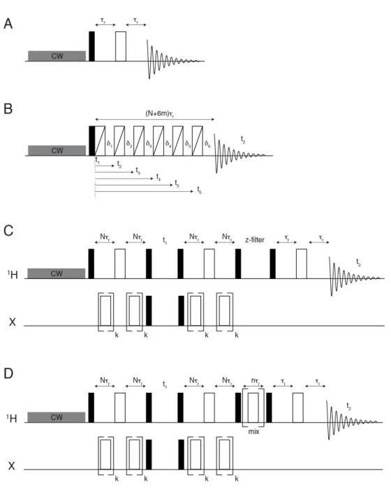
Figure S1. Pulse sequence schemes used in the present work.
(A) Spin echo. τ
r
indicates the rotor period.
(B) aMAT (adapted from ref. (34))
(C)
1
H,X TEDOR (where X indicates either
13
C or
15
N). N is an integer and k is an odd integer.
(D)
1
H,X TEDOR (where X indicates
13
C in this work) with spin magnetization exchange between
S5
Figure S2. Molecular models used for quantum chemistry calculations
Left: the larger model m1 used for DFT structure optimization and hyperfine coupling calculations. Right:
the smaller model m0 used for ab initio calculations of g- and D-tensors, showing the spin-density
distri-bution (from a SA-CASSCF(7,5) calculation, 0.002 a.u. isosurface).
S6
Figure S3. Additional
13
C spectra.
13
C spin-echo spectrum (blue) and projection from the
13
C aMAT spectrum (magenta), with assignment
of observable peaks and comparison with the calculated
13
C shift ranges for model F.
13
C spin-echo
spec-trum was acquired at 100 kHz MAS, ~280 K on a 500 MHz spectrometer (11.7 T) while the
13
C aMAT
spectrum was acquired at 30 kHz MAS, ~280 K on a 500 MHz spectrometer (11.7 T).
S7
Figure S4.
1
H aMAT spectrum.
(A)
1H aMAT spectrum acquired at 40 kHz MAS, ~280 K on a 500 MHz spectrometer (11.7 T). (B) The
S8
Figure S5. Additional
1
H spectra.
1