HAL Id: tel-01968697
https://tel.archives-ouvertes.fr/tel-01968697
Submitted on 3 Jan 2019HAL is a multi-disciplinary open access
archive for the deposit and dissemination of sci-entific research documents, whether they are pub-lished or not. The documents may come from teaching and research institutions in France or abroad, or from public or private research centers.
L’archive ouverte pluridisciplinaire HAL, est destinée au dépôt et à la diffusion de documents scientifiques de niveau recherche, publiés ou non, émanant des établissements d’enseignement et de recherche français ou étrangers, des laboratoires publics ou privés.
Structure and composition of mono- and multispecies
skin and environmental bacteria : effects of cosmectics
and other bilogically active compounds
Andrei Gannesen
To cite this version:
Andrei Gannesen. Structure and composition of mono- and multispecies skin and environmental bacteria : effects of cosmectics and other bilogically active compounds. Bacteriology. Normandie Université; Académie des sciences de Russie, 2018. English. �NNT : 2018NORMR068�. �tel-01968697�
2
Table of content
List of abbreviations ... 3
Acknowledgements ... 4
General introduction ... 5
Key points of the thesis ... 8
The aim of the work ... 8
Scientific novelty of investigations ... 9
Practical implications of the work ... 10
States for defense... 11
Thesis approbation ... 12
Publications ... 12
Bibliography review ... 14
CHAPTER 1. Human skin microbial commensal microorganisms and microorganisms closely related with them, their biofilms and interactions with human organism ... 14
1.1. Human microbiota ... 14
1.2. Human skin microbiota... 15
1.3. Some representatives of the human skin microbiota ... 16
1.4. Binary biofilms of some microorganisms of human skin ... 33
CHAPTER 2. Biofilm matrix ... 38
2.1. Organization and components of biofilm matrix ... 38
2.2. The matrix of biofilms of some skin microorganisms ... 44
Experimental study ... 49
Article 1. Regulation of biofilm formation by Pseudomonas chlororaphis In an in vitro system………...50
Article 2. Niclosamide as a promising antibiofilm agent………..59
Article 3. Effect of two cosmetic compounds on the growth, biofilm formation activity, and surface properties of acneic strains of Cutibacterium acnes and Staphylococcus aureus………..67
Supplementary data for the article 3………...………...79
Article 4. Regulation of formation of monospecies and binary biofilms by human skin microbiota components, Staphylococcus epidermidis and Staphylococcus aureus, by human natriuretic peptides……….……88
Article 5. Regulation of monospecies and mixed biofilms formation of skin Staphylococcus aureus and Cutibacterium acnes by human natriuretic peptides…………...………..…100
Article 6. Composition of biofilm matrix of Cutibacterium acnes acneic strain RT5………...….145
General discussion………...197
Discussion………196
Conclusions………..210
3
List of abbreviations
AHL – acyl homoserine lactone ANP – A-type natriuretic peptide BNP – B-type natriuretic peptide
CAMP-factor (CAMP-фактор) - Christie–Atkins–Munch-Petersen factor CFU – colony-forming unit
CLSM – confocal laser scanning microscopy CNP – C-type natriuretic peptide
GABA – gamma-amino butyric acid ID – inhibition dose
LB – lysogenic broth
MIC – minimal inhibitpry concentration MQ-water – water cleaned with MilliQ system MRSA – meticillin-resistant Staphylococcus aureus MRSE – meticillin-resistant Staphylococcus epidermidis NUP – natriuretic peptide
OD – optical density
PNAG – 6-poly-N-acetyl-glucosamine PS – physiologival saline
PS291® - polysaccharide 291® or teflose QS – quorum sensing
RCM – reinforced clostridial medium TSA – tryptic soy agar
TSB – tryptic soy broth
4
Acknowledgements
The author is deeply grateful to his thesis directors: Ph.D., Professor, Director of the Laboratory of Microbiology, Signals and Microenvironment of LMSM EA4312 Mark Georges Juillen Feuilloley, Doctor of Biological Sciences, Professor and Head of the Department of Microbiology of Lomonosov Moscow State University Alexander Ivanovich Netrusov. The author expresses special gratitude to the scientific adviser, Doctor of Biological Sciences, Chief Scientific Officer of the Laboratory of Petroleum Microbiology of Winogradsky institute of microbiology of FRC of Biotechnology RAS Vladimir Konstantinovich Plakunov for teaching the work, valuable comments, assistance in organizing work and moral support.
The author is grateful to colleagues from the laboratory of petroleum microbiology Ph.D. Zhurina M.V., Martyanov S.V. and Teteneva M.A. for help in work, Ph.D. Tarasov A.L. (posthumously) for help in working with anaerobic microorganisms and training work with anaerostats. Also, the author thanks a colleague from the laboratory of anthropogenic habitats Ph.D. Bochkova E.A. for assistance in the work on the isolation of the matrix of biofilm of C. acnes, establishing of contacts with co-authors from the Gubkin Russian State University of Oil and Gas, assistance in carrying out SERS-studies of the matrix and moral support.
The author is grateful to the staff of the Department of Microbiology: Ryzhkova E.P. for help in finding literature on C. acnes, Popova EA, Kreyer VG, Baranova NA for moral support during work, and especially Ph.D. Osmolovsky A.A. for the help in the organization of educational work and support.
The author thanks the staff of the Department for International Cooperation of the Biological Faculty of Lomonosov Moscow State University, namely, Djaikieva R.K. and the deputy dean of the faculty, Ph.D. Kitashov A.V. for the help in the organization of joint postgraduate study, signing and registration of necessary documents, valuable advice.
The author is grateful to the staff of the Laboratory of Microbiology, Signals and Microenvironment LMSM EA4312 of the University of Rouen: technicians Magalie Robert and Olivier Maillot for assistance in the mastering of equipment and organization of experimental work, postgraduate students Segolene Depayras and Valerie Borrel for assistance in working with C.
acnes and finding suitable experimental protocols for proteomic studies. The author is grateful to
Ph.D. Yoan Konto-Ghiorghi for help in organizing the work and determining the strategic ways to study biofilms and organizing a thesis, Ph.D. Anne Groboillot and separately Ph.D, Professor Sylvie Chevalier for help in establishing contacts with co-authors, organizing proteomic research and a large support during work in France. The author is grateful to Ph.D., Professor Olivier Lesouhaitier and Ph.D. Tomas Clamens for help in working with natriuretic peptides. Especially the
5
author is grateful to Andrea Shane, Ph.D., for his assistance in the arrangement of life in France, support and assistance in the laboratory.
The author is grateful to the co-authors of the work: Doctor of Biological Sciences, Head of Laboratory for Regulation of Expression of Genes of Microorganisms Khmel I.A. for the provision of Pseudomonas chlororaphis strains used in the work. The author is grateful to the staff of the Laboratory of Evolutionary Genomics of the A.N. Belozersky Institute of Physico-Chemical Biology: Ph.D. Shtratnikova V.Yu. and Ph.D., senior researcher Logacheva M.D. for help in determining the species of skin isolates M. luteus S01 and K. schroeteri N01.
The author is grateful to the staff of the Laboratory of Biotechnology in the Oil and Gas Industry of the Department of Physical and Colloid Chemistry of the Gubkin Russian State University of Oil and Gas: Ph.D., M.Sc. D.S. Kopitsyn and the graduate student M.V. Gorbachevsky for conducting the SERS-study of samples of the C. acnes matrix. The author is grateful to the staff of the Laboratory of Polymers, Biopolymers and Surfaces of PBS UMR 6270 (Laboratoire de Polymeres, Biopolymeres, surfaces, PBS) of the University of Rouen (France), Ph.D. Sebastien Massier and Ph.D. Julie Hardouin for proteomic studies of the C. acnes matrix.
Author is especially grateful to LMSM technician Chloe Catovic and Agrohall Evreux researcher Olivier Poultier for help in translating of this synopsis in French, and for their kindness and courtesy to the author when he was in the lab.
General introduction
Humans skin is the biggest organ where hundreds of microbial genera coexist together (Grice et al., 2009). Such coexisting was developed during evolution process: the host organism does not impede incoming and proliferation of commensial microbes, but fights against pathogens (including with help of commensial microbiota, Chiller et al., 2001). Microbial species ratio in human skin depends on sex, age, geographical region etc., but the general regularities inside a single group are similar (Grice et al., 2009; SanMiguel & Grice, 2015). One of important factors for microbial growth is microenvironment, which is based on physiological skin features. Normally three types of skin areas and microenvironment – sebaceous (oily), moist and dry are distinguished depending on the location of skin area, number of sebaceous and sweat gland on it (Grice et al., 2009). Each type of microenvironment is characterized by its own special set of microorganisms. In moist and sebaceous sites Actinobacteria (and especially former genus Propionibacterium) and Firmicutes are dominant, and in dry areas – Betaproteobacteria and Corynebacteria (Grice et al., 2009; SanMiguel & Grice, 2015).
6
injuries and diseases (physical traumas, atopic dermatitis, psoriasis, other immune system disorders) lead to changes in microbial species ratio in skin (SanMiguel & Grice, 2015). This results in complications (overgrowth of pathogens, inflammations etc., Hong et al., 2011). Skin is a barrier which interacts permanently with environment and undergoes effects of exogenous and endogenous factors. These effects can impact also skin microbiota. Endogenous factors are determined by humoral and nervous regulation of skin state. Exogenous factors can be divided on biological (contacts with other living organisms), physical (air, moistening, environmental temperature, radiation etc.) and chemical (effects of different artificial or natural compounds). As chemical factors, in fact, we can classify all chemical compounds which interact with human skin, including drugs, antibiotics and cosmetics, which are used widely all around the world. Impact of these compounds on skin is very significant (Feuilloley & Orange, 2018) .
Microbial biofilms are spatially and metabolically structured communities which are embedded in an extracellular polymeric matrix and usually located on different interfaces (Nikolayev & Plakunov, 2007). Biofilms are the most common life manner of microorganisms on Earth (Nozhevnikova et al., 2017). Biofilms can grow on different interfaces: liquid-air, solid object – air, liquid-liquid and solid object - liquid (Haussler, Fuqua. 2012). Biofilm formation is a universal property of bacteria, biofilms are found virtually everywhere on all artificial or natural surface (Vlamakis et al., 2013). In biofilms cells undergo physical and biological impacts of different kind in comparison with planktonic forms. In biofilm there are sharp gradients of concentration of nutrients, gases and ions, close adjacency of cells leads to their physical contacts, interchange of signal molecules and metabolites. All these phenomena determine special features of biofilms (Nikolayev & Plakunov, 2007).
When biofilms grow, the specific biofilm phenotype is developed (Nikolayev & Plakunov, 2007), when expression level of different genes is changed, which leads to expression of special cell features in comparison with planktonic state (for example, synthesis of the matrix). As a result, biofilms acquires properties close to multicellular organism. Some time ago there was the hypothesis about interkingdom multispecies biofilms of amoebas and bacteria as ancestors of multicellular organisms – sponges (Verhoeven et al., 2010). However, this hypothesis was incorrect: bacteria in biofilms have biofilm phenotype, but not genotype. Gene expression changes are not irreversible, cells can leave biofilms and return into planktonic state, so, there is no true cell differentiation like in multicellular organisms. But, actually, cells of one species inside a biofilm can differ significantly in their gene expression (Boles, Singh, 2008).
There are three general stages of biofilm formation: cell adhesion, growth and dispersal (Plakunov et al., 2017). In its turn, on each stage there are several sub-stages. On the stage of cell adhesion there are sub-stages of reversible (initial) and irreversible adhesion (Nikolayev &
7
Plakunov, 2007; Dutta et al., 2012; Ribeiro et al., 2012). Cells during initial adhesion step can navigate the surface with flagella and pili to find an optimal place. When irreversible adhesion starts, cells lose their motility (Romeo, 2008; Harmsen et al., 2010; Dutta et al., 2012). On growth stage synthesis of matrix and settlement of secondary colonizers occur (Nikolayev & Plakunov, 2007; Vlamakis, 2013). These processes can go simultaneously or step by step, so it is possible to combine them in one stage. After, biofilm enters the stage of maturaton (Nikolayev & Plakunov, 2007; Mann, Wozniak, 2012; Vlamakis, 2013). On this stage complex three-dimensional structures can be formed like mushroom-like structures of Pseudomonas aeruginosa (Hamsen et al., 2010) or round pellicles of Bacillus subtilis (Vlamakis, 2013). On dispersal step a part of cells loses their biofilm phenotype and returns into free planktonic state.
Quorum sensing should be mentioned specially. The term “quorum sensing” (QS) describes intercellular interactions in bacteria, which allow the community to coordinate genes expression in accordance with changes of population density (Camilli & Bassler, 2006). It is known that QS systems contribute biofilm formation, and lots of molecules serve as QS-factors in bacteria (Worthington et al, 2013). QS investigations in Gram-negative bacteria are conducted usually with use of Vibrio fisheri, V. cholerae and P. aeruginosa as objects (Worthington et al, 2013). Generally, QS-system work based on two proteins: the first synthesizes a signal molecule of autoinducer (AI) and the second protein is a receptor of autoinducer (Worthington et al, 2013). QS systems significantly contribute biofilm formation, and modern methods of biofilm eradication more often mean inhibition of QS, for example, with N-(2-pyrimidil)-butanamide (Kalia, 2015; Furiga et al., 2016; Leoni, Rampioni, 2018).
Microbial biofilms nowadays are an object of intensive research because of several reasons. The first is the interest of fundamental sciences to find out how normally monocellular bacteria can exist in multicellular communities. The second, biofilm formation is a serious problem in medicine and industry, because of their many-fold stronger resistance to antibiotics, immune system and biocides (Hamsen et al., 2010) in comparison with planktonic cultures (planktonic cultures are totality of floating single microbial cells in liquid medium). Also, interesting (and dangerous) phenomenon is percisting cells – a small part of cell population in biofilm, which has lower metabolic acticity and many-fold higher resistance to antibiotics than other biofilm cells (Plakunov et al., 2010). Persisting cells or persisters can reestablish the population after death of the most of cells after a strong negative impact (usually exogenous, like physical biofilm destruction or biocide attack), that makes persisters one of the factors of biofilm stability and resistance (Michiels, Fauvart, 2016).
Also, besides negative aspects, microbial biofilms conduct important processes like, for example, destruction of biodegradation-resistant pollutants of soils and wastewaters (Perni et al.,
8
2013). The fundamental problem for science is understanding how bacteria develop their so called group-level properties (Nadell et al., 2013). These properties are spatial structure of biofilms and coordination of cell behavior during interaction with each other and surfaces (Nadell et al., 2013). Also intracellular and intercellular methabolic and signal pathways in biofilms are of scientific interest.
To generalize, it should be stated that study of biofilms is one of priority areas of modern microbiology, and not only monospecies, but also multispecies biofilms. Human microbiota and its biofilms in human body are of special interest because of human microbiota and particularly skin microbiota im many respects determines the state and functioning of different systems of human organism (Thomas et al., 2017). So, study of biofilms of skin commensal, closely related to them microorganisms and how different active compounds affect them (what is the goal of present thesis) is actual. First, as mentioned above, it is interesting for fundamental science to investigate biofilms and their interactions with human organism. Second, it is actual for applied sciences: biotechnology, cosmetology and pharmacology. In cosmetology nowadays there is growing interest in new cosmetic compounds because of progressive rejection of preservative agents and deepening interrelation between cosmetology, dermatology and microbiology. The last requires developing of new compounds to maintain healthy skin microbiota. In pharmacology now search of new antibiofilm agents to combinate with already existing antibiotics is actual. In biotechnology it is necessary to control biofilm growth in processes of biosynthesis of needful compounds or in maintaining of important biochemical process (like, for example, it is now performed in wastewater treatment process with anammox bacteria (Nozhevnikova et al., 2015)). Also, it is necessary to investigate not only effects of different active compounds on biofilms, but also biofilm structure, especially biofilm matrix, which is a strong barrier for different active compounds (Zhurina et al., 2014). Information about matrix structure is important for searching of compounds which are able to pass through it and reach cells inside biofilms.
Key points of the thesis
The aim of the work
The aim of this work was to investigate effects of active compounds of different types on human skin commencial bacteria and their close relatives of natural environments, their monospecies and binary biofilms and investigate probable mechanisms of action of used compounds.
To reach the aim, several tasks were set and performed:
9
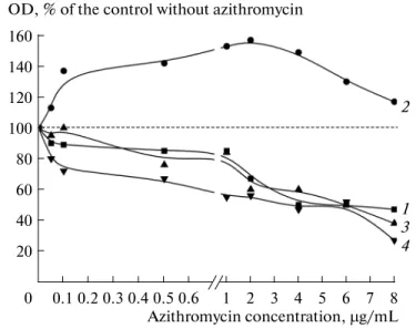
Pseudomonas chlororaphis – nonpathogenic microorganism closely related with pathogenic P. aeruginosa. Azithromycin is an antibiotic widely used in treatment of pseudomonas infections.
2) Investigate effects of antihelmintic drug niclosamide on cultures and biofilms of human skin commensal bacteria Micrococcus luteus, Kytococcus schroeteri and Staphylococcus aureus. Find out if niclosamide can strengthen the effect of azithromycin.
3) Investigate effects of thermal water Uriage™ (TWU) and oligosaccharide PS291® on growth of monospecies and binary planktonic cultures and biofilms of human skin commensal bacteria S. aureus, S. epidermidis and acneic strains of Cutibacterium acnes.
4) Investigate effects of natriuretic peptides A- and C-type on growth of monospecies and binary planktonic cultures and biofilms of S. aureus and acneic strains of C. acnes.
5) Investigate biochemical composition of biofilm matrix of acneic strain Cutibacterium
acnes HL043PA2 (RT5).
Scientific novelty of investigations
It was unveiled that subinhibitory concentrations of azithromycin stimulate growth of P.
chlororaphis, and acylhomoserine lactone-dependent QS-system is involved in this process. It was
unveiled that azithromycin affects P. chlororaphis in a special way: biofilm growth stimulation appears at relatively higher concentrations, while at lower concentrations (several orders lower than minimal inhibitory concentration (MIC)) there is inhibition of biofilm growth. It was shown for the first time also that azithromycin-stimulated biofilms of P. chlororaphis have bigger amount of polysaccharide component of matrix in biofilms and more resistant to heat shock.
Strong inhibitory effects of niclosamide on planktonic cultures and biofilms of S. aureus, M.
luteus and K. schroeteri. It was shown that niclosamide quenches stimulatory effect of subinhibitory
concentrations of azithromycin on biofilms of S. aureus. This makes niclosamide a promissing antibiofilm agent.
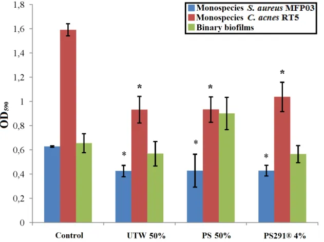
It was shown for the first time that TWU™ ans PS291® significantly decrease biofilm growth of S. aureus and C. acnes without toxic impact on cells. Probably, these cosmetic compounds modify cell adhesion ability, and this makes them a perspective base for new cosmetics development.
It was unveiled that human natriuretic peptides (NUP) have strong effect on biofilms of Gram-positive human skin commencial bacteris. It was established that effects of NUPs A- and C-type on biofilms of S. aureus, S. epidermidis and C. acnes depends significantly on growth conditions that can correlate with different skin status and NUP regulatory functions: enlarge competitive features of C. acnes and S. epidermidis against S. aureus. This approves the suggestion
10
that NUPs have capacity to affect the microorganisms and co-evolution of human skin microbiome and human regulatory molecules. Also it makes NUPs perspective for application in dermatology and cosmetology as normalizing agents for microbiota.
Deep biochemical investigation of biofilm matrix of C. acnes was done for the first time. It was shown that, for investigated strain, the polysaccharides were dominant matrix component. Total proteome analysis of matrix was performed for the first time, more than 400 proteins were shown to be a part of matrix, their functions were suggested. SERS-spectroscopy of matrix and biofilms biomass of C. acnes was made for the first time, spectral fingerprints were obtained.
Practical implications of the work
The universality of the phenomenon of biofilm growth stimulation by antibiotics was shown: azithromycin stimulates growth of biofilms of not only human pathogenic bacteria, but also of soil saprotrophic bacteria. The involvement of QS-system of P. chlororaphis in growth stimulation was shown, this approves good perspectives of QS-quenching agents (especially AHL-dependent QS) in biofilm eradication treatments and quenching of antibiotic-mediated biofilm stimulation. It was shown that in stimulated biofilms, polysaccharidic components of matrix are synthesized more actively, and probably because of that azithromycin-stimulated biofilms are more resistant to heat shock. This is useful for further research.
Good perspectives of niclosamide as potential antibiofilm agent for Gram-positive were shown. At human non-toxic concentrations, it eradicates efficiently biofilms of Gram-positive commensal bacteria, and also quenches stimulatory effect of azithromycin on S. aureus biofilm. Niclosamide can be used in the future in complex antibiofilm dtugs.
TWU™ and PS291® are shown to be perspective base components for novel cosmetics to fight agains biofilms of S. aureus and C. acnes in human skin.
NUPs were shown perspective for use in antibacterial treatment, also the new property of NUPs was shown: ability to regulate skin microbiota. Before this ability was considered as uncharacteristic for NUPs, and this gives broad opportunities for endocrinology and clinical practice.
The robust method of Gram-positive bacteria biofilm matrix extraction was developed. This method is non-chemical, this allows to preserve compounds of matrix unchanged for deep analysis.
Protein composition of matrix was revealed. It will be used in further investigation of C.
11
States for defense
1) Azithromycin at extra-low concentrations inhibits growth of P. chlororaphis 449 biofilms. At higher concentrations stimulation occurs, which disappears with approach to MIC. Stimulation and inhibition also result in increasing and decreasing of the polysaccharide moiety of the matrix. Biofilms of P. chlororaphis which were grown in the presence of stimulating concentration of azithromycin are more tolerant to heat shock than in control without azithromycin. AHL-dependent QS-system is involved in stimulation process: expression of a plasmidic gene encoding for AHL-lactonase totally abolishes the stimulatory effect of azythromycin in genetically modificated strains of P. chlororaphis.
2) Niclosamide already at concentration 0.1 μg/ml strongly inhibits the growth of metabolically active cells of S. aureus, M. luteus and K. schroeteri. At 1 μg/ml, niclosamide in a medium fully inhibits growth of metabolically active bacteria in biofilms. In combination with azithromycin niclosamide neutralizes the stimulating effect of azithromycin on S. aureus biofilms.
3) UTW™ and PS291®, probably because of modification of cell adhesion ability, inhibit efficiently biofilm growth of C. acnes and S. aureus. The effect of thermal water should be correlated to its chemical composition. Moreover, UTW™ and PS291® are able to regulate skin microbiota balance: due to decreasing of competitive advantages of S. aureus, both cosmetics increase the ratio of C. acnes and S. epidermidis in binary biofilms with S. aureus.
4) NUPs A- and C-type affect C. acnes, S. epidermidis and S. aureus, and NUP effects depends on cultivation conditions. This suggests different roles of NUPs in different conditions in skin. NUPs possess regulatory functions and they are able to modulate the CFU balance of microorganisms in binary biofilms.
5) A mean of 54% of the matrix organic matter of C. acnes RT5 biofilms is formed by polysaccharides. Proteins and DNA account for 16% of the organic matrix. About 30% of the matrix organic compounds are molecules characterized as peptidic, carbohydrate and non-DNA. These molecules are low-molecular metabolites including porphyrins and intermediates of porphyrin synthesis.
6) 447 different proteins were found in the matrix of C. acnes RT5. More than 20 hydrolases of different substrate specificity, more than 40 proteins of unknown origin and function, a lot of enzymes and structural proteins. Most of the proteins probably appear in the matrix after cell autolysis during biofilm formation. Hydrolases should be involved in the pathogenicity of C. acnes biofilms in skin and other microenvironments in the human organism.
7) SERS investigation of matrix allowed to find 57 peaks of different intensity, which compose spectral fingerprint of C. acnes matrix. The same was done for C. acnes cells. All spectra
12
need to be deeply investigated by other methods, but presently they will be used in united database of bacterial SERS spectra construction.
Thesis approbation
Results of the thesis were presented in 11 scientific congresses and conferences:
1) III All-Russian scientific and practical conference "The process of life development within abiotic changes on the Earth" (23-30 September 2014, Listvyanka);
2) XXVII Winter school for young scientists “Pespective trends of physico-chemical biology and biotechnology” (9-12thFebruary 2015, Moscow);
3) VIII Moscow international congress “Biotechnology: state of the art and perspectives” (17-20thMarch, 2015, Moscow);
4) XXII International scientific conference of students, PhD-students and young scientists “Lomonosov-2015” (13-17th April, 2015);
5) X International school-conference for young scientists “Actual aspects of modern microbiology” (27-30th October, 2015, Moscow)
6) 4th
International congress “Cosm’innov” (24-25thMay, 2016, Orleans, France)
7) International congress “Antimicrobial resistance in microbial biofilms and options for the treatment” (5-7thOctober 2016, Gent, Belgium)
8) XI International school-conference for young scientists “Actual aspects of modern microbiology” (9-12th November, 2015, Moscow);
9) XXIX Winter school for young scientists “Pespective trends of physico-chemical biology and biotechnology” (7-10thFebruary 2015, Moscow);
10) XXI International Puchshino school-conference for young scientists “Biology – the science of XXI century” (17-21thApril, Puchshino);
11) 9th International Conference on Skin Ageing and Challenges (Porto, Portugal).
Publications
6 scientific articles based on materials of the thesis were already published: 2 reviews and 4 experimental articles. All 6 are in journals indexed in Scopus and Web of Science, 3 are in journal of RSCI core. 2 experimental articles are in preparation for publication.
Individual contribution of the thesis author.
The author conducted experimental13
were noted in all publications and cases.
Thesis structure.
The thesis consists in abbreviations list, bibliographic review,14
Bibliography review
CHAPTER 1. Human skin microbial commensal microorganisms
and microorganisms closely related with them, their biofilms and
interactions with human organism
1.1. Human microbiota
Human microbiota is a community of microorganisms (commensal and pathogens), which live inside and on the surface of human organism and ecologically interrelated with it and with each other (Thomas et al., 2017). The human evolved together with his microbiota (Thomas et al., 2017). Different authors evaluated differently the number of microbial cells in human organism and on its surface (data varies between 1012 and 1020cells, Bianconi et al., 2013). Similarly, dispersion of data concerning the number of human organism own cells was great – several orders (Sender et al., 2016). Some scientists nowadays are in doubt with these data which are widespread in scientific literature. According to Rosner, 2014, the frequently cited ratio of microbial to human cells equal 10:1 origins from the article of Luckey, 1972, where the microbial cell number was estimated by their amount in gut contents. Some authors suppose that the data of this article and following are a partly incorrect because they were based on a too large generalization and averaging of the human body volumes, mass, microbial cell volumes and so on. (Sender et al., 2016). Results of modern metadata analyses, which include calculation of multiple parameters, suggest that in average the human body consists in 3.72х1013 (Bianconi et al., 2013). The number of microbial cells inside and
on the surface of the human organism was estimated about 3.9x10¹³ (Sender et al., 2016), so the actual ratio may be about 1:1.
Nevertheless, the species biodiversity of the human microbiota is great: according to modern estimations, at least 40000 strains are associated with the human organism, these strains belong to 1800 genera and possess more than 9 million genes differing from human genes (Schwiertz & Rusch, 2016). It is a 100-fold bigger amount of genes than in humans (Thomas et al., 2017). Also, the microbiota of each individual possesses an amount of genes which is 10-fold bigger than the proper individual’s genome (Thomas et al., 2017). Microbiota of humans includes representatives of all three life domains – archaea, prokaryotes, eukaryotes (Thursby & Juge, 2017), and also viruses, which form the human virome (Virgin, 2014). The most complete data about the genetic diversity of the human microbiota to date were obtained by metagenomics and metatranscriptomics approaches, which have advantages (total DNA analysis allows to find non-cultivable microorganisms) and disadvantages (dependence on nucleic acids preparation methods, impossibility of complete microorganism description, Walker, 2016). However, microorganisms
15
cultivation methods of on different laboratory media nowadays allow to investigate a very limited amount of microbes, between 0.1 and 1% of the real population (Walker, 2016).
The human microbiota performs a number of important functions: synthesis of needful compounds (Lai et al., 2010; SanMiguel & Grice, 2015; Nakatsuji et al., 2018), defense against pathogens and immune system functioning support (SanMiguel & Grice, 2015). Close interaction of human microbiota with human organism during evolution processes formed finely-tuned and very complexly organized communities, that some authors call “the second brain” (Ochoa-Repáraz & Kasper, 2016), and human organism together with its microbiota – “superorganism” (Thomas et al., 2017).
1.2. Human skin microbiota
Humans skin is the biggest organ where hundreds of microbial genera coexist together (Grice et al., 2009). Such coexistence was developed during the evolutionnary process: the host organism does not impede incoming and proliferation of commensal microbes, but fights against pathogens (including with the help of the commensal microbiota, Chiller et al., 2001). The microbial species ratio in human skin depends on sex, age, geographical region and environment, but the regularities within a single group are generally similar (Grice et al., 2009; SanMiguel & Grice, 2015). One of the important factors for microbial growth is its microenvironment, which is depending on physiological skin features. Normally three types of skin areas and microenvironment are described – sebaceous (oily), moist and dry – following the location of the skin areas and the density of of sebaceous or sweat glands (Grice et al., 2009). Each type of microenvironment is characterized by its own special set of microorganisms. In moist and sebaceous sites Actinobacteria (and especially former members of the genus Propionibacterium) and Firmicutes are dominant, whereas Betaproteobacteria and Corynebacteria are essentially found in dry areas (Grice et al., 2009; SanMiguel & Grice, 2015).
As the first barrier and the biggest human organ, skin is responsible for several important functions: thermoregulation, excretory function, defense function, gases exchange. Microorganisms which inhabit the human skin are involved in processes on skin and significantly determine its status (Achermann et al., 2014; Leung et al., 2018). Skin status changes (genetic distortions, diseases, exogenous factors) are generally leading to skin microbiota changes. For example, in atopic dermatitis a raucous proliferation of S. aureus on affected areas can occur, which results in disease complications (Elfatoiki et al., 2016; Lacey et al., 2016). Similarly S. aureus can overgrow in psoriasis (Totte et al., 2016) and folliculitis. When S. aureus starts to proliferate actively in hair follicles hollows it can rapidly cause inflammation (Balasubramanian et al., 2017). Dandruff is another skin disorder which, according to different estimations, it affects about half of the world population (Xu et al., 2016). Etiology of dandruff remains to date a matter of discussion, but
16
frequently fungi of the Malassezia genus are associated with it and proliferate intensively on affected skin areas. Malassezia fungi themselves are causative agents of tinea versicolor and sometimes proliferates abundantly on skin affected with atopic dermatitis and psoriasis (Xu et al., 2016). Also, Malassezia sp. is a cause of seborrhea (Soarez et al., 2015).
1.3. Some representatives of the human skin microbiota
In this tip some microorganisms of human skin and closely related ones are described.
1.3.1. Pseudomonas
Pseudomonas are not a bacterial genus classically associated to skin although metagenomic
studies have shown that, even on healthy skin, in some moistly area they can account for more than 90% of the bacterial population (Grice et al., 2008). On the human skin, a lot ofof Pseudomonas species can be found, namely P. fluorescence, P. putida and P. aeruginosa are the most common. P.
aeruginosa is a soil saprotroph (Deredjian et al., 2014), but it is much more well-known as a human
pathogen (Loveday et al., 2014). Lots of studies on P. aeruginosa are already published, and lots of works are focused on it each year. That because P. aeruginosa is an opportunistic, fast growing and frequently aggressive pathogen. Its virulence is much higher than that of P. fluorescens and P.
putida. It is involved and can be also the causative agent of a lot of diseases including lung cystic
fibrosis (Fernandez et al., 2013), urinary tract infections (Tajbakhsh et al., 2015), artificial implants fouling (Cole et al., 2014) or otitis (Wang et al., 2005). Recently (before meticillin-resistant S.
aureus abundant spread) P. aeruginosa was the main causative agent of bacteremias with highes
mortality rate (Kreger et al., 1980). In termsof skin disorders, P. aeruginosa is responsible for the green nails syndrome (Chiriac et al., 2015), wounds infections and inflammations (Pfalzgraff et al., 2018), pseudomonas-caused folliculitis or hot tubes folliculitis (because of heating tubes in water pools and other water facilities are frequent sources of transmission, Fowler & Stege, 1990), ecthyma gangrenosum (Mull et al., 2000)…. P. aeruginosa is able to destroy the skin structure in bacteremia which leads to serious exacerbations (Wu et al., 2011). P. aeruginosa is especially dangerous for people with compromised immunity (Bassetti et al., 2018). The difficulties of P.
aeruginosa infections treatment are connected with its ability to form biofilms on skin, in particular,
in wounds (Davis et al., 2008; Karna et al., 2016). Also, because of rather long-term fight against P.
aeruginosa in clinics, dangerous multidrug resistant strains of P. aeruginosa appeared (Potron et al.,
2015). Now P. aeruginosa is considered as the main infectious causative agent amongst Gram-negative rods (Bassetti et al., 2018).
Because of all these facts P. aeruginosa now is a model object for biofilm studies and studies of different antibacterial compound action on biofilms. P. aeruginosa is a very popular object, a number of monographs and lots of scientific articles every year are dedicated to it, so here only a very short review of the key points directly related to the present thesis will be made. Lots of works
17
are dedicated to P. aeruginosa biofilms resistance to antibiotics. For example, resistance to tobramycin and ciprofloxacin (Stewart et al., 2015), ceftazidime and colistin (Furiga et al., 2016) and other. A number of works are dedicated to azithromycin action on P. aeruginosa biofilms and its efficiency (for instance, Phelan et al., 2015, Saini et al., 2015). Concerning azithromycin resistance, MexAB-OrpM and MexCD-OprJ efflux pumps have been described for their role in resistance establishment (Gillis et al., 2005). Azithromycin also interacts with AHL-dependent QS-systems Las and Rlh in P. aeruginosa and blocks their functioning (Tateda et al., 2001), and by this way interferes with biofilm formation (Nalca et al., 2006). On P. aeruginosa a biofilm stimulatory effect of sub-MIC antibiotic concentrations was described. For example, doxycycline and polymyxin B in sub-MIC concentrations stimulate P. aeruginosa biofilms growth (Tote et al., 2009). Gentamicin in sub-MIC concentrations increases the expression of at least 30 genes (including the Lon protease) which are responsible for biofilm formation in P. aeruginosa (Marr et al., 2007).
A number of works concern the interactions of P. aeruginosa with signal molecules of the human organism. It was shown that catecholamines and γ-amino butyric acid (GABA) stimulate the growth of P. aeruginosa and increase its virulence (Lesouhaitier et al., 2009). Interferon-γ and dynorphin can exert similar effects (Lesouhaitier et al., 2009). Recently it was shown that the genome of P. aeruginosa encodes for an enzyme, the amidase AmiC, which is an ortholog of the C-type natriuretic peptide (CNP) receptor (Rosay et al., 2015). This is probably a reason of CNP’s ability to inhibit biofilm growth by P. aeruginosa. In P. aeruginosa,AmiC is a regulator of the amidase operon ami expression and therefore has a large pleiotropic activity (Wilson & Drew, 1991).
P. putida is a saprotrophic microorganism living in soil, water (Fernandez et al., 2015) and
rhizosphere, that makes it useful in the context of plants protection against phytopatogens (Bernal et al., 2017). However, herewith P. putida can also behave as an opportunistic pathogen and can be a causative agent of nosocomial infections (Fernandez et al., 2015). P. putida can form biofilms in skin and change the structure of the epidermis (Fernandez et al., 2015). Despite of the fact that P.
putida infections are rather rare and usually occur in patients with compromised immunity, there are
many data about multidrug resistant strains of P. putida that can represent serious danger (Molino et al., 2014; Hardjo Lugito et al., 2015). For example, strains of P. putida have been now identified which are resistant to carbapenem, ciprofloxacin (Kumita et al., 2009), cefepime (Luczkiewicz et al., 2015) and some other beta-lactams (Trevino et al., 2010). There are described cases of abundant growth of P. putida in patients with diabetic gangrene andfollowingexacerbations (Hardjo Lugito et al., 2015).
18
canprotects plants against phytopathogens (Kremmydas et al., 2013) and was even patented for crops treatment. However, as P. putida, P. fluorescens can colonize the human skin and be a cause of infections which include biofilm infections (Scales et al., 2014). Treatment of such infections can be difficult for multiple reasons and the principal is the high adaptability of this microorganism. For instance, different works describe the resistence of P. fluorescens resistance to biocides used as antiseptics: glutaraldehyde (Simoes et al., 2006), benzyldimethyldodecylammonium (Ferreira et al., 2011). Data concerning P. fluorescens antibiotic resistance are also abundant, in particular againts beta-lactams (Bompard et al., 1988) and cephalosporins (Luczkiewicz et al., 2015). It was also shown that atmospheric pollutants, such asNO2, can increase P. fluorescens resistance to
ciprofloxacine and chloramphenicol through activation of MexEF-OprN efflux pumps genes expression (Kondakova et al., 2016). Moreover, it was shown on P. fluorescens that a range of endogenous molecules produced by the human organism can modulate the virulence and biofilm formation of this bacterium. These compounds can be antibacterial or traditionally considered as non-antibacterial. For example, substance P (a neuropeptide) increases the cytotoxicity of P.
fluorescens, and in common with the hormone epinephrine (adrenaline) increases the swarming
motility of the cells (Biaggini et al., 2015). The hormone serotonin also increases the swimming motility of P. fluorescens(Biaggini et al., 2015). Conversely, in some systems, the nhibitory neurotransmitter GABA inhibits the growth of P. fluorescens in biofilms (Dagorn et al., 2013). If concern antibacterial endogenous agents, it was shown recently that β-defensin-2 is able to increase the virulence of P. fluorescens cells (Madi et al., 2013). Thus, despite of the fact that P. fluorescens and P. putida are traditionally considered as safe rhizosphere microorganisms in comparison with
P. aeruginosa, it seems to be that a number of molecules of human organism can interact with them.
This can suggest at least universal mechanisms of interaction between microorganisms colonizing skin and human organism. Because of than it is interesting to investigate the effects of such active compounds and, in particular, antibiotics, on the growth of microorganisms which are closely related to widespread and well-known pathogens. For example, in amicroorganism such as
Pseudomonas chlororaphis.
P. chlororaphis is a rhizospheric saprotroph which is closely related to opportunistic Pseudomonas inhabiting the human skin – P. aeruginosa and P. fluorescens (Mishra et al., 2009;
Hesse et al., 2018). In databases there are works dedicated to genetics and regulatory processes in P.
chlororaphis biofilms. For instance, it was shown that functioning of phenazine synthesis system
(Wang et al., 2016; Yu et al., 2018), regulatory system GacS/GacA (Li et al., 2015) and AHL-dependent QS-system (Peng et al., 2018) for biofilm formation in P. chlororaphis. However, at the time of present thesis preparation, there were no works found in the literature concerning the effects of active compounds (drugs etc.) on P. chlororaphis. Only Shepelevich et al. made a screening of P.
19
chlororaphis strains resistance to a range of antibiotics: non-effective ones were considered as
perspective as herbicides which will not inhibit P. chlororaphis growth (Shepelevich et al., 2012). But authors did not examined the effects of antibiotics on P. chlororaphis biofilms, this makes present work original and actual.
1.3.2. Micrococcus luteus
M. luteus is a Gram-positive non-motile aerobic cocci, which form tetrads and belong to the
phylum Actinobacteria (Hanafy et al., 2016). M. luteus is a saprotroph and a member of the normal human skin microbiota (Kloos et al., 1974, Daeschlein et al., 2012). It localizes mostly in the stratum corneum (Lange-Asschenfeldt et al., 2011), the upper surface of the skin formed of dehydrated cells containing an abundance of keratine and phospholipids. This microorganism is also found in water and soil (Mauclaire & Egli, 2010). The interest to M. luteus was arising at the end of XX century, when it was isolated from the human skin. Cases of M. luteus-caused bacteremia were described, but those cases were detected in people with health issues: in carcinoma (Albertson et al., 1978) and renal diseases (Peces et al., 1997). In the first case bacteremia occurred after surgical operation, and in the second one because of catheter infection; in both cases M. luteus was introduced into the weakened organism after exogenous surgical impact. After, a case of M.
luteus mediated endocarditis was described in an old woman with compromised immunity. She
underwent an operation for removing breast cancer, chemotherapy and course of drugs (Miltiadous & Elisaf, 2011). Miltiadous & Elisaf established in their work that, at the time they were writing the paper, only 17 cases of M. luteus-mediated endocarditis were described in the literature. They suggested that M. luteus was safe for healthy people, but nevertheless some investigators consider it as a potential opportunistic pathogen (Mauclaire & Egli, 2010). In 2010 the full genome of M.
luteus was sequenced (Young et al., 2010), and it was shown that M. luteus belongs a the group of
free-living microorganisms with the smallest genome (about 2.5 million b.p.). Small length of genome results in a range of metabolic limitations for M. luteus: for example, it is unable to growth on a medium with glucose as only carbon source. Authors suggest that such genome reduction could be an adaptation to the ecological niche of M. luteus in skin (Mauclaire & Egli, 2010). Nevertheless, it is generally considered that a small genome is an indication of potential pathogenic activity. Indeed, as they are unable to synthesize many necessary compounds for their metabolism, these bacteria need to scavenge host molecules with a risk of alteration of eukaryotic cells.
Biofilms of M. luteus are also poorly investigated. It seems that the little interest of researchers can be explained by the general neutrality of M. luteus if it is safe for healthy people. M.
luteus was used as a model in Matsura et al. work where they studied biofilm growth in microfluidic
system and the influence of medium pH-level (Matsura et al., 2013). Coumarins were shown to be active agains M. luteus cultures (Emmadi et al., 2014), but the authors did not tested the activity of
20
coumarins against M. luteus biofilms. An interesting work was performed by Mauclaire & Egli (2010) where they compared biofilm formation and culture growth of M. luteus in microgravity in the ISS and compared to normal gravity conditions. They found that in microgravity M. luteus grows better in planktonic form and worse in biofilms in comparison with normal gravity. Also, on ISS the biofilm matrix of M. luteus contains less proteins and colloid polysaccharides, and the cell surface becomes more hydrophobic.
As other skin microbes, M. luteus can be affected by antibiotics, but in this area there are also little amount of works. There are some data about the activity of some quinolone antibiotics against
M. luteus, which block the activity of the DNA-gyrase (Zweerink & Edison, 1986). There are also
data about M. luteus isolated from human skin and containing the plasmid pMEC2, which encodes for resistance to macrolide antibiotics (Liebl et al., 2002). This plasmid encodes for a resistance factor of peptidic structure which has 50-54% of similarity with the ones of Gram-positive opportunistic pathogens with high G-C pairs amount – Corynebacterium and Cutibacterium (former Propionibacterium). Because of this, authors suggested a probable role of non-pathogenic M. luteus in horizontal transfer of antibiotic resistance genes. At the time of the present thesis preparation there no data were found concerning the potential interaction of M. luteus and endogenous human molecules.
M. luteus can be interesting for researchers as a potential biofuel source: it is potentially able
to synthesize alkenes because of possession of a gene set for metabolism of amino and fatty acids (Young et al., 2010; Surger et al., 2018). However, the little interest to M. luteus in the context of medical research may be unwarranted. Indeed, it was shown that M. luteus isolated from poultry wastewaters possesses a keratinolytic activity (Laba et al., 2015). This is logical with the fact that this bacterium is living in the stratum corneum but it can also suggest a potential effect of M. luteus in skin by metabolizing keratin and it appears that the keratinolytic activity of micrococci can be potentially active in the stratum corneum. Also, cells of M. luteus can function as liposome-like capsules for chlorhexidine, which protects chlorhexidine from lysis inside neutrophils (Wendel et al., 2015). In this context, M. luteus was used as container for transporting chlorhexidine into neutrophils and temporary its conservation inside them. After M. luteus cell lysis chlorhexidine serves as an additional tool for neutrophil fighting against pathogens, for example, like
Fusobacterium cecrophorum. 1.3.3. Kytococcus schroeteri
The genus Kytococcus was separated from the genus Micrococcus in 1995. Kytococcus differs from M. luteus by its phylogenetics, menaquinone composition (MQ 7,8,9 and 10 in M.
luteus and MQ 8 and MQ 8-H2 in Kytococcus), peptide bridges composition in peptidoglycan, polar
21
al., 1995; Becker et al., 2002). This genus consists of three species - K. schroeteri, K. sedentarius,
K. aerolatus, and the genus name can be translated as “skin cocci” (Chan et al., 2012). K. schroeteri
was isolated in 2002 from blood of patient with endocarditis and named in honor of the German microbiologist and mycologist Joseph Schroeter (Becker et al., 2002). K. schroeteri lives on the human skin, but there were lots of described clinical cases of endocarditis after artificial heart valve implantation or heart shunt (Aepinus et al., 2008; Yousri et al., 2010; Schaumburg et al., 2013). Resistance to beta-lactams was noticed in K. schroeteri (Aepinus et al., 2008; Schaumburg et al., 2013). Cases of shoulder prosthetic devices infections and following exacerbations caused by K.
schroeteri were described (Chan et al., 2012). Also, there were described cases of different
bacteremia in patients with leukemia (pneumonia (Blennow et al., 2012; Amaraneni et al., 2015), skin ulcers and crusted papules (Nagler et al., 2011)). Papules and ulcers on skin caused by K.
schroeteri seem to be logically explained by the close relation of K. schroeteri with M. luteus. M. luteus possess a keratinolytic activity (see above), and K. schroeteri probably also. As with M. luteus, problems caused by K. schroeteri normally appear in people with compromised immunity:
after implantation or anti-cancer chemotherapy.
There were no found data concerning signal interaction of K. schroeteri and human organism, in particular, human skin at the time of present work preparing. Also there were no studies found concerning K. shcroeteri biofilms and the effects of different active compounds on K. shcroeteri biofilms. This makes present work actual and original.
1.3.4. Staphylococcus aureus
S. aureus is a Gram-positive non-motile catalase-positive facultative anaerobic cocci
(Masalha et al., 2001), which is able to perform nitrate and nitrite respiration (Balasubramanian et al., 2017). If P. aeruginosa is the most frequent Gram-negative causative agent of nosocomial and other infections, S. aureus is the most frequent Gram-positive infectious agent of humans, if not particularly the most widespread and frequent infection causative in general (Green et al., 2012). Because of this, of S. aureus fast growth and easy simplicity in work with and, as final, its lower danger in comparison to other pathogens like Bacillus anthracis, S. aureus is one of the model microorganisms for Gram-positive bacteria and their biofilms the more frequently employed. S.
aureus causes 10-30 cases of bacteremia per 100000 citizens in developed countries per year (Tong
et al., 2015). Infections caused by S. aureus can affect differen organs and areas in the human organism including the respiratory tract and gut as well as artificial implants and catheters (Tong et al., 2015; Thippeswamy et al., 2011). For example, the colonization and biofilm formation of S.
aureus on the surface of venous and hemodialysis catheters leads to sepsis, this in combination with
the high resistance of S. aureus biofilms to antibacterials leads to complication of treatment process (Tran et al., 2012).
22
Despite of obvious danger for human health, a large percentage of the healthy population (up to 30% in adults and 70% in children) harbours S. aureus as a component of the normal skin microbiota, particularly in moist mucosal areas around nostrils and nose mucosa (Balasubramanian et al., 2017). Also, S. aureus in healthy humans can colonize skin of axillae, groin, arms, chest and stomach (Otto, 2010). If speaking about niches of S. aureus on skin, these niches are sweat glands (Otto, 2010) and fair follicles hollows (Ten Broeke-Smits et al., 2010). Microorganisms on skin form biofilms, and S. aureus is not an exception (Ten Broeke-Smits et al., 2010; Jahns et al., 2012; Khorvash et al., 2012; Matard et al., 2013). In some cases of skin disorders like psoriasis, rosacea (Totte et al., 2016) or atopic dermatitis (Hong et al., 2011) an abundant growth and biofilm formation of S. aureus occurs (Allen et al., 2014). For example, in case of atopic dermatitis biofilms of S. aureus can growth in sweat gland excretion tract, which leads to their occlusion and further inflammatory complications (Allen et al., 2014). It was shown that S. aureus is able to form biofilms on the surface of skin wounds which leads to exacerbations, inflammation and regeneration delays (Estes et al., 2011).
Biofilms of S. aureus nowadays are an object of intensive research because of the reasons mentioned above. The biofilm matrix of S. aureus was deeply studied. It consist of polysaccharides, proteins and extracellular DNA (Lister, Horswill, 2014). QS-system of S. aureus was alsos studied and it is based on autoinducer peptide(s) consisting of thiolactone ring and N-terminal linear part; the receptor is a membrane histidine kinase AgrC which phosphorylates a regulatory protein ArgA (Kim et al., 2017). Signaling pathways of S. aureus biofilm formation were investigated pretty in details; sigma-factor B, additional regulator of staphylococci SarA, regulator MgrA and other molecules are involved in their functioning. Biofilms of S. aureus and their structures were also studied (van de Belt et al., 2001; Archer et al., 2011).
Because S. aureus is a cause of nosocomial infections and a range of other diseases, and in addition because it is a human skin inhabitant, S. aureus undergo frequently the action of antibiotics. This over-exposure to antibiotics was leading to the appearance of multiple resistances. Meticillin-resistant S. aureus (MRSA) resistant to bets-lactams because of presence of lactamases, represent a major threat for humanity (Green et al., 2012). The number of annual infections caused by MRSA in USA in the middle of 2000-s was about 100000, and the death rate was about 18000 per year, which made MRSA the top of the heap infectious agent, higher than AIDS and tuberculosis (Green et al., 2012). Cases of S. aureus resistance to levofloxacin, tetracycline, chloramphenicol, cefoxitin, ciprofloxacin, gentamicin, tetracycline and sulfamethoxazole-trimethoprim were described (Akanbi et al., 2017). One of the resistance mechanisms are multidrug resistance efflux pumps, like Nor, Sdr, TetA and Tet38 (tetracycline efflux pumps), LmrS, Qac and other (Andersen et al., 2015). It was shown in S. aureus, that antibiotics in sub-MIC concentrations
23
stimulate the virulence and biofilm growth. For example, beta-lactams increase the hemolytic activity of S. aureus via enhancement of the expression of a two-component system, SaeRS (Kuroda et al., 2007), which is responsible for the synthesis of fibronectin-binding proteins, nucleases, coagulases and hemolysins (Kuroda et al., 2007). In some strains of MRSA, ceftaroline (5th generation cephalosporine) at sub-MIC concentrations stimulates the biofilm growth. This process was attributed to an increase of agrA (virulence regulating transcription factor, QS) and
icaA (poly-N-acetylglucosamine or polysaccharide intercellular adhesion (PIA) synthesi) genes
expression. However, a correlation between expression of these genes and biofilm growth stimulation was not observed in all cases (Lazaro-Diez et al., 2016). In some MRSA strains, agr genes expression is also increased by sub-MIC concentrations of clindamycin and tetracycline (both suppress protein synthesis), which leads to exacerbations of MRSA-infections (Joo et al., 2010). Clindamycin in sub-MIC concentrations also enhances the expression of a range of genes of biofilm formation processes. Those genes encode fibronectin-binding proteins (Fnb), phenol-soluble modulins (psm), hydrolases of murein (AltA) and other (Schilcher et al., 2016). A stimulatory effect on biofilm growth is was also shown in some MRSA strains after exposure to vancomycin and azithromycin at sub-MIC concentrations (Majidpour et al., 2017). However, MRSA form a very heterogenous group and in some strains sub-MIC concentrations of azithromycin at inhibit α-hemolysin synthesis and biofilm growth in general (Gui et al., 2014). This antibiofilm effect can be increased by combination with some additives, for example, like acrylic polymer DuraSite® (Wu et al., 2010). Such opposite data reveal the complexity of the biofilm formation processes and support deeper investigations of antibiotic impacts on S. aureus and in particulsr on MRSA.
As in the case of P. aeruginosa, in S. aureus the QS systems seem to be essential in biofilm resistance to antibiotics and in interaction with them. Besides the agr-system, S. aureus possesses also a RAP/TRAP system, which affects the agr operon and also regulates cell wall synthesis (Brackman et al., 2016). When the RAP/TRAP system is inhibited, for example by hamamelitannine (2,5-di-O-halloil-D-hamamelose) which interacts with the TraP receptor, the synthesis of the cell wall is indirectly increased, and consequently the extracellular DNA amount is decreased. This is leading to an increase of S. aureus biofilms sensitivity to some antibiotics (for instance to vancomycin), because exracellular DNA is an important factor of antibiotic resistance (Brackman et al., 2016). Savirin (S. aureus virulence inhibitor) or 3-(4-propane-2-ilfenyl)-sulfonil-1H-triazolo-(1,5-a)-quinazoline-5-on is a recently found compound which significantly inhibits the virulence of S. aureus and facilitates immune system fighting against S. aureus through inhibition of the agr-system.
Some works are dedicated to investigating the effects of cosmetic components on S. aureus biofilms. For example, extract of immature fruits of Juglans regia (component of hair dyes) inhibits
24
the adhesion and biofilm formation of S. aureus (Quave et al., 2008). Extracts of leaves of Castanea
sativa which contains pentacyclic triterpenes, inhibit the biofilm growth of S. aureus via
interruption of its agr-dependent QS-system. The natural surfactant glycerol monolaurate, frequentlyused in cosmetics,also inhibits the growth of S. aureus biofilms (Schlievert & Peterson, 2012; Hess et al., 2015). At sub-MIC concentrations, eugenol (4-allyl-2-metoxyphenol), a component of clove oil used in cosmetics, suppresses biofilm formation by S. aureus and inhibits the expression of genes responsible for biofilm assembly. Also, it prevents the colonization of mucosa by S. aureus cells in vivo (Yadav et al., 2015). At MIC and higher, eugenol accelerates mature biofilm dispersal, causes cell deformation, destruction of cell envelope and cell contents outflow (Yadav et al., 2015). Combination of eugenol with carvacrol (an oregano component) possesses a synergistic effect (Yadav et al., 2015). Carvacrol itself shows antimicrobial and antibiofilm effect on S. aureus (Knowles et al., 2005). In binary biofilms of S. aureus and
Salmonella enterica ser. typhimurium on initial stages of biofilm formation carvacrol inhibits more
efficently S. aureus, and in mature biofilms of S. enterica ser typhimurium, extracellular polysaccharides of S. aureus matrix play a key protective role (Knowles et al., 2005). Cinnamon bark oil which includes also phenolic compounds, shows antibacterial activity against S. aureus and some other microorganisms (Nabavi et al., 2015). Vetiver (Chrysopogon zizanioides) essential oil is widely used in perfumery and shows antibacterial and antibiofilm effects on S. aureus (Burger et al., 2017). Results of investigation in the framework or present thesis have shown that TWU™ and PS291® inhibit significantly biofilm growth of S. aureus without toxic effect - probably via cell surface adhesion and co-adhesion interruption (Gannesen et al., 2018).
Tween 80 is widely used in laboratory experiments, and sometimes in cosmetics as emulsifying, stimulates the growth of biofilms and planktonic cultures of S. aureus. The effect is probably based on its surface-active properties: in presence of Tween 80 cell membranes become more permeable, and nutrients and ions – more accessible foe bacteria (Nielsen et al., 2016). However, the data concerning Tween 80 are controversial: a work reports that 0.01% of Tween 80 in the medium inhibits the growth of S. aureus and P. aeruginosa (Toutain-Kidd et al., 2009). This underlines the necessity of additional deeper investigation of Tween 80 effects. Nevertheless, the effects of Tween 80 should be strain-specific. The biocide triclosan is used sometimes in cosmetics and, in sub-MIC concentrations, it stimulates small colonies variant (SCV) phenotype in S. aureus (Forbes et al., 2015).Triclosan-mediated SCV is characterized by a weak biofilm formation activity, lower virulence and colony pigmentation, triclosan resistance and increased sensitivity to some antibiotics – tetracycline, kanamycin, ampicillin etc. (Forbes et al., 2015). Cosmetics components can be not only inhibitors of S. aureus signalling pathways and biofilm formation, but they can also modify the bacterial metabolism without visible lethal effect. For instance, in S. aureus Glycyrrhiza
25
glabra root extracts inhibit the synthesis of diacetyl – the main factor of unpleasant sweat odor
(Hara et al., 2014). G. glabra root extract partially inhibits the membrane lactate dehydrogenase (LDH), thus it interrupts pyruvate synthesis which is essential for the following reactions of acetone and diacetyl synthesis. Associated to G. glabra extracts, α-tocopheryl-l-ascorbate-2-O-phosphate diester potassium salt or EPC-1Kalso partially inhibits the LDH activity and it partially disturbs the fumarate dehydrogenase complex and malonil-KOA-synthase (Hara et al., 2014).
A number of published works are dedicated to the study of the interaction of human endogenous molecular factors with S. aureus. S. aureus, similarly to other mentioned skin microorganisms, interacts with different active compounds. For instance, defensins and cathelicidins show direct antibacterial effect on S. aureus (Brown et al., 2013). Here active compounds without direct antibacterial activity are described. Already at the end of the last century it was shown that colonization of nostrils mucosa by S. aureus correlates in women with estrogen level: the higher cariopicnotic index (and consequently estrogen level) resulted in higher amount of women-carriers of S. aureus (Winkler et al., 1990). Later it was shown that the use of hormonal contraceptives based on estrogens and progestins also leads to an increase of S. aureus carriage on nose mucosa in women (Zanger et al., 2012). This is supporting the idea of a positive effect of estrogens on mucosa colonization by S. aureus. Epinephrine was shown stimulating the growth of
S. aureus in a model of infected skin wound in swine (Stratford et al., 2002). Catecholamines
(epinephrine, norepinephrine) can substitute lacking ligands and iron carriers in mutants of S.
aureus strains lacking staphyloferrin (staphylococcal siderophore, Beasley et al., 2011).
α-melanocyte stimulating hormone shows antimicrobial and antibiofilm action on S. aureus including MRSA in vitro (Madhuri et al., 2009). Substance P (neuropeptide which is realized by nerve endings in skin, Severini et al., 2002) increases synthesis of staphylococcal enterotoxin C2 and increases the ability of S. aureus to adhere to keratinocytes: the target of substance P in S. aureus is the thermo unstable ribosomal elongation factor Ef-Tu (N’Diaye et al., 2016). Earlier it was shown that substance P and also neuropeptide Y do not have any antibacterial activities against S. aureus (Hansen et al., 2006; Karim et al., 2008), but these compounds and also calcitonin-gene related peptide possess antibacterial activity against other microorganisms – P. aeruginosa, Streptococcus
mutans, Candida albicans (Karim et al., 2008). Corticosteroids inhibit the growth of S. aureus. For
instance, a combination of the antibiotic mupirocin and hydrocortisone butyrate efficiently decreases the amount of S. aureus on skin areas affected by eczema and atopic dermatitis (Gong et al., 2006). In vitro some synthetic corticosteroids (fluticasone, mometasone, budesonide) shows antibacterial and antibiofilm activity against S. aureus (Goggin et al., 2013).
In present work it was shown that human NUPs affect significantly the biofilm formation activity of S. aureus and S. epidermidis (Gannesen et al., 2018). The effect of NUPs depend on