HAL Id: cea-03033700
https://hal-cea.archives-ouvertes.fr/cea-03033700
Submitted on 1 Dec 2020HAL is a multi-disciplinary open access archive for the deposit and dissemination of sci-entific research documents, whether they are pub-lished or not. The documents may come from teaching and research institutions in France or abroad, or from public or private research centers.
L’archive ouverte pluridisciplinaire HAL, est destinée au dépôt et à la diffusion de documents scientifiques de niveau recherche, publiés ou non, émanant des établissements d’enseignement et de recherche français ou étrangers, des laboratoires publics ou privés.
Challenges and Perspectives of the Hybridization of PET
with Functional MRI or Ultrasound for Neuroimaging
Nicolas Tournier, Claude Comtat, Vincent Lebon, Jean-Luc Gennisson
To cite this version:
Nicolas Tournier, Claude Comtat, Vincent Lebon, Jean-Luc Gennisson. Challenges and Perspectives of the Hybridization of PET with Functional MRI or Ultrasound for Neuroimaging. Neuroscience, Elsevier - International Brain Research Organization, 2020, �10.1016/j.neuroscience.2020.10.015�. �cea-03033700�
Challenges and perspectives of the hybridization of PET with functional MRI or ultrasound for neuroimaging
Nicolas Tournier, Claude Comtat, Vincent Lebon, Jean-Luc Gennisson
Laboratoire d'Imagerie Biomédicale Multimodale (BioMaps), Université Paris-Saclay, CEA, CNRS, Inserm, Service Hospitalier Frédéric Joliot, Orsay, France
Corresponding author : Nicolas Tournier (PhD, PharmD)
nicolas.tournier@cea.fr
Université Paris-Saclay, CEA, CNRS, Inserm, BioMaps, Service Hospitalier Frédéric Joliot,
4 place du général Leclerc, 91401 ORSAY France Phone +33 1 69 86 77 12 Fax + 33 1 69 86 77 86
Abstract (<250 words)
Hybridization of positron emission tomography (PET) with other functional neuroimaging techniques such as functional magnetic resonance imaging (fMRI) or functional ultrasound (fUS) still raises technical and methodological challenges. Beyond the co-registration of anatomical images with functional data, development of hybrid imaging systems has paved the way for a large field of research based on the concept of bimodal functional neuroimaging such as PET/fMRI. In this framework, comparison of respective performances of brain PET and fUS suggests complementarity and great potential of hybrid PET/fUS for preclinical neuroimaging. Hybridization of functional neuroimaging techniques first offers opportunities to validate or improve measurement made by each modality. Future research may propose and validate hybrid parameters that quantitatively connect the brain molecular environment and the neuro-vascular coupling, which may improve our understanding of brain function in health and disease, with perspectives in neuroscience and neuropharmacology. In the coming years, cross-fertilization of neuroimaging communities and training of young researchers on multiple imaging modalities may foster the development of hybrid neuroimaging protocols that will take the full potential and the limitations of each modality into account.
Highlights : 3 to 5 bullet points (maximum 85 characters, including spaces, per bullet
point
• Hybridization of PET with fMRI or fUS raises technical/methodological challenges
• Hybrid parameters that connect molecular and functional brain data can be obtained
• fUS has a great potential to be hybridized with PET for preclinical neuroimaging
6 keywords
PET/MR, functional MRI, functional ultrasound, pharmacological imaging, translational, multimodal imaging
Introduction
The etymology of the term “hybrid” has received for a long time a negative connotation, conveying the meanings of strangeness, bastardy, even monstrosity. The term probably derived from the latin ibrida used for animal crosses. However, current orthograph, introduced in the 19th century, seems to derive from the ancient Greek
ὕβρις (hubris, "outrage"). The process of “hybridization” has spread into a much positive connotation, when it was understood that controlled and science-driven crossing or inter-breeding of plants and animals may improve our life. The term hybridization is therefore used to qualify the parallel association of imaging techniques in a single device. This clearly shows the expectations of the deep and intimate association of imaging techniques in terms of outcome and synergy (Chen et al., 2018).
It is striking that the hybridization of positron emission tomography (PET) with X-ray computed tomography (PET/CT) has prompted the rise of PET as a major imaging technique in the clinical practice (Beyer et al., 2000). The proposal to combine PET with CT was made in the early 1990s for intrinsic alignment of functional (PET) and anatomical (CT) images, while using the CT images to derive the PET attenuation correction factors (Beyer et al., 2011). For neuroimaging, co-registration (fusion) of brain PET and Magnetic Resonance (MR) images obtained from different scanners is the standard method to localize PET-derived functional data in brain structures identified on anatomical MRI. Co-registration of brain PET and MR images mainly relies on rigid matching which is not considered a major issue in most situations. Simultaneous acquisition was therefore not a critical need (Chen et al., 2018). Things are different when considering MRI has a functional neuroimaging technique (fMRI), which became a standard imaging technique for translational neuroscience. Data obtained from fMRI substantially differ from that of brain PET. Hybridization of PET with MR in a single scanner was therefore proposed to foster the synergy between these two major functional neuroimaging techniques (Chen et al., 2018). Technical feasibility of combining PET and MR in a single hybrid system has been envisioned in early 2000’s (Marsden et al., 2002). It has been a decade between early views and expectations on in vivo imaging of brain function using hybrid PET/MR systems and the concrete availability of such scanners in several imaging centers worldwide (Heiss, 2009; Sander et al., 2020).
In the past 10 years, functional ultrasound (fUS) appeared as a new in vivo modality for preclinical neuroimaging (Tiran et al., 2017; Deffieux et al., 2018; Ferrier et al., 2020). This technique previously developed on rodents (Macé et al., 2011; Demené et al., 2016)
is based on ultrasensitive Doppler imaging that catches in real-time the small change in cerebral blood volume (CBV) assumed to reflect local brain function.
The present review aims at providing an overview on the current knowledge in the hybridization of functional neuroimaging techniques, starting from the experience gained by hybrid PET/MR research protocols. Here we present the challenges encountered, the solutions proposed and the perspectives of the hybridization of functional neuroimaging techniques including PET, fMRI or fUS.
1. Functional neuroimaging techniques
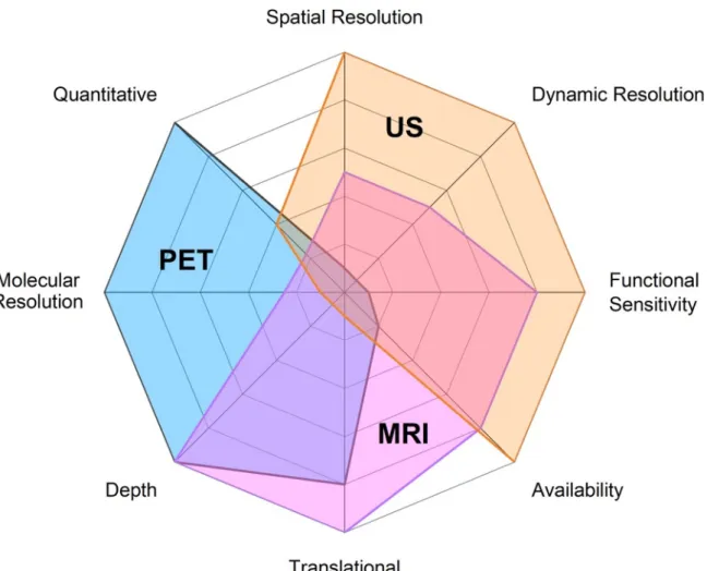
Beyond the anatomical characterization that medical imaging can bring, three main non-invasive modalities are being developed to map brain function in vivo. PET is a molecular imaging technique which allows in vivo estimates of biological processes at the molecular and cellular level. It is based on the injection of a molecular probe labeled with a positron emitter radionuclide, followed by the detection of photons by a ring of detectors. From the detected signals, a quantitative map of the probe concentration within the body is reconstructed with a spatial resolution of the order of 3 to 4 mm in humans and 1 mm in rodents, and a temporal resolution of a few tens of seconds. Compared to other molecular imaging modalities, PET is characterized by a very high molecular sensitivity: it can detect between 10-11 mol/L and 10-12 mol/L
concentrations (Fig. 1).
Functional MRI often refers to the estimation of brain activity by using the endogenous blood-oxygen–level dependent (BOLD) contrast to detect hemodynamic changes. It is characterized by a spatial resolution of ~2 mm in humans and ~0.5 mm in rodents, and a delay of a few seconds between the start of the neuronal activation and the maximum of the BOLD signal. Hence, it cannot capture faster changes. Perfusion MRI can also be considered as a functional modality, with the estimation of quantitative CBV and cerebral blood flow (CBF) maps, unlike for the BOLD contrast. Arterial spin labelling (ASL) is a perfusion technique that does not require the injection of a contrast agent. However, ASL is less advanced than BOLD imaging, the latter being the modality of choice to measure brain activity. Other perfusion techniques like dynamic
susceptibility contrast (DSC) and dynamic contrast enhanced (DCE) are based on the use of suitable contrast agent (McGehee et al., 2012) (Fig. 1).
Functional ultrasound is the fastest imaging modality to detect small hemodynamics changes in the brain (Macé et al., 2011). For now, fUS is mainly used in preclinical protocols to capture rapid change in CBV (~seconds) associated with changes in brain function (Fig. 1) (Osmanski et al., 2014; Sieu et al., 2015). Compared with PET and MRI, this technique is highly invasive, since it requires a craniotomy or reduction of skull thickness to access the brain for animals bigger than mice, which have a very thin skull bone, (Deffieux et al., 2018). Indeed, ultrasound are blocked by the skull bone which distorts the propagating ultrasonic wave front. Methods based on simulation of ultrasonic wave propagation are currently developed to try to solve this issue, but they are animal dependent and time consuming, which limits their applications for real time feedback (Pinton et al., 2012). It was however shown that ultrasonic contrast agents such as inert gas microbubbles may improve ultrasonic signal-to-noise ratio so that fUS without invasive craniotomy can be envisioned (Errico et al., 2016).
Comparison of the technical performances of PET, fMRI and fUS for neuroimaging is represented in figure 1. It is striking that PET and fMRI show great complementary in their performances while sharing minimally invasiveness, allowing for clinical neuroimaging and 3D acquisition of the whole brain, thanks to suitable imaging depth. fUS excels where PET falls short. The limited clinical perspectives of fUS however suggests the relevance of the PET/fUS combination for preclinical research, with potentially better availability than hybrid PET/fMRI (Fig. 1). Functional US benefit from exquisite spatial resolution (~100 µm). Functional sensitivity, which describes the ability to detect moderate change in brain function, is higher for fUS than fMRI. It is widely admitted that fUS can estimate small changes in cerebral blood flow (CBF) with better accuracy that fMRI (Deffieux et al., 2018). The functional sensitivity of PET critically depends on the pharmacokinetic and pharmacologic properties of radiotracers used. The dynamic resolution refers to the time needed to explore one state of brain function. Functional US benefits from excellent dynamic resolution, with a framerate >10 000 frames/s, leading to a real-time feedback (~1 Hz). Compared with fMRI and fUS, PET suffers from a low dynamic resolution, which is not related to the time-resolution and framerate of PET acquisition but critically depends on radiotracer properties: compounds with slower brain kinetics following a bolus injection, with slow
peak radioactivity and slow washout from targeted brain regions, require longer acquisition duration to generate quantitative outcome parameters (Pike, 2016). Availability of imaging devices is more limited for PET, since it requires dedicated radiotracers to be produced or purchased, along with dedicated facilities for radioprotection. Clinical MRI is widely available in most radiology departments. Although fUS is an emerging technique, it can be easily installed in most laboratories with lower cost than preclinical PET or MRI. The translational perspectives MRI and PET are evident, although PET procedures involve injection of radiopharmaceuticals and may require arterial blood sampling which may limit its clinical use. fUS has demonstrated its value for human application in peroperatory (Imbault et al., 2017)and in newborns (Demene et al., 2017) but remains very difficult to be performed in most situations due to skull aberrations. Imaging depth is consistently very limited for fUS without craniotomy or natural windows on the brain. PET has supremacy on molecular resolution since this method provides information regarding biochemical or pharmacological events occurring in the brain rather than change in hemodynamics. Quantitative imaging is also mastered by PET thanks to accurate and absolute radioactivity counting. Recent data suggest that fUS can discriminate different levels of brain function with suitable accuracy (Rabut et al., 2020).
Technical hybridization of neuroimaging techniques
There are many available software solutions for fusing PET data with separately acquired MRI images satisfactorily. However, only hybrid PET/MRI system can provide simultaneous functional information representing disease states (Zhu and Zhu, 2019). The proposal to combine PET and MRI is not recent and goes back to the late 1980s, when Bruce E. Hammer filed in 1989 a patent for a hybrid system where a ring of PET detectors is inserted inside the bore of a MR. Since the beginning, the objective was to realize simultaneous PET and MR acquisitions, which raised major technological challenges to operate a PET detector inside a high magnetic field, in the presence of radiofrequency pulses and gradient switching. The first prototypes were developed for rodent imaging in the late 1990s and early 2000s, with the MR sensitive parts of the PET detectors positioned 4 meters away from the magnet, resulting in severe loss of sensitivity (Marsden et al., 2002). Thanks to major improvements in the technology of solid-state photodetectors, insensitive to the magnetic field, the first prototypes with fully MR compatible PET detectors were developed in the mid-2000s, initially applied to rodent imaging (Judenhofer et al., 2007) and shortly after to human brain imaging (Schlemmer et al., 2008). The first fully integrated whole-body PET/MR system for clinical applications was marketed in 2011 (Delso et al., 2011). Since then, a few hundred systems were installed worldwide from four different manufacturers, both for clinical and research applications, mainly in oncology and neurology. Some of them only permit sequential (non-simultaneous) PET/MR acquisitions.
The hybridization of PET with US is in its infancy although there is no major technological challenge in simultaneously operating both modalities. Jean Provost and colleagues reported on the first hybrid system, PETRUS, allowing for the simultaneous acquisition of PET and ultrafast ultrasound (UUI) data for rodent imaging (Provost et al., 2018). The instrument was assembled from an existing pre-clinical PET-CT system and a clinical UUI device, with the addition of a motorized micro-positioner to operate the US probe within the PET/CT field of view. They showed no significant degradation of PET image quality due to the presence of the US probe (Perez-Liva et al., 2018) and demonstrated the feasibility and interest of hybrid PET/CT/UUI for cancer and cardiac multi-parametric 3D imaging in mice and rats (Facchin et al., 2020). They also mentioned the interest of PET and UUI hybridization for functional brain imaging in rodents. To the best of our knowledge, no simultaneous PET/US acquisition for functional brain imaging has been reported so far in the literature.
Methodological challenge
Practical consideration
The practice of hybrid, multimodal imaging protocols for the study of brain function is still in progress. This situation does not only reflect the recent availability of hybrid PET/MR scanners for clinical practice and research. It also shows that protocols used in single modality imaging systems do directly translate into feasible and/or relevant projects to be used on costlier and less available hybrid systems. Only few teams have so far conceptualized their experience on the design of protocols for hybrid neuroimaging for neuroscience research (Sander and Hesse, 2017; Sander et al., 2020).
There is still a large majority of studies describing the use of hybrid PET/MR systems for studies that could theoretically be performed using independent PET and MR sessions performed in the same individuals. It is worth noting that neuroimaging clearly benefits from recent technological improvement of the performances of PET and MR systems. High-end PET/MR systems often include recent and high-performance PET and/or MR systems that justify their choice over available systems in certain imaging centers. It cannot however be neglected that the presence of the MR magnet complicates the access to the scanner during acquisition. Pharmacokinetic modelling is the gold standard for correct interpretation of brain PET data using many radiotracers, which requires injection of the patients under the PET camera, at the start of PET acquisition, and repeated arterial blood sampling to measure the kinetics of parent (unmetabolized) radiotracer in the plasma (Innis et al., 2007). The staff may need to use metal-containing shielding or systems for blood sampling that are not MR-compatible. Moreover, the staff usually goes in and out during PET acquisition to limit radiation exposure after radiotracer injection which is usually prohibited during fMRI acquisition because it may interfere with the measurement of fMRI parameters. PET imaging is a silent environment, and the noise of most MR sequences may impact brain function assessed using PET (Chonde et al., 2013). This noise requires protection headphones which impact on photon attenuation has to be anticipated in the absence of CT scanner or transmission source in available PET/MR systems (Tellmann et al., 2018; Mackewn et al., 2020).
MRI is assumed to be less invasive than PET because it does not systematically involve injection of pharmaceutical product. In animals however, contrast agents such
as ferumoxytol are often injected at pharmacological dose to improve the functional sensitivity of fMRI to detect change in the BOLD signal. The impact of contrast agents on PET radiotracer PK and binding, as well as the availability of the investigated target has to be systematically investigated to avoid any bias in the estimation of PET-derived parameters (Muehe et al., 2020).
Post-acquisition co-registration of functional PET images on anatomical MR images still remains the standard method to compensate for the limited anatomical performances of PET or PET/CT to delineate brain regions. However, there are situations where PET radioligands show extremely low brain uptake and offer limited anatomical landmarks, simultaneous PET/MR acquisition enables accurate contouring of the regional kinetics of the brain PET signal (Marie et al., 2019). Moreover, some patients with debilitating conditions are not likely to accept or even withstand several imaging sessions, even during the same day.
PET/MR acquisitions are fully justified in many situations encountered during neuroscience research, when the time between acquisitions in each modality becomes a confounding parameter for the interpretation of imaging data. Acquisitions performed several hours to months apart may account for significant difference in brain function because many pathophysiological parameters (stress, motivation, fatigue, circadian rhythm, etc.) may impact the outcome of imaging data so that comparison and correlation between PET and fMRI data may be difficult or even misleading. Cognitive paradigms involving memory or learning tasks cannot be repeated without learning from the first imaging session to the next one. In these situations, sequential (non-simultaneous) imaging systems offer the possibility to drastically reduce the time between PET and MR acquisition which improves bimodal imaging protocols in most situations (Chen et al., 2018).
Hybrid protocols for synchronized neuroimaging
Most current PET-fMRI studies in the literature compared the fMRI-derived “resting-state” brain connectivity with the PET-derived neuroreceptor binding or metabolism in brain regions in the absence of any intervention in healthy and pathophysiological conditions (Muzic et al., 1998; Riedl et al., 2014, 2016; Aiello et al., 2015; Marchitelli et al., 2018; Scherr et al., 2019; Ripp et al., 2020). This paradigm offers the possibility to address neuroreceptor or biochemical correlates underlying disease- or state-induced change in functional connectivity in the same individual, at the same time (Cavaliere et al., 2018).
This multimodal paradigm can theoretically be performed using separated or non-simultaneous PET/MR systems (Karjalainen et al., 2017; Dubol et al., 2018) and does not exploit the full potential of simultaneous hybrid imaging. However, simultaneous acquisition allows to derive correlation maps that come from both modalities to describe the dynamics of the coupling of PET- and MR-derived parameters (Aiello et al., 2016).
Creative designs of PET/MR protocols have to be imagined to make the best with the combination of simultaneously acquired functional data (Sander et al., 2020). The use of simultaneous hybrid scanners is especially relevant to investigate time-dependent change in brain function. Imaging protocols or paradigms have to be defined to study the functional impact of events named “interventions”, hypothesized to precipitate within-scan changes in brain function, to be simultaneously detected by both modalities (Fig. 2). Behavioral intervention involves cognitive task or stimuli which have driven the applicability of fMRI in translational neuroscience over the past 30 years (Bandettini, 2012). Pharmacological interventions involve CNS acting drugs with impact of neurotransmission or expected change in brain physiology, which made the philosophy of pharmacological neuroimaging. Pharmacological challenges are widely used by the PET community to validate new radiotracers or study the impact of investigated drugs on brain function (McCluskey et al., 2020). The use of fMRI to study the effects of CNS-acting drugs, the so-called pharmaco-fMRI (phMRI) aims at determining drug-induced changes on disease relevant networks, and non-invasively prove pharmacological effects induced by acute or chronic administration of investigated drugs at the CNS level (Wandschneider and Koepp, 2016). Although limited molecular information can be gained compared with PET, regional analysis of phMRI data provide a pragmatic mean for early assessment of treatment response which does not depend on the availability of target-specific imaging probe. Pharmacological MRI is therefore increasingly used in CNS drug development to predict mechanisms of drug efficacy and safety in animals and humans, including healthy volunteers and patients (Mandeville et al., 2014). Neuropharmacology is also a highly promising application domain for fUS (pharmaco-fUS) to address the CNS effects of drugs, at least in animals (Rabut et al., 2020; Vidal et al., 2020).
MR-derived BOLD signal measured using fMRI or CBV measured using fUS is very transient and its estimation is only possible thanks to adequate dynamic resolution (Fig. 1). Block intervention paradigm with repeated rest/task cycles can be performed
to improve the functional sensitivity of the technique to detect change in brain function
(Amaro and Barker, 2006). This strategy is difficult to be used for PET imaging protocols because it takes much more time for the radiotracer to reach pharmacokinetic equilibrium and capture a functional state of the brain (Fig. 1). PET imaging is predominantly performed after injection of a single bolus dose of radiotracer. As a consequence, PET captures a functional or biochemical state of the brain that occurs at the time of radiotracer injection, with the assumption that this state does not vary too much while the radiotracer diffuses to the brain and interacts with the investigated biomarker (~20 minutes to hours) (Fig. 1). In most situations, the dynamic resolution of PET is much lower than that of fMRI (~minutes) or fUS (~seconds). Therefore, in pharmacology studies, equilibrium of plasma concentrations is often required to maintain constant drug exposure during the whole scan to estimate receptor occupancy or changes in brain function associated with controlled and stable plasma levels of the investigated drug. The release of endogenous neurotransmitters induced by CNS-acting drugs or cognitive-task can also be captured by PET as it competes with the binding of radiotracers at their specific target receptor. Repeated PET experiments are therefore performed before (baseline) and after (challenge) drug administration or task to compare their impact, which requires at least two consecutive PET experiments (Fig. 2A) (Sander et al., 2020). This common PET design is not optimal for fMRI or fUS, for which high variability in baseline estimate exists, so that conditions have to be preferentially tested within a single imaging session (Jenkins, 2012; Mandeville et al., 2014).
Microdose radioligands used for PET experiments are specifically designed not to exert any pharmacological effect. This property guarantees the toxicological safety of PET experiments which fosters the acceptability of their translation to humans. Microdose PET experiments are therefore not likely to induce change in brain function to be detected by other functional imaging techniques such as fMRI or fUS. Ideal PET radioligands have to freely diffuse across the blood-brain barrier (BBB) and bind to investigated target with limited non-specific and/or off-target binding to the brain (Pike, 2009). Non-specific binding of the radiotracer is assumed to be not saturable and is therefore non-displaceable by pharmacologic doses. Radiopharmaceutical preparations contain a certain amount of unlabeled analogue of the radioligand to be determined to assess the molar activity, a.k.a specific activity, which is the ratio between activity and mass of compound, expressed in MBq/µmol or MBq/µg. High
molar activity is often required for radiotracers to freely bind and map available target in the brain. Using low molar activity preparation, the binding of radiotracer may compete with the binding of unlabeled ligand, leading to reduced uptake in the target region (Fig. 2A). This situation is typically avoided in conventional PET studies because it may induce unwanted but target related pharmacological effects. Interestingly, the use of low specific activity preparation has been steered toward pioneer pharmacological PET/MR protocols (Sander et al., 2013). For short-lived isotopes such as carbon-11 (T1/2 = 20.4 min), controlled decrease in specific activity
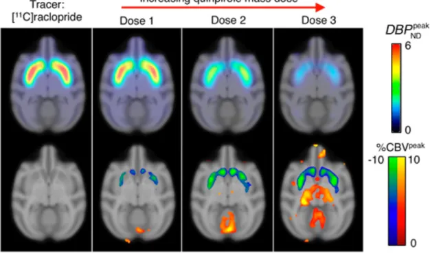
can be obtained by waiting for radioactive decay, thus decreasing the proportion of radiolabeled compound relative to its non-labelled analogue in the preparation. Alternatively, the radiopharmaceutical preparation can be spiked by a selected amount of unlabeled ligand. Many PET radioligands are chemical analogues of drugs which tolerability at pharmacological dose is well described, so that this strategy can theoretically be transferred to humans. Raclopride is a prototypical D2/D3 dopamine receptor antagonist and [11C]raclopride is widely used as PET probe to these specific
receptors. In a seminal paper, Sander and colleagues tested repeated PET/fMRI experiments in nonhuman primate using several preparations of [11C]raclopride with
decreasing molar activity (i.e with increasing mass dose of unlabeled raclopride). Using this strategy, it was possible to correlate MR-derived change in CBV with the PET signal as a function of injected dose, time and space. They showed that the vascular response was tightly coupled to PET-derived changes in receptor occupancy, which provided a minimally-invasive method to quantitatively explore the neurovascular coupling associated with the inhibition of D2/D3 receptors in vivo (Sander et al., 2013).
Several PET protocols have been developed to track change in brain function within a single PET acquisition (Fig. 2). These protocols are theoretically suited for the detection of events occurring during the scan, which enables within-subject studies with lower variability than repeated experiments. This paradigm may better fit the design of functional neuroimaging paradigm used for fMRI or fUS, making the best with the simultaneous acquisition of different imaging modalities (Sander et al., 2020). Displacements studies are widely performed as strategy for the validation of radiotracers, especially those used for neuroreceptor imaging. Pharmacologic dose of the investigated ligand or a reference and target-specific ligand can be injected during the time-course PET acquisition, several minutes to hours after the injection of the
radiotracer (Fig. 2B). In some situations, competition of the pharmacologic levels of exogenous or endogenous ligand with the binding of the PET radioligand leads to in-course change of the PET signal in target containing brain regions (Ceccarini et al., 2012; Pike, 2016). The extent of displacement can be estimated as the relative change in brain radioactivity after displacement relative to baseline (before intervention) or control (experiments without intervention). For some radiotracers, regions with low expression of the investigated target can serve as reference region to ensure the target-specificity of the displacement (Matthews et al., 2012). However, optimal timing for displacement is difficult to define: it should ideally be performed at the maximum of radiotracer binding, which is hardly predictable. For reversible radioligands, level and kinetics of displacement closely depends on the pharmacokinetics of both the tested drug and the radiotracer, and their respective association-dissociation kinetics at the target level (Pike, 2016). Drugs used for pharmacological challenges are therefore preferentially injected IV to achieve controlled levels in plasma and the brain, for improved control of the time-frame of interaction with the CNS target. For irreversible radioligands, displacement has by definition negligible effect. Co-injection or pre-treatment is the only option to highlight the specific binding of irreversible radioligands using a pharmacological challenge.
The sensitivity of PET tracer to detect changes in the concentration of an endogenous competitor via displacement of the tracer, i.e its vulnerability to competition, also closely depends on its pharmacokinetic and receptor-binding properties (Morris and Yoder, 2007). The possibility of within-scan determination of the release of endogenous ligand also depends on the consistency of the phenomenon with the dynamic resolution offered by the selected PET radioligand (Fig. 1). Despite some limitations, within-scan intervention offers a pragmatic and semi-quantitative technique to localize and rank the target-specific binding across different brain regions using PET. This paradigm has however less quantitative properties than full kinetic modelling using two consecutive PET scan (Honer et al., 2014) (Fig. 2A). In the perspective of the design of simultaneous PET/MR studies, within-scan intervention appear to be a good compromise to gain PET-derived information on targeted neuroreceptor while enabling functional neuroimaging data before and after displacement, thus avoiding the test-retest variability associated by repeated experiments (Baumgartner et al., 2018). It is likely that full PET kinetic modelling, which is the gold standard in PET experiments, will not be the standard for most PET/MR protocols. Simplified PET
methods have to be developed for each radioligand to provide spatially localized and quantitative information on target engagement or endogenous neurotransmitter release, to be compared with synchronized fMRI or fUS data (Irace et al., 2020).
Methodological efforts have recently been made to exploit the synchronicity of PET and fMRI acquisition, thus fostering the use of simultaneous PET/MR systems to track changes in brain function with improved dynamic resolution. Radiotracer infusion of [150]H
20 with kinetic modelling is the gold standard method for quantitative
measurement of the CBF. [150]H
20 can measure the resting state CBF in various
disease states, and investigate the response to pharmacological or behavioral intervention (Imaizumi et al., 2004). Continuous infusion of [150]H
20 rather than bolus
injection is mainly justified by the very short radioactive decay of oxygen-15 (~2 min). Today, fMRI has overtaken [150]H20 PET in most situations excepts contra-indication
of MR experiments or the need for silent conditions (Coez et al., 2009). Conventional bolus injection of [18F]FDG provides an average glucose uptake into the brain over an
extended period of ~30 min. Villien et al. (2014) applied continuous infusion of [18F]FDG PET to study the dynamics of glucose uptake in the brain (Villien et al., 2014).
These methods named functional PET (fPET) gained recent interest because they tent to push PET imaging into fMRI-like paradigms, by isolating changes in glucose metabolism induced by single or repeated intervention, with better dynamic resolution
(Hahn et al., 2018; Li et al., 2020). Carson and co-workers have initiated the use of radiotracer infusion for neuroreceptor imaging (Carson, 2000). The basis of constant infusion or bolus/infusion method is to ensure controlled and constant delivery of radiotracer to the brain so that pharmacokinetic equilibrium (steady-state) is reached and maintained during the scan (Fig. 2C). Time-dependent change in the PET signal induced by intervention is therefore more specifically related to change occurring at the molecular target level, which is intended to simplify the interpretation of PET data, while improving dynamic resolution and functional sensitivity to track change in target availability (Carson, 2000). This method requires specific development for each radiotracer and is not as convenient as bolus injection for the clinical practice. In the perspective of PET/MRI protocols, the bolus/infusion strategy was optimized for quantitative determination of the serotonin transporter (SERT) binding in humans using [11C]DASB (Gryglewski et al., 2017). Quantification of SERT occupancy
associated with a single IV dose of citalopram, injected during radiotracer infusion, was then validated (Gryglewski et al., 2019; Silberbauer et al., 2019). Prolonged
steady-state enabled by infusion lets room for within-scan fMRI sequences before, during and after intervention, which may theoretically allow for block studies of brain function when studying transient phenomenon (Fig. 2C). In theory, this paradigm may be better suited for task intervention rather than pharmacological imaging due to cumulative levels of drugs associated with repeated administrations.
Scientific outcome of PET-fMRI studies
The availability of hybrid PET/MR systems have paved the way for a large field of research based on the concept of bimodal functional neuroimaging. Hybridization of PET and fMRI first offered many opportunities to validate or improve measurement made by each modality. Dynamic brain PET studies can last more than 1 h. Within scan voluntary and involuntary head motions are almost inevitable and have to be monitored to avoid head-motion artifacts (Bloomfield et al., 2003). This is particularly true in the elderly, children or in patients with dementia or movement disorders. Head restraints can be used to reduce head motion but head movements still cannot be completely eliminated (Bloomfield et al., 2003). The possibility for repeated anatomical MR sequences with the development of MR-active markers has been proposed to track head motion in real time during a simultaneous PET/MR brain scan, and directly incorporate the motion measured by the markers in the reconstruction of dynamic brain PET images (Huang et al., 2014).
PET offers quantitative imaging for many neurochemical and neurophysiological events such as brain perfusion or glucose metabolism. Several studies astutely used hybrid imaging to validate MR-derived protocols to estimate these parameters. PET with 15O-radiolabeled water ([150]H20), which requires technically demanding arterial
blood sampling, is considered the gold-standard method in quantification of CBF in brain regions (Herscovitch et al., 1983). Different image-derived input function (IDIF) methods have been proposed to limit the invasiveness of the procedure. Time-of-flight (TOF) MRI is useful to localize brain feeding arteries and measure corresponding time-activity curves on co-registered PET images. However, the quantitative value of kinetic modelling using IDIF is limited given the low spatial resolution of PET relative to the small diameter of carotid and vertebral arteries. Khaligi and coworkers (2018) took advantage of the simultaneously acquired combination of MR-TOF-derived angiogram and initial [150]H20 PET data to improve the robustness of CBF estimation in patients
(Khalighi et al., 2018). Phase-contrast MRI (PC-MRI) can measure blood flow velocity and vessel area quickly and noninvasively, which was previously investigated as an index of the global brain perfusion (Vestergaard et al., 2017). Ssali and coworkers combined PC-MRI with simultaneous [150]H
20-PET data to create quantitative
perfusion maps on a PET/MRI scanner. In pigs, they validated a minimally-invasive PET/MRI approach using global CBF determined by phase-contrast MRI as a normalization factor (Ssali et al., 2018). The clinical relevance of this strategy was recently demonstrated in healthy volunteers and patients with Moyamoya disease as a model of chronic ischemic disease (Ishii et al., 2020).
Estimation of CBF is also possible by Doppler ultrasound (Oktar et al., 2006; Han et al., 2007; Purkayastha and Sorond, 2012) or the clinically feasible MR-derived ASL (Jezzard et al., 2018). However, many factors may impair the quantitative value and reproducibility of ASL (Mutsaerts et al., 2020). Availability of hybrid PET/MR scanner has made it possible to directly compare quantitative regional CBF estimated using ASL-MRI and absolute quantitative [150]H20-PET (bolus injection), simultaneously
obtained in the same subjects in rest, during hyperventilation, and after acetazolamide challenge, a pharmacological intervention aiming at increasing CBF (Vorstrup et al., 1984; Puig et al., 2019). The authors showed that ASL-MRI and [150]H20-PET
measurements of regional CBF were highly correlated across different perfusion states. However, they showed poorer correlation within individual states, in particular in resting state measurements, and only moderate correlation in hemodynamic reactivity (Puig et al., 2019).
Brain [18F]FDG PET with kinetic modelling provides a biochemically relevant
estimation of brain function because glucose metabolism is tightly linked to neuronal activity (Varrone et al., 2009; Mergenthaler et al., 2013). However, radiation exposure and the relatively low spatial resolution of PET have made fMRI the current standard for functional neuroimaging experiments. fMRI-BOLD provides an indirect and poorly quantitative estimation of neuronal activity. Indeed, the BOLD signal depends on the complex interplay of hemodynamic parameters including the cerebral metabolic rate of oxygen, CBF and CBV changes (Gauthier and Fan, 2019). Interestingly, comparison of simultaneously acquired [18F]FDG and BOLD-fMRI have been used to
untangle the metabolic correlate for fMRI signal in rats in resting and activated state (whisker pad stimulation) (Wehrl et al., 2013). The authors observed a regional mismatch, which was explained by the different biochemical basis of each technique,
suggesting complementarity rather than competition of both modalities to explore brain function in vivo. More recently, human studies confirmed this observation and revealed the potential of the hybridization of [18F]FDG-fPET (infusion protocol) with fMRI to
provide insights into dynamics of hemodynamic and metabolic interactions during visual stimulus in a block design paradigm (Jamadar et al., 2019).
Full pharmacokinetic modelling requires dynamic PET acquisition from injection to binding equilibrium, which is difficult for clinical routine in patient, especially for radioligands with slow kinetics such as the β-amyloid binding radioligand [18F]florbetapir (Auvity et al., 2020). In these situations, binding can be estimated from
late static PET images acquired several minutes or hours after radiotracer injection. In the absence of early PET data, it is not possible to estimate the extent of initial delivery of the radiotracer, which is highly dependent on CBF and may confound the estimation of [18F]florbetapir binding. Scott et al. therefore incorporated CBF information from
ASL-MRI into the PET pharmacokinetic modelling of [18F]florbetapir. They showed that
ASL-corrected PET data acquired from 20 to 50 min post injection were strongly correlated with [18F]florbetapir binding estimated using the standard 60 min
acquisition, thus allowing for reduced length of PET acquisition in patients (Scott et al., 2019).
Translational molecular imaging is so far dominated by PET. However, magnetic resonance spectroscopy (MRS) can estimate brain concentration of metabolites in vivo. PET/MRS therefore offers perspectives for the hybridization of molecular information simultaneously acquired in the same subjects. This strategy can be used to demonstrate associations between PET-derived marker of neuroinflammation and MRS-derived markers for neuronal integrity, levels of neurotransmitters or the redox status in patients with various neuropsychiatric diseases (Hafizi et al., 2018; Ratai et al., 2018; Da Silva et al., 2019). O'Gorman Tuura and colleagues ingeniously used this approach to test the sensitivity (or vulnerability) of the glutamatergic radioligand [18F]PSS232 to detect shifts in cerebral glutamate levels precipitated by IV injection of
N-acetyl-cystein (O’Gorman Tuura et al., 2019).
Seminal [11C]raclopride PET/MR work performed by Sander and colleagues in
anesthetized nonhuman primates revealed the synchronicity of levels of receptor blocking by dopamine antagonist and consequent decrease in CBV (Sander et al., 2013). More recently, they used a bolus-infusion strategy for [11C]raclopride to track
dopamine agonists in monkeys (Sander et al., 2016). They showed that selective D2/D3 agonists elicited dose-dependent increases in receptor occupancy, together with change in CBV, in caudate and putamen (Fig. 3). Using this strategy, it was moreover possible to perform a longitudinal monitoring of the pharmacological response to repeated pharmacological challenges performed within a single imaging session (Sander et al., 2016). Birn and coworkers used [18F]fallypride, another
D2/D3-specific radioligand, to track change in the synaptic content of endogenous dopamine induced by increasing doses of methylphenidate (Birn et al., 2019). Interestingly, it was shown that dramatic changes in CBF induced by hypercapnic challenges, assessed using MR-derived ASL, did not affect the brain PET kinetics of [11C]raclopride or
[18F]fallypride. Change in CBF, that often occurs due to behavioral tasks or
pharmacological challenges, was therefore not likely to affect the outcome or PET data using these specific radiotracers (Sander et al., 2019). Altogether, pioneer work performed around dopamine receptors as model CNS target illustrates the invaluable potential of pharmacological PET/MRI to study the neurovascular-coupling mechanism during receptor-specific modulation by endogenous and exogenous ligands (Sander et al., 2020).
Perspectives
The relevance of brain PET/MR systems for diagnostic is mainly justified by the co-acquisition of multiple parameters derived from both modalities. In a pragmatic way, multiparametric imaging enhances the chance to detect change in brain function relevant to the investigated phenomenon since functional sensitivity of each modality is difficult to predict (Fig. 1). This is a competitive advantage to explore multiple hypothesis in a limited number of individuals. In the clinical practice, it is likely that the combination of multimodal parameters may improve the accuracy of diagnostic compared with each individual modalities (Tahmasian et al., 2016; Chen et al., 2018). So far, the literature on hybrid PET/MR in neuroscience research is still dominated by methodological and proof-of-concept studies. This suggests that the philosophy of PET/MR protocol design is maturating. Developments are therefore needed to make the best with the hybridization of functional neuroimaging techniques (Sander et al., 2020).
Simultaneous PET/MR was shown a relevant tool to validate or improve the performance of each other modality. PET-derived outcome parameters theoretically
benefit from absolute quantification, although experimental variability is a daily undertaking (Fig. 1) (Baumgartner et al., 2018). Cross-calibration studies may thus enable validation of MRI-based functional estimates (Zhu and Zhu, 2019). Normalization of fMRI remains a challenge. Today, it is still difficult to quantify the magnitude of the BOLD fMRI signal between MR scanner, between individuals and even between sessions in the same individual. Normalization methods for fMRI have been proposed to allow for large multicentric studies using different scanners and/or repeated scans in the same individual in a longitudinal way, thus matching the design of most PET protocols (Cohen et al., 2004; Driver et al., 2017). ASL becomes more widely adopted in research and clinical settings, efforts have sought to standardize the method and validate its CBF estimates against the PET-based gold-standard. The importance of such differences relies upon the specific context and experimental setting, and whether expected error is acceptable or systematic. Future studies could take advantage of PET/MRI cross-comparison to validate standardized ASL protocols or at least, to more clearly identify the populations or situations were ASL-MRI can be faulted to correctly estimate CBF. Once ASL is validated, clinicians will gain a noninvasive, radiation-free, available, robust and repeatable imaging method with to investigate quantitative perfusion in the brain (Fan et al., 2016).
It is also likely that hybrid PET/MRI systems could be used to reduce the invasiveness of PET or improve its quantitative performances. IDIF is an attractive but challenging noninvasive alternative to arterial blood sampling, it is also a very challenging field associated with diverse problems that impede its broader use in PET-alone studies (Zanotti-Fregonara et al., 2011). Precise brain co-registration MRI, with improved contrast in brain vessels and perfusion data may be used as an alternative or a complement to arterial input function to derive hybrid PET/MR kinetic models that would take information from both modalities to elucidate the brain kinetics of radiotracers (Sari et al., 2014; Zhu and Zhu, 2019). This strategy may be particularly suited for PET radioligand which initial brain uptake closely depends on brain perfusion. For such radioligands, preliminary [150]H
20-PET imaging can be performed
for quantitative determination of CBF and implementation in kinetics models (Deo et al., 2014). It is much likely that MRI-guided estimation of IDIF and CBF may be increasingly employed as methods to improve the accuracy of PET kinetic modelling, or for normalization purpose (Sander et al., 2019; Zhu and Zhu, 2019).
The design and validation of fMRI paradigms is a complex research field. Once validated, fMRI paradigms can apply to various pathophysiological states to test corresponding hypotheses on brain circuitry and function (Bandettini, 2012). The PET equivalent is the validation of radioligands for which valid and feasible kinetic models are needed to study corresponding brain function in multiple situations (Honer et al., 2014). Task-related or pharmacological interventions are shared by both modalities as mean to precipitate change in brain function (Fig. 2). Intuitively, it is likely that future PET/MR protocols for neurosciences will have to meet the expectations of both modalities, which may dangerously narrow the diversity of investigated parameters. Today, the most advanced dynamic PET/MR paradigms, including intervention, are designed to investigate the neurovascular coupling associated with the dopaminergic receptors (Sander et al., 2020). This probably reflects the reliability of dopaminergic radioligands for which simplified models exist, and can be customized for simultaneous PET/MR acquisition. It should be kept in mind when developing new radiotracers for PET that their applicability for PET/fMRI studies should be considered for in-depth and multimodal exploration of brain function associated with the investigated target.
Molecular fMRI is a new concept based on the development of MRI-detectable molecular probes to study brain function in vivo (Sinharay and Pagel, 2016; Ghosh et al., 2018). Dual-modality imaging probes, that can be simultaneously detected by several modalities, may therefore provide a multiparametric and multiscale mapping of brain function, making the best with the performances of each modality (Yang et al., 2018a). However, noninvasive brain delivery of MRI contrast agents requires a strategy for getting them across the BBB, which is a major limitation compared with small-molecule radiotracers used for PET. For this reason, the most advanced MR-dedicated contrast agents for molecular neuroimaging aim at studying the integrity and function of the blood-brain barrier. Physical integrity of the BBB can be studied in vivo using contrast-enhanced MRI using gadolinium-chelates that do not cross the intact BBB (Magnin et al., 2015). MRI provided many information regarding the functional impact of transient disruption of the BBB induced by focused ultrasound with microbubbles. MR and PET have successfully monitored the dynamics of BBB disruption (Conti et al., 2019), its impact for brain delivery of therapeutics (Goutal et al., 2018), as well as the consequences on brain metabolism (Arif et al., 2020) or glial activation (Sinharay et al., 2019). MR-molecular imaging of endothelial cell activation
is also highly relevant, at least in a preclinical setting, since molecular targets expressed by the brain vasculature facing the blood are easily accessible by large contrast-carrying particles (Gauberti et al., 2018). The increasing diversity of MR-dedicated molecular imaging probes can be combined with MRS and PET to provide a larger and multiparametric landscape of the molecular environment in certain brain regions, which may be useful to untangle complex signaling processes, in a longitudinal way. In this framework, magnetic particle imaging (MPI) is increasingly regarded as an alternative imaging technique to selectively track and quantify magnetic nanoparticles in the brain (Wu et al., 2019). Human-sized MPI has been recently proposed as mean to explore the neurovascular system, with great potential for hybridization with complimentary neuroimaging techniques (Graeser et al., 2019) . The clinical pharmacodynamics of many CNS-acting drugs is difficult to predict (Srinivas et al., 2018). PET and phMRI have played an unevaluable role to elucidate the mechanism of action of drugs at the CNS level, with rapid clinical perspectives (Suridjan et al., 2019). In neuropharmacology, a large variability in response exist, which mechanisms remains misunderstood. This variability may involve the downstream processes of pharmacokinetics (i.e ability of the drug to reach the target tissue), interaction with the CNS target, or transduction of the neuroreceptor signaling (Srinivas et al., 2018). Conventional PET studies using target-specific radioligands are useful to estimate baseline target availability and drug induced target engagement. This strategy offers an indirect but target-restricted insight into the effects of drugs to the brain. Isotopic radiolabeling of drugs is sometime possible, which enables direct consideration of the brain kinetics, including transport across the BBB, as factor of variability for pharmacodynamics (Tournier et al., 2018; Bauer et al., 2019). In this framework, hybridization of PET imaging with other functional neuroimaging techniques has a great potential to unveil neurovascular coupling in complex situations such the desensitization of neuroreceptor (Sander et al., 2016) or the action of biased agonists (Vidal et al., 2018), during which a disconnection between regional target engagement and signal transduction may exist.
The novelty and the poor translational perspectives of fUS compared with PET and MRI probably justify the absence of hybrid systems for neuroimaging. To the best of our knowledge, hybridization of UUI with MR has not been performed and requires technological development. MRI has been associated with ultrasound to monitor focused ultrasound therapy (Larrat et al., 2010; Ozenne et al., 2020) of
neurostimulation (Yang et al., 2018b). However, association of fUS imaging with fMRI has not been reported to our knowledge. Functional MRI and fUS offer relatively similar outcome parameters describing neurovascular coupling, with limited molecular information (Fig. 1). fUS may nonetheless be used to validate new fMRI sequences in a translational perspective. Comparison of the performances of fUS with PET strikingly shows the complementarity of these functional neuroimaging techniques (Fig. 1). Recent work on fUS suggest that it is less labor intensive and has great potential to overtake fMRI for preclinical neuroscience (Deffieux et al., 2018). Technical feasibility of hybrid PET/fUS systems has been demonstrated for cardiac and oncologic applications and may gain popularity in the near future (Perez-Liva et al., 2018; Provost et al., 2018). The technical feasibility to perform brain PET/fUS it still complicated by skull aberrations. It can however be hypothesized that PET/fUS may become a new player in hybrid functional neuroimaging in the coming years.
Once hybridization of PET with fUS will be possible, the combination of the three modalities should be envisioned to monitor and interpret the same physiological phenomenon with complimentary biomarkers at multiple temporal scale. As an example, the classical brain activation paradigm, which consists in stimulating whiskers in rodent has been explored using BOLD-fMRI, [18F]FDG-PET and fUS (Fig.
4). Such theoretical trimodal PET/fMRI/fUS study may provide simultaneous information on the brain function in response to intervention, and track change in brain metabolism relative to fMRI-derived blood oxygenation and fUS-derived CBV (Macé et al., 2011; Wehrl et al., 2013).
Conclusion
There are still technical, methodological and cultural challenges to be addressed to accelerate the use of hybrid functional neuroimaging protocols (Table 1). Efforts are being made to develop hybrid imaging systems that enable multiparametric exploration of brain function in a single imaging session in animals and humans. Improving the availability of such systems in preclinical and clinical research centers is a prerequisite. PET/MRI has proven its relevance for translational neuroimaging and PET/fUS has great potential, at least for preclinical application. We think that original acquisition paradigms, that take the full potential and the limitations of each modality into account, remain to be developed to make the best with hybrid neuroimaging systems. Simultaneous acquisition of PET with other neuroimaging techniques may generate hybrid parameters that quantitatively connect the brain structural properties, molecular environment and the neuro-vascular coupling, which may better describe the contexture and synchronicity of brain function than each individual modality. Cross-fertilization of neuroimaging communities and training of young researchers on multiple imaging modalities is therefore essential. This may foster the development of hybrid neuroimaging protocols with synergistic rather than additional outcomes to investigate brain function in health and disease.
Acknowledgement
This work was performed on a platform member of France Life Imaging network (grant ANR-11-INBS-0006). Nicolas Tournier received funding from the french national research agency (ANR-19-CE17-0027-01). We thank Matteo Tonietto and Michel Bottlaender for their kind help on the figures.
Declarations of interest
References
Aiello M, Cavaliere C, Salvatore M (2016) Hybrid PET/MR Imaging and Brain Connectivity. Front Neurosci 10:64.
Aiello M, Salvatore E, Cachia A, Pappatà S, Cavaliere C, Prinster A, Nicolai E, Salvatore M, Baron J-C, Quarantelli M (2015) Relationship between simultaneously acquired resting-state regional cerebral glucose metabolism and functional MRI: a PET/MR hybrid scanner study. NeuroImage 113:111– 121.
Amaro E, Barker GJ (2006) Study design in fMRI: basic principles. Brain Cogn 60:220– 232.
Arif WM, Elsinga PH, Gasca-Salas C, Versluis M, Martínez-Fernández R, Dierckx RAJO, Borra RJH, Luurtsema G (2020) Focused ultrasound for opening blood-brain barrier and drug delivery monitored with positron emission tomography. J Controlled Release 324:303–316.
Auvity S, Tonietto M, Caillé F, Bodini B, Bottlaender M, Tournier N, Kuhnast B, Stankoff B (2020) Repurposing radiotracers for myelin imaging: a study comparing 18F-florbetaben, 18F-florbetapir, 18F-flutemetamol,11C-MeDAS, and 11C-PiB. Eur J Nucl Med Mol Imaging 47:490–501.
Bandettini PA (2012) Twenty years of functional MRI: the science and the stories. NeuroImage 62:575–588.
Bauer M, Tournier N, Langer O (2019) Imaging P-glycoprotein function at the blood-brain barrier as a determinant of the variability in response to CNS drugs. Clin Pharmacol Ther.
Baumgartner R, Joshi A, Feng D, Zanderigo F, Ogden RT (2018) Statistical evaluation of test-retest studies in PET brain imaging. EJNMMI Res 8 Available at: https://www.ncbi.nlm.nih.gov/pmc/articles/PMC5809632/ [Accessed July 9, 2020].
Beyer T, Townsend DW, Brun T, Kinahan PE, Charron M, Roddy R, Jerin J, Young J, Byars L, Nutt R (2000) A combined PET/CT scanner for clinical oncology. J Nucl Med Off Publ Soc Nucl Med 41:1369–1379.
Beyer T, Townsend DW, Czernin J, Freudenberg LS (2011) The future of hybrid imaging-part 2: PET/CT. Insights Imaging 2:225–234.
Birn RM, Converse AK, Rajala AZ, Alexander AL, Block WF, McMillan AB, Christian BT, Filla CN, Murali D, Hurley SA, Jenison RL, Populin LC (2019) Changes in Endogenous Dopamine Induced by Methylphenidate Predict Functional Connectivity in Nonhuman Primates. J Neurosci Off J Soc Neurosci 39:1436– 1444.
Bloomfield PM, Spinks TJ, Reed J, Schnorr L, Westrip AM, Livieratos L, Fulton R, Jones T (2003) The design and implementation of a motion correction scheme for neurological PET. Phys Med Biol 48:959–978.
Carson RE (2000) Pet physiological measurements using constant infusion. Nucl Med Biol 27:657–660.
Cavaliere C, Kandeepan S, Aiello M, Ribeiro de Paula D, Marchitelli R, Fiorenza S, Orsini M, Trojano L, Masotta O, St Lawrence K, Loreto V, Chronik BA, Nicolai E, Soddu A, Estraneo A (2018) Multimodal Neuroimaging Approach to Variability of Functional Connectivity in Disorders of Consciousness: A PET/MRI Pilot Study. Front Neurol 9:861.
Ceccarini J, Vrieze E, Koole M, Muylle T, Bormans G, Claes S, Van Laere K (2012) Optimized in vivo detection of dopamine release using 18F-fallypride PET. J Nucl Med Off Publ Soc Nucl Med 53:1565–1572.
Chen Z, Jamadar SD, Li S, Sforazzini F, Baran J, Ferris N, Shah NJ, Egan GF (2018) From simultaneous to synergistic MR-PET brain imaging: A review of hybrid MR-PET imaging methodologies. Hum Brain Mapp 39:5126–5144.
Chonde DB, Abolmaali N, Arabasz G, Guimaraes AR, Catana C (2013) Effect of MRI acoustic noise on cerebral fludeoxyglucose uptake in simultaneous MR-PET imaging. Invest Radiol 48:302–312.
Coez A, Zilbovicius M, Ferrary E, Bouccara D, Mosnier I, Ambert-Dahan E, Kalamarides M, Bizaguet E, Syrota A, Samson Y, Sterkers O (2009) Processing of voices in deafness rehabilitation by auditory brainstem implant. NeuroImage 47:1792–1796.
Cohen ER, Rostrup E, Sidaros K, Lund TE, Paulson OB, Ugurbil K, Kim S-G (2004) Hypercapnic normalization of BOLD fMRI: comparison across field strengths and pulse sequences. NeuroImage 23:613–624.
Conti A, Mériaux S, Larrat B (2019) About the Marty model of blood-brain barrier closure after its disruption using focused ultrasound. Phys Med Biol 64:14NT02. Da Silva T, Hafizi S, Rusjan PM, Houle S, Wilson AA, Prce I, Sailasuta N, Mizrahi R (2019) GABA levels and TSPO expression in people at clinical high risk for psychosis and healthy volunteers: a PET-MRS study. J Psychiatry Neurosci JPN 44:111–119.
Deffieux T, Demene C, Pernot M, Tanter M (2018) Functional ultrasound neuroimaging: a review of the preclinical and clinical state of the art. Curr Opin Neurobiol 50:128–135.
Delso G, Fürst S, Jakoby B, Ladebeck R, Ganter C, Nekolla SG, Schwaiger M, Ziegler SI (2011) Performance measurements of the Siemens mMR integrated whole-body PET/MR scanner. J Nucl Med Off Publ Soc Nucl Med 52:1914–1922.
Demene C, Baranger J, Bernal M, Delanoe C, Auvin S, Biran V, Alison M, Mairesse J, Harribaud E, Pernot M, Tanter M, Baud O (2017) Functional ultrasound imaging of brain activity in human newborns. Sci Transl Med 9.
Demené C, Tiran E, Sieu L-A, Bergel A, Gennisson JL, Pernot M, Deffieux T, Cohen I, Tanter M (2016) 4D microvascular imaging based on ultrafast Doppler tomography. NeuroImage 127:472–483.
Deo AK, Borson S, Link JM, Domino K, Eary JF, Ke B, Richards TL, Mankoff DA, Minoshima S, O’Sullivan F, Eyal S, Hsiao P, Maravilla K, Unadkat JD (2014) Activity of P-Glycoprotein, a β-Amyloid Transporter at the Blood-Brain Barrier, Is Compromised in Patients with Mild Alzheimer Disease. J Nucl Med 55:1106– 1111.
Driver ID, Wise RG, Murphy K (2017) Graded Hypercapnia-Calibrated BOLD: Beyond the Iso-metabolic Hypercapnic Assumption. Front Neurosci 11 Available at: https://www.frontiersin.org/articles/10.3389/fnins.2017.00276/full [Accessed July 1, 2020].
Dubol M, Trichard C, Leroy C, Sandu A-L, Rahim M, Granger B, Tzavara ET, Karila L, Martinot J-L, Artiges E (2018) Dopamine Transporter and Reward Anticipation in a Dimensional Perspective: A Multimodal Brain Imaging Study. Neuropsychopharmacol Off Publ Am Coll Neuropsychopharmacol 43:820–827. Errico C, Osmanski B-F, Pezet S, Couture O, Lenkei Z, Tanter M (2016) Transcranial functional ultrasound imaging of the brain using microbubble-enhanced ultrasensitive Doppler. NeuroImage 124:752–761.
Facchin C, Perez-Liva M, Garofalakis A, Viel T, Certain A, Balvay D, Yoganathan T, Woszczyk J, De Sousa K, Sourdon J, Provost J, Tanter M, Lussey-Lepoutre C, Favier J, Tavitian B (2020) Concurrent imaging of vascularization and metabolism in a mouse model of paraganglioma under anti-angiogenic treatment. Theranostics 10:3518–3532.
Fan AP, Jahanian H, Holdsworth SJ, Zaharchuk G (2016) Comparison of cerebral blood flow measurement with [15O]-water positron emission tomography and arterial spin labeling magnetic resonance imaging: A systematic review. J Cereb Blood Flow Metab 36:842–861.
Ferrier J, Tiran E, Deffieux T, Tanter M, Lenkei Z (2020) Functional imaging evidence for task-induced deactivation and disconnection of a major default mode network hub in the mouse brain. Proc Natl Acad Sci U S A 117:15270–15280. Gauberti M, Fournier AP, Docagne F, Vivien D, Martinez de Lizarrondo S (2018)
Molecular Magnetic Resonance Imaging of Endothelial Activation in the Central Nervous System. Theranostics 8:1195–1212.
Gauthier CJ, Fan AP (2019) BOLD signal physiology: Models and applications. NeuroImage 187:116–127.
Ghosh S, Harvey P, Simon JC, Jasanoff A (2018) Probing the brain with molecular fMRI. Curr Opin Neurobiol 50:201–210.
Goutal S, Gerstenmayer M, Auvity S, Caillé F, Mériaux S, Buvat I, Larrat B, Tournier N (2018) Physical blood-brain barrier disruption induced by focused ultrasound does not overcome the transporter-mediated efflux of erlotinib. J Controlled Release 292:210–220.
Graeser M, Thieben F, Szwargulski P, Werner F, Gdaniec N, Boberg M, Griese F, Möddel M, Ludewig P, van de Ven D, Weber OM, Woywode O, Gleich B, Knopp T (2019) Human-sized magnetic particle imaging for brain applications. Nat Commun 10 Available at: http://www.nature.com/articles/s41467-019-09704-x [Accessed September 29, 2020].
Gryglewski G et al. (2019) Modeling the acute pharmacological response to selective serotonin reuptake inhibitors in human brain using simultaneous PET/MR imaging. Eur Neuropsychopharmacol J Eur Coll Neuropsychopharmacol 29:711–719.
Gryglewski G, Rischka L, Philippe C, Hahn A, James GM, Klebermass E, Hienert M, Silberbauer L, Vanicek T, Kautzky A, Berroterán-Infante N, Nics L, Traub-Weidinger T, Mitterhauser M, Wadsak W, Hacker M, Kasper S, Lanzenberger R (2017) Simple and rapid quantification of serotonin transporter binding using [11C]DASB bolus plus constant infusion. NeuroImage 149:23–32.
Hafizi S, Da Silva T, Meyer JH, Kiang M, Houle S, Remington G, Prce I, Wilson AA, Rusjan PM, Sailasuta N, Mizrahi R (2018) Interaction between TSPO-a neuroimmune marker-and redox status in clinical high risk for psychosis: a PET-MRS study. Neuropsychopharmacol Off Publ Am Coll Neuropsychopharmacol 43:1700–1705.
Hahn A, Gryglewski G, Nics L, Rischka L, Ganger S, Sigurdardottir H, Vraka C, Silberbauer L, Vanicek T, Kautzky A, Wadsak W, Mitterhauser M, Hartenbach M, Hacker M, Kasper S, Lanzenberger R (2018) Task-relevant brain networks identified with simultaneous PET/MR imaging of metabolism and connectivity. Brain Struct Funct 223:1369–1378.
Han JH, Ho SSY, Lam WWM, Wong KS (2007) Total cerebral blood flow estimated by color velocity imaging quantification ultrasound: a predictor for recurrent stroke? J Cereb Blood Flow Metab 27:850–856.
Heiss W-D (2009) The potential of PET/MR for brain imaging. Eur J Nucl Med Mol Imaging 36 Suppl 1:S105-112.
Herscovitch P, Markham J, Raichle ME (1983) Brain blood flow measured with intravenous H2(15)O. I. Theory and error analysis. J Nucl Med 24:782–789. Honer M, Gobbi L, Martarello L, Comley RA (2014) Radioligand development for
molecular imaging of the central nervous system with positron emission tomography. Drug Discov Today 19:1936–1944.
Huang C, Ackerman JL, Petibon Y, Normandin MD, Brady TJ, El Fakhri G, Ouyang J (2014) Motion compensation for brain PET imaging using wireless MR active
markers in simultaneous PET-MR: phantom and non-human primate studies. NeuroImage 91:129–137.
Imaizumi M, Kitagawa K, Oku N, Hashikawa K, Takasawa M, Yoshikawa T, Osaki Y, Matsushita K, Matsumoto M, Hori M, Hatazawa J (2004) Clinical significance of cerebrovascular reserve in acetazolamide challenge -comparison with acetazolamide challenge H2O-PET and Gas-PET. Ann Nucl Med 18:369–374. Imbault M, Chauvet D, Gennisson J-L, Capelle L, Tanter M (2017) Intraoperative
Functional Ultrasound Imaging of Human Brain Activity. Sci Rep 7:7304. Innis RB et al. (2007) Consensus nomenclature for in vivo imaging of reversibly
binding radioligands. J Cereb Blood Flow Metab 27:1533–1539.
Irace Z, Mérida I, Redouté J, Fonteneau C, Suaud-Chagny M-F, Brunelin J, Vidal B, Zimmer L, Reilhac A, Costes N (2020) Bayesian Estimation of the ntPET Model in Single-Scan Competition PET Studies. Front Physiol 11:498.
Ishii Y, Thamm T, Guo J, Khalighi MM, Wardak M, Holley D, Gandhi H, Park JH, Shen B, Steinberg GK, Chin FT, Zaharchuk G, Fan AP (2020) Simultaneous Phase-Contrast MRI and PET for Noninvasive Quantification of Cerebral Blood Flow and Reactivity in Healthy Subjects and Patients With Cerebrovascular Disease. J Magn Reson Imaging JMRI 51:183–194.
Jamadar SD, Ward PG, Li S, Sforazzini F, Baran J, Chen Z, Egan GF (2019) Simultaneous task-based BOLD-fMRI and [18-F] FDG functional PET for measurement of neuronal metabolism in the human visual cortex. NeuroImage 189:258–266.
Jenkins BG (2012) Pharmacologic magnetic resonance imaging (phMRI): Imaging drug action in the brain. NeuroImage 62:1072–1085.
Jezzard P, Chappell MA, Okell TW (2018) Arterial spin labeling for the measurement of cerebral perfusion and angiography. J Cereb Blood Flow Metab 38:603–626. Judenhofer MS, Catana C, Swann BK, Siegel SB, Jung W-I, Nutt RE, Cherry SR, Claussen CD, Pichler BJ (2007) PET/MR images acquired with a compact MR-compatible PET detector in a 7-T magnet. Radiology 244:807–814.
Karjalainen T, Karlsson HK, Lahnakoski JM, Glerean E, Nuutila P, Jääskeläinen IP, Hari R, Sams M, Nummenmaa L (2017) Dissociable Roles of Cerebral μ-Opioid and Type 2 Dopamine Receptors in Vicarious Pain: A Combined PET-fMRI Study. Cereb Cortex N Y N 1991 27:4257–4266.
Khalighi MM, Deller TW, Fan AP, Gulaka PK, Shen B, Singh P, Park J-H, Chin FT, Zaharchuk G (2018) Image-derived input function estimation on a TOF-enabled PET/MR for cerebral blood flow mapping. J Cereb Blood Flow Metab 38:126– 135.
Larrat B, Pernot M, Montaldo G, Fink M, Tanter M (2010) MR-guided adaptive focusing of ultrasound. IEEE Trans Ultrason Ferroelectr Freq Control 57:1734–1737.