HAL Id: tel-02340971
https://tel.archives-ouvertes.fr/tel-02340971
Submitted on 31 Oct 2019
HAL is a multi-disciplinary open access
archive for the deposit and dissemination of sci-entific research documents, whether they are pub-lished or not. The documents may come from teaching and research institutions in France or abroad, or from public or private research centers.
L’archive ouverte pluridisciplinaire HAL, est destinée au dépôt et à la diffusion de documents scientifiques de niveau recherche, publiés ou non, émanant des établissements d’enseignement et de recherche français ou étrangers, des laboratoires publics ou privés.
therapeutic vectors through in situ ellipsometry
Elisa Bindini
To cite this version:
Elisa Bindini. Understanding in vivo degradation of mesoporous silica therapeutic vectors through in situ ellipsometry. Medicinal Chemistry. Sorbonne Université, 2018. English. �NNT : 2018SORUS115�. �tel-02340971�
THÈSE DE DOCTORAT
DE SORBONNE UNIVERSITÉ
École doctorale : Physique et Chimie des Matériaux
réalisée
au Laboratoire de Chimie de la Matière Condensée de Paris
présentée par
Elisa BINDINI
Sujet de la thèse :Understanding in vivo degradation of mesoporous silica
therapeutic vectors through in situ ellipsometry.
soutenue le 6 juillet 2018
devant le jury composé de :
Pr.
Mika LINDEN
Rapporteur
Dr.
Jacques LENG
Rapporteur
Pr.
Jean-Marc FRIGERIO Examinateur
Dr.
Marco FAUSTINI
Membre invité
Pr.
Clément SANCHEZ
Membre invité
Dr.
Andrea CATTONI
Co-encadrant
The research conducted in this Ph.D. thesis was performed between the Laboratoire de Chimie de la Matière Condensée de Paris and the Centre for Nanoscience and Nanotech-nology C2N. The work was financially supported by the Région île de France and the Collège de France.
Contents
I Nanocarriers for targeted drug delivery 1
I.1 Nanotechnology for health care . . . 1
I.2 Nanomedicine and Drug Delivery . . . 3
I.2.1 Multifunctional NPs . . . 9
I.3 Biocompatibility and Immunoreaction . . . 17
I.3.1 Accelerated Blood Clearance . . . 17
I.3.2 Making stealth nanoparticles . . . 17
I.4 Targeting strategies . . . 18
I.4.1 Passive targeting . . . 18
I.4.2 Active targeting . . . 21
I.5 In vivo degradation and toxicity . . . 24
I.5.1 The nano-bio interface . . . 25
I.5.2 Protein corona . . . 25
I.5.3 Cell uptake . . . 28
I.5.4 Biodegradation . . . 30
I.6 Mesoporous Silica Nanoparticles . . . 32
I.6.1 Synthesis . . . 32
I.6.2 Toxicity . . . 36
I.7 Hybrid organic-inorganic mesoporous silica particles . . . 39
I.7.1 Functionalization through post-synthetic grafting . . . 40
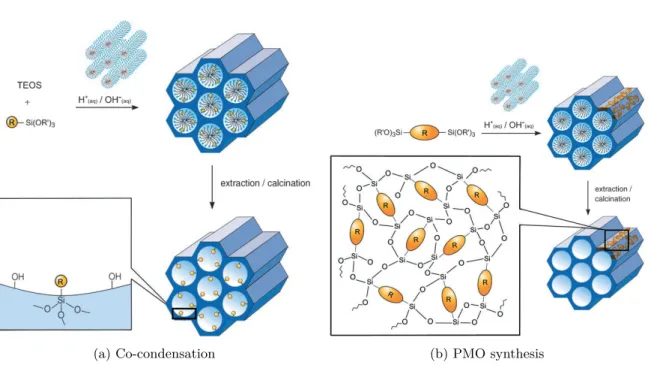
I.7.2 Co-condensation . . . 41
I.8 Conclusions and perspectives . . . 42
II Mesostructured sol-gel silica thin films 47 II.1 Sol-gel chemistry of silicates . . . 47
II.2 Mesostructuration . . . 49
II.3 Dip-Coating . . . 52 iii
II.4 Surface functionalization . . . 53
II.4.1 Atom-transfer radical polymerization. . . 54
II.4.2 Growing PEGylated polymer brushes on silica thin films. . . 56
II.5 Thin films characterization . . . 68
II.5.1 Variable Angle Spectroscopic Ellipsometry. . . 68
II.5.2 Environmental Ellipsometric Porosimetry . . . 68
II.5.3 Small-angle X-ray scattering (SAXS) . . . 73
II.5.4 Scanning-electron microscopy (SEM) and Transmission Electron microscopy (TEM) . . . 74
II.5.5 X-ray photoelectron spectroscopy (XPS). . . 74
III Study of dip-coating deposition process 75 III.1 Fundamentals of dip-coating. . . 76
III.2 Irreproducibility issues . . . 78
III.3 The role of solvent relative vapor pressure . . . 81
III.4 Towards controlled gradients . . . 91
IV Ellipsometry as a tool to study thin films dynamics in biological fluids 93 IV.1 Spectroscopic ellipsometry . . . 93
IV.2 Light and materials. . . 97
IV.3 Total Internal Reflection Ellipsometry . . . 100
IV.4 Development of TIRE setup including a microfluidic device . . . 103
IV.4.1 Microfluidics . . . 106
IV.4.2 Microfluidic cell fabrication . . . 106
IV.4.3 Sample fabrication . . . 108
IV.4.4 Bonding . . . 109
IV.4.5 Oligomers release . . . 111
IV.4.6 Setup issues . . . 112
IV.4.7 Concluding remarks . . . 114
V Mesoporous silica dissolution in physiological conditions 117 V.1 State of the art . . . 117
V.2 Dissolution of mesoporous silica thin films . . . 119
V.3 Dissolution in PBS . . . 122
V.3.1 The Noyes-Whitney model of dissolution. . . 124
Contents
V.3.3 Mesoporous Silica Nanoparticles film . . . 136
V.4 Role of surface adsorbed biomolecules . . . 139
V.5 Hybrid silica films . . . 146
V.5.1 Synthesis and characterization . . . 146
V.5.2 Dissolution in PBS . . . 147
V.5.3 Role of surface adsorbed proteins . . . 149
V.5.4 Surface chemistry. . . 150
V.6 Monitoring Gold clusters embedded in mesoporous silica . . . 152
V.6.1 Gold QDs synthesis and characterization. . . 154
V.6.2 Gold QDs confined in mesoporous silica . . . 157
V.6.3 Dissolution of a quantum rattle structure in PBS . . . 159
V.7 Considerations about in vivo conditions: shear stress and particle flow dynamics . . . 163
V.8 Flow influence on dissolution rate . . . 166
V.9 Dissolution in real biological fluids . . . 171
V.9.1 Serum . . . 171 V.9.2 Blood . . . 174 V.10 Conclusions . . . 178 V.11 Future perspectives . . . 179 Bibliography 183 v
Acknowledgments
There are many people I would like to thank for being a part of this great adventure.
First of all, my supervisor Cédric Boissière for giving me a free hand to explore the world of mesoporous materials and much more. For listening, encouraging, teaching me to raise always new questions and to look old problems from a new angle, pushing me to raise always new possibilities and take new challenges.
My co-supervisor Andrea Cattoni for essential help and support. For inspirational dis-cussions and being always available also if he had three deadlines the day after.
I would like to thank my thesis committee: Mika Linden, Jacques Leng and Jean-Marc Frigerio for accepting to evaluate and discuss my work.
L’équipe procedés: Marco, Guillaume, Igor, Quentin, Benjamin, Zeinab, Chirine.. be-cause we struggled a lot, but together was easier. For all the great chats, all your input and ideas, coffee breaks and funny moments which made these three years special. Honor-able mention to Olivier who was the best flat-mate I could ever have and the best friend, too.
All you guys in the lab who welcome me in the best way, making Paris my second home and who during the years shared with me all the joy and all the pain of research. Thank you also for the many discussions, parties, pétanques and saint-hilaire evenings, for making life lighter. With a special thought to Anael, Anne, Natacha, Francisco, César, Jessie, Guillame G., Johanna, Guillaume M. and Elham.
The Marcoussis crew: Juan, Niccolò, Carlos, Marijana, Kostantinos for all your help and support and for making my time in the Marcoussis desert a great time. Thank you also for the many great parties and endless discussions. Juan I will never thank you enough for all your help while I was struggling with microfluidics.
Omar Azzaroni and all the people of the soft-matter laboratory in Argentina: Lorena, Juan, Matias, Waldemar, Gonzalo, Juan, Facundo, Doris, Antonieta, Esteban, Agustin (the two of them) for the infinite discussions at lunch, for being great scientists and amazing people. I spent one of the best moment of my life with you guys.
The great family: Angi, Sofi, J, Simo, Lore. Friends who are always there for you even if we are scattered all over he world (and God bless WhatsApp). You are my strength. Thank you Poggio for the XPS accelerated course and all the moral support, the beers and the fun. Thank you Marco, everything started because of you.
My family for your love and support. For listening and encouraging me, even when you don’t understand what i’m talking about.
Finally, Fred for always being on my side supporting me in life, even when i’m really
chiante. For love and patience. Merci beaucoup.
Introduction
Nanoparticles-based controlled drug-delivery systems are one of the most promising tool for human health care. In the last decades, many platforms based on nanoparticles have been developed for diagnosis and therapy of major diseases, especially cancer. Porous materials able to load and release drugs in a controlled manner have been widely inves-tigated as biocompatible vectors. Ideally, a targeted drug delivery vector should be able to target specific areas in the body and release its cargo at controlled rate when it is needed, avoiding secondary effects due to overdose and maximizing the therapeutic effi-ciency. Porous particles with gate-keeping mechanisms responding to stimuli such as pH, redox potential, light-triggering, temperature, etc. have been developed but many porous carriers still rely on diffusion to release the encapsulated drugs.
Among these platforms, silica NPs provide many advantages, such as highly controllable size and shape, high drug payload, low toxicity, and excellent biocompatibility. They dissolve in physiological conditions and their degradation products, namely orthosilicic acid, can be easily excreted through kidneys. Mesoporous silica NPs have a porous structure with pores diameters from 2 to 50 nm and can bond active biomolecules and drugs by physical adsorption or covalent binding.
However, a detailed understanding of the behavior of mesoporous silica nanoparticles in biological environments is needed to push the technology towards a clinical standard. In particular, dissolution of mesoporous silica NPs, influences their biodurability, their po-tential toxicity and their drug release kinetics and should be investigated deeply to predict nanoparticles fate in vivo. Attention should be paid to carefully identify the parameters that affect the dissolution of particles such as surface area, size, and surrounding media. Indeed, these studies should be designed to mimic the relevant in vivo environment. In this thesis we propose a study of mesoporous silica degradation in biological relevant conditions realized through in situ ellipsometry on model thin films, in complex media containing proteins and in dynamic flow.
Ellipsometry provides the thickness and the optical constants of the analyzed thin film and ix
studied by ellipsometry. Nevertheless, ellipsometry can’t be performed in opaque liquids so we use two different setups for transparent buffer-solutions and for real biological fluids such as serum and blood. In the latter case we employ a setup for total internal reflection ellipsometry (TIRE), able to work in opaque fluids.
The thesis is organized in five chapters as illustrated in the figure above.
Chapter 1 aims to present the state-of-the-art regarding drug delivery systems based on
nanoparticles, discussing biocompatibility and toxicity, targeting mechanisms and in vivo degradation. Particular attention is brought on mesoporous silica nanoparticles and their biomedical applications, which are the focus of this work. Chapter 2 reviews sol-gel chemistry and nanostructuration methods, along with all the techniques involved in the preparation and characterization of mesoporous silica thin films and extensively employed in this thesis. A detailed study on dip-coating deposition technique was carried out since the good control of the layer thickness is fundamental for degradation experiments reproducibility, and it is reported in Chapter 3.
Chapter 4 describes the ellipsometry technique and the total internal reflection mode
(TIRE) employed for analysis in biological fluids. We discuss the design and optimization of TIRE setup, which we propose to couple with a microfluidic channel to perform dissolu-tion experiments in flow condidissolu-tions. Results are presented and discussed in Chapter 5 in which dissolution rate of silica at 37◦C are reported for several physiological fluids. First, a general model for mesoporous silica dissolution is proposed and parameters affecting
dissolution such as surface area and porous structure are discussed. Finally, the influence of protein adsorption and liquid flow rate on mesoporous silica dissolution kinetics are highlighted.
Chapter I
Nanocarriers for targeted drug
delivery
I.1
Nanotechnology for health care
Probably one of the biggest innovations brought by nanotechnology is its application to medical science: Nanomedicine, which has radically changed the way we approach medicine. In the lecture "There’s plenty of room at the bottom" given by Richard Feynman in 1959 at Caltech [1], which is usually referred to as the origin of nanotechnology, there were the first revolutionary ideas of nanomedicine. In his speech Feynman considered the possibility that patients may in the future swallow the surgeon, and talked about designing and manipulating very small robots to be introduced into the human body for repairing altered cellular processes or for healing injuries. Feynman pointed out that the biological systems and the cellular mechanisms work on very tiny scale and he said:"Consider the
possibility that we too can make things very small, which does what we want, when we want - and that we can manufacture an object that maneuvers at that level". At that
time all of this was just imagination, but with the technical advances of the 1970s and the 1980s nanotechnology moved the first steps. A very important technical progress was the invention of the scanning tunneling microscope which allowed, for the first time, the visualization of individual atoms. Latterly, significant technical advances as atomic force microscopy, atomic layer deposition and nanocrystal synthesis made nanotechnology evolve quickly and the first nanomaterials appeared, along with their applications. The understanding of molecular and supramolecular world developed with nanotechnol-ogy allowed to unravel the complexity of biointeractions, and the possibility to create devices for imaging and manipulation of biological structures became real. A
ity of biological processes occur at the nanoscale: DNA, proteins, antibodies, lipids and self-assembled structures made of these building blocks are nanosized materials. Under-standing the relation between the structures and the properties of these nanomaterials is mandatory if we want to be able to successfully fight against diseases and injuries, given that biology uses nanostructures to manage cellular processes.
Figure I.1 – Nanoparticle drug delivery systems and the related scales.
Nowadays we produce many artificial nanostructures able to interact with complex, self-assembled biological nanosystems, allowing the manipulation of biological processes. Nanofibers and nanopatterned substrates are used to produce biomimetic scaffolds [2,3] where cells spread and proliferate to form bone tissue [4], muscle [5] or epithelial tissue [6]. Solutions of nanoparticles have been used as tissue glues or as hemostatic materials to stop internal bleeding [7–9].
path-I.2. Nanomedicine and Drug Delivery
ways which produce specific signals, for example circulating biomarkers. Detecting these signals is crucial for rapid and early diagnosis which, until now, has been limited by the lack of biosensors capable of probing the concerned zone. Nanomaterials can interact with biological signals through their chemical, optical or magnetic properties, becoming a powerful tool for in vivo diagnostics. Several types of nanoparticles are being used for imaging and labeling, because they can be detected with different techniques exploiting their peculiar properties such as fluorescence, optical absorption, magnetic force or Raman scattering. A commonly used imaging technology rely on magnetic resonance (MRI) and allows to monitor magnetic nanomaterials in living tissues. These nanoparticles are gener-ally made up of a magnetic core, often in iron oxides Fe3O4 or γ-Fe2O3 or metals, and an hydrophilic surface coating [10–14]. Fluorescent nanoparticles are also extensively used for cells imaging. They can consist in polymeric [15,16] or silica particles [17,18], labeled with fluorophores, or surface-stabilized quantum-dots [19–22]. Moreover, nanoparticles are noninvasive and avoid tissue disruption and consequent complications.
The key feature of nanoparticles for imaging and diagnostics is that their properties de-pend on their size and size can be easily tuned during synthesis. Furthermore the surface of nanomaterials can be engineered to control immunoreaction, biodistribution and cel-lular uptake or to make them interact with specific biomarkers, for example conjugating a precise antibody on the surface to target the antigen.
I.2
Nanomedicine and Drug Delivery
Nanoparticles are powerful therapeutic and diagnostic tools, in particular they can be used as drug delivery carriers, improving the effectiveness of healing treatments. In fact, conventional drugs are administered systemically and distributed to the whole body: lack-ing specific targetlack-ing, they are largely degraded and excreted before reachlack-ing the diseased tissue. The inability to establish which organs are concerned by the drugs effects is one of the main limitations of medicine. In cancer treatment, for example, the therapeutic agent needs to be delivered to individual tumor cells in sufficient quantity to be effective but without harming healthy cells. This is indeed the biggest challenge of anticancer therapies: delivering drugs to the pathological areas and confine them there, which allows exploiting their therapeutic effect while limiting detrimental side effects due to overdose. Reducing the uncontrolled dispersal of drugs all over the body, targeted delivery keeps drug concentration in the therapeutic window, maximizing its efficacy, with no need to inject high doses of therapeutics which bring adverse reactions.
The key characteristic of nanovectors is their versatility. They are multicomponent sys-tems which can be designed to accumulate preferentially in some parts of the body. They can include biosensors which target some specific cells and integrate stimuli responsive drug release mechanisms to deliver the drug only under specific environmental conditions. If the particles need to be used as diagnostic tools they have to show peculiar magnetic or optical properties, while if they are supposed to be drug carriers they have to be able to host their cargo, either by covalent bonding or, in the case of porous particles, by absorption in the pores.
Figure I.2 –The design of nanoparticles for biomedical applications involves numerous aspects: composi-tion, size and shape can be tuned to control physical properties and biodistribution; the surface chemistry can make NPs stealth, to avoid accelerated clearance by the immune system (with PEGylation for exam-ple) and can modify the particle surface charge to improve stability and cell uptake; the functionalization with specific ligands allows targeted delivery and drugs can enhance their stability and solve solubility problems through bonding/encapsulation in NPs. Image reported from [23]
Over the past few decades, many nanoparticle platforms have been exploited as drug delivery vehicles, belonging to a wide range of materials such as polymers [24,25], metals
I.2. Nanomedicine and Drug Delivery
[26] and ceramics [27].
Figure I.3 –Growth of the number of publication with keywords ’drug delivery’ and ’nanoparticle’. Data from Web of Science.
The number of publications concerning nanoparticles employed as therapeutic vectors is spreading (figure I.3), and, to date, 50 nanopharmaceuticals have been approved by US FDA (Food and Drug Administration) and are available for use in clinical practice, while even more are being tested in clinical trials [28]. Most of them are previously existing drugs encapsulated in nanoparticles and, in the majority of cases, they rely on passive targeting through EPR effect, which involves nonspecific accumulation in diseased tissue (see sectionI.4.1). Nanoparticles used in approved nanodrug formulations or under evaluation in clinical trials currently include liposomes, polymers, micelles, nanocrystals, metals/metal oxides and other inorganic materials, proteins and dendrimers (figure I.4). A huge part of nanopharmaceutical approved or in clinical trials is made by anticancer and antimicrobial nanodrugs. However, there are also formulations being developed for autoimmune conditions, psychiatric disorders, anesthesia, arthritis, metabolic disorders, ophthalmic conditions, and others (see table I.1).
Most of the formulations approved haven’t demonstrated improved efficacy or targeting compared to the free drug, but they have shown reduced toxicity. In the case of cancer therapy, which is still the main field of application for nanodrugs, the chemotherapeutic agents are often insoluble in water and they require toxic solubilizing excipients, as in the case of paclitaxel. The dose of such chemotherapeutics has to be limited to avoid systemic toxicity, necessarily reducing their efficacy. Encapsulation in nanoparticles offers
Figure I.4 – Types of nanoparticles in A. approved nanodrugs and B. nanoformulations currently in clinical trials. Adapted from [29]
a viable solution to administrate higher doses of hydrophobic drugs without employing toxic agents, thus many chemotherapies have been approved as nanoformulation and more are in clinical development. Abraxane (nab-paclitaxel, Celgene) is a formulation of paclitaxel conjugated to albumin nanoparticles and was approved by the FDA in 2005 for metastatic breast cancer. It has now been indicated as treatment also for other cancers. It is more tolerable than conventional paclitaxel, because of the absence of toxic solvents and can be administered to patients at a considerably higher dose, increasing its efficacy. The first approved nanodrug was Doxil (doxorubicin hydrochloride, Janssen) in 1995, to treat Kaposi’s sarcoma. It showed reduced cardiotoxicity compared to the free drug and nowadays it is employed also to treat breast and ovarian cancer [29].
Anyway, the number of nanodrugs which survives clinical development is still small, cause often they are unable to demonstrate a significant improvement in efficacy and be-cause improved toxicity could be achieved employing other drugs with simpler production processes. For example, liposomal formulations of cisplatin (L-NDDP, SPI-77, lipoplatin, and Li-PlaCis) proved to be less toxic than free cisplatin but brought no benefit in terms of efficacy. Since other less nephrotoxic platinum alternatives (such as carboplatin) already exist, there was no further development of liposomal cisplatin [30].
In fact, research on nanotherapeuticals is flourishing but the transition from academia to production lines is still very poor. There are multiple obstacles and challenges in bringing drug delivery nanocarriers to the market, which can be resumed in three main groups: safety and toxicity issues, production and cost issues and regulation problems.
I.2. Nanomedicine and Drug Delivery
Safety and toxicity issues
To avoid unpredictable side effects, characterization of nanomaterials in biological envi-ronments is mandatory. There is a huge amount of data concerning polymers, liposomes and micelles (which is the reason of their dominance in nanodrugs available for use in clinical practice), but the problem with nanoparticles is that for every new formulation (change of size, charge, shape, surface coating) new tests are necessary. Moreover, NPs can interact with many different organs and living tissues, triggering potentially harmful responses. Thus, the analysis of the interactions between nanoparticles and biological systems is a very long process, which needs many tests for every modification of the ma-terial employed. This procedure can’t be avoided and results in a huge amount of time and money to bring the product on the market.
Regulation issues
One of the greater problems is the lack of standard protocols to characterize nanodrugs and their toxicity, which causes a spreading of informations difficult to compare. Stan-dards for characterization have yet not been defined but major efforts have been made. The US Nanotechnology Characterization Laboratory (NCL), in collaboration with FDA has published guidelines on nanomaterials characterization that include some standards to evaluate their toxicity [31].
Nanotherapeutic products are currently regulated within a conventional regulatory frame-work but they need additional expert evaluations about their safety and efficacy, because of their structural complexity.
From the regulatory perspective, the main challenges concern the applicability of current methods and tools to assess characterization and biodistribution of emerging nanothera-peutics, particularly related to their and impact on the living systems. A huge work needs to be done to obtain classifications of converging technologies and to define critical prod-uct characteristics predictive of prodprod-uct performance in vivo (e.g. size , shape, surface chemistry and porosity). Therefore, both the EMA (European Medicines Agency) and the FDA constantly update their guidelines to evaluate nanomaterials, in the attempt to shape a safe and efficient regulatory framework for nanotherapeutics.
Production and cost issues
One of the factors that limits the commercialization of nanodrugs is that the production costs of nanoparticles for biomedical use are very high. In fact, their synthesis is often
challenging and made of multiple steps to fulfill all the requirements needed. They have to be stable enough to be processed further and to be stocked in large quantities and their synthesis must avoid toxic reagents which traces can be harmful in their final application. They have to be biodegradable and biocompatible, and they need to be masked from the immune system through a surface coating (usually made of hydrophilic polymers, or lipids). Then, if they are used for diagnostics they need special optical, magnetic or thermic properties, which often depends on size. Thus, the NPs size distribution during the production process must be very narrow. If they serve as drug carriers they need to charge an high quantity of load and possibly a mechanism to release it in a controlled way or in special conditions.
Moreover, NPs production process needs industrial infrastructures which include a nanochar-acterization laboratory (for physical, chemical, and biological charnanochar-acterization of terials intended for medical use), a pilot line to scale up laboratory preparation of nanoma-terials, according to industrial and regulatory standards, and a coordination with clinical organizations to perform pre-clinical tests. Compared with conventional formulations, the control of nanomaterials often presents greater scientific and technical challenges, to achieve reproducibility from batch to batch with respect to particle size distribution, charge, and porosity and to have the product stable enough to be stocked and to re-move undesired nanostructures. This implies very high fabrication costs which needs to be justified by an important benefit over conventional formulations to have the product commercialized.
In fact, the nanodrugs currently available are all very simple formulations, often made of polymers, micelles or liposomes, which avoid further steps of surface coating to mask them from the immune system, and they rely on passive targeting through EPR effect, not bringing any ligand to target specific cells. This avoid delicate functionalization steps and expensive reagent such as anti-bodies. More the architecture of the nanoparticle is complex and more is production is expensive and complicated. This means that to be produced and sold it has to be very efficient in his task. To date, NPs based on active targeting (specific binding of a ligand on the NP surface to a receptor on the cells surface) haven’t shown an important gain in therapeutic effects compared to conventional drugs, but the latest trends in approval and clinical trials of nanodrugs saw an increasing percentage of active targeted nanoparticles [29]. One example is is SGT-53 (SynerGene Therapeutics), which contains an antitransferrin antibody fragment that binds with a transferring glycoprotein receptor on cancer cells and it is being studied for the treatment of solid tumors and metastatic pancreatic cancer [32].
I.2. Nanomedicine and Drug Delivery
I.2.1 Multifunctional NPs
Figure I.5 – The growth in the number of publications (vertical axis scale on the right) and number of citations per year (vertical axis scale on the left), concerning theranostic platforms which combine diagnostic and therapeutic properties in a single nanoparticle-based system. Data from Web of Science.
In the last years another important research trend is the development of multi-functional nanoparticles which can integrate multiple functions such as diagnosis, imaging, targeted delivery and controlled release in one individual platform. Such efforts to combine diag-nostic and therapeutic capabilities into a single agent are commonly defined with the term "theranostics" and represent a very active research field which counts currently around 1000 publications per year (figureI.5).
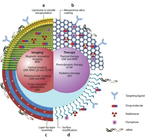
An overview on multi-functional nanoparticles is given in figure I.6 and extensively dis-cussed in a review by Bao et al. [33] recently. Such theranostic platforms include very often inorganic functional components such as metals, metal oxides or semiconductors. They have peculiar optical, magnetic and plasmonic properties which can be easily tuned controlling their size and shape.
Gold nanoparticles, for example, are good contrast agent for optical imaging but they can also be used for photo-thermal therapy, so one can visualize exactly where nanopar-ticles are accumulated and treat the area thermally [34,35]. Gold nanoparticles owe they peculiar optical properties to localized surface plasmon resonance, which absorbs light at specific frequency. The light absorbed is mainly emitted by gold nanoparticles as scattered light, which makes them suitable tools for optical imaging, while the remaining energy is converted into heat. For in vivo application, the absorption frequency should be within
Figure I.6 –Schematic representation of multifunctional nanoparticles for drug delivery and diagnostics. They can be designed as a combination of an inorganic nanocrystal and a coating layer which can in-clude (a) mycelles or liposomes, (b) mesoporous silica, (c) layer-by-layer assembly and (d) surface grafted moieties. Reproduced from [33]
the optical transparent window of human tissues (between 650 and 1300 nm) so light can penetrate deep into the tissue without being rapidly attenuated. Luckily the absorption spectra of gold nanoparticles is tunable with their size and shape [36] and can be shifted from the visible to the near infrared region, allowing the use of gold nanostructures for
in vivo imaging and photo-termal therapy [37]. These nanoparticles can also be loaded with drugs and provide the combined effects of chemotherapy and thermal therapy in one singular platform. To date, the FDA has not yet approved any gold-based nanodrugs, but they are under evaluation as delivery vehicles for the extremely toxic antitumor agent TNFα. In the clinical trials they demonstrated reduced toxicity but they were rapidly cleared by the reticuloendothelial system, to date a new nanoformulation of gold NPs con-jugated with PEG is being developed to deliver TNFα (Aurimune, from CytImmune) and has been found to decrease clearance rate and improve accumulation in tumors through
I.2. Nanomedicine and Drug Delivery
EPR effect.
Not every type of nanoparticle is naturally multi-functional, as gold nanoparticles are, but often they are composites in which each part brings a different functionality. For example, it is common to find core-shell structures in which an inorganic core is encapsulated into a mesoporous silica shell or into a lipid bilayer or a biopolymer coating. In this case the inorganic core is used for imaging, due to his physical properties, while the coating layer allows including drug molecules, fluorescent tracers and targeting ligands. Moreover the nanoparticles surface can be engineered to provide biocompatibility and stability.
Iron oxide NPs have been studied in numerous clinical trials as contrast enhancement reagents for magnetic resonance imaging (MRI). However, FDA-approved iron oxide nanoparticles are employed as iron replacement therapies for the treatment of anemia associated with chronic kidney disease (CKD). Some of them are reported in table I.1. These formulations are made by an iron oxide core, coated with hydrophilic polymers (e.g., dextran, sucrose), and provide a slow dissolution of the iron, allowing administration of large doses without exceeding free iron levels in the blood.
Superparamagnetic iron oxide nanoparticles (SPIONs), particularly iron oxide and mag-netite have been used as non-targeted contrast agents for MRI. Some SPION drug formu-lations have received FDA approval, such as Feraheme (ferumoxytol, AMAG Pharmaceu-ticals), even if it is currently indicated mainly as an iron substitute therapy for anemia condition associated with CKD. However, this nanoformulation is also being studied as an imaging agent in numerous clinical trials [32]. SPIONs can be also used for hyper-thermia treatments, thanks to the energy that they release when excited in a magnetic field and several SPIONS are currently being investigated as hyperthermia agents against tumors, showing promising clinical results. For example, Nanotherm (MagForce AG) is a formulation of SPIONs coated with aminosilanes for local hyperthermia treatment of glioblastoma tumors. It is injected directly into the tumor, and heated through the appli-cation of an alternating magnetic field. The tumor microenvironment reaches in this way a temperature of 40-45 ◦ C and cell death is provoked. Nanotherm is currently awaiting FDA approval.
Among silica-based systems, Cornell Dots (C Dots) are fluorescent core-shell silica nanopar-ticles that are being developed at Cornell University as a diagnostic and therapeutic tool for cancer treatment. They have a silica core labeled with a near-infrared organic dye surrounded by a pure silica shell, which is coated with polyethylene glycol (PEG). Com-pared to free dye equivalents in solution, the C Dots are 20 to 30 times brighter and they showed improved photo-stability. They were originally designed as diagnostic tools
to map tumors, however they showed ability to induce cancer cell death in vitro when cancer cells are in a state of nutrient deprivation, while high concentrations were well-tolerated under normal conditions. When administrated to mice, tumors also reduced. They are undergoing human clinical trial since 2010. [38] The same research group at Cornell University is developing mesoporous C dots to host drugs, and C-dots conjugated with antibodies to target specific cancer cells [39,40].
Organic nanoparticles have been widely explored as well, constituted by polymers, pro-teins, dendrimers or lipids, especially liposomic systems.
Liposomes are lipid vesicles with a large aqueous center in which they can host hydrophilic compounds, while hydrophobic molecules can be embedded in the lipid bilayer; they carry drugs across biological barriers overcoming cellular uptake and improving biodistribution, being suitable vectors for many kinds of drugs [41,42]. FDA approved many formula-tions where drugs are encapsulated in liposomes and they showed improved stability and bioavailability (see tableI.1).
Polymeric nanoparticles are also widely investigated as nanocarriers because of their good stability in vitro and in vivo and the possibility to easily graft multiples molecules and ligands on their surface, making them very versatile. Moreover, they’re usually made up of biodegradable polymers such as poly(lactic acid) (PLA), poly(glycolic acid), poly(ε-caprolactone) and their copolymers, avoiding problems of toxicity; poly(ethylene glycol) (PEG), poly(ethylene oxide) PEO, and poly(propylene oxide) (PPO) are also used to synthesize nanoparticles which have great biocompatibility [43–45]. Polycyanoacrylate nanoparticles have proved themselves effective for brain delivery [46,47].
I.2. Nanomedicine and Drug Delivery
Table I.1 – List of nanodrugs approved by US FDA and available for clinical practice [29]
Trade name (Manufac-turer)
Generic name Indications Benefit of NP*
Liposome NPs Curosurf (Chiesi USA)
Poractant alfa Respiratory distress syndrome
Increased delivery with smaller volume, decreased toxicity Doxil (Janssen) Doxorubicin
HCl liposome
injection
Karposi’s sarcoma, ovarian cancer, multi-ple myeloma
Increased delivery to disease site, decreased systemic toxicity Abelcet (Sigma-Tau) Liposomal am-photericin B lipid complex
Fungal infections Decreased toxicity
Depodur (Pacira Pharmaceuticals) Liposomal mor-phine sulphate Postoperative analge-sia Extended release Marquibo (Spec-tral Pharmaceuti-cals) Liposomal vin-cristine
ALL Increased delivery to
tumor site, decreased systemic toxicity Onivyde (Ipsen Biopharmaceuti-cals) Liposomal irinotecan
Pancreatic cancer Increased delivery to tumor site, decreased systemic toxicity Visudyne(Bausch and Lomb) Liposomial verteporfin Ocular histoplasmo-sis, myopia Increased delivery to site of diseased vessels, photosensitive release Vyxeos(Jazz Pharmaceuticals) Liposomal daunorubicin and cytarabine
AML Increased efficacy
through synergis-tic delivery of co-encapsulated agents
Trade name (Manufac-turer)
Generic name Indications Benefit of NP*
Polymer NPs Adagen (Leadiant Biosciences)
Pegademase bovine
SCID Longer circulation
time, decreased immunogenicity Adynovate (Shire) Antiemophilic factor, pegylated
Hemophilia Greater protein
stabil-ity, longer half-life Cimzia (UCB) Certolizumab
pe-gol
Crohn’s disease,
rheumatoid arthritis, psoriatic arthritis
Longer circulation time, greater stability
in vivo
Copaxone (Teva) Glatimer acetate Multiple sclerosis Controlled clearance Eligard (Tolmar) Leuprolide
ac-etate and poly-mer
Prostate cancer Longer circulation
time, controlled
payload delivery Mircera (Vifor) Methoxy
polyethylene glycol-epoetin beta
Anemia Greater aptamer
stability Pegasys (Genen-tech) Pegylated IFN alpha-2a Hepatitis B, Hepatitis C Greater protein stability Plegridy (Biogen) Pegylated IFN
beta 1-a
Multiple sclerosis Greater protein
stability Renvela; Renagel (Genzyme) Sevelamer car-bonate; Seve-lamer HCl CKD Longer circulation
time and therapeutic delivery Zilretta (Flexion Therapeutics) Triamcinolone acetonide Osteoarthritis knee pain extended release
I.2. Nanomedicine and Drug Delivery
Trade name (Manufac-turer)
Generic name Indications Benefit of NP*
Micelle NPs Estrasorb (No-vavax)
Micellar estradiol Vasomotor symptoms in menopause
Controlled delivery
Nanocrystal NPs
Avinza (Pfizer) Morphine sulfate Psychostimulant Greater drug loading and bioavailability EquivaBone
(Zimmer Biomet)
Hydroxyapatite Bone substitute Mimics bone structure
Emend (Merck) Aprepitant Antiemetic Greater absorption
and bioavailability Invega Sustenna (Janssen) Paliperidone palmitate Schizophrenia, schizoaffective dis-order
Slow release of low-solubility drug
NanOss (RTI
Surgical)
Hydroxyapatite Bone substitute Mimics bone structure
Rapamune
(Wyeth
Phar-maceuticals)
Sirolimus Immunosuppressant Greater
bioavailability
Ritalin NA (No-vartis)
Methylphenidate HCl
Psychostimulant Greater drug loading and bioavailability
Tricor (AbbVie) Fenofibrate Hyperlipidemia Greater
bioavail-ability, simpler
administration Zanaflex
(Acorda)
Tizanidine HCl Muscle relaxant Greater drug loading
Trade name (Manufac-turer)
Generic name Indications Benefit of NP*
Inorganic NPs Dexferrum
(American Re-gent)
Iron dextran Iron deficiency in
CKD
Increased dose
Feraheme
(AMAG Pharma-ceuticals)
Ferumoxytol Iron deficiency in
CKD
Prolonged, steady re-lease with less fre-quent dosing
Ferrlecit (Sanofi-Aventis)
Sodium ferric glu-conate complex in sucrose injection Iron deficiency in CKD Increased dose Protein NPs Abraxane (Cel-gene) Albumin-bound paclitaxel
Breast cancer, pancre-atic cancer
Greater solubility, increased delivery to tumor, decreased toxicity
Ontak (Eisai) Denileukin difti-tox
Cutaneous T-cell lym-phoma
Targeted T-cell speci-ficity, lysosomal es-cape
* Compared with conventional formulations
ALL = acute lymphoblastic leukemia; AML = acute myeloid leukemia; CKD = chronic kidney disease;
HCl = hydrochloride; IFN = interferon; SCID = severe combined immunodeficiency disease.
I.3. Biocompatibility and Immunoreaction
I.3
Biocompatibility and Immunoreaction
For using nanoparticles as nanomedicine tools they need to be stable in vivo and to have good biocompatibility. A surface coating of polymers or lipids is usually an effective way to avoid particles aggregation and fast dissolution, providing them stability in biological media [14,48–51]. Hydrophilic polymer such as poly(ethylene glicol) (PEG) can constitute a protective shell for nanoparticles and lower the non-specific adsorption of proteins, hindering the organism immunoreaction and thus insuring longer circulation times.
I.3.1 Accelerated Blood Clearance
In order for a drug delivery device to achieve its goals, it must remain in the bloodstream long enough to reach its therapeutic target, but nanoparticles undergo immune recognition due to their relatively large size: they have the same dimensions as viruses, against which our immune system developed a very effective defense. The phenomenon of accelerated blood clearance (ABC) is responsible of fast elimination of nanoparticles from bloodstream and it is due to a rapid uptake of NPs by macrophages of the reticuloendothelial system (RES). The macrophages of the RES remove nanoparticles from the circulation through phagocytosis, but they can identify them only through specific proteins, called opsonines, bonded or adsorbed onto the particle surface. The opsonines are blood proteins which start to bind very quickly to every particle injected in the bloodstream, in a process called opsonization, which triggers recognition by the phagocytes and following elimination. Once internalized by the macrophage, the nanoparticles are rapidly cleared through the liver within few hours. Thus, masking the nanoparticles to avoid opsonization is critical to enhance nanocarriers blood circulation times.
I.3.2 Making stealth nanoparticles
The simplest strategy to overcome this problem is to play on the size of NPs. In fact, particles bigger than 200 nm are removed very efficiently by the RES, and colloids smaller than 6 nm are quickly cleared through the kidneys. So, in order to be retained as long as possible, the size of nanocarriers can range from 6 to 200 nm, with an optimum between 50 and 100 nm, which is also the size range to exploit the EPR effect to reach diseased tissues, as described in section I.4.1. The anisotropy of the nanoparticles is another pa-rameter playing a role in the kinetics of cellular uptake process. It has been observed that a little anisotropy can promote the cellular uptake while a strong anisotropy can facilitate renal clearance [52].
The mentioned properties are important to extend circulation times of NPs but they are not sufficient to retain the carriers in the bloodstream long enough to reach their target cells. Some surface engineering needs to be performed to hide the nanoparticles from the immune system.
One widely used method to slow down opsonization is covering the nanoparticles with molecules which reduce the hydrophobic and electrostatic interactions between opsonines and the particle surface. These coatings are usually made up of long hydrophilic polymer chains, the most employed being PEG (poly ethylene glycol) and PEG-containing copoly-mers, which demonstrated their effectiveness in minimizing blood protein adsorption and extend the circulation time of nanoparticles [53–55]. The results are promising but still a huge amount of nanoparticles accumulates in the liver and doesn’t target the tumor. An interesting strategy to improve the targeting is to cover the nanoparticles with PEG and then add cationic polymers containing amine moieties: the particle remains hydrophile but gains a positive surface charge which favors its interaction with cellular membranes and its subsequent uptake, as described in section I.4.1. However the synthesis needs to be properly tuned to avoid a too high charge density which would destroy the cellular membranes and reduce the circulation times of nanoparticles. This type of coating has been tested in vivo by Meng et al., showing a significant improvement in targeting tumors in comparison with a simple PEG coating [56].
I.4
Targeting strategies
The innovation about nanocarriers is the possibility of reaching injured tissue, such as cancer cells, avoiding healthy tissues and the ability of releasing cytotoxic molecules only where they are needed. This allows the formulation of drugs that couldn’t be employed in conventional chemotherapy because either highly toxic or insoluble in water, for example. The goal of medical research on drug delivery devices is to make them passively or ac-tively target tumors or diseased tissues. The two approaches are detailed in the following sections.
I.4.1 Passive targeting
In order to improve the preferential accumulation of nanoparticles in tumors, one strategy is to take advantage of the existing differences between pathological and healthy tissues. Tumors show leaky blood vessels, presenting many large fenestrations (from 100 nm to several hundreds of nm) due to fast and defective angiogenesis (the formation of new blood
I.4. Targeting strategies
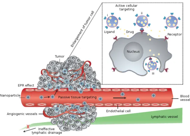
Figure I.7 –Nanoparticles can reach tumors through Enhanced Permeation and Retention effect (EPR), exploiting the leakiness of angiogenic blood vessels, in an approach called passive targeting. Besides, nanocarriers can identify their target cells through ligand-receptor interactions, through an active targeting approach. Image reproduced from [57]
vessels from existing ones) typical of pathological processes. The leakiness of tumor vas-culatures is due to the rapid proliferation of endothelial cells and the lack of pericytes; this condition enhances vessels permeability allowing nanoparticles and large macromolecules to pass into tumors. Moreover, nanoparticles are retained in the cancer because of tumors inefficient lymphatic drainage, while in normal tissues macromolecules are cleared by the lymphatic system through the constant drainage of the extracellular fluid. The interstitial fluid is thus renewed very often and the solutes and colloids contained in it are recycled to the circulation. In tumors, this important lymphatic function is defective and the extracellular fluid is retained, along with the nanoparticles and macromolecules within it. Molecules smaller than 6 nm are able to diffuse back to the blood circulation but bigger objects accumulate. This phenomenon is referred to as enhanced permeation and reten-tion effect (EPR) and it is the main responsible of passive accumulareten-tion of nanoparticles in tumors.
The EPR effect greatly depends on the physicochemical properties of nanocarriers, such as size, shape, charge and surface chemistry. In the first place, a high blood concen-tration of nanoparticles is important to ensure extravasation into tumors and prevents the efflux back to the blood stream. Consequently, it is mandatory to extend circulation times of nanoparticles, hiding them from the immune system through stealth coatings which decrease their interaction with opsonins and consequently delay their uptake by macrophages (see section I.3.2).
The size of the nanovectors is a crucial parameter to have an efficient accumulation in tumors through EPR effect: the average fenestration in tumor blood vessels being around 100 nm, nanoparticles bigger than this cut-off usually are not able to penetrate in tumors. Among nanoparticles which are smaller than 100 nm, the larger ones diffuse in the tumors more slowly than smaller ones but if their size is below 6 nm they undergo rapid renal clearance, being not suitable for drug delivery application. The rapid elimination through the kidneys of small-size imaging probes have been demonstrated, showing poor accumulation in the main organs and a clearance of more than 50% of the injected dose within 24h. Many contrast agents used for MRI or CT imaging or the near-infrared dyes used for optical imaging have dimensions in this range (< 6 nm) and are rapidly cleared via the kidneys [58,59]. Thus the optimal size of nanoparticles for tumor targeting through EPR effect is between 6 and 100 nm. Furthermore, not all the tumors have the same permeability and knowing the specific tumor biology is important to design an effective nanocarrier. For example, if the tumor is hyper-permeable, NP of 30, 50, 70 or 100 nm show similar distributions, however only particles smaller than 70 nm can efficiently penetrate and accumulate in poorly permeable tumors [60]. It is important to remind that not only cancer is concerned by EPR effect, in fact there are other pathologies which show similar characteristic to tumor vasculature, such as cardiovascular conditions. Thus, size is for sure a very important parameter, but the nanoparticles shape may also play a role: spherical objects will not interact with the surrounding environment the same way as rod-shaped or disc-shaped particles. In fact, it has been observed that nanorods tumor distribution kinetics differs from biodistribution of their spherical counterparts: nanorods with an aspect ratio of 10 show similar blood circulation profiles compared to spherical nanoparticles with equivalent hydrodynamic radius, however they extravasate in tumors 4 times faster. Moreover, they diffuse deeper in the cancer [61], even if they could penetrate cells less efficiently, as discussed in section I.5.3. In many biological processes the shape and the deformability of nanosized objects are crucial to ensure the proper working of the whole system, therefore these parameters need to be considered
I.4. Targeting strategies
when designing a drug delivery device, in order to maximize its effectiveness.
Surface charge have a significant impact on all the nanoparticles biointeractions, partic-ularly on protein adsorption and cellular uptake of nanocarriers. Indeed, several studies seem to confirm that positively charged nanoparticles are internalized much faster than neutral or negatively charged ones [56,62] despite their shorter blood circulation times [63]. Such an effect could be related to the negatively charged character of cell membranes, which favors adhesion and up-take of positive nanoparticles.
The discovery of EPR effect paved the way for future cancer therapy, however we still need to understand the importance of many parameters to be able to entirely exploit it. For example, EPR shows a big variability between patients and its effectiveness greatly depends on tumor biology and microenvironment. Besides, EPR effect increases the accumulation of drug nanocarriers in tumors but not necessarily improves their ability to penetrate inside cells. In order to enhance the affinity of drug delivery vehicles for their targeted cells, the surface of NPs is modified with targeting ligands in the so-called active targeting approach, presented in the next section.
I.4.2 Active targeting
Although passive targeting serves as basis for nanocarriers drug delivery, it still suffers lack of control. For this reason, many efforts have been made to design nanocarriers which can selectively bind to target cells. In order to achieve this goal, nanoparticles are functionalized with molecules called ligands, which bind to specific receptors onto the cell surface: through ligand-receptor interactions nanovectors find their target cells and bind to them. Once bound, nanoparticles are quickly internalized by cells and release their cargo. Commonly employed ligands include macromolecules such as antibodies, nucleic acids, proteins, sugars, peptides and small molecules such as vitamins. Their target molecules, often also called receptors, can be proteins, lipids or sugars located on the surface of cells. Actively-targeted NPs have increased affinity for their target cells but they need to be in their proximity to interact with them, so this type of NPs still relies on EPR effect to reach the tumors and need to be designed to have long blood circulation times. Active targeting doesn’t change much the biodistribution of nanomaterials but increases the NPs uptake, enhancing therapeutic efficacy.
In order to be effective, this strategy needs to target receptors or antigens which are over-expressed on the surface of target cells in comparison with normal cells. For example, folate receptor (FR) is a membrane receptor over-expressed on the surface of many cancer cells including lung, brain, breast and ovarian cancer cells, while it shows limited
sion on healthy cells, on which it is poorly accessible from the bloodstream. The ligand used to target FR is Folic acid (FA, also called vitamin B9 or folate), a nutrient required for cell proliferation and involved in the biosynthesis of nucleotides. Cancer cells prolif-erate very fast and they need nutrients to maintain their fast-growing metabolism, thus they overexpress receptors for nutrition as FR and Transferrin receptor (TfR). Folic acid and transferrin (a protein which delivers iron to cells) have been bound to nanocarriers outer surface improving intracellular delivery in vivo [64,65].
Ligands are often linked to the surface of NPs through covalent bonds which can be per-formed directly on the surface (Gold NPs reaction with thiols) or on functional groups specifically added to improve the reactivity of the material (e.g. OH and NH2 on inor-ganic NPs). The density of ligands on the surface of nanoparticles greatly affects their affinity for targets and thus, their uptake rate. Multiple ligands can show cooperative ef-fects: once a ligand binds to its substrate, the subsequent binding of the adjacent ligands are thermodynamically favored. Moreover, the multiple interactions of NPs with the cell force a local concentration of the receptors which triggers the wrapping of the cellular membrane, beginning nanoparticles internalization (see sectionI.5.3). Nevertheless, high density of ligands not always improves the cellular uptake, in fact the surface decora-tion with ligands changes the properties of NPs: surface charge and hydrophobicity are modified, and hydrodynamic size is also different. This alteration modifies the biodistribu-tion and circulabiodistribu-tion times, in some cases improving the macrophage clearing. Therefore, the ligand density has to be properly tuned to optimize the efficacy of actively-targeted nanocarriers and its influence on the nanoparticles in vivo interactions has always to be considered.
An interesting approach to this issue is given by Ashley et al. [14] encapsulating a porous silica nanoparticle in a lipid bilayer through liposome fusion, creating a so-called photo-cell (figure I.8). In this configuration, the outer surface is dynamic and, when one ligand identifies its target and binds to it, the other ligands can migrate on the lipid bilayer to maximize the exposure to cell receptors. With this method, the advantage of multiple cooperative binding is retained employing a small density of ligands, avoiding the com-plications linked to the presence of many ligands on the surface. The protocells charged with Doxorubicine (a widely employed anti cancer drug) proved themself very effective, they target tumors much better than liposomes-Doxorubicine systems and they are less toxic to healthy cells.
Many antibodies have been studied as targeting ligands but they face some important limitations which narrow their application in vivo. First of all, they are large
macro-I.4. Targeting strategies
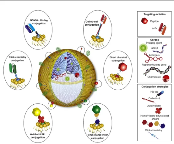
Figure I.8 –Representative design of a functionalized protocell, constitued by a mesoporous silica core (blue in figure) and a lipid bilayer outer shell. Tumor-targeting ligands (peptides or proteins, such as recombinant human scFvs) can be conjugated to functional groups on the surface of the protocell lipid bilayer. The mesoporous core can host a wide variety of cargos such as chemotoxins, genes, siRNA, aptamers or imaging agents. Polyethylene glycol can be conjugated on the surface (green coil) to enhance circulation times. Image reproduced from [66]
molecules of about 150 kDa and their size complicates conjugation, especially on small nanoparticles, besides they increase the hydrodynamic radius of the NPs remarkably. Moreover, their physiological role is to recognize antigens and signaling them to immune cells or macrophages to be cleared from the bloodstream, so they compromise circulation times of NPs. Other proteins, such as transferrin (Tf) [67], have been used as targeting ligands because of their three-dimensional shape, which provides them high affinity with specific substrates but, as antibodies, they also enhance the size of NPs because of their bulky nature. Moreover, their tertiary structure being modified, they can lose the affinity for their target after conjugation. Smaller molecules as peptides have also been studied as targeting ligands. In this case they doesn’t impact much on the nanoparticles size and they have simpler three-dimensional interactions. The most used are peptides contain-ing the sequence RGD (arginine-glycine-aspartic acid) which bind to integrine receptors
overexpressed on angiogenic endothelial cells. Another family of ligands is represented by short, single-stranded oligonucleotides (DNA or RNA) but, if their specificity is high, their stability in biological environment is easily threaten by nucleases. On the contrary, small molecules such as folic acid have good stability and small size and they overcome many of the limitations previously described. Nevertheless, small molecules ligands are limited in number, because the screening process to identify new efficient affinity ligands for interested substrates are long and difficult. The most used ligands of small size are folic acid to target folate receptor (FR), triphenylphosphonium (TPP) to target mitho-condria and several carbohydrates to target lectins (cellular membrane proteins). To conclude, actively-targeted nanoparticles are under evaluation and some formulation are already in clinical development, they still face many restraints but they could open the path for a more efficient therapy of many diseases, allowing optimization of drug delivery and limitation of side effects.
I.5
In vivo degradation and toxicity
Nanomaterials have unique characteristics due to their size and in the last decades they have been employed in a wide range of applications such as electronics, coatings, batter-ies, optics, composites, cosmetics, paints, medicine etc. The human and environmental exposure to nanomaterials have reached a level which makes mandatory to assess its pos-sible hazard to human health. When employed in medicine, nanomaterials are designed to follow specific routes, mainly they are injected in the blood circulation. Anyway, nanoparticles produced on an industrial scale for applications other than medicine can also reach the human body, via the airways, through ingestion or passing through the skin. Because of their high surface-to-volume ratio, materials at the nano scale are very reactive and interact with living systems and this makes it necessary to understand not only the nanomaterial dynamics but also the surrounding environment, because nanopar-ticles properties are modified when introduced in a biological system. Nanotoxicology studies became necessary, given the exponential growth of nanotechnology, to prevent a harmful, unregulated use of nanomaterials. Evaluation of the in vivo hazard of nano-materials is still at an early stage but key factors involved in nano-bio interactions have been identified in the last years, along with the main mechanisms of toxicity including protein misfolding, production of reactive oxygen species and cellular membrane damage. A quick overview is given in the following sections about nanoparticles pathway within the body and how the biological environment reacts to NPs presence.
I.5. In vivo degradation and toxicity
I.5.1 The nano-bio interface
When a nanomaterial comes in contact with biological components, a complex dynamic interface is formed: the interactions with biomolecules modify the forces which usually control nanoparticles in colloidal suspension, for their part, nanomaterials interfere in biomolecules reactions and functions, causing a great impact. The interface between nanomaterials and biological systems is influenced by many parameters including physic-ochemical characteristics of the nanoparticles and properties of the suspending media, besides membrane and biomolecules interactions. What makes the characterization of this interface so difficult is that many parameters are involved and it’s not simple to sepa-rate their contributions; considering that the nanoparticle properties such as size, shape , surface charge, porosity, surface roughness, crystallization and surface hydrophobicity are all important characteristic to determine interaction at the bio-interface. Moreover chem-ical composition and surface functionalization have of course a huge impact. The liquid media is equally important to shape the interface: its ionic strength, pH and the presence of biological macromolecules (e.g. proteins) control the NPs aggregation and dissolution, furthermore ions and macromolecules can adsorb onto the surface of the nanoparticles, changing their hydrodynamic radius and surface charge, promoting or hindering agglomer-ation and modifying the forces acting on particle-medium interface (electrostatic repulsive forces and attractive Van der Waals forces).
Modeling the nano-bio interface presents one more difficulty: the system is not at steady-state. It evolves continuously, due to the presence of cells which actively interact with the NPs through binding, uptake, production of proteins and ions transfers. The inter-nalization of NPs trough ligand-receptor identification and membrane wrapping further complicates the interactions involved and expose NPs to a different environment inside the cell.
When it comes to the interface between biological systems and nanomaterials all of this must be considered to make this field evolve and to perform a safe use of nanomaterials. A nanoparticle entering a biological medium is not the same object that in synthesis solvent. Its surface is modified and the forces through which it interacts with the medium and the other particles completely depend on this new state.
I.5.2 Protein corona
Physiological fluids, such as blood and interstitial fluid, contain a complex mixture of proteins, so when nanoparticles enter a physiological environment they are rapidly coated by proteins which adsorb on their surface, forming what is known as "protein corona".
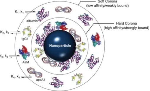
The corona modifies the size, surface chemistry and aggregation state of the nanomaterials dramatically, shaping them as they will be "seen" by cells and biological interfaces. It is formed of an inner layer of strongly bound proteins, called hard corona, and an outer layer of weakly adsorbed protein in fast exchange with the environment, known as soft corona. It is this new hybrid object (nanoparticle + protein corona) which determines transport kinetics and interactions of nanomaterials in a biological environment.
Figure I.9 – Schema of protein corona formation on a nanoparticle surface. The corona formation is a kinetic (k) and thermodynamic (K) process which depends on the surface characteristic of the nanopar-ticles, such as functionalization and charge. It exist an inner layer of strongly bound proteins and an outer layer of adsorbed proteins which exchange rapidly with the environment. Serum proteins commonly observed in NP coronas are shown: serum albumin, immunoglobulin G1 (IgG1), alpha-2 macroglobulin (A2M), and apolipoprotein A-1 (apoA1). Reproduced from [68].
Physicochemical properties of the nanoparticles, such as surface functionalization, charge and size influence the composition and evolution of the protein corona which, in turn, controls particle’s bioactivity, determining their interaction with cells, targeting activity and circulation times [69,70]. Size in particular defines the curvature of the surface which is responsible for the adsorbed protein amount and composition, smaller particles generally showing higher protein adsorption, due to their bigger curvature, which decrease steric hindrance between adsorbed macromolecules [69,71]. Surface charge is also relevant for the adsorption of biomolecules, but it will never avoid completely the formation of protein corona, as demonstrated by Qiu et al. using nanoparticles differently charged: the positive ones adsorbed a greater amount of protein in comparison to negative ones
I.5. In vivo degradation and toxicity
but a corona is formed around all the systems investigated [72].
Protein corona is a dynamic interface which exchanges over time with the surrounding environment; in a first time, higher-abundance protein with fast kinetics are adsorbed preferentially on the NP’s surface, to be displaced later by lower-abundance proteins with higher affinity. Understanding the evolution of protein corona in vivo is critical to obtain effective and non toxic nanocarriers. Corona composition also depends on the biological fluid investigated; in blood, human serum albumine (HSA) and fibrinogen are the main components, no matter particle size or material, because they are the most abundant blood proteins. Anyway, they seem to be in great part on the soft corona, in fact when washed and centrifuged, NPs show a very different corona, where the amount of HSA is drastically reduced and there is an enrichment of apolipoprotein A-I and antithrombine-III, both low abundance proteins, as reported by Kokkinopoulou et al. [73] and an high amount of immunoglobulins. Binding to immunoglobulins leads to particle opsonization and phagocytosis.
Anyway, when particles are internalized by cells they are exposed to a different environ-ment in comparison with bloodstream, and their corona can undergo modification. There are several studies, well discussed in the review of Feliu et al. [74], and the majority of them showed that even passing though different biological environments, the protein corona keeps a stable fingerprint of the first fluid encountered. It has been demonstrated that protein corona can hinder the aggregation of nanoparticles, stabilizing them in bio-logical media [70,75] and it can influence dissolution rates of nanocarriers and drug release kinetics, as reported by Shahabi et al. in the case of mesoporous silica particles [70], where drug release rate has been found lower in presence of protein corona, probably due to a diffusion barrier formed by the protein layer, which act as a sort of protective shell. The presence of protein corona has to be considered when designing a targeted nanocarrier, the layer of biomolecules could indeed shield the ligands bound on the surface, limiting the access to them, moreover proteins could compete with the interaction between ligands and receptors [76,77].
On the other hand, particles have reverse effects on biomolecules which can be harmful, and it’s important to understand this type of interactions to design safe nanoparticle drug delivery systems. In fact, binding to nanoparticles can affect the structure and function of proteins, they can unfold and be denaturated by the contact with the particle’s surface, losing their function such as enzymatic activity, with catastrophic effects on cellular methabolism, and they can also undergo fibrillation due to the contact with nanomaterials [78,79]. The modification of protein structure is one of the main mechanism
of toxicity associated with nanoparticles in vivo.
The so-called "biological identity" of nanoparticles controls all the particle’s interactions in
vivo and strongly depends from many parameters, that’s why, to be relevant,
characteri-zation of nanomaterials should be done within the investigated biological medium and not in cell culture medium or buffers, which usually have a much lower protein ratio, produc-ing a significantly different protein corona and, consequently, different bio-interactions. In fact, despite many studies, it is still very difficult to predict the behavior of nanoparticles
in vivo starting from their intrinsic properties, because when NPs enter physiological
envi-ronments their physicochemical properties critically change and the system’s complexity doesn’t allow easy representative modeling.
I.5.3 Cell uptake
For particle uptake to occur, the free energy at the interface must be lowered enough to overcome the resisting forces which inhibit the internalization. These forces include the stretching of cellular membrane, the diffusion of receptors until the binding site and the hydrophobic exclusion of polar moieties. Particle adhesion depends on specific (ligand-mediated) and nonspecific binding interactions with the cellular membrane, which can, subsequently, wrap around the particle in a process called receptor-mediated endocytosis and start the particle uptake, as illustrated in figure I.10. In order to start the process of internalization, a critical number of ligand-receptor interaction must be achieved, and receptors need to migrate through the membrane towards the binding site, in a process that has its own diffusion rate constant, with an optimal value to achieve wrapping. Then, the energy released from these cooperative interactions must be enough to overcome the folding and stretching of the membrane around the particle.
The nanoparticle properties such as surface charge, hydrophobicity and size influence the nonspecific interactions with the membrane, and it has always to be considered that these characteristics change once particles enter in a biological environment, as highlighted in section I.5.2. Surface coatings can optimize nonspecific interaction to favor particles up-take, for example adding positive charges to be attracted by the negative charged domains on the cell membrane, anyway this strategy has to be properly tuned because positive charged particles are identified and eliminated faster by the macrophages. The role of surface charge is mainly linked to the corona formation before internalization, in fact it controls the amount of protein which adsorb more on less strongly on the nanoparticle surface. When cells interact with NPs the latest are already covered with proteins and their surface change has been modified, in some cases inversed. So, if positive charges
![Table I.1 – List of nanodrugs approved by US FDA and available for clinical practice [29]](https://thumb-eu.123doks.com/thumbv2/123doknet/13606785.424468/28.892.148.776.226.1086/table-list-nanodrugs-approved-fda-available-clinical-practice.webp)