HAL Id: hal-01270639
https://hal.sorbonne-universite.fr/hal-01270639
Submitted on 8 Feb 2016
HAL is a multi-disciplinary open access
archive for the deposit and dissemination of
sci-entific research documents, whether they are
pub-lished or not. The documents may come from
teaching and research institutions in France or
abroad, or from public or private research centers.
L’archive ouverte pluridisciplinaire HAL, est
destinée au dépôt et à la diffusion de documents
scientifiques de niveau recherche, publiés ou non,
émanant des établissements d’enseignement et de
recherche français ou étrangers, des laboratoires
publics ou privés.
Distributed under a Creative Commons Attribution| 4.0 International License
Lobophora (Dictyotales, Phaeophyceae) and
scleractinian corals
Christophe Vieira, Olivier P. Thomas, Gérald Culioli, Grégory Genta-Jouve,
Fanny Houlbreque, Julie Gaubert, Olivier de Clerck, Claude Payri
To cite this version:
Christophe Vieira, Olivier P. Thomas, Gérald Culioli, Grégory Genta-Jouve, Fanny Houlbreque,
et al..
Allelopathic interactions between the brown algal genus Lobophora (Dictyotales,
Phaeo-phyceae) and scleractinian corals. Scientific Reports, Nature Publishing Group, 2016, 6, pp.18637.
�10.1038/srep18637�. �hal-01270639�
the brown algal genus Lobophora
(Dictyotales, Phaeophyceae) and
scleractinian corals
Christophe Vieira
1,2,3, Olivier P. Thomas
4,5, Gérald Culioli
4,6, Grégory Genta-Jouve
7,
Fanny Houlbreque
1, Julie Gaubert
1,4, Olivier De Clerck
2& Claude E. Payri
1Allelopathy has been recently suggested as a mechanism by which macroalgae may outcompete corals in damaged reefs. Members of the brown algal genus Lobophora are commonly observed in close contact with scleractinian corals and have been considered responsible for negative effects of macroalgae to scleractinian corals. Recent field assays have suggested the potential role of chemical mediators in this interaction. We performed in situ bioassays testing the allelopathy of crude extracts and isolated compounds of several Lobophora species, naturally associated or not with corals, against four corals in New Caledonia. Our results showed that, regardless of their natural association with corals, organic extracts from species of the genus Lobophora are intrinsically capable of bleaching some coral species upon direct contact. Additionally, three new C21 polyunsaturated alcohols
named lobophorenols A–C (1–3) were isolated and identified. Significant allelopathic effects against
Acropora muricata were identified for these compounds. In situ observations in New Caledonia,
however, indicated that while allelopathic interactions are likely to occur at the macroalgal-coral interface, Lobophora spp. rarely bleached their coral hosts. These findings are important toward our understanding of the importance of allelopathy versus other processes such as herbivory in the interaction between macroalgae and corals in reef ecosystems.
Like many other groups of organism macroalgae are known to influence the growth, survival, and reproduction of other organisms in their vicinity by producing allelochemicals. Early studies on macroalgal allelopathy predomi-nantly focused on four main categories of effects: (1) regulation of algal populations, (2) regulation of invertebrate colonization, (3) lethal and sublethal effects on fishes, and (4) antimicrobial activities1,2. By far, allelopathic defensive functions against herbivores have been the most extensively studied role for macroalgal secondary metabolites over the past 30 years3. More recent studies also revealed the role of allelopathy in the competition with benthic competitors other than algae and notably with corals4. A series of studies demonstrated that some macroalgae possess allelochemicals with bleaching properties on specific coral species5–8. Allelopathy against corals has been suggested in the brown algal genus Lobophora J. Agardh (Dictyotales, Phaeophyceae). But while Lobophora exhib-its a wide array of bioactivities (e.g. antibacterial, antifungal, antiviral) see9for review, a limited number of studies were directed towards understanding the ecological roles of Lobophora natural products e.g.8,10–13. Nevertheless,
Lobophora remains an important benthic component of tropical coral reefs and species of this genus are commonly
observed interacting with scleractinian corals in the Caribbean14,15 and in the Pacific16,17. Among the macroalgae present in the southwestern lagoon of New Caledonia, Lobophora is most commonly encountered in association 1UMR ENTROPIE, LabEx-CORAIL, U227 “Biocomplexité des écosystèmes coralliens”, Institut de Recherche pour
le Développement, Nouméa, Nouvelle-Calédonie, France. 2Phycology Research Group and Center for Molecular
Phylogenetics and Evolution, Ghent University, Gent, Belgium. 3Sorbonne Universités, UPMC Univ Paris 06, IFD,
Paris, France. 4Institut de Chimie de Nice-EEIC, UMR 7272 Université Nice Sophia Antipolis, CNRS, Faculté des
Sciences, Nice, France. 5Institut Méditerranéen de Biodiversité et d’Ecologie marine et continentale (IMBE), Aix
Marseille Université, CNRS, IRD, Avignon Université, Station marine d’Endoume, rue de la Batterie des Lions, Marseille, France. 6Université de Toulon, MAPIEM, EA 4323, La Garde, France. 7Laboratoire de Pharmacognosie
et de Chimie des Substances Naturelles- UMR CNRS 8638 COMETE - Université Paris Descartes, Paris, France. Correspondence and requests for materials should be addressed to C.V. (email: [email protected])
received: 10 March 2015 accepted: 20 November 2015 Published: 05 January 2016
with scleractinian corals. A review on the species diversity in New Caledonia indicated that the genus is a lot more diverse than reported in the literature18 with at least 31 species, present in New Caledonia. Furthermore, species closely associated with scleractinian corals predominantly belong to a specific clade. Lobophora species have apparently developed very specific ecological niches together with morphologies. For instance, four species of Lobophora with decumbent to encrusting growth forms are in direct contact with corals (i.e. L. hederacea, L.
monticola, L. rosacea, L. undulata), while other species with different morphologies were found growing in different
habitats and substrates18. Association with corals, except in some rare cases19, did not represent an apparent threat for corals, but rather a shelter for algae from herbivores20. Nevertheless, Lobophora has been considered a potent competitor against corals, particularly following the dramatic regime shift in the Caribbean and Great Barrier Reef16,21–23. Subsequently, several studies have aimed at studying Lobophora-coral interactions and understanding the mechanisms by which species of Lobophora may outcompete corals. Dead coral surface is generally a prereq-uisite for the algal settlement while only a limited number of living coral species seem vulnerable to Lobophora overgrowth24–27. However, two studies showed that Lobophora allelochemicals presented bleaching properties against three coral species, Porites astreoides, P. cylindrica and Montastraea cavernosa8,11. Conversely, one study demonstrated that Lobophora waterborne compounds enabled coral recruitment12. Overall, Lobophora association with corals has been largely interpreted as negative, even though only a limited number of studies convincingly demonstrated that Lobophora could pose an important threat to corals.
Taking into account that: (1) some Lobophora species are naturally occurring associated with coral species on healthy reefs without apparent signs of competition towards their coral “hosts”, and; (2) that Lobophora organic extracts displayed allelopathy against some coral species in bioassay experiments, we address the following ques-tions: Do Lobophora species naturally found in association with corals present negative allelopathy against the latter; are all Lobophora species, regardless of their association with corals, equally susceptible to bleach corals; and last, if allelopathic interactions are at play, which compounds mediate these interactions? To tackle these questions, we implemented a multi-level approach of allelopathic bioassays starting from a multi-species and crude extract level to a single species and isolated compounds level. We first tested and compared allelopathy effects of several species of
Lobophora crude extracts against several species of corals. Then, we compared the negative allelopathy of numerous
semi-purified fractions and purified compounds from a single Lobophora species on the most vulnerable coral.
Results
Importance of Lobophora–corals associations in New Caledonia.
Association between Lobophora and corals occurs in a variety of habitats, ranging from coral-dominated to algal-dominated communities. We monitored 78 transects in the southwest lagoon and detected Lobophora species associated with corals in 54 tran-sects (69%) (Table 1). Restricting ourselves to trantran-sects in which Lobophora was present, the average percentage of associations of this species ranged from 7 to 24%. Three species, L. abscondita, L.crassa and L. nigrescens were never associated with corals. Instead these species grew on a variety of substrates such as dead coral rubble and bedrock (Table 1).Lobophora species are associated with a limited number of coral genera. Association between Lobophora and Acropora is by far the most common. Except in the case of L. hederacea where the alga appears to have deleterious
effects on the Seriatopora coral19, living parts of other corals were not overgrown by Lobophora nor presented evident traces of bleaching. Lobophora predominantly grew at the dead basal parts of branching coral colonies. In the case of L. rosacea, the alga forms dense rosettes niched within the coral branches. In the case of L. hederacea and L. monticola the alga attaches itself to the coral base and adopts decumbent forms, while L. dimorpha adopts a procumbent form.
Effects of Lobophora spp. extracts on corals.
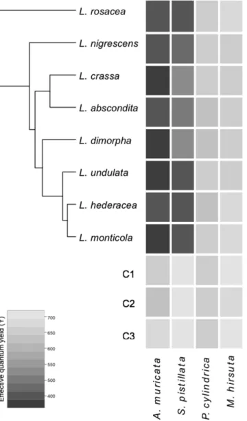
All extracts prepared from Lobophora species caused sig-nificant visual bleaching on the corals A. muricata and S. pistillata and suppression of photosynthetic efficiencyin situ, relative to controls (p < 0.001), while no significant bleaching effects were detected in P. cylindrica and M. hirsuta (Fig. 1). In general, A. muricata was more pronouncedly bleached than S. pistillata (Fig. 1). No significant
difference was observed between the Lobophora species (Fig. 1). Consequently, A. muricata was selected as a target coral for the identification of allelopathic compounds, and the alga L. rosacea was chosen as it is the most
Percentage
of transectsa associationsAverage b Acropora Montipora Stylophora Porites Seriatopora Turbinaria Non-coral substrate
L. abscondita 9 0 0 0 0 0 0 0 100 L. crassa 9 0 0 0 0 0 0 0 100 L. dimorpha 11 15 100 0 0 0 0 0 0 L. hederacea 23 23 15 0 6 22 42c 15 0 L. monticola 11 24 82 12 0 6 0 0 0 L. nigrescens 9 0 0 0 0 0 0 0 100 L. rosacea 42 22 45 22 15 12 0 0 0 L. undulata 19 7 50 42 8 0 0 0 0
Table 1. Association of Lobophora species with corals in the southwest lagoon of New Caledonia.
apercentage of transects where the species were observed in the vicinity of corals. baverage percentage of associations as assessed by the stratified random point count method in transects where the species was present. cLobophora - coral associations with visible deleterious effects (bleaching and or overgrowth).
common and abundant species in the southwest lagoon of New Caledonia, allowing collection of enough material for subsequent analytical identification of purified allelopathic compounds.
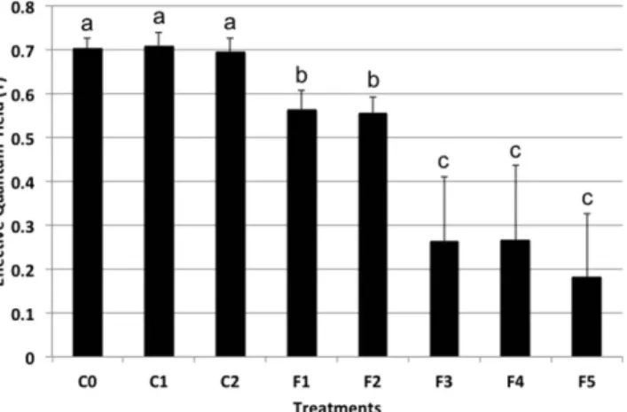
Bioassay-guided fractionation.
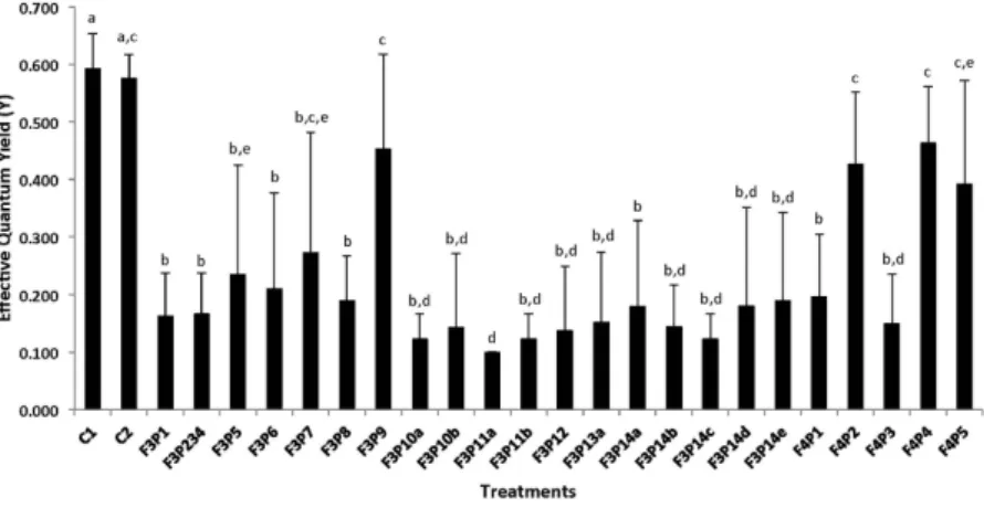
The L. rosacea extract was fractionated by VLC into five fractions of con-trasting polarity. Out of the five fractions tested against the coral A. muricata, the less polar ones (F3 to F5) caused significant visual bleaching and suppression of photosynthetic efficiency relative to controls (Fig. 2), with a decrease of the photosynthetic efficiency of ca. 50% for F3 and F4, and of 70% for F5. The most polar fractions (F1 and F2) significantly suppressed coral photosynthetic efficiency (25% decrease) but less than F3–F5. F4 and F5 displayed very similar HPLC-DAD-ELSD-MS profiles and consequently only F3 and F4 were chemically studied.A first fractionation of F3 by reversed phase HPLC resulted in 14 sub-fractions named F3P1 to F3P14. Because most of them were still identified as mixtures of compounds by 1H NMR, the most bioactive sub-fractions were further purified to identify compounds responsible for the bioactivity. Therefore, the final purification of F3P13, F3P10 and F3P11 led to the pure compounds 1 (F3P13a), 2 (F3P10a) and 3 (F3P11b) respectively (Fig. 3). The structure of the chemical components of the other sub-fractions was not identified due to the low amount available or complexity of the mixture. Reversed phase HPLC fractionation of F4 resulted in five sub-fractions (F4P1-F4P5) from which no pure compound was identified.
Figure 1. Heatmap representation of the bioassay results of eight species of Lobophora, viz. L. rosacea,
L. nigrescens, L. crassa, L. abscondita, L. dimorpha, L. undulata, L. hederacea and L. monticola, crude
extracts tested against four coral species, viz. Acropora muricata, Stylophora pistillata, Porites cylindrica and Montipora hirsuta. The color is indicative of the coral effective quantum yield (Y) measurement under the
patch surface after 24 h of exposure. C1 (no patch), C2 (patch without solvent) and C3 (patch with solvent) are the three controls. The phylogenetic tree is the maximum clade credibility tree obtained from BEAST analysis of the concatenated alignment of four genes (rbcL, cox3, psbA and LSU) from Vieira et al.18.
Subfractions and pure compounds caused contrasting effects, with ca. 80% of them causing significant bleach-ing and suppression of photosynthetic efficiency relative to controls (Fig. 4). The suppression of photosynthetic efficiency ranged from ca. 40 to 80%, relative to the coral effective quantum yield baseline, depending on the sub-fractions. Based on the Tukey HSD post hoc test results, six significantly different groups of allelopathic sub-fractions or pure compounds stood out. Three allelopathic compounds were selected for structure identifica-tion, as they were considered sufficiently pure.
Structure identification of compounds 1–3.
Compound 1 was isolated as colorless oil and its molecular formula was proposed as C21H31ClO by HRESIMS analysis ([M + NH4]+ at m/z 352.2407 and 354.2382 with iso-topic ratio 3:1). The 1H NMR analysis started with a terminal vinyl group at δH 5.34 (dt, H-1a), 5.21 (dt, H-1b) and 6.02 (ddd, H-2) which was COSY coupled to a deshielded methine at δH 4.38 (ddt, H-3) (Table 2). Even if we first suspected the presence of a secondary alcohol at this position, the chemical shift of the corresponding carbon was more shielded than expected at δC 67.8 (C-3) for an allylic alcohol. In agreement with MS data, we then deduced the presence of a chlorine atom at this position which was COSY correlated to a oxygenated methine (δH 3.70 ddd, H-4; δC 75.2, C-4). The spin coupled system was then extended to an ABXM system at δH 2.49 (H-5a) and 2.25 (H-5b) which was further coupled to an alternate polyunsaturated carbon chain composed of four double bonds separated by three methylenes. The configurations of the double bonds were assigned as Z by interpreta-tion of the chemical shifts of allylic carbons. All these connecinterpreta-tions were later confirmed using HSQC and HMBC spectra. The other end of the compound was deduced to be composed of a second terminal vinylic system cou-pled to the polyunsaturated core trough three COSY correlated methylene units. Unfortunately no similar allylic chlorohydrine was found in the literature that could allow us to conclude on the relative configuration of 1. We then decided to compare the 13C NMR experimental values with the calculated values obtained on the most stable conformers of the like and unlike diasteroisomers. Working on the most stable conformer, the Mean Absolute Error (MAE) was found to be lower for the unlike configuration (Fig. 5).
For 2, the isotopic pattern of the HRESIMS spectrum evidenced the absence of a chlorine atom in this mole-cule and the molecular peak at m/z 334.2744 ([M + NH4]+) suggests the replacement of this atom by an alcohol. Inspection of the 1H and 13C NMR spectra allowed us to localize the structural changes in the vicinity of the first
Figure 2. Barplot representation of the bioassays results with the five fractions of L. rosacea on A.
muricata. The statistical analyses, comparing the fractions treatment patches to MeOH-treated patch and
untreated patch controls, were performed using Kruskal-Wallis and Tukey’s HSD post-hoc test. Letters indicate distinct groupings based on post-hoc statistical comparison among sub-fractions. Asterisks indicate significance in relation to controls (MeOH-treated or untreated, accordingly) with P < 0.001, n = 10 assays, ≥ 5 fractions per assay for all experiments. Error bars represent standard deviation of the mean. C1 (no patch), C2 (patch without solvent) and C3 (patch with solvent) are the three controls.
Figure 3. Chemical structure of Compounds 1–3. Compound 1 (F3P13):
(6Z,9Z,12Z,15Z)-nonadeca-1,6,9,12,15,18-hexaene-3,4-diol; Compound 2 (F3P10a): (6Z,9Z,12Z,15Z)-nonadeca-6,9,12,15,18-pentaene-3,4-diol; Compound 3 (F3P11b): (6Z,9Z,12Z,15Z)-4-chlorononadeca-6,9,12,15,18-pentaen-3-ol.
Figure 4. Barplot representation of the allelopathic bioassay results with the 23 compounds isolated from the fractions 3 and 4 of L. rosacea on A. muricata. The statistical analyses, comparing the compounds-treated
patchs to MeOH-treated patch and untreated controls, were performed using Kruskal-Wallis and Steel-Dwass-Critchlow-Fligner post-hoc test. Letters indicate distinct groupings based on post-hoc statistical comparison among sub-fractions. Asterisks indicate significance in relation to controls (MeOH-treated or untreated, accordingly) with P < 0.001, n = 10 assays, 23 sub-fractions per assay. Error bars represent standard deviation of the mean. Letters indicate significant differences (Kruskal–Wallis test, P < 0.01; Steel–Dwass post-hoc test, P < 0.05, mean + s.d., n = 10).
Compound
Lobophorenol A (1) Lobophorenol B (2) Lobophorenol C (3) δC δH, mult. (J in Hz) δC δH, mult. (J in Hz) δC δH, mult. (J in Hz)
1a 118.4 5.34, dt (17.0, 1.0) 116.7 5.31, dt (17.0, 1.0) 10.8 0.97, t (7.5) 1b 5.21, dt (10.0, 1.0) 5.18, dt (10.0, 1.0) 2 137.3 6.02, ddd (17.0, 10.0, 8.5) 139.4 5.92, m 26.8 1.57, m 1.47, m 3 67.8 4.38, ddt (8.5, 4.5, 1.0) 76.6 3.94, m 76.2 3.32, m 4 75.2 3.70, ddd (9.5, 5.5, 4.5) 75.5 3.48, m 74.9 3.45, m 5a 32.8 2.49, br dt (14.5, 5.5) 31.8 2.36, m 32.2 2.36, dt (14.5, 7.0) 5b 2.25, ddd (14.5, 8.0, 5.5) 2.14, m 2.24, dt (14.5, 7.0) 6 126.4 5.48, m 127.3 5.51, m 127.6 5.52, m 7 131.4 5.49, m 130.8 5.45, m 130.6 5.45, m 8 26.8 2.87, t (6.0) 26.7 2.85, m 26.8 2.87, t (6.0) 9 128.7 5.37, m 128.7 5.37, m 128.7 5.37, m 10 129.4 5.37, m 129.4 5.37, m 129.4 5.37, m 11 26.6 2.86, t (6.0) 26.6 2.86, m 26.6 2.86, t (6.0) 12 129 5.37, m 129 5.37, m 129 5.37, m 13 129.1 5.37, m 129.1 5.37, m 129.1 5.37, m 14 26.6 2.82, t (6.0) 26.6 2.82, m 26.6 2.82, t (6.0) 15 129.3 5.37, m 129.3 5.37, m 129.3 5.37, m 16 130.8 5.38, m 130.8 5.38, m 130.8 5.38, m 17 27.6 2.10, br q (7.0) 27.6 2.07, m 27.6 2.08, m
18 30.1 1.46, quint (7.0) 30.1 1.46, quint (7.0) 30.1 1.46, quint (7.0)
19 34.4 2.07, br q (7.0) 34.4 2.06, br q (7.0) 34.4 2.07, m
20 139.8 5.82, ddt (17.0, 10.0, 7.0) 139.8 5.82, ddt (17.0, 10.0, 7.0) 139.8 5.82, ddt (17.0, 10.0, 7.0) 21a 115.1 5.00, dq (17.0, 2.0) 115.1 5.00, dq (17.0, 2.0) 115.1 5.00, dq (17.0, 2.0)
21b 4.94, dt (10.0, 1.0) 4.94, dt (10.0, 1.0) 4.94, dt (10.0, 1.0)
Table 2. 1H (500 MHz) and 13C NMR (125 MHz) chemical shifts (in ppm) for compounds 1–3 in CD 3OD.
vinylic system. Indeed, the methine signals at δH 4.38 (ddt, H-3) and δC 67.8 (C-3) in 1 were replaced by signals at
δH 3.94 (H-3) and δC 76.6 (C-3) that are reminiscent of an allylic secondary alcohol. Therefore, the chlorine atom placed at C-3 in 1 was replaced by a second alcohol in 2 at this position. The relative configuration of compound
2 was deduced to be unlike applying the same method as for 1 (Fig. 5). In this case, hydrogen bonds between the
two vicinal alcohols render the gauche conformer more stable than the anti obtained for 1. Both compounds may be produced by an anti opening of a common epoxide intermediate with water or a chloride ion.
The HR-(+ )ESIMS data obtained for compound 3 with a molecular peak at m/z 336.2895 ([M + NH4]+) suggested that this natural product corresponds to a dihydrogenated derivative of 2. The location of the reduced double bond was unambiguously deduced from 1H NMR data that showed the lack of a terminal vinylic system. The appearance of a methyl at δH 0.97 (t, H-1) definitely placed the new ethyl group at the beginning of the chain. We assume the same relative configuration for this compound as those previously proposed for 1 and 2, being linked biosynthetically. The low amounts of compounds isolated prevented any attempts to assign their absolute configuration at C-3 and C-4.
Discussion
Proliferation of Lobophora in coral reef environments has repeatedly caused concern among biologists16,28. There is, however, considerable uncertainty regarding the causes underlying such proliferations as well as of the threat that Lobophora poses to corals. In addition, given the recent progress in understanding species-level diversity in the genus, it is not known whether all or only a subset of Lobophora species compete with corals for space. Our survey of Lobophora – coral associations in the southwest lagoon of New Caledonia demonstrates that not all species of Lobophora associate with corals. Out of eight Lobophora species, three never associated with a coral, but instead grew attached to scattered hard substrate in seagrass beds, shallow wave-washed habitats or coral rubble. Nevertheless, perhaps some interactions may have been detected with greater observations. The other five species were associated with living coral colonies, but grew at the dead bases adopting procumbent to decumbent forms (e.g. L. dimorpha, L. undulata, L. hederacea, L. monticola), or a fasciculate morphology niched within coral branches such as L. rosacea. The latter species is also the most commonly encountered, being observed in 42% of the transects. Most Lobophora species, however, were only observed in 10 to 20% of the transects. Even then, these numbers tend to overestimate the prevalence of Lobophora on the entire reef since the sites where the transects were laid out were precisely those locations where Lobophora – coral interactions were most conspicuous during initial surveys. Per transect, the presence of Lobophora never surmounted 25%. Acropora species were clearly the preferred partner, but all but one Lobophora species displayed a broader range of hosts. Corals associated with Lobophora did not present traces of bleaching (Fig. 6), except in the case of L. hederacea associated with Seriatopora caliendrum. Based on the ecological niche and the morphological differentiation between Lobophora species we investigated if the species found in direct contact with corals have developed specific allelochemicals capable of impairing corals. Our results demonstrate that all Lobophora species, usually found in contact or not with corals, displayed similar bleaching effects on the tested corals. In other words, naturally found in contact or not, extracts of the eight
Lobophora species show similar effects on corals: they are equally capable or not of bleaching specific corals. These
results are of significant importance as it implies that species of the genus Lobophora are intrinsically capable of bleaching some corals upon direct contact. In an evolutionary context, this either means that Lobophora has devel-oped: (1) allelopathic compounds targeted towards competing benthic organisms or (2) allelopathy against corals, or other benthic organisms, may be a side-effect (i.e. a secondary unintentional effect) of secondary metabolites with different ecological roles, such as antimicrobial properties (e.g. biofilm deterrents). Recent findings by Rasher and Hay29 showing that the red alga Galaxaura filamentosa uses different compounds to compete with corals versus to resist herbivores would refute the side-effect hypothesis. However, it is unknown at present if a differentiation between allelochemicals and anti-herbivory chemicals is the rule rather than the exception. At least the large frac-tion of pure or mixed compounds (80% of the isolated compounds from two fracfrac-tions) that result in a significant suppression of photosynthetic efficiency on corals, would argue against such differentiation.
Among the four coral species tested, A. muricata and M. hirsuta were the most significantly bleached corals. These results indicated a differential susceptibility to Lobophora allelopathy depending on the coral species. In this aspect our results echoed those of Rasher et al.5 who also noticed differential susceptibility across coral species to algal allelopathy. Rasher et al.5 found that A. millepora and P. damicornis were more sensitive to macroalgal allelopathic damage than M. digitata and P. cylindrica. We shared three genera (Acropora, Montipora, and Porites)
Figure 5. Mean Absolute Errors (MAE) and Corrected Mean Absolute Errors (CMAE) obtained between the 13C NMR experimental and theoretical values for the two possible diasteroisomers of compounds 1
and one species (P. cylindrica) with Rasher et al.5. Also, we had similar results across those genera, although in our case M. hirsuta and P. cylindrica were not damaged at all. P. damicornis, which belongs to the same family as S. pistillata, i.e., Pocilloporidae, was also quite sensitive5. However, Acropora and Montipora, which belong to the same family (Acroporidae), were differentially susceptible in both studies. Partly agreeing with our findings, Lesser et al.30 showed that Acroporids are not as resilient in the face of environmental perturbation compared to other species on the same reef. Nugues and Bak27 also showed that Caribbean corals had differential competitive abilities against Lobophora.
We then proceeded to the isolation and structure identification of the chemicals from L. rosacea exhibiting bleaching properties against A. muricata, the most susceptible coral out of the four tested. Results from the bio-assays with the five fractions showed that allelopathy against coral correlates with the polarity of the compounds, with the less polar fractions displaying the highest allelopathic activity. These results concur with the findings of Rasher and Hay8, showing that lipidic extracts from several algal species, including Lobophora variegata, resulted in significant bleaching, while hydrophilic compounds from Chlorodesmis fastigata (Udoteaceae, Chlorophyta) and
Galaxaura filamentosa (Galaxauraceae, Rhodophyta) were not active. These results corroborate the importance of
direct contact, which is preferable for hydrophobic allelochemicals transfer.
Most of the purified compounds from L. rosacea displayed a significant bleaching effect on A. muricata. The three new C21 polyunsaturated alcohols, named lobophorenols A–C (1–3) were among the most active fractions and sub-fractions were identified after NMR and MS analyses. These compounds were identified as three new C21 polyunsaturated alcohols. All these compounds may originate after opening of a common epoxide intermediate formed from a polyene. Similar C21 apolar polyenes have been reported only once from the alga Fucus vesiculosus31. It is worth highlighting the presence of a chlorinated analogue 1, which is particularly rare and represent less than 1% of all the secondary metabolites isolated from species of the Phaeophyceae family32,33. Although, we may point out that De Nys et al.8 also isolated halogenated allelochemicals, the presence of the chlorine atom may however not be related to the bleaching properties of the molecule, since both compounds 2 and 3, deprived of this halogen atom, present similar adverse properties. Furthermore, the isolated allelochemicals do not belong to the terpene family of natural products, as somewhat expected from De Nys et al.6 and Rasher and Hay5 but polyunsaturated alcohols. It shows that allelopathy against corals may involve a variety of families of compounds as already reported by Slattery and Lesser11, and strongly supported by the diversity of compounds displaying bleaching properties in this study. The lack of ability to correlate bioactivity with classes of compounds is well known and been discussed numerous times for anti-herbivore compounds, antibiotics, etc.34–36. It is worth pointing out that we were expect-ing to find terpenes, given the richness in terpenoids of the Dictyotaceae family to which Lobophora belongs37.
Figure 6. Pictures of natural association between Lobophora spp. and coral species in New Caledonia.
(A) L. rosacea at next to Acropora sp., (B) L. undulata at the base of Acropora sp., (C) L. rosacea at the bases of
However, much to our surprise this family of compounds was not detected by NMR. Yet, the genus Zonaria, which is sister to Lobophora, did not present terpenes either (authors’ unpublished data).
Lobophora bioactivity against corals does not come as a surprise as in the literature Lobophora extracts (crude,
hydrophilic or hydrophobic extracts) and isolated compounds have been shown to display a broad spectrum of activities and in particular antimicrobial (e.g. fungi, bacteria, protozoa) bioactivities e.g.10,38,39. The exact bleach-ing mechanisms are unaddressed here and could very well be targetbleach-ing either the polyp or the Symbiodinium. Nonetheless, it is worth mentioning that after two weeks following the bioassays, the surface area which bleached in contact with the patches, recovered their original coloration.
Present field assays would suggest that Lobophora has the potential to chemically impair some coral spe-cies by direct contact. Nevertheless, in situ observations indicate that although apparently chemically potent,
Lobophora do not or rarely bleach coral hosts in a natural setting (this study). Slattery and Lesser11 also questioned if Lobophora presented allelopathic effects on corals in the Bahamas. Yet, while Lobophora extracts and a purified compound bleached the coral Montastrea cavernosa, contact experiments between Lobophora and the coral did not11. Furthermore, no claim of coral bleaching as a result of contact with Lobophora in natural setting was made by the authors11. Even though it would be tempting to conclude that allelopathy is ecologically important in the competition between Lobophora and corals, there is no strong evidence from field observations. Herbivory on the other hand, clearly appears as an important factor preventing competition to occur11,17,26,28. Therefore, the ques-tion remains: what explains the inconsistency between field observaques-tions and bioassay experiments? A possible explanation for this discrepancy would be the localization of the bioactive compounds within the endometabo-lome. Bioassays artificially expose corals to chemicals, a situation that would only occur as a result of abrasion or herbivory under natural conditions. Alternatively, the compounds may be part of the exometabolome, present on the surface of the alga, but external factors (e.g. herbivory) or a defense system by the coral itself may ward off allelopathic interactions, thereby preventing Lobophora from outcompeting corals. Several investigators have demonstrated that bioactive lipids distributed on the surfaces of algae, including Lobophora, were capable of dam-aging corals5,7,8, 40. This forms a strong indication that at least some compounds are present on the algal surface. Given the presence of allopathic compounds, most coral species prevent the overgrowth of crustose Lobophora species owing to a set of defense mechanisms24,27. Additionally, field observations and experiments showed that herbivory is a major factor preventing increase in Lobophora abundance11,26,28. In New Caledonia, only one spe-cies of Lobophora, L. hederacea, was observed overgrowing a coral spespe-cies, Seriatopora caliendrum19. In the latter case, coral overgrowth appears to be possible owing to a combination of factors including the coral vulnerability and the inhibition of grazing19, supporting the important role of coral defense and herbivory in preventing nega-tive allelopathic interactions. In the Great Barrier Reef, Jompa and McCook17 showed that a crustose Lobophora species was capable of overgrowing the coral Porites cylindrica when herbivory was reduced. In damaged reefs, however, coral morbidity and mortality in addition to shifts in herbivory pressure result in whole different setting where macroalgal allelopathy may have harmful effects on corals. Although not yet explored, it is possible that allelopathy in damaged reef may results from the synergetic effects of macroalgal exudates/allelochemicals acting in combination with a number of environmental parameters/stressors such as seawater pH, oxygen depletion, and or temperature maxima1.
The role of chemical interactions between macroalgae and corals initially evinced in the early 90s in form of positive allelopathy41, has regained interest only recently, yet this time in form of negative allelopathy8. Most studies on the subject have disclosed deleterious effects (e.g. bleaching, recruitment inhibition) in damaged reefs. The present work, focusing on a healthy reef ecosystem, provided evidence that allelopathic defense is not restricted to Lobophora species that are naturally found in close contact with corals. These findings are important toward our understanding of the importance of allelopathic competition and defense systems versus herbivory in the interaction between macroalgae and corals in reef ecosystems.
Materials and methods
Quantification of Lobophora–corals association.
Eight species of Lobophora, commonly encountered in the southwest lagoon of New Caledonia were selected to quantify their association with corals and for the bioassays, i.e. L. abscondita, L. crassa, L. dimorpha, L. hederacea, L. monticola, L. nigrescens, L. undulata, and L.rosacea (Fig. 6). 78 belt transects, as described by English et al.42, each 10 m long, were deployed across coral dom-inated reefs in the southwest lagoon of New Caledonia. Within a belt transect, a 2500 cm2 quadrat (50 × 50 cm) was placed consecutively left and right along a defined line, and photographs were taken directly above each quadrat using a Lumix Panasonic digital camera (12 megapixels) mounted on a photoquadrat framer. In each quadrat, the frequency of Lobophora – coral associations was assessed by placing 16 points per quadrat using a stratified random point count method using the software “Coral Point Count with Excel extensions”43. Details of the sampling locations and quantification methods are outlined in Supplementary information. From these data we calculated the percentage of transects in which Lobophora was associated with corals. The average percentage of associations of each species was calculated per transect where the species was observed.
Preparation of the extracts and fractions of Lobophora for bioassays.
Algal samples for bio-assays were collected by SCUBA in January 2013 in the southwest lagoon of New Caledonia (Supplementary information). Samples were cleaned from epiphytes and stored at − 20 °C until freeze-drying. Four coral species were selected as targets of the bioassays, i.e. Acropora muricata (Linnaeus, 1758; Acroporidae),Montipora hirsuta (Nemenzo, 1967; Acroporidae), Stylophora pistillata (Esper, 1797; Pocilloporidae)
and Porites cylindrica (Dana, 1846; Poritidae). Algal specimens were identified at species-level using mitochondrial cox3 gene sequences see18. The area of each individual was estimated using the alumi-num foil technique44. Then, the specimens were freeze-dried and the dried samples were ground with a mortar and pestle using liquid nitrogen. One gram of ground powder was exhaustively extracted, by
out in terms of bioactivity against A. muricata or any of the other corals (cf. results), L. rosacea was chosen for subsequent analytical identification of purified allelopathic compounds as it is the most common and abundant species in the southwest lagoon of New Caledonia, thus allowing collection of enough material for subsequent analytical identification of purified allelopathic compounds. The biomass (209 g of dry mass) of L. rosacea was exhaustively extracted, by adding consecutively five times MeOH/CH2Cl2 (1:1, v/v; 1.2 L of solvent), leaving it 10 min in an ultrasonic bath and 5 min to decant, and then retrieving the supernatant liquid. The resulting extract was concentrated under vacuum to yield a homogeneous dry powder (8.3 g). The extract was then mixed with an equal amount of C18 silica powder (Polygoprep
®
60-50) and fractionated by Vacuum Liquid Chromatography (VLC) into five fractions (F1–F5), eluting with the five organic solvents aforementioned for SPE. An additional elution was done with CH2Cl2 in order to ensure exhaustive compounds extraction from the crude extract, and was additionally tested as a sixth fraction (F6). The resulting filtrates were evaporated under vacuum, resuspended into MeOH to reach a concentration of 10 mg∙mL−1, filtered through 0.22 μ m PTFE syringe filters (Phenomenex, UK) and filled into HPLC vials for subsequent Ultra-High Performance Liquid Chromatography-Diode Array Detection (UHPLC-DAD) analyses and High Performance Liquid Chromatography (HPLC) purification.According to the results on the ecological activity (cf. next paragraph), F3 and F4 were selected for compounds isolation and purification. The HPLC purification was performed on a Jasco (Groß-Umstadt, Germany) prepara-tive HPLC system (pump PU-2087 plus; diode array detector MD 2018 plus; column thermostat CO 2060 plus; autosampler AS 2055 plus; LC Net II ADC Chromatography Data Solutions; sample injection loop: 250 μ L) on a phenyl-hexyl reversed phase column (XSelect CSH
™
, 5 μ m, 19 × 250 mm; Waters, France), using for F3 an isocratic elution mode [acetonitrile (CH3CN) + 0.1% trifluoroacetic acid (TFA)/H2O + 0.1% TFA; 69/31, v/v] and a flow rate of 10 mL/min. Fourteen sub-fractions (from F3P1 to F3P14) were obtained. Fraction F4 was fractionated on the same column with a CH3CN/H2O + 0.1% TFA gradient on a 30 min run (0–5 min: 90% CH3CN; 5–10 min: 90 at 100% CH3CN, 10–25 min: 100% CH3CN) at 10 mL/min, leading to five sub-fractions (F4P1 to F4P5). The purification of compounds from four sub-fractions of F3 (F3P10, F3P11, F3P13, and F3P14) were performed on a C18 semi-preparative column (XSelect CSH™
C18, 5 μ m, OBD, 19 × 250 mm; Waters, France) with a CH3CN/ H2O + 0,1% TFA gradient (UV detection: 210 nm, flow rate: 10 mL/min).Among all the fractions and sub-fractions only the three major, pure and bioactive compounds 1–3, corre-sponding to fractions F3P13a (18.4 mg), F3P10a (3.8 mg) and F3P11b (3 mg) respectively, were identified on the basis of NMR and MS data.
NMR analyses were performed in CD3OD on a Bruker Avance 500 spectrometer using signals of the residual peaks of the solvent for calibration of the chemical shifts in ppm (δH 3.31 for 1H NMR and δC 49.0 for 13C NMR). LC-DAD-ELSD-ESI/MSn analyses were carried out on a LaChrom Elite HPLC (VWR-Hitachi) composed of a L-2130 quaternary pump, a L-2200 autosampler, and a L-2300 column oven. Detection was performed with a L2455 DAD and an ELSD (Chromachem model, Eurosep) coupled to an Esquire 6000 spectrometer. UHPLC-HRMS were performed on a UHPLC U3000 (Dionex) coupled to a QqToF Impact II (Bruker).
In situ allelochemical assays.
Field experiments, conducted in situ were designed to keep the coral under natural field conditions, thus limiting pre-experimental stress usually resulting from cutting, gluing and trans-plantation. The bioassays were conducted in Sainte Marie Bay (22° 17.863′ S, 166° 28.898′ E) with three of the coral genera, i.e. Acropora muricata, Porites cylindrica and Montipora hirsuta and on genus in Maitre Islet Reef for Stylophora pistillata (22° 20.446′ S, 166° 24.108′ E). A series of three bioassay experiments were successively performed. The first experiment evaluated the bioactivity of the crude extract of the Lobophora species previ-ously selected (L. abscondita, L. crassa, L. dimorpha, L. hederacea, L. monticola, L. nigrescens, L. undulata, and L.rosacea) on four coral species (Acropora muricata, Porites cylindrica, Stylophora pistillata and Montipora hirsuta).
The second experiment tested the bioactivity of the five fractions obtained from the extracts of L. rosacea on A.
muricata. The final experiment tested the bioactivity of the sub-fractions and compounds from two of the most
bioactive fractions of L. rosacea identified in the previous experiment (F3 and F4). All bioassay experiments were performed in situ directly on coral colonies at approximately natural concentration (i.e. concentration per surface area previously estimated), the latter being critical for bioassays assessing allelopathic interactions. Thereto, we determined the amount of crude extracts, fractions, sub-fractions, and pure compounds per unit of algal surface area (i.e. 1 cm2) and reported it to the surface of the agarose patch applied on the coral (i.e. 2 cm2).
A replicate was defined by one colony of coral on which all the extracts, fractions or sub-fractions (including in some cases pure isolated compounds) were tested. A total of 10 replicates were implemented. The methodology was adapted from Rasher and Hay8. The chemical samples (crude extracts, fractions, sub-fractions or pure compounds)
were resuspended in 1 mL MeOH and added at natural concentration into a 4% agarose gel (Conda Pronadisa, Spain). The mix chemical sample/agarose was poured into a polyvinyl chloride mold, composed of 10 times 2-cm2 wells. Before that, tulle bands, of 20 × 2 cm, were disposed at the bottom of the wells onto which the gel mixture will adhere while gelifying. The strips were prepared the day before field application and refrigerated until then at 5 °C. They were applied onto the coral by knotting the tulle bands to the branches, and removed after 24 h of exposure. Agarose strips with and without MeOH were additionally made as controls, to ensure the non-effect of either the agarose strips itself or the solvent on the coral. Gel strips were applied on the corals between 09:00 and 11:00 AM.
Coral photosynthesis measurements.
Pulse Amplitude Modulated (PAM) fluorometry measurements were performed with a Diving-PAM (Walz) right after removal of the strips. PAM fluorometry measures the photosynthetic efficiency of photosystem II within the endosymbiotic Symbiodinium spp. that may be used as a quantitative measure of photo-inactivation during coral bleaching45. PAM fluorometry values of healthy corals are ranging between 0.5 to 0.8, depending on the coral species and time of the day. Values between 0 to 0.2 are indicative of severe bleaching or mortality46. As outlined in Rasher and Hay8 PAM fluorometry measurements were performed where the strips were applied and 5-cm next to it, as a spatial control to have a coral health base-line for comparison.Statistical analyses.
Normality of distribution of the coral responses for all the bioassay experiments was tested with the normality Shapiro-Wilk test. If the responses violated parametric assumptions, coral responses were evaluated using the Kruskal-Wallis H test followed by the Tukey honestly significant difference (HSD) post hoc comparisons test for significant Kruskall-Wallis findings. If the data respected the parametric assumptions, a one-way ANOVA was performed followed by the Tukey post hoc HSD test for significant ANOVA findings. Statistical analyses were performed using the computing environment R47.References
1. Harlin, M. M. & Rice, E. L. Allelochemistry in marine macroalgae. Crit. Rev. Plant Sci. 5, 237–249 (1987). 2. Amsler, C. D. Algal chemical ecology. Springer (2008).
3. Paul, V. J. & Puglisi, M. P. Chemical mediation of interactions among marine organisms. Nat. Prod. Rep 21, 189–209 (2004). 4. Bonaldo, R. M. & Hay, M. E. Seaweed-coral Interactions: variance in seaweed allelopathy, coral susceptibility, and potential effects
on coral resilience. PLoS One 9, e85786 (2014).
5. Rasher, D. B., Stout, E. P., Engel, S., Kubanek, J. & Hay, M. E. Macroalgal terpenes function as allelopathic agents against reef corals.
Proc. Natl. Acad. Sci. U.S.A. 108, 17726–17731 (2011).
6. De Nys, R., Coll, J. & Price, I. Chemically mediated interactions between the red alga Plocamium hamatum (Rhodophyta) and the octocoral Sinularia cruciata (Alcyonacea). Mar. Biol. 108, 315–320 (1991).
7. Andras, T. D. et al. Seaweed allelopathy against coral: surface distribution of a seaweed secondary metabolite by imaging mass spectrometry. J. Chem. Ecol. 38, 1203–1214 (2012).
8. Rasher, D. B. & Hay, M. E. Chemically rich seaweeds poison corals when not controlled by herbivores. Proc. Natl. Acad. Sci. U.S.A.
107, 9683–9688 (2010).
9. Vieira, C., Gaubert, J., De Clerck, O., Payri, C., Culioli, G. & Thomas, O. P. Biological activities associated to the chemodiversity of brown algae belonging to the genus Lobophora (Dictyotales, Phaeophyceae). Phytochem. Rev., (in press).
10. Kubanek, J., Jensen, P. R., Keifer, P. A., Sullards, M. C., Collins, D. O. & Fenical, W. Seaweed resistance to microbial attack: a targeted chemical defense against marine fungi. Proc. Natl. Acad. Sci. U.S.A. 100, 6916–6921 (2003).
11. Slattery, M. & Lesser, M. P. Allelopathy in the tropical alga Lobophora variegata (Phaeophyceae): mechanistic basis for a phase shift on mesophotic coral reefs ? J. Phycol. 50, 493–505 (2014).
12. Birrell, C. L., McCook, L. J., Willis, B. L. & Harrington, L. Chemical effects of macroalgae on larval settlement of the broadcast spawning coral Acropora millepora. Mar. Ecol. Prog. Ser. 362, 129–137 (2008).
13. Kuffner, I. B., Walters, L. J., Becerro, M. A., Paul, V. J., Ritson-Williams, R. & Beach, K. S. Inhibition of coral recruitment by macroalgae and cyanobacteria. Mar. Ecol. Prog. Ser. 323, 107–117 (2006).
14. Mumby, P. J., Foster, N. L. & Fahy, E.A.G. Patch dynamics of coral reef macroalgae under chronic and acute disturbance. Coral Reefs
24, 681–692 (2005).
15. De Ruyter Van Steveninck, E. & Bak, R. Changes in abundance of coral-reef bottom components related to mass mortality of the sea-urchin Diadema antillarum. Mar. Ecol. Prog. Ser. 34, 87–94 (1986).
16. Diaz-Pulido, G. et al. Doom and boom on a resilient reef: climate change, algal overgrowth and coral recovery. PLoS One 4, e5239 (2009).
17. Jompa, J. & McCook, L. J. The effects of nutrients and herbivory on competition between a hard coral (Porites cylindrica) and a brown alga (Lobophora variegata). Limnol. Oceanogr. 47, 527–534 (2002).
18. Vieira, C., D’hondt, S., De Clerck, O. & Payri, C. E. Toward an inordinate fondness for stars, beetles and Lobophora ? Species diversity of the genus Lobophora (Dictyotales, Phaeophyceae) in New Caledonia. J. Phycol. 50, 1101–1119 (2014).
19. Vieira, C., Payri, C. & De Clerck, O. Overgrowth and killing of corals by the brown alga Lobophora hederacea (Dictyotales, Phaeophyceae) on healthy reefs in New Caledonia: a new case of the epizoism syndrome. Phycol. Res. 63, 152–153 (2015). 20. Bennett, S., Vergés, A. & Bellwood, D. Branching coral as a macroalgal refuge in a marginal coral reef system. Coral Reefs 29, 471–480
(2010).
21. Hughes, T. P. Catastrophes, phase shifts, and large-scale degradation of a Caribbean coral reef. Science 265, 1547–1551 (1994). 22. Cheal, A. et al. Coral–macroalgal phase shifts or reef resilience: links with diversity and functional roles of herbivorous fishes on the
Great Barrier Reef. Coral Reefs 29, 1005–1015 (2010).
23. Done, T., Turak, E., Wakeford, M., DeVantier, L., McDonald, A. & Fisk, D. Decadal changes in turbid-water coral communities at Pandora Reef: loss of resilience or too soon to tell? Coral Reefs 26, 789–805 (2007).
24. De Ruyter van Steveninck, E., Van Mulekom, L. & Breeman, A. Growth inhibition of Lobophora variegata (Lamouroux) Womersley by scleractinian corals. J. Exp. Mar. Biol. Ecol. 115, 169–178 (1988).
25. Diaz-Pulido, G. & McCook, L. J. Effects of live coral, epilithic algal communities and substrate type on algal recruitment. Coral Reefs
23, 225–233 (2004).
26. Jompa, J. & McCook, L. J. Effects of competition and herbivory on interactions between a hard coral and a brown alga. J. Exp. Mar.
Biol. Ecol. 271, 25–39 (2002).
27. Nugues, M. M. & Bak, R. P. Differential competitive abilities between Caribbean coral species and a brown alga: a year of experiments and a long-term perspective. Mar. Ecol. Prog. Ser. 315, 75–86 (2006).
(Phaeophyceae). Rev. Bras. Farmacogn. 21, 216–228 (2011).
38. Cantillo-Ciau, Z., Moo-Puc, R., Quijano, L. & Freile-Pelegrín, Y. The tropical brown alga Lobophora variegata: A source of antiprotozoal compounds. Mar. Drugs 8, 1292–1304 (2010).
39. Engel, S., Puglisi, M. P., Jensen, P. R. & Fenical, W. Antimicrobial activities of extracts from tropical Atlantic marine plants against marine pathogens and saprophytes. Mar. Biol. 149, 991–1002 (2006).
40. Lane, A. L. et al. Desorption electrospray ionization mass spectrometry reveals surface-mediated antifungal chemical defense of a tropical seaweed. Proc. Natl. Acad. Sci. U.S.A. 106, 7314–7319 (2009).
41. Morse, A. Role of algae in the recruitment of marine invertebrate larvae. In: Plant-animal interactions in the marine benthos (eds John D. M., Hawkins S. J., Price J. H.). Oxford Science Publications (1992).
42. English, S. S., Wilkinson, C. C. & Baker, V. V. Survey manual for tropical marine resources. Australian Institute of Marine Science (AIMS) (1994).
43. Kohler, K. E. & Gill, S. M. Coral Point Count with Excel extensions (CPCe): A Visual Basic program for the determination of coral and substrate coverage using random point count methodology. Comput Geosci-UK 32, 1259–1269 (2006).
44. Marsh, Jr, J. A. Primary productivity of reef-building calcareous red algae. Ecology 51, 255–263 (1970).
45. Warner, M. E., Fitt, W. K. & Schmidt, G. W. Damage to photosystem II in symbiotic dinoflagellates: a determinant of coral bleaching.
Proc. Natl. Acad. Sci. U.S.A. 96, 8007–8012 (1999).
46. Fitt, W. K., Brown, B. E., Warner, M. E. & Dunne, R. P. Coral bleaching: interpretation of thermal tolerance limits and thermal thresholds in tropical corals. Coral Reefs 20, 51–65 (2001).
47. R Development Core Team. R: a language and environment for statistical computing. Vienna, Austria: R Foundation for Statistical Computing; 2012. Open access available at: http://cranr-projectorg (last accessed date January 15, 2015) (2013).
Acknowledgements
M. Gaysinski and S. Greff are deeply acknowledged for recording NMR and HRMS data respectively. C. Vieira is a PhD fellow of the University of Pierre and Marie Curie and Ghent University and is part of MARES, a Joint Doctorate programme selected under Erasmus Mundus coordinated by Ghent University (FPA 2011-0016).
Author Contributions
C.V., O.D.C., C.P., O.T. and G.C. designed research; C.V., J.G. and F.H. performed bioassays experiments; C.V., J.G. and F.H. analyzed bioassays data; O.T., J.G. performed extraction and fractionation; G.C. and G.G.-J. performed the NMR characterization and mass spectrometry; C.V. prepared Figures 1–3 and 5; O.T. prepared Figures 4 and 6; all authors contributed to the writing of the manuscript.
Additional Information
Supplementary information accompanies this paper at http://www.nature.com/srep Competing financial interests: The authors declare no competing financial interests.
How to cite this article: Vieira, C. et al. Allelopathic interactions between the brown algal genus Lobophora
(Dictyotales, Phaeophyceae) and scleractinian corals. Sci. Rep. 6, 18637; doi: 10.1038/srep18637 (2016).
This work is licensed under a Creative Commons Attribution 4.0 International License. The images or other third party material in this article are included in the article’s Creative Commons license, unless indicated otherwise in the credit line; if the material is not included under the Creative Commons license, users will need to obtain permission from the license holder to reproduce the material. To view a copy of this license, visit http://creativecommons.org/licenses/by/4.0/