HAL Id: hal-02135082
https://hal.archives-ouvertes.fr/hal-02135082
Submitted on 21 May 2019
HAL is a multi-disciplinary open access
archive for the deposit and dissemination of
sci-entific research documents, whether they are
pub-lished or not. The documents may come from
teaching and research institutions in France or
abroad, or from public or private research centers.
L’archive ouverte pluridisciplinaire HAL, est
destinée au dépôt et à la diffusion de documents
scientifiques de niveau recherche, publiés ou non,
émanant des établissements d’enseignement et de
recherche français ou étrangers, des laboratoires
publics ou privés.
Posterior shoulder instability managed by arthroscopic
acromial pediculated bone-block. Technique
Pierre Métais, Jean Grimberg, Philippe Clavert, Jean-François Kouvalchouk,
François Sirveaux, Geoffroy Nourissat, Jérome Garret, Pierre Mansat, Arnaud
Godenèche
To cite this version:
Pierre Métais, Jean Grimberg, Philippe Clavert, Jean-François Kouvalchouk, François Sirveaux, et
al.. Posterior shoulder instability managed by arthroscopic acromial pediculated bone-block.
Tech-nique. Orthopaedics and Traumatology - Surgery and Research, Elsevier, 2017, 103 (8), pp.S203-S206.
�10.1016/j.otsr.2017.09.001�. �hal-02135082�
O
pen
A
rchive
T
oulouse
A
rchive
O
uverte
(OATAO)
OATAO is an open access repository that collects the work of some Toulouse
researchers and makes it freely available over the web where possible.
This is
an author's
version published in:
https://oatao.univ-toulouse.fr/23102
Official URL :
https://doi.org/10.1016/j.otsr.2017.09.001
To cite this version :
Any correspondence concerning this service should be sent to the repository administrator:
[email protected]
Métais, Pierre and Grimberg, Jean and Clavert, Philippe and Kouvalchouk, Jean-François
and Sirveaux, François and Nourissat, Geoffroy and Garret, Jérome and Mansat, Pierre
and Godenèche, Arnaud Posterior shoulder instability managed by arthroscopic acromial
pediculated bone-block. Technique. (2017) Orthopaedics & Traumatology: Surgery & Research,
103 (8). S203-S206. ISSN 1877-0568
OATAO
O
pen
A
rchive
T
ou
l
ouse
A
rchive
O
uverte
Posterior
shoulder instability managed by arthroscopic acromial
pediculated
bone-block. Technique
P.
Métais
a,∗,
J. Grimberg
b,
P. Clavert
c,
J.-F. Kouvalchouk
d,
F. Sirveaux
e,
G. Nourissat
f,
J.
Garret
g,
P. Mansat
h,
A. Godenèche
i,
the French Arthroscopy Society
a Service de chirurgie du membre supérieur, hôpital privé La Châtaigneraie, 63110 Beaumont, France b IRCOS, 6, avenue Alphonse-XIII, 75016 Paris, France
c Service de chirurgie du membre supérieur, avenue Baumann, 67400 Illkirch, France d Hôpital Foch, 40, rue Worth, 92151 Suresnes, France
e Centre chirurgical Emile-Gallé, CHRU de Nancy, 49, rue Hermite, 54000 Nancy, France f UPMC Inserm UMRS938, clinique des Maussins, 67, rue de Romainville, 75019 Paris, France g Clinique du Parc, 155 ter, boulevard Stalingrad, 69006 Lyon, France
h Département d’orthopédie-traumatologie, hôpital Riquet, CHRU-Purpan, place du Dr-Baylac, 31059 Toulouse, France i Hôpital privé J.-Mermoz (Ramsay-GDS), centre orthopédique Santy, 69008 Lyon, France
Keywords: Acromial bone-block Technique Shoulder Instability Posterior
a b s t r a c t
In posterior shoulder instability (recurrent dislocation, involuntary posterior subluxation or voluntary subluxation that has become involuntary), surgery may be considered in case of failure of functional treatment if there are no psychological contraindications. Acromial bone-block with pediculated deltoid flap, as described by Kouvalchouk, is an alternative to iliac bone-block, enabling triple shoulder locking by the blocking effect, the retention hammock provided by the deltoid flap and posterior capsule repair. Arthroscopy allows shoulder joint exploration and diagnosis of associated lesions, with opening and conservation of the posterior capsule; it greatly facilitates bone-block positioning and capsule reinsertion. The present report describes the procedure in detail.
Level of evidence: Technical note.
1. Introduction
In posterior shoulder instability (recurrent dislocation, invol-untary posterior subluxation or volinvol-untary subluxation that has become involuntary), the first-line option is functional treatment by adapted rehabilitation based on recentering the humeral head and on muscle reinforcement[1,2].
In case of failure of functional treatment, if there are no psy-chological contraindications, surgery may be considered to treat capsulolabral lesions, which may or may not be associated with bone lesions. The main procedures are capsulolabral reconstruction
[3–5], filling reverse Hills-Sachs lesion[6,7], and iliac bone-block
[8–13].
Acromial bone-block with pediculated deltoid flap, described by Kouvalchouk[14], is an alternative to iliac bone-block, presenting the same mechanical requirements as the coracoid bone-block
∗ Corresponding author.
E-mail address:[email protected](P. Métais).
described by Latarjet and Patte and Debeyre[15–17]for anterior instability. It enables double shoulder locking by bone augmenta-tion and a muscular hammock effect. The glenoid is reconstructed or augmented and the capsule is reinforced. Triple locking can be achieved by associating capsule reinsertion on the posterior edge of the glenoid.
With the advent of arthroscopic surgery, it seemed useful to suggest an improvement to this technique:
• minimal posterior approach, to dissect and harvest the bone-muscle graft;
• arthroscopy, to explore, open and conserve the posterior capsule; • to facilitate bone-block positioning and capsule reinsertion.
The first arthroscopic acromial pediculated bone-blocks for pos-terior instability were performed as part of the 2016 symposium of the French Society of Arthroscopy (SFA).
2. Technique
The SFA symposium demonstrated that posterior shoulder instability could be managed by bone-block stabilization, of which acromial pediculated bone-block is one variant.
2.1. Patient positioning
Surgery is performed under general anesthesia and locoregional anesthesia by interscalene nerve block, with the patient semi-seated (45◦) with the shoulder off the table so as to leave the scapular belt free (Fig. 1). The arm can be positioned according to the surgeon’s habits, on a simple support, with or without trac-tion, or on an articulated arm. The sterile drapes are as in standard shoulder arthroscopy.
Bone relief is outlined: acromion and acromial spine, acromio-clavicular joint and glenohumeral joint line projection.
2.2. Harvesting step
The harvesting step is basically as described by Kouvalchouk
[14].
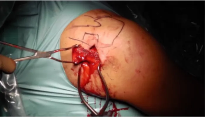
A 5 cm longitudinal incision is made over the joint line. After dissection of subcutaneous tissue, the superficial sides of the del-toid muscle and acromion are easily released. The bone-harvesting site is marked. Harvesting begins at the posterolateral angle of the acromion, taking a 2.5 cm long block. Harvesting may be shifted slightly medially, depending on acromion and shoulder morphol-ogy, but in that case the site is closer to the spine, with greater risk of fracture. Depth is about 1.5 cm and thickness about 0.5 cm, although this may be varied as the anterior and inferior acromial cortex has to be spared. Based on these marks, the deltoid flap is dissected and freed with a 2.5 cm width from the acromion toward the humeral insertion, with a height of 5 cm. It is essential to spare the deltoid insertion on the acromion.
Harvesting is cautious, using a saw and/or bone chisel, respec-ting an inferior tablet and the anterior cortex. The bicortical graft is mobilized to complete release of the deep side of the deltoid from the subacromial-subdeltoid bursa (Fig. 2).
2.3. Arthroscopy step
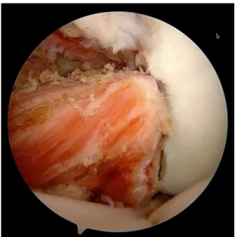
The posterior portal is classical, using the posterior incision made for the bone-block harvesting. After thorough exploration, an anterior optical portal is performed through the rotator interval.
Lesions causing posterior instability are identified. The posterior capsule is released from the posterior edge of the glenoid cavity and conserved. The bone surface is rasped, to be flat and regular and congruent with the acromial bone-block.
Fig. 1. Drawings of anatomic landmarks.
Fig. 2. Acromial bone-block and deltoid pedicle.
Fig. 3. Bone-block fixed to ancillary handle.
2.4. Positioning step
Arthroscopy facilitates positioning the bone-block, which is fixed onto the glenoid by 2 titanium screws (HCS 4.5, Synthes). An ancillary (Arthro-Latarjet, DepuyMitek, Raynham, MA) may be used, especially in the early learning curve, but is not essential.
The posterior portal serves to pass the bone-block and should be enlarged by simple soft-tissue dissection along the infraspinatus muscle fiber axis, by finger, forceps or dissection scissors.
Two K-wires are positioned in the bone-block, not penetrating beyond the cancellous side. The bone-block is drilled by a 3.2 mm cannulated bit and fixed to the double cannula by the 2 dedicated long screws of the ancillary. It can thus be easily mobilized (Fig. 3). The bone-block is passed through the posterior plane under extra- and intra-articular control and positioned touching or slightly overlapping the glenoid. The height can be that of the lesions observed on arthroscopy, or the mid-third and inferior third of the glenoid. Once it has been well positioned, the 2 K-wires are pushed into the posterior glenoid cortex and fixed to the anterior cortex, without perforating it. Drilling uses the 3.2 mm cannulated bit and screwing is performed after measuring the length of the screws (HCS 4.5 Synthes), providing compression and locking, until the heads are sunk. Distal screwing is performed only in the second cortex and should not penetrate beyond (Fig. 4).
Screw-head position is checked with a finger or by the arthro-scope introduced via the posterior portal until it touches the screwdriver to allow extra-articular visualization if the flap pre-vents an intra-articular view of the screws (Figs. 5–6).
2.5. Capsule reinsertion step
The arthroscope is introduced via the anterior portal, using the existing approaches. One or 2 anchors are positioned on the poste-rior side of the glenoid, slightly involving the cartilage surface, to
Fig. 4. Intra-articular view of acromial bone-block and deltoid pedicle.
Fig. 5. CT, sagittal slice.
Fig. 6. CT, horizontal slice.
allow capsuloplasty or capsule reconstruction by anchored sutures, following the usual arthroscopic technique.
2.6. Closure and postoperative care
We do not perform closure of the muscle plane, but suture the cutaneous and subcutaneous planes, without drainage. The patient
is immobilized on a cushion in 20◦abduction and 0◦external rota-tion for 3 weeks, then without abducrota-tion or any particular rotarota-tion for a further 3 weeks: i.e., 6 weeks in all. Early protected rehabili-tation is initiated on the day after surgery, without exceeding the pain threshold or any passive or active internal rotation for the first 6 weeks.
3. Discussion
Posterior instability surgery depends on the patient (age, hyper-laxity, sports, occupation, history, etc.), the anatomic lesions (ligament, bone) and the surgeon’s experience.
There are several treatment options: capsulolabral reconstruc-tion[3–5], filling reverse Hills-Sachs lesion[6,7], glenoid osteotomy
[18,19], posterior iliac bone-block[8–13], non-pediculated acro-mial bone-block harvested from the spine [20,21], or acromial pediculated bone-block[14,22,23].
Acromial bone-block with deltoid pedicle for posterior stabiliza-tion was first described in 1993 by Kouvalchouk[14]. It reorients the joint surface and prolongs or replaces the posterior glenoid edge, overlapping it or not[22]. Half of the cancellous thickness is harvested and the cancellous surface is in contact with the glenoid. There is only one surgical site, avoiding the morbidity associated with iliac harvesting[24]. The muscular pedicle may ensure vascu-larization, favoring bone healing and limiting bone lysis, although this is only a hypothesis.
The deltoid flap is intended to be the posterior equivalent of the conjoint tendon, actively promoting humeral head recenter-ing. Adduction and internal rotation (backward dislocation) tenses the deltoid band, which thus acts as a retention hammock and rein-forcement of the posterior capsule. Several studies have shown that the conjoint tendon hammock effect increases joint stability and is essential to anterior stabilization[25,26].
Triple locking is possible, by posterior capsulolabral reinsertion, and is recommended in case of hyperlaxity and associated postero-inferior instability[23]. If the notch exceeds 25% of the size of the humeral head, Kouvalchouk recommends an associated procedure
[22].
There are only two articles, reporting clinical results in small cohorts:
• Kouvalchouk et al.[22]: 11 patients operated on, with 2 failures (1 psychiatric case and 1 with previous posterior Bankart procedure then glenoid osteotomy). Nine patients were stable at 5.5 years’ follow-up;
• Sirveaux et al.[23]: 9 acromial pediculated bone-block proce-dures (mean 3.5 years’ follow-up) compared to 9 iliac bone-block procedures (mean 13.5 years’ follow-up). Duplay score and return to sport seemed better with acromial pediculated bone-block.
This technique, improved by arthroscopy, entails certain diffi-culties one needs to be aware of:
• bone harvesting should respect acromial architecture, to avoid graft fracture. Postoperative acromial fracture may occur, due to fatigue in the residual bone hinge if harvesting is excessive or too medial;
• the muscular flap should remain attached to the bone-block. Painstaking surgery easily ensures a fine pedicle;
• deltoid and infraspinatus dissection should protect and spare the axillary and suprascapular nerves[27–29];
• bone-block positioning on the posterior edge of the glenoid requires close attention. Lesions of the glenoid rim and poste-rior labrum should be analyzed and corrected. Screwing requires close attention. Glenoid retroversion should be anticipated, to
avoid intra-articular damage. The anterior cortex should not be pierced, so as not to injure the brachial plexus. Screw-heads should be sunk sufficiently to avoid inducing pain.
4. Conclusion
The present study is part of an exhaustive project by the French Society of Arthroscopy assessing management of posterior insta-bility; the 2016 symposium showed the benefit of bone-block stabilization, of which acromial pediculated bone-block is one vari-ant.
There are too few reported series for it to be possible to analyze the advantages of acromial pediculated bone-block, but published results suggest that the technique should be considered and that arthroscopy undoubtedly provides precious help and renewed attractiveness, reducing surgery time by facilitating bone-block positioning on the glenoid and capsulolabral reconstruction, and allowing exploration and treatment of associated pathologies.
Acromial pediculated bone-block meets the conditions for good-quality stabilization by triple locking: glenoplasty, hammock effect and capsulolabral reconstruction. It is an option for treating poste-rior instability of the shoulder.
Disclosure of interest
P.M.: occasional consultancy for DepuyMitek.
The authors J. Grimberg, P. Clavert, J.-F. Kouvalchouk, F. Sirveaux, G. Nourissat, J. Garret, P. Mansat, A. Godenèche have not supplied their declaration of competing interest.
Acknowledgments
With warmest thanks to J.F. Kouvalchouk References
[1]McIntyre K, Bélanger A, Dhir J, Somerville L, Watson L, Willis M, et al.
Evidence-based conservative rehabilitation for posterior glenohumeral instability: a systematic review. Phys Ther Sport 2016;22:94–100.
[2]Burkhead Jr WZ, RockwoodCA Jr. Treatment of instability of the shoulder with
an exercise program. J Bone Joint Surg Am 1992;74:890–6.
[3]Bradley JP, McClincy MP, Arner JW, Tejwani SG. Arthroscopic capsulolabral
reconstruction for posterior instability of the shoulder: a prospective study of 200 shoulders. Am J Sports Med 2013;41:2005–14.
[4]Leivadiotou D, Ahrens P. Arthroscopic treatment of posterior shoulder
insta-bility: a systematic review. Arthroscopy 2015;31:555–60.
[5]Wooten CJ, Krych AJ, Schleck CD, Hudgens JL, May JH, Dahm DL. Arthroscopic
capsulolabral reconstruction for posterior shoulder instability in patients 18 years old or younger. J Pediatr Orthop 2015;35:462–6.
[6]Lavender CD, Hanzlik SR, Pearson SE, Caldwell PE. Arthroscopic reverse
rem-plissage for posterior instability. Arthrosc Tech 2016;5:e43–7.
[7]Krackhardt T, ScheweB, Albrecht D, Weise K. Arthroscopic fixation of the
sub-scapularis tendon in the reverse Hill-Sachs lesion for traumatic unidirectional posterior dislocation of the shoulder. Arthroscopy 2006;22:227.
[8]Simone C, Enrico V, Brent JM, Katia C. Bone block procedures in posterior
shoul-der instability. Knee Surg Sports Traumatol Arthrosc 2016;24:604–11.
[9]Smith T, Pastor MF, Goede F, Struck M, Wellmann M. Arthroscopic posterior
shoulder stabilization with an iliac bone graft and capsular repair. Oper Orthop Traumatol 2015;27:63–73.
[10]Barbier O, Ollat D, Marchaland J-P, Versier G. Iliac bone-block autograft
for posterior shoulder instability. Orthop Traumatol Surg Res 2009;95: 100–7.
[11]Smith T, Goede F, Struck M, Wellmann M. Arthroscopic posterior shoulder
stabi-lization with an iliac bone graft and capsular repair: a novel technique. Arthrosc Tech 2012;1:e181–5.
[12]Lafosse L, Franceschi G, Kordasiewicz B, Andrews WJ, Schwartz D.
Arthro-scopic posterior bone block: surgical technique. Musculoskelet Surg 2012;96: 205–12.
[13]Levigne C, Garret J, Walch G. Posterior bone block for posterior instability. Tech
Should Elb Surg 2005;6:26–35.
[14]Kouvalchouk JF, Coudert X, WatinAugouard L, Da Silva Rosa R, Paszkowski
A. Treatment of posterior instability of the shoulder joint using an acromial stop with a pediculated deltoid flap. Rev Chir Orthop Reparatrice Appar Mot 1993;79:661–5.
[15]Latarjet M. À propos du traitement des luxations récidivantes de l’épaule. Lyon
Chir 1954;49:994–1003.
[16]Patte D, Debeyre J. Luxations récidivantes de l’épaule. Encycl Med Chir. In:
Tech-niques chirurgicales–orthopédie–traumatologie. Paris: Ed. Elsevier; 1982. p. 44–265.
[17]Molé D, Walch G. Traitement chirurgical des instabilités de l’épaule.
Articula-tion gléno-humérale. In: Techniques Chirurgicales-Orthopédie Traumatologie. Paris-France: Editions Techniques- Encycl. Med. Chir; 1993 [44-265, 19 p.].
[18]Metcalf MH, Duckworth DG, Lee SB, Sidles JA, Smith KL, Harryman DT,
et al. Postero-inferior glenoplasty can change glenoid shape and increase the mechanical stability of the shoulder. J Shoulder Elbow Surg 1999;8: 205–13.
[19]Bessems JH, Vegter J. Glenoplasty for recurrent posterior shoulder
instabil-ity. Good results in 13 cases followed for 1–16 years. Acta Orthop Scand 1995;66:535–7.
[20]Scapinelli R. Posterior addition acromioplasty in the treatment of
recur-rent posterior instability of the shoulder. J shoulder Elbow Surg 2006;15: 424–31.
[21]Arciero RA, Mazzocca AD. Posterior acromial bone block augmentation for the
treatment of posterior glenoid bone loss associated with recurrent posterior shoulder instability. Tech Should Elb Surg 2006;7:210–7.
[22]Kouvalchouk JF. Traitement des luxations postérieures récidivantes de l’épaule.
J Traumatol Sport 2006;23:170–6.
[23]Sirveaux F, Leroux J, Roche O, Gosselin O, De Gasperi M, Molé D. Surgical
treat-ment of posterior instability of the shoulder joint using an iliac bone block or an acromial pediculated bone block: outcome in eighteen patients. Rev Chir Orthop Reparatrice Appar Mot 2004;90:411–9.
[24]Costa Mendes L, Sauvigné T, Guiol J. Morbidity of autologous bone
harvest-ing in implantology: literature review from 1990 to 2015. Rev Stomatol Chir Maxillofac Chir Orale 2016;117:388–402.
[25]Giles JW, Boons HB, Elkinson I, Faber KJ, Ferreira LM, Johnson JA, et al. Does
the dynamic sling effect of the Latarjet procedure improve shoulder stability? A biomechanical evaluation. J Shoulder Elbow Surg 2013;22:821–7.
[26]Dines JS, Dodson CC, McGarry MH, Oh JH, Altchek DW, Lee TQ.
Contribu-tion of osseous and muscular stabilizing effects with the Latarjet procedure for anterior instability without glenoid bone loss. J Shoulder Elbow Surg 2013;22:1689–94.
[27]Longo UG, Forriol F, Loppini M, Lanotte A, Salvatore G, Maffuli N, et al. The
safe zone for avoiding suprascapular nerve injury in bone block procedures for shoulder instability. A cadaveric study. Knee Surg Sports Traumatol Arthrosc 2014.
[28]UzA, Apaydin N, Bozkurt M, Elhan A. The anatomic branch pattern of the axillary
nerve. J shoulder Elbow Surg 2007;16:240–4.
[29]Valenti PetSoFEC. Anatomie chirurgicale des principaux nerfs à l’épaule : nerf
![[PDF] Les bases de Python : les Variables, les boucles et les conditions | Cours informatique](data:image/gif;base64,R0lGODlhAQABAIAAAP///wAAACH5BAEAAAAALAAAAAABAAEAAAICRAEAOw==)