HAL Id: hal-03224023
https://hal.archives-ouvertes.fr/hal-03224023
Submitted on 11 May 2021
HAL is a multi-disciplinary open access archive for the deposit and dissemination of sci-entific research documents, whether they are pub-lished or not. The documents may come from teaching and research institutions in France or abroad, or from public or private research centers.
L’archive ouverte pluridisciplinaire HAL, est destinée au dépôt et à la diffusion de documents scientifiques de niveau recherche, publiés ou non, émanant des établissements d’enseignement et de recherche français ou étrangers, des laboratoires publics ou privés.
Chrysosporium multifidum, a fungus with moderate
antimicrobial activity isolated from Hermetia illucens
gut microbiota
Yesenia Correa, Billy Cabanillas, Valérie Jullian, Daniela Álvarez, Denis
Castillo, Cédric Dufloer, Beatriz Bustamante, Elisa Roncal, Edgar Neyra,
Patricia Sheen, et al.
To cite this version:
Yesenia Correa, Billy Cabanillas, Valérie Jullian, Daniela Álvarez, Denis Castillo, et al.. Identification and characterization of compounds from Chrysosporium multifidum, a fungus with moderate antimi-crobial activity isolated from Hermetia illucens gut microbiota. PLoS ONE, Public Library of Science, 2019, 14 (12), pp.e0218837. �10.1371/journal.pone.0218837�. �hal-03224023�
RESEARCH ARTICLE
Identification and characterization of
compounds from Chrysosporium multifidum, a
fungus with moderate antimicrobial activity
isolated from Hermetia illucens gut microbiota
Yesenia CorreaID1, Billy CabanillasID1*, Vale´rie JullianID2, Daniela A´ lvarez1,Denis Castillo1, Ce´dric Dufloer2, Beatriz Bustamante3, Elisa Roncal1, Edgar Neyra1, Patricia Sheen1, Michel Sauvain1,2
1 Laboratorios de Investigacio´ n y Desarrollo, Universidad Peruana Cayetano Heredia, Lima, Peru, 2 Unite´ Mixte de Recherche 152 Pharmacochimie et Biologie pour le De´veloppement, Institut de Recherche pour le De´veloppement, Universite´ Toulouse III–Paul Sabatier, Toulouse, France, 3 Clinical Mycology Laboratory, Instituto de Medicina Tropical Alexander von Humboldt, Universidad Peruana Cayetano Heredia, Lima, Peru
*bjcabanillas@hotmail.com
Abstract
The gut microbiota of insects is composed of a wide range of microorganisms which pro-duce bioactive compounds that protect their host from pathogenic attack. In the present study, we isolate and identify the fungus Chrysosporium multifidum from the gut of Hermetia illucens larvae. Extract from C. multifidum culture broth supernatant showed moderate activ-ity against a strain of methicillin-resistant Staphylococcus aureus (MRSA). Bioguided isola-tion of the extract resulted in the characterizaisola-tion of sixα-pyrone derivatives (1–6) and one diketopiperazine (7). Of these compounds, 5,6-dihydro-4-methoxy-6-(1-oxopentyl)-2H-pyran-2-one (4) showed the greatest activity (IC50= 11.4±0.7μg/mL and MIC = 62.5μg/
mL) against MRSA.
Introduction
Hermetia illucens, or the black soldier fly (BSF), is an insect native to the Americas whose
lar-vae quickly colonize decomposing matter. Unaffected by their pathogen-rich diet, BSF effi-ciently produces a range of substances relevant to the animal feed and biodiesel industries [1–
2]. This suggests that BSF larvae have a potent immune system capable of defeating a wide range of pathogens present in their environment [3–5]. Furthermore, the gut of BSF larvae may also harbor beneficial microbes and fungi that also control pathogens. Therefore, BSF and the microbes and fungi associated with it are good targets in the search for new types of anti-microbial compounds relevant for human and animal medicine. Recently, a study discussed the diversity of fungi isolated from the gut of BSF larvae. Among the fungi characterized, Tri-chosporon asahii was shown to be active on strains of Candida glabrata and Candida lusitaniae
[6]. Other antibacterial substances such as peptides [7,8] and lipids [9] have also been isolated from BSF, but information is still limited. The purpose of this work is to isolate and identify
a1111111111 a1111111111 a1111111111 a1111111111 a1111111111 OPEN ACCESS
Citation: Correa Y, Cabanillas B, Jullian V, A´lvarez
D, Castillo D, Dufloer C, et al. (2019) Identification and characterization of compounds from
Chrysosporium multifidum, a fungus with
moderate antimicrobial activity isolated from
Hermetia illucens gut microbiota. PLoS ONE 14
(12): e0218837.https://doi.org/10.1371/journal. pone.0218837
Editor: Horacio Bach, University of British
Columbia, CANADA
Received: June 3, 2019 Accepted: December 4, 2019 Published: December 20, 2019
Copyright:© 2019 Correa et al. This is an open access article distributed under the terms of the
Creative Commons Attribution License, which permits unrestricted use, distribution, and reproduction in any medium, provided the original author and source are credited.
Data Availability Statement: All relevant data are
within the manuscript and its Supporting Information files.
Funding: This research was funded by the program
Ciencia Activa (FONDECYT-CONCYTEC) for the project 108-2015-FONDECYT “Research of new antibiotic molecules isolated from the microbial diversity of the gut of a saprophagous Peruvian fly”.
fungi with antimicrobial activity from the gut of BSF larvae, as well as to isolate and identify antimicrobial compounds from thein vitro culture of antimicrobial fungi.
Material and methods
Larvae rearing
The BSF larvae used in this experiment were obtained from the breeding colony established at the Universidad Peruana Cayetano Heredia (Lima, Peru) maintained at 28± 1˚C and 70% rel-ative humidity. Specimens were fed fresh unsterilized chicken guano for 11 days. After this time, larvae exhibited lengths between 1.5–2 cm.
Extraction of gut from larvae and isolation of active fungi
Samples were collected in triplicate; each collection corresponded to larvae obtained from a different breeding cycle. Ten larvae were washed with 70% ethanol/water and sterilized for 15 min with UV light. Each larva was dissected longitudinally using sterile scalpels and the entire gut was removed using sterile tweezers. Portions (0.5 cm) of the midgut from each larva were pooled and combined with 200μL of saline solution 0.89% w/v. The pooled sample was diluted (10−1–10−4) and 10μL was seeded on 15 mL of potato dextrose agar (PDA) (BD-Difco1) or 15 mL Sabouraud agar (SBA) (BD-Difco1) both supplemented with chloramphenicol (100 mg/ L) and gentamicin (50 mg/L). To discount external contamination, two controls were pre-pared: one consisted of the saline diluent; the other was prepared from swabs of the larvae sur-face after disinfection and before dissection. Controls were treated in the same manner as pooled samples. The agar plates with sample dilutions were incubated for 21 days at 30± 1˚C. Yeast and mold strains with different morphology were separated repeatedly and grown on the same fresh media to obtain pure colonies. A previously described method [10] with small modifications (S1 Method), was used to evaluate preliminary antimicrobial activity of fungi colonies against the pathogenic bacteriaStaphylococcus aureus subsp. aureus ATCC 43300 and Salmonella enterica subsp. enterica var. Typhimurium ATCC 13311.
Extraction of DNA from the active fungus
A 100 mg sample of mycelium, 700μL of extraction buffer (0.1 M Tris-HCl (pH 8), 20 mM EDTA (pH 8), 1.4 M NaCl, 0.2% (v/v) 2-mercaptoethanol and 2% (w/v) CTAB), and acid-washed 150–212μm glass beads were combined in a 2 mL tube. The mycelium was lysed using Qiagen Tissue Lyser II for 30 seconds, and an aliquot of 15μL RNAse A (20 mg/mL) was added to the tube and mixed at 55˚C and 850 rpm for 30 min. Chloroform:isoamyl alcohol (24:1, 700μL) was added, and the mixture was centrifuged at 14,000 rpm for 10 min at room temperature. The supernatant was mixed with 50μL of 10% (w/v) CTAB and 600 μL of chloro-form and centrifuged at 14,000 rpm for 10 min. The resulting supernatant was transferred to a clean 1.5 mL tube. An equal volume of ice-cold isopropanol was added, incubated at -20˚C overnight and then centrifuged at 14,000 rpm for 20 min. The pellet was rinsed twice with 1 mL of 80% ethanol (4˚C) and centrifuged at 14,000 rpm for 10 min. The resulting pellet was left to dry at room temperature for 3 hours. DNA recovery was quantified using a Nanodrop spectrophotometer. The isolated DNA was separated by electrophoresis on a 1.5% agarose gel to verify integrity. The extracted DNA was stored at -20˚C until use.
PCR amplification and sequencing of the active fungus DNA
Two zones of fungal DNA were amplified by conventional PCR: ITS1-2 rRNA with the univer-sal primers ITS1 (TCCGTAGGTGAACCTGCGGG) and ITS4 (TCCTCCGCTTATTGATGGC); and
Competing interests: The authors have declared
the D1/D2 domains of large sub-unit (LSU) ribosomal DNA (rDNA) with the universal prim-ers NL1 (GCATCAATAAGCGGGAGGAAAG) and NL4 (GGTCCGTGTTTCAAGGGG). Reactions were prepared by combining 25μL of KOD Hot start Master Mix (Sigma Aldrich1), 1.5μL of each primer (10μM), 18.5 μL of nuclease free water, and 3.5 μL DNA extract (20 ng/μL). DNA was amplified using the following conditions on a thermocycler: 94˚C for 5 min, followed by 35 amplification cycles (94˚C for 30 seconds, 55.7˚C for ITS1-2 or 58.1˚C for D1/D2 for 30 sec-onds, and 72˚C for 60 seconds), with a final incubation of 72˚C for 7 min. The PCR product was verified using 1.5% agarose gel electrophoresis and sequenced (Macrogen USA). The sequence of each PCR product was analyzed using Sequencher 5.4.6 Software (Gen Codes Cor-poration). Subsequently, the Nucleotide BLAST tool (NCBI) was used to align the observed sequence to known reference sequences.
Preparation of the active fungus broth extract
A culture of the active fungus (1x10⁵spores/mL) was used to inoculate 50 mL of Sabouraud broth (BD-Difco1). The inoculated broth was incubated at 30˚C and 150 rpm for 2 days. The resulting culture was divided into two parts and transferred to flasks containing 500 mL of dextrose broth which was then incubated at 30˚C and 150 rpm for 3 days. This operation was repeated until 10 L of culture were obtained. Mycelium was separated from the broth by vac-uum filtration. The broth was extracted with one volume of ethyl acetate. The organic layers were collected and a rotavapor was used to remove the ethyl acetate solvent, resulting in 1.5 g of crude extract.
TLC-direct bioautography—dot-blot assay
Activity of fungus broth extract was tested using a thin layer chromatography-direct bioauto-graphy (TLC-DB) dot-blot assay described by Jesionek et al. [11] with some modifications. Duplicates of 100, 200, or 300μg of extract and 0.25 μg of tetracycline control was spotted on 8 x 3 cm aluminum-backed silica gel 60 F264 (Merck1) plates. Plates were added to 90 mm Petri dishes and sterilized under UV light for 15 minutes to avoid contamination. Then, 5 mL of nutritive agar (BD-Difco1) containing the pathogenic bacteria (1x10⁶CFU/mL) was dis-tributed over each plate. After solidification of the medium, the Petri dishes were incubated at 37˚C for 18 hours. Bacterial growth was revealed by adding 200μL of a 5 mg/mL solution of MTT (3-(4, 5-dimethylthiazol-2-yl)-2, 5-diphenyl tetrazolium bromide, Sigma-Aldrich1) in PBS 1X (pH 7.4) over the agar followed by an incubation at 37˚C for 3 hours. Inhibition zones were revealed as a yellow color against a purple background (S1 Fig). The same method was used to test the activity of fractions obtained from chromatography assays.
Compound isolation
The crude extract (1.5 g) was separated on silica gel by MPLC with a gradient of CH2Cl2– MeOH (v/v, 0:1 to 1:0) to provide 16 fractions (CM1-CM16). Fraction 5 showed the highest activity on the TLC-DB test. This fraction was separated again using silica gel and a gradient of petroleum ether-ethyl acetate (v/v, 90:10 to 80:20) resulting in 8 fractions (CM5.1 –CM5.8). Fraction CM5.3 was identified as 2 (6.4 mg), fraction CM5.5 as 4 (1.7 mg) and fraction CM5.8 as 6 (8 mg). Fraction CM5.7 was filtered on a Sephadex LH-20 column using CH2Cl2as eluent to yield 1 (4.2 mg). Compounds 3 (16.3 mg) and 5 (4.9 mg) were obtained from the purifica-tion of fracpurifica-tion CM9 on silica gel using a gradient of petroleum ether-ethyl acetate (v/v, 80:20 to 60:40). A fractionation of CM10 on silica gel with a solvent system of MeOH-CH2Cl2(v/v, 80:20 to 100:0) resulted in the isolation of 7 (5 mg).
Compound identification
Optical rotations were measured on a JASCO P-2000 polarimeter. Mass spectra were obtained using a Thermo Scientific LTQ Orbitrap XL mass spectrometer. NMR data were obtained using a Bruker AVANCE 500 NMR spectrometer for compounds 1, 4, 6 and 7 and on Bruker AVANCE 300 NMR spectrometer for compounds 2, 3 and 5. Analysis of spectroscopic data were carried out using Xcalibur1and MestreNova1software. A detailed description of the spectroscopic information of each compound as well as a copy of the spectra are presented as supplementary material (S1 Data,S2 Data).
Antibacterial activity assays
The half-maximal inhibitory concentration (IC50) was determined using microdilution [12,13]. Mueller Hinton Broth (MHB, 7 mL) was inoculated with the pathogenic bacteria and incubated at 37˚C for 24 hours. Meanwhile, isolated compounds were dissolved in DMSO. Dilutions were prepared in 96-well plates mixing prepared DMSO solutions with MHB medium to a final volume of 50μL. Then, an inoculated aliquot (50 μL) was added to the dilu-tions. Final concentrations of compounds in each well ranged from 500 to 0.98μg/mL, bacte-rial density was 5 x 10⁵CFU/mL and the final concentration of DMSO was less 1%. After 24 h of incubation at 37˚C the optical density (OD) was read at 595 nm. Tetracycline was used as a positive control in a range of 0.3–0.025μg/mL. A Probit analysis was performed to determine the IC50of the compounds. The minimum inhibitory concentration (MIC) was determined as the lowest concentration that did not present turbidity or bacterial growth.
Results and discussion
Isolation and identification of the active fungus
Culture of solutions prepared fromH. illucens gut resulted in the isolation of 25 cultivable
fun-gal strains with different morphotypes. The most active strain (HGU11_8) was selected by sub-mitting all isolated fungal strains to a preliminary antimicrobial test againstS. aureus and S.
Typhimurium (S1 Method,S1 Table). We then proceeded to identify this active strain by DNA sequencing. An NCBI Blast search of the resulting sequence resulted in 100% identity and cov-erage with GenBank accession numbers AB861747.1 and AB359438.1, which correspond to
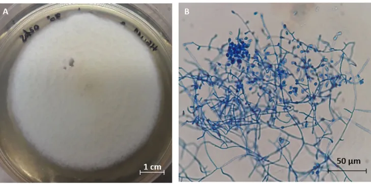
Arthroderma multifidum, a telomorphic ascomycete [14,15]. However, no telomorphic stage was observed in the prepared cultures. Instead, abundant pyriform microconidia and hyaline septate hyphae (Fig 1) were observed, which indicate the presence of the anamorphic form of
A. multifidum named Chrysosporium multifidum (GenBank accession numbers: MK982149
and MK982181). The use of this fungal growth stage allowed us to determine the antimicrobial activity of its culture supernatant with different experimental methods.
This species is an opportunistic or possibly pathogenic saprotroph commonly found in soil. However, it seems to behave as an endosymbiont withH. illucens. Chrysosporium has been
iso-lated from chicken guano samples [16], which makes it likely that it BSF larvae acquired it from their diet. It is also possible that this fungus was selected by the BSF’s biological system, as the fungus may provide enzymes or even beneficial antimicrobial substances to the larvae in exchange for an environment with enough nutrients for growth and development [17]. It is known that some of these selected fungi can survive in glandular cavities or cuticular invagina-tions called mycangia where they can develop and reproduce while being transported to new hosts by the insects [18,19].
Isolation of chemicals from
C. multifidum broth extract
Bioguided analysis of the ethyl acetate extract ofC. multifidum broth resulted in the isolation
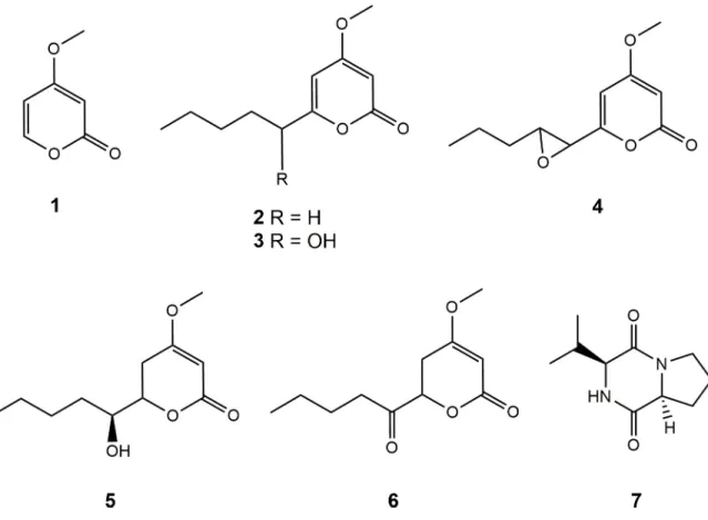
of 4-methoxy-2H-pyran-2-one (1) [20], 4-methoxy-6-pentyl-2H-pyran-2-one (2) [21], 6-(1-hydroxypentyl)-4-methoxy-pyran-2-one (3) [21,22], 6-[8-propyloxiran-1-yl]-4-methoxy-pyran-2-one (4) [23], pestalotin (5) [24,25], 5,6-dihydro-4-methoxy-6-(pentanoyloxy)-2H-pyran-2-one (6) [22,25] and cyclo-(L-Pro-L-Val) (7) [26]. All the compounds (Fig 2) were identified by comparison of their spectroscopic data (HRMS and1H and13C NMR) with liter-ature-reported values, as well as careful examination of their 2D NMR spectra (COSY, HSQC, HMBC). Optical rotations were also coherent with published data, except for (4), for which we found an optical rotation close to zero indicating the possible isolation of a racemic mixture (found [α]20
D-4.3, c = 0.16, MeOH/CH2Cl29/1; published [α]25D-98.7, c = 0.6, MeOH) [23]. This study is the first published chemical characterization ofC. multifidum. The
litera-ture describes several chemical prospecting works carried out on species of the Chrysos-porium genus, which led to the discovery of groups of compounds such as adenopeptines
[27], nucleosides [28], zearalenone derivatives [29], benzoquinones [30], naphthoqui-nones [31], anthraquinones [32], benzolactones [33], naphthopyrones [34], naphthalenes [35], phenyl-2(1H)-pyridinones [36], alkylphenols [37], bisdechlorogeodins [38], sterols [39,40], and caryophillenes [41]. However, there is no prior record of anyα-pyrone deriva-tives, so this would be the first report of these compounds within the genus. Compound 7 is also reported here for the first time within theChrysosporium genus; but it has also been
reported from cultures of other fungi and bacteria [42,43].
Biological analyses
Theα-pyrone 4 was the most active compound in the bioautography test. The antimicrobial activity of this compound was further quantified (Table 1), which indicates only moderate activity compared with the tetracycline control. Other known compounds isolated from
Fig 1.Chrysosporium multifidum isolated from H. illucens gut after 7 days of incubation at 30˚C. Macroscopic view (A). Microscopic view of pyriform
microconidia and hyaline septate hyphae (B).
https://doi.org/10.1371/journal.pone.0218837.g001
Chrysosporium spp. have displayed biological activities as antitumor [36], antifungal
[28,29,40,41] and cytotoxic [33] agents. However, only naphthoquinone-type compounds iso-lated fromC. queenslandicum [31] have been shown to be active against the gram-positive bac-teriaMicrococcus luteus and Bacillus subtilis with MIC values close to those obtained in this
work. On the other hand, both natural and syntheticα-pyrones have shown antimicrobial and antifungal activity on a variety of species [44,45]. Substitutions in positions 4 and 6 of the pyr-one ring may be related to this activity. Subsequent trials should be carried out to test the activ-ity of all derivatives of isolatedα-pyrones on other groups of bacteria including gram-negative species.
Conclusion
A total of 25 different fungi were isolated from the gut ofH. illucens larvae fed with chicken
guano. These colonies were tested on methicillin-resistantStaphylococcus aureus (MRSA)
ATCC 43300 andSalmonella Typhimurium ATCC 13311. The fungal specimen with the
great-est activity was subsequently named asChrysosporium multifidum. A broth culture of the
fun-gus was prepared and seven compounds present in the broth were characterized using
Table 1. Antimicrobial activity of compound 4 against MRSA.
Compound IC50(μg/mL) MIC (μg/mL)
4 11.4± 0.7 62.5
Tetracycline 0.1± 0.02 0.4
https://doi.org/10.1371/journal.pone.0218837.t001
Fig 2. Structures of compounds of 1–7 isolated fromC. multifidum broth extract.
chromatographic methods and TLC-DB. Theα-pyrone derivative 4 was shown to have moder-ate activity (MIC 62.5μg/mL) against MRSA. These first results from the exploration of the microbiota ofH. illucens indicate that it may be a useful source of antimicrobial compounds
that have activity against otherwise resistant pathogens. Furthermore, this opens a window to explain howH. illucens larvae control the pathogenic microbes ingested in contaminated food
with an endosymbiotic fungus.
Supporting information
S1 Methods. Preliminary evaluation of antimicrobial activity of yeast and molds.
(DOCX)
S1 Fig. Results of bioautography test forC. multifidum.
(TIF)
S1 Table. Results of preliminary antimicrobial test of yeast and molds isolated fromH.
illucens against S. aureus and S. Typhimurium.
(XLSX)
S1 Data. RMN and MS spectra of isolated compounds.
(DOCX)
S2 Data. Spectroscopic data of compounds isolated from the culture broth ofC.
multifi-dum.
(DOCX)
Author Contributions
Conceptualization: Yesenia Correa, Denis Castillo, Michel Sauvain. Funding acquisition: Michel Sauvain.
Investigation: Yesenia Correa, Billy Cabanillas, Vale´rie Jullian, Daniela A´ lvarez, Denis
Cas-tillo, Ce´dric Dufloer, Beatriz Bustamante, Michel Sauvain.
Methodology: Ce´dric Dufloer, Elisa Roncal, Edgar Neyra, Patricia Sheen. Supervision: Yesenia Correa, Billy Cabanillas, Vale´rie Jullian, Michel Sauvain. Validation: Yesenia Correa, Billy Cabanillas, Vale´rie Jullian, Michel Sauvain.
Writing – original draft: Yesenia Correa, Billy Cabanillas, Daniela A´ lvarez, Michel Sauvain.
Writing – review & editing: Yesenia Correa, Billy Cabanillas, Daniela A´ lvarez, Michel
Sauvain.
References
1. Wang YS, Shelomi M. Review of Black Soldier Fly (Hermetia illucens) as animal feed and human food. Foods. 2017; 6(10):91.https://doi.org/10.3390/foods6100091
2. Mu¨ller A, Wolf D, Gutzeit HO. The black soldier fly, Hermetia illucens–a promising source for sustain-able production of proteins, lipids and bioactive substances. Z Naturforsch C. 2017; 72(9–10): 351– 363.https://doi.org/10.1515/znc-2017-0030PMID:28742526
3. Jeon H, Park S, Choi J, Jeong G, Lee SB, Choi Y, et al. The intestinal bacterial community in the food waste-reducing larvae of Hermetia illucens. Curr Microbiol. 2011; 62(5): 1390–9.https://doi.org/10. 1007/s00284-011-9874-8PMID:21267722
4. Bruno D, Bonelli M, De Filippis F, Di Lelio I, Tettamanti G, Casartelli M, et al. The intestinal microbiota of Hermetia illucens larvae is affected by diet and shows a diverse composition in the different midgut Compounds with moderate antimicrobial activity from a fungus isolated from Hermetia illucens gut microbiota
regions. Appl Environ Microbiol. 2019; 85(2):e01864–18.https://doi.org/10.1128/AEM.01864-18
PMID:30504212
5. Choi WH, Yun JH, Chu JP, Chu KB. Antibacterial effect of extracts of Hermetia illucens (Diptera: Stratio-myidae) larvae against Gram-negative bacteria. Entomol Res. 2012; 42(5): 219–226.https://doi.org/ 10.1111/j.1748-5967.2012.00465.x
6. Varotto Boccazzi I, Ottoboni M, Martin E, Comandatore F, Vallone L, Spranghers T, et al. A survey of the mycobiota associated with larvae of the black soldier fly (Hermetia illucens) reared for feed produc-tion. PLoS ONE. 2017; 12(8): e0182533.https://doi.org/10.1371/journal.pone.0182533PMID:
28771577
7. Park SI, Kim JW, Yoe SM. Purification and characterization of a novel antibacterial peptide from black soldier fly (Hermetia illucens) larvae. Dev Comp Immunol. 2015; 52(1): 98–106.https://doi.org/10. 1016/j.dci.2015.04.018PMID:25956195
8. Elhag O, Zhou D, Song Q, Soomro AA, Cai M, Zheng L, et al. Screening, expression, purification and functional characterization of novel antimicrobial peptide genes from Hermetia illucens (L.). PloS ONE. 2017; 12(1): e0169582.https://doi.org/10.1371/journal.pone.0169582PMID:28056070
9. Won HC, Jiang M. Evaluation of antibacterial activity of hexanedioic acid isolated from Hermetia illucens larvae. J Appl Biomed. 2014; 12(3): 179–89.https://doi.org/10.1016/j.jab.2014.01.003
10. Pereira E, Santos A, Reis F, Tavares RM, Baptista P, Lino-Neto T, et al. A new effective assay to detect antimicrobial activity of filamentous fungi. Microbiol Res. 2013; 168(1): 1–5.https://doi.org/10.1016/j. micres.2012.06.008PMID:23041377
11. Jesionek W, Mo´ricz A, Ott P, Kocsis B, Horva´th G, Choma IM. TLC-direct bioautography and LC/MS as complementary methods in identification of antibacterial agents in plant tinctures from the Asteraceae family. J AOAC Int. 2015; 98(4): 857–861.https://doi.org/10.5740/jaoacint.SGE2-ChomaPMID:
26268962
12. Wiegand I, Hilpert K, Hancock RE. Agar and broth dilution methods to determine the minimal inhibitory concentration (MIC) of antimicrobial substances. Nat Protoc. 2008; 3(2): 163–175.https://doi.org/10. 1038/nprot.2007.521PMID:18274517
13. Zgoda JR, Porter JR. A Convenient Microdilution method for screening natural products against bacte-ria and fungi. Pharm Biol. 2001; 39(3): 221–225.https://doi.org/10.1076/phbi.39.3.221.5934 14. Chabasse D, Guiguen C, Couatarmanac’h A, Launay H, Reecht V, De Bièvre C. Contributionàla
con-naissance de la flore fongique ke´ratinophile isole´e des petits mammifères sauvages et du lapin de gar-enne en France-Discussion sur les espèces fongiques rencontre´es. Ann Parasit Hum Comp. 1987; 62 (4): 357–368.https://doi.org/10.1051/parasite/1987624357
15. Metin B, Heitman J. Sexual reproduction in dermatophytes. Mycopathologia. 2017; 182(1–2): 45–55.
https://doi.org/10.1007/s11046-016-0072-xPMID:27696123
16. Allimuthu V. Implication of fungal growth in poultry management and biogas production. PhD Thesis, Periyar University, 2015. Available from:http://hdl.handle.net/10603/151954.
17. Moubasher AH, Abdel-Sater MA, Soliman Z. Yeasts and filamentous fungi inhabiting guts of three insect species in Assiut, Egypt. Mycosphere. 2017; 8(9): 1297–316.https://doi.org/10.5943/ mycosphere/8/9/4
18. Beaver R. Insect-fungus relationships in the bark and ambrosia beetles. In: Wilding N, Collins NM, Ham-mond PM, Webber JF, editors. Insect-fungus interactions. 14th Symposium of the Royal Entomological Society of London in collaboration with the British Mycological Society; 1987 Sept 16–17; London; England. London: Academic Press; 1989. p.121-43.https://doi.org/10.1016/B978-0-12-751800-8. 50011–2
19. Stone W, Nebeker T, Monroe W. Ultrastructure of the mesonotal mycangium of Xylosandrus mutilatus (Blandford), an exotic ambrosia beetle (Coleoptera: Curculionidae: Scolytinae) by light, scanning, and transmission electron microscopy. Microsc Microanal. 2005; 11(S02): 172–173.https://doi.org/10. 1017/S143192760550015
20. Rao KB, Reddy GCS. A new reaction of patulin. J Nat Prod. 1989; 52(6): 1376–1378.https://doi.org/10. 1021/np50066a039
21. Evidente A, Zonno MC, Andolfi A, Troise C, Cimmino A, Vurro M. Phytotoxicα-pyrones produced by Pestalotiopsis guepinii, the causal agent of hazelnut twig blight. J Antibiot. 2012; 65: 203–206.https:// doi.org/10.1038/ja.2011.134PMID:22293915
22. Strunz GM, Heissne CJ, Kakushima M, Stillwell MA. Metabolites of an unidentified Fungus: a new 5,6-dihydro-2-pyrone related to pestalotin. Can J Chem. 1974; 52(5): 825–826.https://doi.org/10.1139/ v74-128
23. Yang XL, Huang L, Li HY., Yang DF, Li ZZ. Two new compounds from the plant endophytic fungus Pes-talotiopsis versicolor. J Asian Nat Prod Res. 2014; 17(4): 333–7.https://doi.org/10.1080/10286020. 2014.961918PMID:25290251
24. Ellestad GA, McGahren WJ, Kunstmann MP. Structure of a new fungal lactone, LL-P880.alpha., from an unidentified Penicillium species. J Org Chem. 1972; 37(12): 2045–2047.https://doi.org/10.1021/ jo00977a044PMID:5037459
25. Kimura Y, Susuki A, Tamura S. 13C-NMR Spectra of pestalotin and its analogues. Agr. Biol. Chem. 1980; 44(2): 451–452.https://doi.org/10.1080/00021369.1980.10863966
26. Sansinenea E, Salazar F, Jime´nez J, Mendoza A, Ortiz A. Diketopiperazines derivatives isolated from Bacillus thuringiensis and Bacillus endophyticus, establishment of their configuration by X-ray and their synthesis. Tetrahedron Lett. 2016; 57(24): 2604–2607.https://doi.org/10.1016/j.tetlet.2016.04.117 27. Hayakawa Y, Adachi H, Kim JW, Shin-ya K, Seto H. Adenopeptin, a new apoptosis inducer in trans-formed cells from Chrysosporium sp. Tetrahedron. 1998; 54(52): 15871–15878.https://doi.org/10. 1016/S0040-4020(98)00996-X
28. Yamashita M, Kawai Y, Uchida I, Komori T, Kohsaka M, Imanaka H, et al. Chryscandin, a novel peptidyl nucleoside antibiotic. II. Structure determination and synthesis. J Antibiot. 1984; 37(11): 1284–1293.
https://doi.org/10.7164/antibiotics.37.1284PMID:6549001
29. Hoshino Y, Ivanova VB, Yazawa K, Ando A, Mikami Y, Zaki SM, et al. Queenslandon, a new antifungal compound produced by Chrysosporium queenslandicum: production, isolation and structure elucida-tion. J Antibiot. 2002; 55(5): 516–519.https://doi.org/10.7164/antibiotics.55.516PMID:12139022 30. Fredenhagen A, Petersen F, Tintelnot-Blomley M, Ro¨sel J, Mett H, Hug P. Semicochliodinol A and B:
inhibitors of HIV-1 protease and EGF-R protein tyrosine kinase related to asterriquinones produced by the fungus Chrysosporium merdarium. J Antibiot. 1997; 50(5): 395–401.https://doi.org/10.7164/ antibiotics.50.395PMID:9207909
31. Ivanova VB, Hoshino Y, Yazawa K, Ando A, Mikami Y, Zaki SM, et al. Isolation and structure elucidation of two new antibacterial compounds produced by Chrysosporium queenslandicum. J Antibiot. 2002; 55 (10): 914–918.https://doi.org/10.7164/antibiotics.55.914PMID:12523825
32. Slater GP, Haskins RH, Hogge LR. Metabolites from a Chrysosporium species. Can J Microbiol. 1971; 17(12): 1576–1579.https://doi.org/10.1139/m71-252PMID:5168359
33. Jeon JE, Julianti E, Oh H, Park W, Oh DC, Oh KB, et al. Stereochemistry of hydroxy-bearing benzolac-tones: isolation and structural determination of chrysoarticulins A–C from a marine-derived fungus Chrysosporium articulatum. Tetrahedron Lett. 2013; 54(24): 3111–3115.https://doi.org/10.1016/j. tetlet.2013.04.006
34. Ogawa H, Hasumi K, Sakai K, Murakawa S, Endo A. Pannorin, a new 3-hydroxy-3-methylglutaryl coen-zyme A reductase inhibitor produced by Chrysosporium pannorum. J Antibiot. 1991; 44(7): 762–767.
https://doi.org/10.7164/antibiotics.44.762PMID:1880066
35. Tsipouras A, Goetz MA, Hensens OD, Liesch JM, Ostlind DA, Williamson JM, et al. Sporandol: a novel antiparasitic binaphthalene from Chrysosporium meridarium. Bioorg Med Chem Lett. 1997; 7(10): 1279–1282.https://doi.org/10.1016/S0960-894X(97)00226-6
36. Hirano N, Kohno J, Tsunoda S, Nishio M, Kishi N, Okuda T, et al. TMC-69, a new Antitumor antibiotic with Cdc25A inhibitory activity, produced by Chrysosporium sp. TCI068. J Antibiot. 2001; 54(5): 421– 427.https://doi.org/10.7164/antibiotics.54.421PMID:11480885
37. Sekhar Rao KC, Divaka S, Karanth NG, Sattur AP. 14-(20,30,50-trihydroxyphenyl)tetradecan-2-ol, a
novel acetylcholinesterase inhibitor from Chrysosporium sp. J Antibiot. 2001; 54(10): 848–849.https:// doi.org/10.7164/antibiotics.54.848PMID:11776443
38. Tanaka Y, Matsuzaki K, Zhong CL, Yoshida H, Kawakubo T, Masuma R, et al. Dechlorogeodin and its new dihydro derivatives, fungal metabolites with herbicidal activity. J Antibiot. 1996; 49(10): 1056–9.
https://doi.org/10.7164/antibiotics.49.1056PMID:8968402
39. Van der Pyl D, Cans P, Debernard JJ, Herman F, Lelievre Y, Tahraoui L, et al. RPR113228, a novel far-nesyl protein transferase inhibitor produced by Chrysosporium lobatum. J Antibiot. 1995; 48(7): 736– 737.https://doi.org/10.7164/antibiotics.48.736PMID:7649878
40. Yang SW, Buevich A, Chan TM, Terracciano J, Chen G, Loebenberg D, et al. A new antifungal sterol sulfate, Sch 601324, from Chrysosporium sp. J Antibiot. 2003; 56(4): 419–22.https://doi.org/10.7164/ antibiotics.56.419PMID:12817816
41. Yang SW, Chan TM, Terracciano J, Boehm E, Patel R, Chen G, et al. Caryophyllenes from a fungal cul-ture of Chrysosporium pilosum. J Nat Prod. 2009; 72(3): 484–487.https://doi.org/10.1021/np8006414
PMID:19183048
42. Chen YS. Studies on the metabolic products of Rosellinia necatrix. I. Isolation and characterization of several physiologically active neutral substances. Bull Agr Chem Soc Japan. 1960; 24(4): 372–381.
https://doi.org/10.1080/03758397.1960.10857680
43. Takeda Y, Fujita T, Shingu T, Ogimi C. Studies on the bacterial gall of Myrica rubra: isolation of a new [7.0]-Metacyclophan from the galland dl-β-phenyllactic acid from the culture of gall-forming bacteria. Chem Pharm Bull. 1987; 35: 2569–2573.https://doi.org/10.1248/cpb.35.2569
44. Fairlamb IJ, Marrison LR, Dickinson JM, Lu FJ, Schmidt JP. 2-pyrones possessing antimicrobial and cytotoxic activities. Bioorg Med Chem. 2004; 12(15): 4285–4299.https://doi.org/10.1016/j.bmc.2004. 01.051PMID:15246105
45. Bhat ZS, Rather MA, Maqbool M, Lah HU, Yousuf SK, Ahmad Z.α-pyrones: Small molecules with ver-satile structural diversity reflected in multiple pharmacological activities-an update. Biomed Pharmac-other. 2017; 91: 265–277.https://doi.org/10.1016/j.biopha.2017.04.012PMID:28460229