HAL Id: hal-01567829
https://hal.archives-ouvertes.fr/hal-01567829
Preprint submitted on 24 Jul 2017HAL is a multi-disciplinary open access archive for the deposit and dissemination of sci-entific research documents, whether they are pub-lished or not. The documents may come from teaching and research institutions in France or abroad, or from public or private research centers.
L’archive ouverte pluridisciplinaire HAL, est destinée au dépôt et à la diffusion de documents scientifiques de niveau recherche, publiés ou non, émanant des établissements d’enseignement et de recherche français ou étrangers, des laboratoires publics ou privés.
Universal pulse design of refocusing kT-point pulses
using parallel transmission: application to
three-dimensional T2-weighted sequences at 7T
Vincent Gras, Franck Mauconduit, Alexandre Vignaud, Alexis Amadon, Denis
Le Bihan, Tony Stöcker, Nicolas Boulant
To cite this version:
Vincent Gras, Franck Mauconduit, Alexandre Vignaud, Alexis Amadon, Denis Le Bihan, et al.. Uni-versal pulse design of refocusing kT-point pulses using parallel transmission: application to three-dimensional T2-weighted sequences at 7T. 2017. �hal-01567829�
Universal pulse design of refocusing k
T-point pulses using parallel
transmission: application to three-dimensional T
2-weighted sequences at 7T
Vincent Gras
1, Franck Mauconduit
2, Alexandre Vignaud
1, Alexis Amadon
1,
Denis Le Bihan
1, Tony Stocker
3, Nicolas Boulant
11
CEA, DRF, Joliot, NeuroSpin, Unirs, CEA Saclay, Gif sur Yvette, France
2
Siemens Healthcare, Saint-Denis, France
3
DZNE, Bonn, Germany
Corresponding author: Dr. Nicolas Boulant, CEA Saclay, 91191 Gif sur Yvette
Cedex, France. Email: [email protected]
Abstract
T2-weigthed sequences are particularly sensitive to the RF field inhomogeneity problem due to the errors accumulated by the imperfections of the train of refocusing pulses. In this work we report a universal RF pulse design for 3D refocusing pulses utilizing parallel transmission (pTx) to mitigate the RF inhomogeneity problem at ultra-high field.
In this work Average Hamiltonian Theory was used to synthetize with pTx a single non-selective refocusing pulse able to mitigate the RF field inhomogeneity problem at 7T while optimizing its scaling properties in the presence of static field offsets. The design was performed under explicit power and specific absorption rate constraints on a database of 10 subjects. Simulations were conducted to support the theory and in-vivo brain imaging experiments using the Nova 8Tx-32Rx coil were finally carried out on additional test subjects to validate the proposed design with variable flip angle three-dimensional T2-weighted sequences at 7T. The simulation and experimental data demonstrate great improvement with the proposed calibration-free universal pulses, reaching on average ∼8 % normalized root mean square rotation angle error over the test subjects, compared to the standard, non-pTx, circularly-polarized mode of excitation.
This work completes further the spectrum of 3D universal pulses to mitigate the RF field inhomogeneity problem throughout all 3D MRI sequences and without any pTx calibration. The approach returns a single pulse which can be scaled to match the desired flip angle train, thereby increasing the modularity of the proposed plug and play approach.
Introduction
Parallel transmission (pTx) (1) is a key technology to mitigate the RF field inhomogeneity problem encountered in MRI at Ultra-High-Field (UHF). Unless this problem is solved, the potential of UHF imaging is seriously hampered by the sub-optimal signal-to-noise and contrast-to-noise ratios in addition to potential zones of shades, all detrimental to medical diagnosis. With ever more powerful algorithms employed for RF pulse design and dedicated techniques, many experimental achievements have been demonstrated in the community, with applications ranging from proton density-weighted (2), T1-weighted (3,4), T2-T1-weighted (5,6) and T2*-weighted (7–9) sequences. However, despite the great achievements made to ensure safety (10–14), speed up the transmit RF field (B1) and static field offset (∆B0) map measurements (15–17) and accelerate RF pulse designs with for instance Graphics Processing Units (4,9,18), the technique has barely been exploited in clinical routine, due to a still considered cumbersome workflow which is furthermore prone to delays and human errors. In (19), a new calibration-free concept using pTx was presented to greatly mitigate the RF inhomogeneity problem at 7T while skipping the entire calibration step. The concept, named universal pulses (UP), consisted of first building a database of subject-based field maps and to perform offline the RF pulse design simultaneously on the same subjects to achieve robustness with respect to intersubject variability. The first demonstration was performed with a home-made pTx coil and with the Magnetization Prepared Rapid Gradient Echo (MPRAGE) sequence while the number of database and test subjects was 6 in each case. To demonstrate broader applicability, universal pulses were then designed for the MPRAGE and multi-slice GRE2D sequences with commercially available coils, namely the Rapid (Rapid Biomedical, Rimpar, Germany) 8Tx-8Rx and the Nova (Nova medical Inc, Wilmington, MA, USA) 8Tx-32Rx head coils (20), this time with 10 database and test subjects. The results confirmed the robustness and safety of the proposed approach with respect to intersubject variability, with great mitigation of the RF field inhomogeneity problem compared to the single-channel conventional Circularly-Polarized (CP) mode of excitation, and only at a mild cost in performance compared to the subject-based tailored approaches.
The pulses employed in the MPRAGE sequence consisted of an inversion (180°) pulse and a train of small tip angle (5°) excitations (20), which both used the kT-points parametrization approach
(2). While these pulses represent a good portion of needed non-selective pulses, it does not yet cover all pulse possibilities encountered throughout 3D MRI sequences. T2-weighted sequences such as turbo spin echo (21), SPACE (Sampling Perfection with Application of optimized Contrasts using different flip angle Evolution) (22), weighted FLAIR (Fluid Attenuation Inversion Recovery) (23) and T2-weighted DIR (Double Inversion Recovery) (24) sequences indeed make extensive use of refocusing pulses to better visualize gray and white matter lesions such as strokes, ischemia and multiple sclerosis (25,26). As opposed to excitation pulses, the initial magnetization prior to refocusing pulses cannot be assumed longitudinal so that rotation matrices with purely transverse axis of rotation and given rotation angle must be considered and designed. Although successful attempts based on optimal control (5) were obtained for synthetizing target rotation matrices with subject-based tailored pTx approaches, the technique suffered from inefficient pulses, especially at small flip angles (FA). This can be explained by the challenging task of generating purely transverse axis rotations, as it is normally required, when the pulses are relatively long and the spins are exposed to significant ∆B0 offsets, while trying to homogenize the spins excitation. From previous experience in NMR spectroscopy (27), and aside from Specific Absorption Rate (SAR) considerations, such a task is possible if the transmit B1 field can be made significantly larger than the chemical shifts or, in our case, the spread of ∆B0. But this cannot unfortunately be the case in MRI throughout an extensive volume such as the human brain. The attempt to circumvent the problem in Ref. (5) hence was made by including many degrees of freedom in the pulse design and by exploiting nonlinearities in Bloch’s equation, thus causing SAR and power issues and increasing acquisition time. Lastly, because the rotation angle of the rotation matrices did not scale with the RF amplitude, and because the axis of rotation was not always purely transverse, the RF pulse design was performed individually for each pulse of the echo train, increasing computation and pulse preparation time while leading to greater sequence complexity. Alternatively, a signal-based metric can be used (28) to minimize the signal deviation from the nominal one, which has yielded good results at 3T with pulse-dependent RF-shimming. This approach, however, leads to a new optimization whenever T1 and T2 values or other sequence parameters (echo-spacing, repetition time, FA train) are changed which makes at first sight a universal pulse version of it inconvenient.
Despite the apparent need for synthetizing target rotation matrices, in Ref. (6) Eggenschwiler and colleagues obtained good mitigation of the RF field inhomogeneity problem at 7T in the design of refocusing pulses while only considering the action of the pulses on longitudinal magnetization. For the variable flip angle pulse sequence investigated in their work, one single pulse was designed and its FA scaling properties were investigated depending on different kT-point parametrizations. Numerical results showed the benefits of some particular gradient and RF waveforms symmetry, but no analytical tools were actually provided to rigorously explain the success of the retained approach. Moreover the impact of ∆B0 was ignored at the RF pulse design level, which can lead to remaining B1 and ∆B0 artefacts. In this work, we use Average Hamiltonian Theory (AHT) (29) to design 3D refocusing pulses, which provides an adequate analytical framework to describe rotation matrices. We furthermore show that this representation nicely and rigorously explains the symmetry principles proposed in Ref. (6). That theory is further used to guide the 3D refocusing pulse design to improve pulse performance in the presence of static field offsets and for large FAs. The technique developed in this work is then applied to mitigate the RF inhomogeneity problem in the T2-weighted SPACE and FLAIR-SPACE sequences at 7T. Importantly, this work integrates the concept of universal pulse design which has, to our knowledge, never been proposed for the design of refocusing pulses at UHF using pTx. For experimental validation, this study includes in-vivo brain imaging experiments at 7T using pTx universal pulses and focuses on the comparison with the conventional non pTx excitation CP mode.
Theory
In this section, we derive analytically the spins’ dynamics by using the AHT framework (29), which provides a description of rotation matrices, as well as their scaling properties, in the presence of ∆B0 offsets. We first follow Warren’s approach in his application of AHT to compute excitation profile approximations (30) and adapt the theory to the kT-point framework used in MRI (2). Given the history of this field, we use the SU(2) spinor quantum mechanical formalism to calculate rotation matrices. We refer the reader to the relevant literature (29,30) for further details.
We consider a spin ½ particle located at position r. Assuming ℏ = 1, the Hamiltonian in Schrodinger’s equation is:
H = ω + γG t . r σ + ω t σ + ω t σ , [1]
where ω0 = γ∆B0 is the Larmor frequency in the frame rotating at the carrier frequency, γ is the gyromagnetic ratio, G t is the three-dimensional time-dependent gradient vector, σx,y,z denote the Pauli matrices while ω1,x (= γB1,x) and ω1,y (= γB1,y) represent the nutation frequency respectively along the x and y axis. In pTx, with Nc transmit channels, one simply has: ω t = γ ∑ B , t , B , denoting the transmit RF field of channel n. We now move into an interaction frame described by the following unitary transformation:
U" # t = e"%&' ()#*+,.- . / 0/
1
) . [2]
In the new frame, the Hamiltonian becomes:
H" # = U" #HU" #2 − iU" #05671 8 0# , =12 ω t σ + ω t σ cos ω t + k 0 . r − k t . r +12 −ω t σ + ω t σ sin ω t + k 0 . r − k t . r , [3]
where we have used of the conventional k-space definition used in the Small Tip Angle (STA)
approximation (31): γr. - G u du = k 0 . r − k t . r# . This Hamiltonian only contains the RF terms which are modulated by the ∆B0 offset and the time integral of the gradient waveforms. In this new frame, the rotation matrix in SU(2) is:
UB = eC" D)* DE*⋯
, [4]
where A(i), i ≥ 0, are the Magnus expansion terms (29,30). In Equation [4], the overall term in the exponential takes the form of an infinite series where its truncation corresponds to different orders of approximation. For the applications considered in (9,30), it was shown that the first two terms already returned an excellent approximation of the full Bloch dynamics, and we will assume this to be true for
this application as well. In the appendix, we show that the zero-order approximation obtained by retaining only the term A(0) in Equation [4] reads:
UB T = U2 T eC"HIJKLMU T , [5]
where U = eC"%&' ()N*O .,, θQND= R- Ω t e*T "() NC# dt
CT R, φ = arg X- Ω t eCT*T "()NC# dtY, Ω t = Xω t + iω t Y e"O # .,, T is the total duration of the pulse and σZ = σ cos φ + σ sin φ . Remarkably, θQND is nothing more than the STA formula (31,32). It is important to note,
however, that the corresponding expression here describes a rotation angle and not a transverse magnetization (9,30,32). As a result, it is not equivalent to linear response theory and to the STA approximation used to describe the dynamics of the transverse magnetization (30). Assuming a
self-refocused trajectory, i.e. k 0 = 0, and after undoing the interaction frame transformation Uint(T), one finally obtains for the propagator, or rotation matrix, in the initial frame:
U T = U" #2 T UB T = eC"HIJKLMU T = U T/2 eC"HIJKLM\])J/'U T/2 . [6]
This equation is one of the main results of the paper. The overall rotation matrix U(T) is not a rotation about a purely transverse axis, as normally specified for refocusing pulses. But conveniently, it can be decomposed into a free precession during half the duration of the pulse (U T/2 ), followed by a rotation with purely transverse rotation axis and rotation angle dictated by the STA approximation
(eC"HIJKLM\])J/'), and finally ended by the same free precession (see Figure 1 for illustration). It thus looks as if the rotation around the transverse axis acts instantaneously, at the middle of the pulse, and hence should have the desired refocusing properties. It may be seen that the phase of the axis of rotation in the transverse plane varies with ω0T/2 and thus can vary within one voxel. But for a 100 Hz maximum spread of the Larmor frequency and a pulse duration of 1 ms, the phase dispersion would only be 18°, thus preserving phase coherence at echo time to a high level. The phase ϕ furthermore depends on ω0 and to some extent may cancel the action of ∆ω0T/2 in some circumstances.
Figure 1. Illustration of the zero order approximation derived from Average Hamiltonian Theory. The
rotation matrix arising from a kT-points waveform of duration T can be decomposed into a free-evolution during T/2, followed by an instantaneous transverse rotation with rotation angle θSTA evaluated with the STA approximation and, finally, the same free precession during T/2. The overall rotation matrix is not purely transverse but the approximation justifies the good refocusing properties of the RF pulse when the right conditions are met.
So far the zero order term in the AHT description does not pose any scaling problem as A(0) depends linearly on the RF amplitude. Additional nonlinearities arise with the next Magnus expansion terms. The next leading term A(1) in general is given by (29,30):
A = −"- dtN - dt#' _H
" # t , H" # t `, [7]
where the bracket denotes the commutator operation, i.e. [A, B] = AB – BA. By defining τ = t2 – t1, one can show after somewhat tedious algebra that:
A = −L&a - Γ
cE τ eC"()edτ
N
, [8]
where ΓcE τ denotes the autocorrelation function of Ω1(t), i.e. ΓcE τ = - Ω t ΩN ∗ t + τ dt (30). Note that the first order AHT term is along the z-axis. The presence of this term would not be so problematic if it was the same for each pulse of the echo train since, to a reasonable approximation, it could be again factored out symmetrically on both sides of the purely transverse rotation, as in Equation [6]. But this term being power-dependent, its contribution becomes variable along a variable flip angle train and leads to accumulated dephasing.
We are now in the position to analyze the design proposed by Eggenschwiler and colleagues (6). When ω0 = 0 and in the presence of the following symmetries: 1) temporal symmetry in the RF waveform: ω1x(t) + iω1y(t) = ω1x(T-t) + iω1y(T-t), and 2) temporal anti-symmetry in the gradient waveform G(t) = -G(T-t) (leading to a symmetric k-space trajectory k(t)), it can be shown that the integral of ΓcE τ is real and thus A(1) is zero. It thus explains the good scaling properties in the absence of ∆B0 when enforcing those symmetries, since the error done by doing a simple STA analysis is led by A(2). It can also be shown that when the symmetries above are present, and still when ∆B0 = 0, the axis of rotation of the rotation matrix is purely transverse, despite the k-space modulation induced by the gradient waveforms, a result fully consistent with Levitt’s demonstration in the case of palindromic
sequences (33) and with Pruessman’s double angle method (34). The assumption that ∆B0 = 0 everywhere in the brain, however, is incorrect as it can greatly affect the calculation of the FA in the STA approximation (35) while it can also lead to a larger, pulse-dependent, A(1) term. One can derive an upper-bound on the magnitude of A(1) by Taylor-expanding eC"()e in Equation [8]:
gA g = Ra X- ΓcE τ eC"()edτ N YR ≤ ∑ |()|7 ! - gΓcE τ gτ dτ N *T , ≤ ∑ |()|7 ! |cEklm |'N7n' * * *T , [9]
where |Ω op | is the maximum amplitude over the kT-point waveform. Note here that the zero order term of the expansion vanishes due to the imposed symmetry. Aside from power and SAR limit considerations, |Ω op | is expected to vary as 1/T to achieve a desired rotation angle. As a result when T tends towards 0, all terms in Equation [9] tend to 0 as well and so does A(1). Interestingly, this result implies that shorter pulses lead to a better STA approximation.
The results of this analysis are exploited in the pulse design methods below to synthetize valid rotation matrices for refocusing. A justification of using the conventional STA analysis for rotation angles was indeed demonstrated and arguments to increase the quality of the approximation and to favor scalability, e.g. with short pulse durations and symmetries, were provided. The symmetries discussed above fortunately constitute a sufficient but not necessary condition to cancel A(1) when ∆B0 = 0. As a
result, this observation is also exploited to further optimize numerically the scaling properties in the pulse design with additional knowledge of the ∆B0 distribution.
Methods
Measurements were performed on a Magnetom 7T scanner (Siemens Healthcare, Erlangen, Germany) equipped with the Nova 8TX-32RX head coil and a SC72 whole body gradient insert (70 mT/m maximum amplitude and 200 T/m/s maximum slew rate). All scans were run under local SAR supervision mode (Siemens step 2.3). The study was approved by the local ethics committee and all volunteers gave written informed consent.
Parallel transmit RF pulse design
In this work, as justified by the theory the SPACE refocusing pulse train was built from a unique pTx kT-point pulse, repeated at each new excitation with the RF waveform scaled in order to produce the
desired rotation. The set of RF coefficients qr∈ ℂ c× kT and kT locations v ∈ ℝx× kT of the so-called
generating kT-point pulse was obtained by minimizing over qr and v the FA normalized root mean
square error (NRMSE), for a target FA y equal to the largest FA used in the SPACE protocol. Let us denote by z the pair v , q and introduce the notation: λ ∗ z ≡ v , λ q , for any complex number
λ (k-space trajectory left unchanged, RF coefficients scaled with λ). In the approach based on a unique
generating pulse to form the refocusing pulse train z}, … , z•€• as well as the starting 90° excitation pulse z‚ ƒ, the nth pulse of nominal FA α then is defined as z =…7
…)∗ z while the 90° excitation pulse
simply is z‚ ƒ = X†‡ °…
)Y ∗ z (note here the multiplication by the pure imaginary number which rotates
the rotation axis by 90° around z to achieve the Carr-Purcell-Meiboom-Gill (CPMG) condition). Consistently with the theory, the generating pulse is designed by using the STA approximation to homogenize the FA around the target value y , although y is not small. An adequate framework thus is the standard spatial domain method (35) which expresses the FA-NRMSE as a quantity proportional to ‖|Š vr qr| − α ‖ , where Š vr is the complex spatial encoding matrix. For the design of a universal pulse, where a multiplicity (the number of database subjects, NŒ) of spatial encoding matrices
needs to be considered simultaneously, a unique objective function still can be defined by taking the average FA-NRMSE across all database subjects (20). Assuming that the database “samples” reasonably well the variability of field maps across the population, the proposed metric is expected to provide an estimation of the expectation value of the FA-NRMSE across the same population.
Let us now seek to define the constraints one should observe on the RF coefficients qr and the k-space trajectory vr to ensure good refocusing properties. To reduce the spatial phase variation of A(0) within one voxel, we impose self-refocussed trajectories: v j, 1 = 0 for j = 1,2,3. Without any further constraint, pulse optimization leads to a solution whose rotation matrix presents a non-vanishing first order Magnus term A along the z-direction (see Equation [8]) when ∆B0 = 0, and may lead to a progressive violation of the CPMG condition. In the following, this solution is termed “non-symmetric solution”.
Keeping the same objective function but this time also imposing temporal-symmetry on the k-space trajectory and the RF coefficients: v . , NOJ− m + 1 = v . , m , 1 ≤ m ≤ NOJ and
q . , NOJ− m + 1 = q . , m , 1 ≤ m ≤ NOJ, suppresses the term A from the AHT expression in
regions where ΔB = 0. The solution obtained with symmetric RF waveforms and k-space trajectories is termed below the “symmetric solution”. Starting from this solution, we finally investigated the possibility to optimize further the scaling behavior of the pulse in regions where off-resonance appears, i.e. by minimizing in a second step the so-called scaling error, defined as:
SE v , q = “”U T XU" #2 T eC"D) N Y C
− Id” –“
[10]
where |. |– denotes the Fröbenius norm, removing here the above symmetry constraints, and under the constraint to not increase the FA-NRMSE of the symmetric solution. That way, in order to maintain coherence along the echo train we attempt to find a solution that minimizes on average over the whole volume of interest the distance between the “true” rotation matrix (U T obtained with full numerical Bloch simulations) and its zeroth order, STA, approximation (U" #2 T eC"D) N) at large FAs. Below, the resulting solution is termed “scaling-optimized solution”.
To take into account hardware limits and patient safety, the following constraints were enforced explicitly in the design of the generating kT-point pulse z : i) RF amplitude (160 V peak amplitude), ii) average RF power per channel (3 W), iii) total average RF power (16 W), iv) 10-g SAR and global SAR (10 and 3.2 W/kg respectively) and v) gradient slew-rate limitations for the blips (200 T/m/s). Having the same, scaled, pulse in the train of refocusing pulses makes SAR analysis easy as SAR becomes additive and simply scales with the square of the FA. As a result, z is designed with the above power and SAR limits divided by —X‡ °…
)Y + ∑ X
…˜
…)Y
™Nš
O ›, where ETL is the Echo Train Length.
The database used for the design of the generating kT-point pulse was composed of RF and static field offset maps acquired on ten healthy adults (5 males, 5 females, 25 ≤ age ≤ 44 years) in a separate study (20). For each subject, the static field offsets and the eight complex transmit field distributions were obtained through a three-dimensional multiple gradient recalled echo (GRE) sequence (2.5 mm isotropic resolution, matrix size 64 × 96 × 128, TR = 25 ms, 3 echoes, TE = 5, 6.5, 8 ms, TA = 3 min) and a multi-slice interferometric turbo-FLASH acquisition (5 mm isotropic resolution, matrix size 40 × 64 × 40, TR = 20 s, TA = 4 min 40 s) (15).
For the optimization of the SPACE refocusing pulse generator, the number of kT-points was set to 9 and the RF pulse total duration was 1100 µs for the excitation and the refocusing pulses. The RF pulse and k-space trajectory optimizations were performed simultaneously using the Active-Set algorithm implemented in Matlab (MathWorks, Natick, MA) (18,36).
In-vivo experiments
T2-weighted SPACE and FLAIR-SPACE protocols were first implemented respectively on five (3 males, 2 females, 22 ≤ age ≤ 50 years old) and three (2 males, 1 female, 22 ≤ age ≤ 50 years old) healthy adult volunteers in the CP transmission mode, i.e. equivalent to the transmission mode used on non-pTx systems. The two protocols were subsequently repeated with exactly the same sequence parameters, but using the optimized kT-point pulses. The sequence parameters for the SPACE acquisition were: TR = 3 s, ES = 8.6 ms, readout bandwidth = 290 Hz/pixel, ETL = 117, matrix size = 224 × 256 × 208, 0.8 × 0.8 × 0.8 mm3 voxels and TA = 7:47 min, and for the FLAIR-SPACE acquisition:
TR = 8 s, TI = 2.25 s, ES = 7.5 ms, readout bandwidth = 300 Hz/pixel, ETL = 96, matrix size = 224 × 256 × 120, 1 × 1 × 1.3 mm3 voxels and TA = 11:55 min. To reduce the ETL, a GRAPPA acceleration factor of 2 and partial Fourier acquisition of 6/8 was used for both protocols. The FA evolution of the refocusing pulse train, displayed in Figure 2 for the FLAIR-SPACE protocol, was computed using the approach of Mugler and colleagues (22), with T1 = 1400 ms and T2 = 40 ms as nominal values for the relaxation times. Following the two protocols, the same transmit RF field and static field offset mapping protocols as the ones used to construct the field map database were used. Those data were not used to design RF pulses but were exploited for retrospective control of RF pulse performance.
Both the SPACE and FLAIR-SPACE pTx protocols were based on the same scaling-optimized generating pulse zr (1.1 ms pulse duration) whose characteristics are described in Section “Parallel transmit RF pulse design”. The CP implementations of the excitation and refocusing pulses used 1.1 ms long rectangular pulses. The inversion pulse used for FLAIR preparation was designed independently from the generating pulse of the SPACE pulse train, using again the point parameterization (9 kT-points, total pulse duration of 4 ms). The CP implementation counterpart used an overdriven 10 ms long adiabatic pulse.
Figure 2. Flip angle evolution of the refocusing pulse train used for the FLAIR-SPACE protocol and
optimized for T1 = 1400 ms, T2 = 40 ms (22). The overlaid red curve displays the nominal SPACE signal evolution during the application of the refocusing pulse train.
RF pulse performance simulations
A benchmarking of the different approaches to design the generating pulse zr (the non-symmetric, the symmetric, and the scaling-optimized design) was performed retrospectively in simulation as follows: as a first step, for each test subject their specific B1 and ∆B0 maps were used to compute the propagator maps of the CP pulse and the differently designed universal kT-point pulses with full Bloch simulations. The simulations were done at different scaling values of the generating pulse to implement 5° up to 120° pulses. They were further exploited to report the NRMSE values of a FA profile over the whole brain in the STA regime (10°), as well as the large FA regime (120°, i.e. slightly above the maximum FA of the SPACE RF pulse train). We also investigated the scaling property of the pulses by computing for each scaling value λ = α /α and each generating pulse zr the scaling error SE λ ∗ zr (Equation [10]). As a perhaps more intuitive and geometrical feature of the scaling error, we also computed the elevation
e λ ∗ zr of the rotation axis of the pulse propagator U(T) after cancelling the free evolutions during
T/2: U′ T = U2XNY U T U2XNY. According to Equation [6], the generator of U′ T to zero order has a vanishing σ component, and therefore, e λ ∗ zr ≈ 0 when λ is small. In this regime and after undoing the free evolutions above, the rotation axis hence is purely transverse. But for large FAs, higher order terms may not be negligible and may lead to a non-transverse rotation axis, i.e. to |e λ ∗ zr | > 0. That quantity depicts one particular deviation of the refocusing pulse train from the ideal behavior which leads to a deviation from the CPMG condition.
With the non-symmetric solution, we expect the scaling behavior to be poor, i.e. a rapid increase of SE λ ∗ zr and e λ ∗ zr with λ. For symmetric designs (note here that the CP pulse is symmetric), the behavior is expected to improve, at least in regions with small ∆B0 values. The scaling optimized solution yet is expected to improve further since the scaling error SE zr becomes the objective function itself in a second optimization step. Note however that the simulation tests are performed on the RF and static field offset maps of the subjects on whom the SPACE protocols were applied, which were obviously independent of the database subjects on which the pulse design was performed. Finally, to validate the theoretical analysis of the effect of pulse duration, the scaling errors were investigated for the same pulses but stretched in time to 3.3 ms while preserving the area of each sub-pulse.
In vivo image comparisons
A visual qualitative comparison of the SPACE and FLAIR-SPACE images acquired with the CP pulses and the scaling optimized universal pTx pulses was performed for each subject to identify the regions where significant gain in terms of signal and contrast occurred and to assess whether the introduction of universal pulses introduces some specific artifact or not, as compared with the default transmission CP mode. Image comparisons were performed on raw native images, i.e. without correction of the reception profile of the head coil.
Results
Refocusing pulse performance
The FA-NRMSE of the pTx inversion pulse used in the FLAIR-SPACE sequence was 6 % on average over all subjects. The performance of the generating refocusing pulses, obtained retrospectively on each subject is displayed in Table 1 for the small FA (10°) and large FA (120°) regimes.
CP (10°,120°) Non-symmetric (10°,120°) Symmetric (10°,120°) Scaling-optimized (10°,120°) Subject 1 25.8, 25.5 5.0, 5.1 6.8, 6.4 5.3, 5.2 Subject 2 24.9, 24.9 8.8, 10.0 8.6, 8.9 9.4, 9.7 Subject 3 27.1, 26.9 7.1, 6.4 8.3, 7.7 6.4, 6.1 Subject 4 27.0, 26.9 7.1, 8.2 6.2, 6.5 6.8, 7.1 Subject 5 27.6, 26.9 12.5, 13.6 11.9, 12.2 13.1, 13.3 Average 26.5, 26.4 8.1, 8.7 8.4, 8.3 8.2, 8.3
Table 1. FA NRMSE values obtained in each subject in the small FA (10°) and large FA (120°) regimes
with the CP pulse and the non-symmetric, the symmetric, and the scaling optimized SPACE RF pulse train generating pulses. According to the theory, to zeroth order, the calculated flip angle in fact is the rotation angle of a purely transverse rotation matrix, when sandwiching ∆B0 evolutions are unwound (Equation [6]).
With the CP pulse, the FA-NRMSE reaches on average 26.5% and is, as expected, nearly independent of the target FA. Average FA-NRMSE values for the non-symmetric, the symmetric and the scaling optimized solutions are in the small FA regime 8.1, 8.4 and 8.2%. The performance remains very good with the symmetric (8.3%) as well as the optimized scaling (8.3%) design but starts degrading with the non-symmetric design (8.7%) when increasing the target FA (especially for subjects 2 and 5). At this point it is worth stressing that not only the FA accuracy matters to ensure good performance of the refocusing pulse train, but also the coherence with respect to the leading 90° excitation, essential to satisfy the CPMG condition.
The latter requirement can now be inspected in Figure 3, which displays the scaling error SE λ ∗
zr (see Equation [10]) as a function of the nominal FA for the CP pulse, the non-symmetric UP, the symmetric UP and the scaling optimized UP and for a different scaling factor λ so as to cover the range FA = 0° up to 120°. The dashed lines represent the scaling error respectively for the symmetric and scaling optimized solutions, with pulse durations increased from 1.1 to 3.3 ms. Figure 3a represents the scaling error averaged over all voxels (all subjects “pooled” together) while Figure 3b represents the scaling error averaged over the voxels where | ΔB | > 200 Hz (high-ΔB voxels). In Figure 3c, the elevation e λ ∗ zr of the free-evolution factorized propagator U’(T) is reported as well for the
high- ΔB voxels. The scaling error and the elevation analysis indicate that the non-symmetric UP returns the
worst performance. Here, the error is driven by the first order AHT term A which grows as λ . In contrast, symmetric solutions (symmetric UP as well as CP) exhibit a much better behavior, at least on
average, because the symmetry of the RF pulse ensures A = 0 when ΔB = 0. However, the resonance condition is not fulfilled for all brain voxels, as it may reach values larger than 300 Hz at the different air-tissue interfaces. Looking at the high-ΔB voxels highlights this relatively important performance degradation of the symmetric pulses (the CP and the symmetric kT-point). The scaling
optimized solution helps mitigating this effect as it indirectly minimizes the A term and it results in a better performance of the refocusing pulse train near air-tissue interfaces. Finally, still from Figure 3 and consistently with the result of Equation [9], shortening the pulse duration helps maintaining
coherence along the train of refocusing pulses, especially when the off-resonance map is not known accurately, as it is the case with universal pulses.
Figure 3. Scaling error simulations for the 1.1 ms-long CP pulse (gray), non-symmetric (black),
symmetric (red) and scaling-enforced (blue) 1.1 long 9-kT point universal pulses and the 3.3 ms-long symmetric (red dashed line) and scaling-enforced (blue dashed line) 9-kT point pulses. The average error on all voxels pooled together over the five subjects is shown in a) while b) returns the same error this time on average over the voxels satisfying |∆B0| > 200 Hz. In a), the CP curve is almost indistinguishable from the one of the short scaling optimized pulse. Subplot c) shows for the same voxels as in b) the average deviation in degrees of the rotation axis from the transverse plane, after undoing the correct free ∆B0 evolutions symmetrically (Equation [6]). The 3.3 ms long pulses were obtained from the original 1.1 ms-long solutions by increasing the duration of each sub-pulse while preserving the area of the sub-pulses.
In vivo image comparisons
In Figures 4 and 5, we report a CP versus UP comparison for the SPACE protocol, implemented on 5 subjects. For the CP mode, the sagittal (Figure 4) and coronal (Figure 5) slices selected illustrate severe signal dropouts in the cerebellum, the occipital and temporal lobes, and occasionally in the midbrain
(top rows). Those RF inhomogeneity-induced artifacts are typically observed in T2-weighted protocols at UHF with single channel transmission and standard pulses (5,6). In all five subjects, the use of the scaling optimized UPs (bottom rows) allowed excellent suppression of those signal drops to make the SPACE protocol at UHF “truly” whole brain.
Figure 4. Comparison of the SPACE images acquired in CP mode (top row) and with pTx UP pulses
(bottom row) and in all five subjects in a representative sagittal slice. The sagittal view demonstrates a clear signal recovery in the cerebellum and the occipital cortex in all subjects and in the mid-brain in subjects 2 and 4. The reception profile of the coil was not removed.
Figure 5. Comparison of the SPACE images acquired in CP mode (top row) and with pTx UP pulses
(bottom row) in a representative coronal slice. A clear signal enhancement in the cerebellum and also, here, in the temporal lobes can be observed. The reception profile of the coil was not removed.
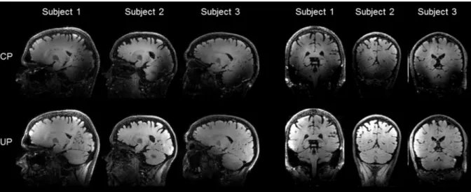
In Figure 6, a similar comparison for the FLAIR-SPACE protocol (three subjects) demonstrates again excellent recovery of the signal in the cerebellum, the occipital and the temporal lobes of the brain. The contrast between white-matter and gray-matter in the FLAIR-SPACE was admittedly weak due to the use of a simple inversion recovery preparation.
Figure 6. Comparison of the FLAIR-SPACE CP (top row) and pTx-UP (bottom row) acquisitions in
representative sagittal and coronal slices for 3 subjects (subjects 1 to 3). As for the SPACE sequence, excellent signal recovery was obtained in the cerebellum, the occipital lobe and the temporal lobes. The reception profile of the head coil was not removed. The weak contrast between white-matter and gray-matter is due to the use of a simple inversion recovery preparation.
Discussion
In this work we have reported a universal RF pulse design for 3D refocusing, universal, pulses. The benefits of the proposed pTx method compared to the standard CP-mode of excitation were explicitly reported with simulations and with in-vivo brain imaging experiments on five and three test volunteers for the T2-weighted SPACE and FLAIR-SPACE sequences respectively. A novelty in the method is the theoretical framework enabled by AHT which justifies, non-trivially, a simple STA analysis to achieve desired refocusing properties. The combined action of RF fields and gradient waveforms can hardly generate a purely transverse rotation axis given the extent of ∆B0 and the relatively weak B1 fields encountered in MRI. The strategy adopted in this work thus was instead to
symmetrize the action of ∆B0 (Equation [6]). With AHT, we derived that the overall rotation matrix indeed could be approximately decomposed into an effective free precession during the first half of the pulse, followed by the instantaneous action of the RF pulse leading to a rotation angle evaluated with the STA approximation and a purely transverse axis of rotation, followed by the same free precession during the second half of the pulse. As long as this apparent free precession is the same on both sides of the instantaneous pulse, refocusing works. Efforts in the numerical optimization then consisted in homogenizing the FA over our database of 10 subjects with their respective B1 and ∆B0 maps (20). Due to the presence of many voxels with reasonably small ∆B0 offsets, the strategy employed was first to minimize the average NRMSE over the database subjects with the right symmetries enforced to guarantee good scaling behavior, i.e. by suppressing A(1) when ∆B0 = 0. To avoid severe local artefacts near brain-air interfaces where ∆B0 is large, the second step then consisted in taking the previous result as a starting point and optimizing its scaling behavior by comparing the rotations in the small and large FA regimes (Equation [10]). A break of symmetry in the k-space trajectory and RF waveforms was then allowed while the NRMSE returned in the previous step was set as an upper-bound constraint. Efforts were also indirectly made to yield good scaling behavior by decreasing the pulse duration (Equation [9]). As shown by the experimental results, a duration of 1.1 ms for the kT-points waveform used in this work appeared to be short enough to not detect shades at the tissue-air interfaces at 7T. While it leads to a better STA approximation and better scalability, minimizing pulse duration also imposes more drastic power and SAR constraints in the pulse design. This may then engender a deterioration of the overall performance so that a compromise must be found. Alternatively to the 2 steps approach proposed here, a pulse duration minimization under power, SAR and NRMSE constraints could be attempted as well.
Due to time constraints in total scan duration, experimental comparison with tailored solutions (static RF shim, kT-points) could not be performed. Nevertheless, since the proposed design made use of the STA approximation, and that T2-weighted sequences are particularly sensitive to the RF field inhomogeneity, it is expected that the static RF shim would perform poorly and that only a mild improvement would be obtained with subject-tailored kT-point waveforms, as shown numerically and
experimentally elsewhere (19,20). Specific knowledge of the ∆B0 map yet could lead to an improved scaling behavior as the knowledge on ∆B0 would then be more accurate.
In previous works, the pulses along the train were designed individually using an optimal control approach (5). Sbrizzi and colleagues (28) also proposed a signal-based approach to design the refocusing RF pulses, thereby relaxing to a certain degree the CPMG condition. Although promising, the latter approach appears to lack the modularity and simplicity that was sought in this work for universal pulse applications, since any change of TR, T1 or T2 value then leads to a new nominal signal evolution and thus requires a new pulse optimization. In addition, separately designed RF pulses in the echo train lead to greater RF pulse design complexity due to their non-additive SAR contributions in pTx, which would likely require some heuristics to solve the constrained optimization problem. For the SPACE sequence investigated in this work, a single pulse was designed to homogenize the rotation angle over the whole spin ensemble. This considerably simplifies SAR and power budget analysis for refocusing-trains since the SAR and power simply scale with the square of the FA and the pulse contributions are additive. The pulse can be duplicated, scaled and incorporated in any train of refocusing pulses, as long as SAR and power limits are not exceeded.
While using universal pulses in the FLAIR-SPACE sequence efficiently suppressed CSF signal, the signal in the temporal and occipital lobes and in the cerebellum was also recovered compared to the CP pulses. As it has been reported in the literature, the white matter and gray matter contrast was weak while using a simple FLAIR inversion preparation at 7T (5). Better contrast could be achieved by designing more complex T2 preparation modules (37,26), possibly with the use of universal pulses.
Conclusion
We have reported an RF pulse design framework for 3D refocusing pulses guided by Average Hamiltonian Theory. The presented experimental results and simulations suggest that excellent mitigation of the RF field inhomogeneity problem in 3D T2-weighted protocols like SPACE and FLAIR-SPACE sequences can be achieved with universal pulses at UHF, while skipping entirely the conventional pTx calibration step and online pulse design. With the previous work done on small and
large flip angle excitation pulses (20), this further completes the spectrum of non-selective universal pulses to be used in 3D MRI sequences, thereby making pTx with universal pulses one step closer to clinical routine.
Acknowledgements
The research leading to these results has received funding from the European Research Council under the European Union’s Seventh Framework Program (FP7/2016-2017), ERC Proof Of Concept Grant Agreement n. 700812.
Appendix
The leading, zero order, term in the Magnus expansion is simply the time integral of Hint:
A =σ2 Ÿ ω t cos ω t + k 0 . r − k t . r*T CT
+ ω t sin ω t + k 0 . r − k t . r ¡dt + ⋯
L¢- ω t cos ω t + k 0 . r − k t . r − ω t sin ω t + k 0 . r − k t . r ¡dt*T
CT .
By denoting T the total pulse duration of the pulse, using the even and odd parity of the cosine and sine
functions, and making use of the following identity: eC"M'L& σ cos θ + σ sin θ e"M'L& = σ cos θ +
φ + σ sin θ + φ , A(0) at the end of the pulse becomes:
A = U2£Lm- ℜ Ω t e*T "()NC# dt
CT +
L¢- a Ω t e*T "()NC# dt
CT ¥ U , [6]
where Ω t = Xω t + iω t Y e"O # ., and U = eC"%&' ()N*O .,. After defining the rotation angle
θQND= R- Ω t eCT*T "()NC# dtR, phase φ = arg X- Ω t eCT*T "()NC# dtY, operator σZ = σ cos φ + σ sin φ , and making use of e5&8¦5& = U2e¦U for any matrix C, the rotation matrix in SU(2) using
the zeroth order approximation of AHT and in the interaction frame is:
References
1. Katscher U, Börnert P, Leussler C, van den Brink JS. Transmit SENSE. Magn Reson Med 2003;49:144–150. doi: 10.1002/mrm.10353.
2. Cloos MA, Boulant N, Luong M, Ferrand G, Giacomini E, Hang M-F, Wiggins CJ, Le Bihan D, Amadon A. kT-points based inversion pulse design for transmit-sense enabled MP-RAGE brain imaging at 7 T. Proc. 20th Annu. Meet. ISMRM 2012:634.
3. Cloos MA, Boulant N, Luong M, Ferrand G, Giacomini E, Hang M-F, Wiggins CJ, Bihan DL, Amadon A. Parallel-transmission-enabled magnetization-prepared rapid gradient-echo T1-weighted imaging of the human brain at 7T. NeuroImage 2012;62:2140–2150. doi: http://dx.doi.org/10.1016/j.neuroimage.2012.05.068.
4. Gras V, Vignaud A, Mauconduit F, Luong M, Amadon A, Le Bihan D, Boulant N. Signal-domain optimization metrics for MPRAGE RF pulse design in parallel transmission at 7 tesla: Signal-Domain Optimization Metrics for MPRAGE RF Pulse Design. Magn. Reson. Med. 2016;76:1431–1442. doi: 10.1002/mrm.26043.
5. Massire A, Vignaud A, Robert B, Le Bihan D, Boulant N, Amadon A. Parallel-transmission-enabled three-dimensional T2-weighted imaging of the human brain at 7 Tesla. Magn. Reson. Med. 2014:n/a– n/a. doi: 10.1002/mrm.25353.
6. Eggenschwiler F, O’Brien KR, Gruetter R, Marques JP. Improving T2-weighted imaging at high field through the use of kT-points. Magn. Reson. Med. 2014;71:1478–1488. doi: 10.1002/mrm.24805. 7. Wu X, Schmitter S, Auerbach EJ, Moeller S, Uğurbil K, Van de Moortele P-F. Simultaneous multislice multiband parallel radiofrequency excitation with independent slice-specific transmit B1 homogenization: Simultaneous Multislice Parallel RF Excitation. Magn. Reson. Med. 2013;70:630– 638. doi: 10.1002/mrm.24828.
8. Tse DHY, Wiggins CJ, Poser BA. High-resolution gradient-recalled echo imaging at 9.4T using 16-channel parallel transmit simultaneous multislice spokes excitations with slice-by-slice flip angle homogenization. Magn. Reson. Med. 2016:n/a-n/a. doi: 10.1002/mrm.26501.
9. Gras V, Vignaud A, Amadon A, Mauconduit F, Le Bihan D, Boulant N. In vivo demonstration of whole-brain multislice multispoke parallel transmit radiofrequency pulse design in the small and large flip angle regimes at 7 tesla: Joint Multislice Multispoke Pulse Design. Magn. Reson. Med. 2016. doi: 10.1002/mrm.26491.
10. Eichfelder G, Gebhardt M. Local specific absorption rate control for parallel transmission by virtual observation points. Magn Reson Med 2011;66:1468–1476. doi: 10.1002/mrm.22927.
11. Lee J, Gebhardt M, Wald LL, Adalsteinsson E. Local SAR in parallel transmission pulse design. Magn Reson Med 2012;67:1566–1578. doi: 10.1002/mrm.23140.
12. Graesslin I, Homann H, Biederer S, Börnert P, Nehrke K, Vernickel P, Mens G, Harvey P, Katscher U. A specific absorption rate prediction concept for parallel transmission MR. Magn Reson Med 2012;68:1664–1674. doi: 10.1002/mrm.24138.
13. de Greef M, Ipek O, Raaijmakers AJE, Crezee J, van den Berg CAT. Specific absorption rate intersubject variability in 7T parallel transmit MRI of the head. Magn Reson Med 2013;69:1476–1485. doi: 10.1002/mrm.24378.
14. Le Garrec M, Gras V, Hang M-F, Ferrand G, Luong M, Boulant N. Probabilistic analysis of the specific absorption rate intersubject variability safety factor in parallel transmission MRI: Probabilistic Analysis of Specific Absorption Rate Intersubject Variability. Magn. Reson. Med. 2016. doi: 10.1002/mrm.26468.
15. Fautz H-P, Vogel MH, Gross P, Kerr A, Zhu Y. B1 mapping of coil arrays for parallel transmission. Proc. 16th Annu. Meet. ISMRM 2008:1247.
16. Nehrke K, Börnert P. DREAM—a novel approach for robust, ultrafast, multislice B1 mapping. Magn. Reson. Med. 2012;68:1517–1526. doi: 10.1002/mrm.24158.
17. Brenner D, Stirnberg R, Pracht E, Stöcker T. Rapid MRI System Calibration using 3DREAM. Proc. 23rd Annu. Meet. ISMRM 2015:491.
18. Hoyos-Idrobo A, Weiss P, Massire A, Amadon A, Boulant N. On Variant Strategies to Solve the Magnitude Least Squares Optimization Problem in Parallel Transmission Pulse Design and Under Strict SAR and Power Constraints. IEEE Trans. Med. Imaging 2014;33:739–748. doi: 10.1109/TMI.2013.2295465.
19. Gras V, Vignaud A, Amadon A, Le Bihan D, Boulant N. Universal pulses: A new concept for calibration-free parallel transmission. Magn. Reson. Med. 2017;77:635–643. doi: 10.1002/mrm.26148. 20. Gras V, Boland M, Vignaud A, Ferrand G, Amadon A, Mauconduit F, Le Bihan D, Stöcker T, Boulant N. Homogeneous non-selective and slice-selective parallel-transmit excitations at 7 Tesla with universal pulses: a validation study on two commercial RF coils. PLOS ONE 2017.
21. Hennig J, Nauerth A, Friedburg H. RARE imaging: a fast imaging method for clinical MR. Magn. Reson. Med. 1986;3:823–833.
22. Mugler JP. Optimized three-dimensional fast-spin-echo MRI. J. Magn. Reson. Imaging 2014;39:745–767. doi: 10.1002/jmri.24542.
23. Visser F, Zwanenburg JJ, Hoogduin JM, Luijten PR. High-resolution magnetization-prepared 3D-FLAIR imaging at 7.0 Tesla. Magn. Reson. Med. 2010;64:194–202.
24. Madelin G, Oesingmann N, Inglese M. Double Inversion Recovery MRI with Fat Suppression at 7 Tesla: Initial Experience. J. Neuroimaging 2010;20:87–92. doi: 10.1111/j.1552-6569.2008.00331.x. 25. van der Kolk AG, Zwanenburg JJ, Brundel M, Biessels G-J, Visser F, Luijten PR, Hendrikse J. Intracranial vessel wall imaging at 7.0-T MRI. Stroke 2011;42:2478–2484.
26. de Graaf WL, Zwanenburg JJM, Visser F, Wattjes MP, Pouwels PJW, Geurts JJG, Polman CH, Barkhof F, Luijten PR, Castelijns JA. Lesion detection at seven Tesla in multiple sclerosis using magnetisation prepared 3D-FLAIR and 3D-DIR. Eur. Radiol. 2012;22:221–231. doi: 10.1007/s00330-011-2242-z.
27. Fortunato EM, Pravia MA, Boulant N, Teklemariam G, Havel TF, Cory DG. Design of strongly modulating pulses to implement precise effective Hamiltonians for quantum information processing. J. Chem. Phys. 2002;116:7599–7606. doi: 10.1063/1.1465412.
28. Sbrizzi A, Hoogduin H, Hajnal JV, van den Berg CAT, Luijten PR, Malik SJ. Optimal control design of turbo spin-echo sequences with applications to parallel-transmit systems: Optimal Control of TSE Sequences. Magn. Reson. Med. 2016:n/a–n/a. doi: 10.1002/mrm.26084.
29. Haeberlen U, Waugh JS. Coherent Averaging Effects in Magnetic Resonance. Phys Rev 1968;175:453.
30. Warren WS. Effects of arbitrary laser or NMR pulse shapes on population inversion and coherence. J. Chem. Phys. 1984;81:5437–5448. doi: http://dx.doi.org/10.1063/1.447644.
31. Pauly J, Nishimura D, Macovski A. A k-space analysis of small-tip-angle excitation. J. Magn. Reson. 1969 1989;81:43 – 56. doi: http://dx.doi.org/10.1016/0022-2364(89)90265-5.
32. Boulant N, Hoult DI. High tip angle approximation based on a modified Bloch–Riccati equation. Magn Reson Med 2012;67:339–343. doi: 10.1002/mrm.23270.
33. Levitt MH. Symmetry in the design of NMR multiple-pulse sequences. J. Chem. Phys. 2008;128:52205. doi: 10.1063/1.2831927.
34. Pruessmann KP, Golay X, Stuber M, Scheidegger MB, Boesiger P. RF Pulse Concatenation for Spatially Selective Inversion. J. Magn. Reson. 2000;146:58–65. doi: 10.1006/jmre.2000.2107.
35. Grissom W, Yip C, Zhang Z, Stenger VA, Fessler JA, Noll DC. Spatial domain method for the design of RF pulses in multicoil parallel excitation. Magn Reson Med 2006;56:620–629. doi: 10.1002/mrm.20978.
36. Gras V, Luong M, Amadon A, Boulant N. Joint design of kT-points trajectories and RF pulses under explicit SAR and power constraints in the large flip angle regime. J. Magn. Reson. 2015;261:181 – 189. doi: http://dx.doi.org/10.1016/j.jmr.2015.10.017.
37. Nezafat R, Stuber M, Ouwerkerk R, Gharib AM, Desai MY, Pettigrew RI. B1-insensitive T2 preparation for improved coronary magnetic resonance angiography at 3 T. Magn. Reson. Med. 2006;55:858–864. doi: 10.1002/mrm.20835.