HAL Id: tel-02003596
https://tel.archives-ouvertes.fr/tel-02003596
Submitted on 1 Feb 2019HAL is a multi-disciplinary open access archive for the deposit and dissemination of sci-entific research documents, whether they are pub-lished or not. The documents may come from teaching and research institutions in France or abroad, or from public or private research centers.
L’archive ouverte pluridisciplinaire HAL, est destinée au dépôt et à la diffusion de documents scientifiques de niveau recherche, publiés ou non, émanant des établissements d’enseignement et de recherche français ou étrangers, des laboratoires publics ou privés.
and imaging of cell membranes
Janah Shaya
To cite this version:
Janah Shaya. Fluorene-based fluorescent markers : new insights in synthesis and applications into labeling of nucleic acids and imaging of cell membranes. Other. Université Côte d’Azur, 2016. English. �NNT : 2016AZUR4066�. �tel-02003596�
UNIVERSITE DE NICE‐SOPHIA ANTIPOLIS − UFR Sciences
Ecole Doctorale de Sciences Fondamentales et Appliquées
THESE
Pour obtenir le titre de
Docteur en Sciences
de l'UNIVERSITE de Nice – Sophia Antipolis
Discipline : Chimie
Présentée et soutenue par
Janah Shaya
« Capteurs fluorescents à base de fluorène : nouvelles
perspectives en synthèse et applications pour le marquage des
acides nucléiques et en imagerie de la membrane cellulaire »
« Fluorene‐based fluorescent markers; new insights in
synthesis and applications into labeling of nucleic acids and
imaging of cell membranes »
Thèse dirigée par le Prof. Alain BURGER et co‐encadree par le Dr. Benoît Y.
MICHEL
Soutenue le 21 Septembre 2016
Jury:
Pr. Demchenko Alexander Professeur, Académie Nationale
des Sciences de l'Ukraine
Rapporteur
Pr. Smietana Michael
Professeur, Université de
Montpellier
Rapporteur
Dr. Jazzar Rodolphe
Chercheur CNRS, Université de
Californie
Rapporteur
Dr. Michel Y. Benoît
Maître de conférences, Université
Nice Sophia Antipolis
Co‐encadrant
de thèse
Pr. Burger Alain
Professeur, Université Nice Sophia
Antipolis
Directeur de
thèse
ﻰﻟﺇ
"
ﻲﻧﺎﻨﺒﻟ
"
ﻥﺍﺮﺒﺟ ﻥﺎﻨﺒﻟ
ﻱﺭﻮﺧﻭ
ﻭ
....
ﻪﻴﻤﺤﻳ ﻦﻣ ﻞﻛ
To my “Lebanon”, the country of Gibran and Corey,
And everyone who keeps it safe….
A toi mon “Liban”, Le pays de Gibran et Corey,
Et à tous ceux qui le protègent….
Acknowledgements
First, I would like to express my sincere thankfulness to the “French ministry of education” and the “Doctoral school of Nice‐Sophia Antipolis university (ED‐SFA)” for offering me this Ph.D grant for three years. I am equally thankful to Pr. Alain Burger for the fruitful leadership of this thesis. In fact, Alain is present and evenly invested in all the work in his lab, especially the difficult starting of new projects like “my fluorenes”. His guidance, knowledge, resources, and ambition were indispensable. An in‐line thank you might go here to Dr. Benoit Michel for his useful contributions. He is one of few chemists I’ve met with an inspiring talent in the art of “organic synthesis”. Dr. Rachid Benhida has the portion of the lion especially in the front lines of this project. I owe him for his support and the 3‐year stay in the “molecules bioactives” group. I would also like to acknowledge all the permanent staff of our group and the ICN; in particular, the director of the ecole doctorale Dr. Elisabeth Taffin‐de‐Givenchy, Dr. Audrey Di Giorgio, Dr. Stephane Azouly and Dr. Sophie Poulain‐Martini for the teaching experience, Dr. Maria Duca, Dr. Luc Demange, Dr. Cyril Ronco, Dr. Guilhem Godeau, Dr. Thierry Darmanin for setting the example of a researcher for me, Dr. Christope Di Giorgio for all the synthesis advices, the director of the ICN Dr. Elisabet Duñach for the GCMS facilities, Dr. Lionel Massi for the mass spectrometry, Dr. Marc Gaysinski for the NMR spectroscopy, and Dr. Nadine Martinet for her tips and advices.
The list of acknowledgments will continue and I hope I do not forget anyone as research is a collaborative effort and many people play a direct or indirect role; Dr. Anthony Martin for sharing his knowledge on catalysis and for the pleasant talks, Dr. Sylvain Antoniotti for his precious remarks on sustainable chemistry, Dr. Fabien Fontaine‐Vive for the pleasure of working with him on DFT calculations, and Prof. Nadia Patino for plenty of teachings and fun. Pr. Iyad Karamé, Dr. AbdelRehman Sidani, Pr. Peter Goekjian, and Dr. Rodolphe Jazzar deserve to be appreciated for being influential chemists that have planted the first eagerness for this domain in me. Iyad is a person to whom I am deeply indebted. A big thank you goes also to Dominique Bonhomme (Domi) and Frederic Benailli (Fredo) for the coffee and work time we spent. I hope that our work together will be validated in publications the soonest.
I have to warmly thank Pr. Alexander Demchenko, Pr. Michael Smietana, and Dr. Rodolphe Jazzar for revising this manuscript and being members of the thesis committee.
I would likewise address the great team in Strassbourg for the warm, helpful, and productive welcoming in their lab where we performed the cell imaging; Pr. Yves Mely, Dr. Andrey Klymchenko, Dr. Mayeul Collot, and the young researchers there; Dr. Yosuke‐Niko, Bohdan, Dorian, Hassan, Iryna, Rajhan and others for the fun trips and nights we passed during my stay. Nice lab has a list of young researchers that enriched this journey with good memories as well: Florian, Gabreilla, Hamza, Alexandra, Hellene, Coralie, Antoine, Sokaina, Loraine, Anne, Lauri, Sasha, Oleg, Malika, Nelli, Cecile. I wish you all the best in your future endeavors. Few words to some should be mentioned: Cate; Sursum Corda ou Janah va… we really missed you in the ICN, Niko; it was a pleasure to share the same lab with you, Anh; you will be truly missed my
dearest, Mauro; you lightened up fluorescence a bit more dude. Merci également à Marie le grand prof. de français pour les produits qu'elle a fait au cours de M1 stage.
I think I forgot Hella, not really! You will be remembered for long time. All the first translations, hang‐outs, fun, and shouting have spiced “Nice” stay. Assia also did her share in translations and joy, merci! Altogether with Diaa, Ilheme, and Najiba, we had a great deal of time and food that I will definitely miss. Dr. Ilheme, despite your early departure from the ICN, the short time was enough to reveal a chemist and person of high standards and a friend for life. Outside the lab, there are many people who had put up with me in all stages. They are a major highlight in my life. The Janwa; you witnessed it all and are still doing patiently, I will simply quote from your thesis: “Behind each work, there is a secret rider, you were always there”, see you soon in Strass. George; an ever‐lasting support and a melody for every occasion, laughing is the key of success as you say. Kawsar; the chief of chiefs, for god’s sake we need to stop running and making rules; not everyone has your potential. Together, we made the perfect KG group of nice. Ramez; you left your space when you moved. Abed, Tharwat; travelling with you is beyond fun. Habeeb and Hawraa; you added a source of joy with our future chemist, Mariam. Finally Pascal; we will hopefully always meet or run away bro, I am so happy that I have met you.
Back home, the gratitude can go endless and I still have too many experiments to do, so I will cut it short. Thanks for everyone who supported me when I decided to change careers. I am very lucky to have a family that trusts my choices without questioning. You are too many, but I love you all. Mostafa and Iman; you are my residence and comfort. Jinan; you did all what I have to do for years. You, Naim, and your three ugly musketeers are blessings. Suzan and Samah, your craziness makes you so special and we are proud of your success with each day passing, Roy, Iyad, and Karam; you will be one of a kind (chemical) engineers, you know it. Jihan, Souaad, Makram, Fares, David, Osama, Wido…. Silvana; don’t get lost in Cuba, I can’t live without you all. Distance did not make some any less of great friends: Rawad, a real brother; we will keep on roaming and reach, we shared all the crazy and wisdom times, all the fun, joy, plans, and determination‐I hope you make it to my defense, Bilal, Bahaa; thanks for all and cheers, Naveen, Fadi, George, Anis, Samer, Dani, and the group leader in Dubai, Khodr; here it is man, our chemistry is finally in action, Lamece the royal; I will sum up the 10 pages of jokes you make of me into one line of don’t stop because I will miss it, Jihan the one and only; from Lyon to Lebanon, and Marah; a real source of aspiration for many years, I will buy a house in Monaco and you will be invited. If anyone is forgotten, be sure it is for lack of time. I appreciate an equal list of people that I could not name.
Finally, there is nothing better than quoting from the French Nobel Prize winner Jacques Monod: “In science, self‐satisfaction is death. Personal self‐satisfaction is the death of the scientist. Collective self‐satisfaction is the death of the research. It is restlessness, anxiety, dissatisfaction, agony of mind that nourish science.” So here it is, a small plan of an unsatisfied scientist who lost his first grant in a war back in 2006, and kept the same target of research till July 2016.
Nice, July, 2016
Table of Contents
•Dedication ... 2
•
Acknowledgments ... 3
•
Table of contents ... 5
•
List of abbreviation ... 9
•
Thesis statement and general presentation ... 11
Chapter One : Bibliographical Review
I. LUMINESCENCE ... 15 I.1
FLUORESCENCE ... 15
I.1.1 Stage 1: Absorption of light (excitation) ... 15
I.1.2 Stage 2: Excited state and fluorescence lifetime ... 17
I.1.3 Stage 3‐Fluorescence emission ... 18
II. FLUOROPHORES ... 20 II.1
INTRINSIC FLUOROPHORES ... 21
II.1.1 Fluorescent proteins
...
21II.1.2 Chlorophyll ... 21
II.2
EXTRINSIC FLUOROPHORES ... 22
II.2.1 Genetically encoded proteins ... 22
II.2.2 Fluorescent nanocrystals ... 22
II.2.3 Small‐molecule fluorophores
...
23II.3
SOLVATOCHROMIC FLUOROPHORES. ... 26
II.3.1 What is “solvatochromism” in fluorescence context (solvatofluorochromism)? ... 26
II.3.2 Negative solvatochromism:
...
26II.3.3 Positive solvatochromism: ... 27
II.4
PUSH‐PULL DYES ... 32
III. FLUORENE AND ITS DERIVATIVES ... 33 III.1
PARTICULAR FEATURES OF FLUORENE AND ITS DERIVATIVES ... 35
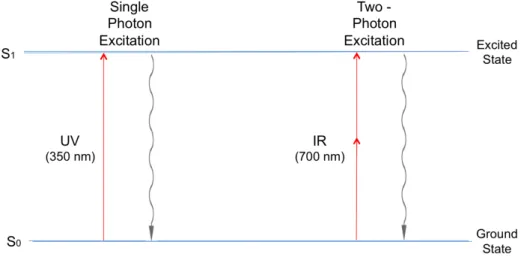
III.1.1 Two‐photon absorption
...
35III.1.2 Aggregation‐Induced Emission Enhancement (AIEE)
...
36III.1.3 Excimer formation
...
36III.1.4 Twisted Intramolecular Charge Transfer (TICT)
...
37III.1.5 Profluorescence of fluorene derivatives for turn‐on applications ... 37
III.1.6 Polyfluorenes (PF) ... 37
III.2
ARRAY OF USES OF FLUORENE AND ITS DERIVATIVES ... 39
III.2.1 Fluorescence imaging and application related to biology
...
39III.2.2 Optical data storage ... 39
III.2.3 Organic Light‐Emitting Devices (OLEDs) ... 40
III.2.4 Dye‐sensitized solar cells (DSSCs) ... 40
III.2.5 Non linear optical dyes in telecommunication ... 40
III.3
SYNTHESIS OF AMINOFLUORENES ... 40
Conventional routes ... 40
Buchwald‐Hartwig amination ... 41
IV. FLUOROSENSING ... 43 IV.1
FLUORESCENCE LABELING AND DETECTION ... 44
IV.2
FLUORESCENCE SENSING ... 45
IV.2.1 FRET ... 46
IV.2.2 Anisotropy ... 47
V.1
CLASSIFICATIONS OF MEMBRANE LIPIDS ... 48
V.1.1 Glycerophospholipids ... 48
V.1.2 Sphingolipids ... 48
V.1.3 Sterols ... 48
V.2
HETEROGENEITY OF LIPID MEMBRANES AND RELATED FLUOROPHORES ... 49
V.2.1 Level One: Lipid raft theory ... 49
Fluorophores related to raft investigations and model membranes ... 50
V.2.2 Level two: Transmembrane lipid asymmetry ... 50
Annexin V detection assays ... 51
V.2.3 Level three: heterogeneity among biological membranes ... 52
V.3
FLUORESCENCE PROBING OF MEMBRANE LIPIDS ... 52
V.3.1 Membrane probes ... 52
VI. NUCLEIC ACIDS ... 54
VI.1
STRUCTURE OF DNA ... 54
VI.2
DNA SYNTHESIS: ... 56
VI.3
THE CHEMISTRY BEHIND DNA SYNTHESIS ... 56
VI.3.1 The phosphotriester approach ... 57
VI.3.2 The phosphite triester and H‐phosphoramidite approach ... 57
VI.3.3 H‐phosphonate approach ... 58
VI.4
CHEMICAL SYNTHESIS OF DNA ... 59
VI.4.1 Solution‐phase synthesis ... 59
VI.4.2 Solid‐phase DNA synthesis ... 59
VI.5
FLUORESCENT LABELING OF NUCLEIC ACIDS ... 61
VI.5.1 Non‐covalent or non‐specific labeling ... 61
VI.5.2 Covalent labeling ... 63
VI.6
SYNTHETIC APPROACHES USED IN COVALENT LABELING OF ODNS ... 67
VI.6.1 Pre‐synthetic modification ... 67
VI.6.2 Post‐synthetic modification ... 67
VI.7
SELECTED SAMPLE APPLICATIONS OF FLUORESCENT NUCLEOSIDES ... 68
VI.7.1 DNA sequencing ... 68
VI.7.2 Single nucleotide polymorphism (SNP) ... 68
VI.8
FLUORENE DERIVATIVES IN THE CONTEXT OF DNA APPLICATIONS ... 68
Chapter Two : Air‐stable palladium catalytic systems for sequential one‐pot
synthesis of unsymmetrical aminoaromatics
I. OPTIMIZATION STUDIES ... 71 II. SCOPE OF THE SELECTIVE MONOAMINATION REACTION ... 74 III. DIAMINATION OF FLUORENES ... 75 IV. SYNTHESIS OF UNSYMMETRICAL FLUORENES ... 75 V. SCOPE OF THE SELECTIVE CONDITIONS ON DIBROMONAPHTHALENE, DIBROMOPYRIDINE, AND DIBROMOTHIPHENE ... 77 VI. APPLICATION: A NEAR‐IR PUSH‐PULL SENSOR ... 78 VII. CONCLUSION ... 79
Chapter three : Push‐Pull fluorene dyes: synthesis and structure‐photophysics
relationship
I. SYNTHESIS ... 82
I.1
VARIATION OF THE ELECTRON‐WITHDRAWING GROUP (EWG) ... 82
I.2
VARIATION OF THE ELECTRON‐DONATING GROUP (EDG) ... 84
II. SPECTROSCOPIC STUDIES ... 84
II.1
‐ ACCEPTOR SERIES ... 84
II.2
‐ VARIED‐AMINE SERIES ... 91
II.3
‐
HAMMETT CORRELATION ... 97III. CONCLUSION ... 99
Chapter Four : Fluorene‐based membrane probes and cell imaging
I. INTRODUCTION ... 101 II. SYNTHESIS ... 102 III. PHOTOPHYSICAL CHARACTERIZATION OF F1‐F3 ... 102 III.1HYDRATION STUDY OF F1 ... 105
III.2
PHOTOPHYSICAL CHARACTERIZATION IN LARGE UNILAMELLAR VESICLES OF DIFFERENT LIPID COMPOSITIONS ... 106
III.3
PHOTODEGRADATION STUDIES ... 109
IV. IMAGING ... 111
IV.1
IMAGING GIANT UNILAMELLAR VESICLES (GUV) WITH F3 ... 111
IV.2
IMAGING CELLS WITH F1‐F3 ... 112
V. CONCLUSION: ... 114
Chapter Five : Fluorene‐based DNA fluorescent markers; synthesis, DNA labeling,
and applications
I. FLUORENE LABEL BY ANULCLEOSIDE APPROACH ... 116 II. FLUORENE LABEL CONNECTED TO DEOXYRIBOSE SUGAR ... 122 II.1SYNTHESIS OF FM2‐4 ... 122
III. CHARACTERIZATION OF THE FREE FLUORESCENT MARKER (X) AND THE X‐LABELED ODN SEQUENCES ... 126 III.1
SPECTROSCOPIC PROPERTIES OF THE NAKED FLUORESCENT NUCLEOSIDE (197) ... 126
III.2
PKA DETERMINATION OF (197) ... 128
III.3
PHOTOPHYSICAL CHARACTERIZATION OF THE X‐LABELED ODNS ... 129
III.4
CD AND THERMAL DENATURATION STUDIES ... 132
III.5
FRET ... 134 IV. CONCLUSION ... 137
Conclusions and perspectives
I. GENERAL CONCLUSION ... 138 II. PERSPECTIVES ... 139
Résumé long
... 141Experimental part and SI
I. GENERAL METHODS ... 146 II. LIPID VESICLES ... 147 III. CELL LINES, CULTURE CONDITIONS, TREATMENT, AND IMAGING ... 148 IV. ODN SYNTHESIS AND PURIFICATION: ... 148 V. MALDI PROCEDURE ... 149 VI. PREPARATION OF SS AND DS LABELED DNA ... 149 VII. DENATURATION STUDIES AND MELTING TEMPERATURES ... 150 VIII. CIRCULAR DICHROISM ... 150 IX. CHAPTER 2 SYNTHETIC PROCEDURES ... 151 X. CHAPTER 3 SYNTHETIC PROCEDURES ... 164 XI. CHAPTER 4 SYNTHETIC PROCEDURES ... 174 XII. CHAPTER 5 SYNTHETIC PROCEDURES ... 179 XIII. SUPPLEMENTARY CHARACTERIZATION OF CHAPTER 3 ... 195XIII.1
TRANSITION DIPOLE MOMENT OF PUSH‐PULL DYES (IN DEBYE): ... 195
XIII.2
DFT AND TDDFT CALCULATIONS. ... 195
XIV. SUPPLEMENTARY CHARACTERIZATION OF CHAPTER 4 ... 199
XIV.1
PHOTODEGRADATION DECAY CURVES OF F1‐3 AS A FUNCTION OF TIME IN ETHANOL ... 199
XIV.2
RATIO IMAGES FOR F2 PROBE IN CELLS ... 200
XIV.3
COMPARISON OF THE INTENSITY OF DYES; F1, F2, AND F3 RESPECTIVELY. ... 200
XV. SUPPLEMENTARY CHARACTERIZATION OF CHAPTER 5 ... 202 XV.1
PHOTOPHYSICAL CHARACTERIZATION OF DYE 190 ... 202
XV.2
HPLC PROFILES: ... 203
XV.3
EXCITATION SPECTRA ... 203
XV.4
MELTING TEMPERATURE CURVES ... 203
XV.5
CD SPECTRA ... 204
XV.6
ACCEPTOR DYES USED IN FRET ... 204
Referenes………..………...……….
205List of abbreviations
• � wavenumber • 2‐AP 2‐aminopurine • 2PA two‐photon absorption • 3‐D three dimensions • 9‐CHF 9‐cycloheptatrienylidene fluorenes • A absorbance • a radius of the Onsager’s cavity • a.u. arbitrary unit • ACN acetonitrile • ACQ aggregation‐caused quenching • Ac acetone
• AIEE aggregation‐induced emission enhancement • asap as‐soon‐as‐possible • BDF base‐discriminating fluorescence • BLA bond length alternation • C concentration • c speed of light • CD circular dichroism • CL cholesterol • cm centimeter • CuAAC copper(I)‐catalyzed alkyne‐ azide cycloaddition • D Debye • DAPI 6‐diamidino‐2‐phenylindole • DBU 1,8‐diazabicycloundec‐7‐ene • DCE dichloroethane • DCM dichloromethane • DCPC 2‐cyanoethyl‐N,N‐diisopropyl‐ chlorophosphoramidite • DMF dimethylformamide • DMSO dimethylsulfoxide • DMTr dimethoxytrityl group • DNA deoxyribonucleic acid • DOPC dioleoylphosphatidylcholine • DOS diversity‐oriented synthesis • DSSC dye‐sensitized solar cell • E energy • EA ethyl acetate • EDG/D electron‐donating group/donor
• ESIPT excited‐state intramolecular proton transfer • EtOH ethanol • eV electronvolt • EWG/A electron‐withdrawing group/acceptor • Ɛ molar absorption coefficient • F fluorene membrane probe • FC flash chromatography • FLS fluorescence lifetime spectroscopy • FM fluorescent ODN marker • FRET Förster Resonance Energy Transfer
• G, A, T, C, U guanine, adenine, thymine, cytosine, uracil respectively • GFP green fluorescent protein • GUV giant unilamellar vesicle • h Planck’s constant • H‐bond Hydrogen bonding • HGP human genome project
• HOMO highest occupied molecular orbital • I inductive effect • ICT intramolecular charge transfer • IR infrared • J spectral overlap in FRET • kDa kilodalton • knr sum of non‐radiative decay rates • kr radiative decay rate • L ligand (tertiary phosphine) • l the light path length in a sample • Ld liquid‐disordered phase • Lo liquid‐ordered phase • LUMO lowest unoccupied molecular orbital • LUV large unilamellar vesicle • M mesomeric effect • MeOH methanol • MO molecular orbital • MW microwave • N* normal excited state • NIR near‐infrared • nm nanometer • nM nanomolar • ODN oligonucleotide • OLED organic light‐emitting device • PC phosphatidyl choline • PCR polymerase chain reaction • PE phosphatidyl ethanolamine • PE solvent Petroleum ether • PF polyfluorenes • PP push‐pull dye • PS phosphatidyl serine • Qdots Quantum dots • r anisotropy • R donor‐acceptor distance in FRET • R0 Förster distance where
fluorescence transfer efficiency is 50 % • RNA ribonucleic acid • RP HPLC reversed‐phase high‐ performance liquid chromatography • rt room temperature • s second • S singlet multiplicity • SI supplementary information • SM sphingomyelin • SMS single molecule spectroscopy • SNP single nucleotide polymorphism • T triplet multiplicity • T* ESIPT tautomer • THF tetrahydrofuran
• TICT twisted internal charge transfer • UV ultraviolet • vs. versus • X Y Z M fluorene ODN labels (FM) • Δf Lippert’s parameter • ΔλSS Stokes shift • λ wavelength • λabs absorption maximum • λem emission maximum
• μE and μG dipole moments of the
excited and ground states
• π aromatic core
• σP Hammett value
• τ fluorescence lifetime
Thesis statement and presentation of the manuscript
The cutting‐edge development of technology often takes place at interfaces of supposedly distinct disciplines of science. Research involving interdependent areas of synthetic chemistry, computations, photophysics, biophotonics, and imaging has evolved as a modern mean of analysis to unravel the scientific enigmas. “Fluorescence” is a domain that integrates virtually all the mentioned disciplines and extends to other frontiers of environmental detections, life, medicinal, and pharmaceutical sciences.“Fluorescence” was observed first by Herschel in 1845 as a phenomenon from quinine solution under sunlight and was further described by Stokes in 1852. A century later, this novelty process was put in the service of science with the invention of “commercial fluorometers” and “fluorescence microscopes”. The advances proceeded fast with “confocal microscopy” and more sophisticated Nobel‐Prize instruments like the “Zewail’s femtosecond (x10‐15 s) spectroscopy”. Fluorescence instrumentations are nowadays advanced, not expensive, and easy‐to‐manipulate.
Eventually, fluorescence techniques have become crucial analytical toolbox for research and industry owing their success to many exquisite characteristics. These techniques are the most sensitive in the field of molecular sensing especially in probing intrinsic biological dynamics. Their sensitivity has attained the limit of single molecule detection with signal recordings that can be controlled at a distance. They demonstrate high degree of accuracy, non‐invasive character in living cells, and ultrahigh spatial and temporal resolutions. Furthermore, fluorescence sensing is extremely versatile providing detections in solid, liquid, gas, and even at the interface of these phases. In addition to instrumentations, the growth in the “design and synthesis of fluorophores” undoubtedly remains the hallmark of progress in fluorescence applications.
An optimal fluorophore is essentially a molecular framework that compiles the required attributes from optical properties such as photostability, brightness, selective excitation and emission to chemical versatility such as tolerance to introduce substituents for bioconjugation and tuning solubility. Such rational design is a challenge by itself. From “organic chemistry” perspectives, this will at least involve a multi‐step synthetic route to produce such a smart molecule; hence, adding more challenges of the known limitations of synthesis. Consequently, chemists are highly urged to bring forth new robust, step‐ and atom‐economic routes to synthesize innovative probes and generate libraries of dyes that elucidate structure‐property relationships.
12
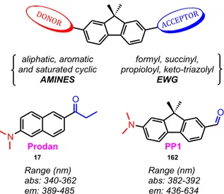
In particular, our work has focused on metal‐catalyzed reactions such as Buchwald‐Hartwig aminations and copper(I)‐catalyzed alkyne‐azide cycloaddition (CuAAC) “click” reactions to create more viable alternatives to otherwise long employed routes. The developed in‐house methodologies allowed accessing a fluorene‐based library of push‐pull dyes as a captivating class of environment‐sensitive probes. A push‐pull dye consists of a π‐scaffold functionalized with an electron donor (D) and acceptor (A). The D‐π‐A system undergoes a photo‐induced intramolecular charge transfer (ICT) state forming a low‐energy molecular orbital (MO). Visible light provides sufficient energy to excite the electrons within this new MO, making these compounds generally colored. The ICT also generates dipole moments that augment the sensitivity of these fluorophores in response to polarity and changes of their environments. The aromatic core (π) plays a key role in the design. Thus, came the choice of the fluorene family in this project. Fluorenes manifest desirable spectroscopic attributes, low cytotoxicity, and appreciable two‐photon absorption cross‐sections allowing cell imaging with reduced photodamage. Fluorene derivatives are extensively used in biomedical research and material sciences such as optoelectronics.
Two central investigations were explored using our fluorene probes, 1) labeling of oligonucleotides (ODNs), and 2) imaging of lipids in biomembranes.
In fact, despite the complete determination of the 3.5x10⁹ base‐pair DNA sequences in the human genome project (HGP) in 2001, this explosive acquisition of structural data has paraded very limited information about the complex biological function of DNA. To address these aspects, fluorescence spectroscopy offers an exquisite tool to characterize nucleic acids and to study their interactions, which are fundamentals to understand cellular events such as DNA repair, DNA methylation, and gene silencing. The search of suitable fluorescent markers that can monitor these interactions without affecting the structure and function of the labeled ODN sequences is still at its peak.
Likewise, the heterogeneous lipid distribution in cell membranes is a subject of intense investigations. Three levels of heterogeneity are distinguished. The lateral heterogeneity, described by the lipid raft hypothesis, is under debate till now. Rafts are believed to be behind many processes such as signal transduction and neurodegenerative diseases. Transmembrane lipid asymmetry is the second heterogeneity that is lost in programmed cell death (apoptosis). Defective apoptosis is the origin of detrimental diseases such as atrophy and cancer. Lastly, the differences between the plasma and intracellular membranes represent the third, least‐explored heterogeneity. Thus, designing highly sensitive probes will help to understand the structure and
dynamics of biomembranes by sensing different parameters like hydration, viscosity, polarity, and order. This will in turn reflect in understanding the cellular processes and in finding solutions to futile medical problems such as cancer.
In conclusion, the purpose of this thesis was to advance a step in the areas of designing and synthesizing novel fluorescent probes that meet the rigorous and demanding conditions of the above‐mentioned biological applications.
The body of the dissertation consists of 5 chapters and a conclusion stating the future perspectives.
• Chapter one is the introduction that states the background of this work.
• Chapter two describes a concise synthetic methodology to prepare a spectrum of aminoaromatics. The methodologies involve selective functionalization of dibromoaromatic scaffolds using air‐stable palladium catalytic systems. In particular, rapid mono‐ and di‐aminations were carried out in addition to sequential couplings of different moieties in one‐pot. The synthesis was scaled up to prepare the essential probes used in the next chapters. The content of chapter 2 has recently been published online in The Journal of Organic Chemistry.
• Chapter three builds on the developed catalytic conditions of Buchwald‐Hartwig amination to explore the effect of the donor part on push‐pull molecules. Moreover, it describes various synthetic pathways to study the acceptor part including CuAAC, peptide couplings, and metal‐halogen exchange reactions. In effect, we concluded the theoretical and experimental characterization and structure‐photophysics relationships of 17 synthesized fluorophores including near‐infrared (NIR) probes. The content of chapter 3 has recently been published in Chemistry: A European Journal.
• Chapter four takes the advantage of the rational design and the new “ynone” anchoring point of “chapter three” in bioconjugating the fluorene probes. This chapter describes the synthesis of three probes designed to image membrane lipids. The photophysical characterization of the dyes, their ability to probe large and giant unilamellar vesicles (LUV and GUV) of different lipid compositions, and the imaging of HeLa cells are reported. The optmized dye is the first plasma membrane‐specific fluorene‐based probe that can be a powerful tool for studying membranes surpassing the features of commonly used probes.
• Chapter five illustrates the synthesis of four emissive nucleobase analogues (phosphoramidites) exploring different linkers and sugars to rationalize the design of a fluorene‐based DNA sensor. Then, it covers the journey of site‐specific ODN
14
incorporations of the biosensors via solid‐phase DNA synthesis, the difficulties encountered, the characterization and the hybridization studies of the labeled ODN sequences, and finally the preliminary application of the optimal biosensor as a mega‐ Stokes shift donor in a FRET pair. The recorded shift seems exceptional reaching up to 300 nm and very promising to validate the dye in future applications.
• A brief conclusion and future perspectives are stated at the end of the manuscript, followed by the experimental section.
Chapter 1:
Bibliographical Review
I. Luminescence
Luminescence is the process of emission of light quanta upon excitation by any source of external energy such as a chemical reaction (chemiluminescence), thermal energy (thermoluminescence), mechanical action on a solid (mechanoluminescence), light (photoluminescence), etc..
In particular, photoluminescence is defined as an optical property of a substance which absorbs photons and then releases the absorbed energy radiatively from its excited state.[1,2] Chemical functionalities that absorb light in the ultraviolet or visible region are referred as chromophores. Absorption in the visible gives colored compounds.
Depending on the nature of the excited state, photoluminescence is classified into 2 forms, fluorescence and phosphorescence. A molecule or a molecular fragment that has the ability to fluoresce is called a fluorophore. A fluorophore is also commonly termed as a dye in reference to its color property.[3,4]
I.1 Fluorescence
Fluorescence can be viewed as a three‐stage process. Each stage and its corresponding optical measurements will be discussed in the following sections.I.1.1 Stage 1: Absorption of light (excitation)
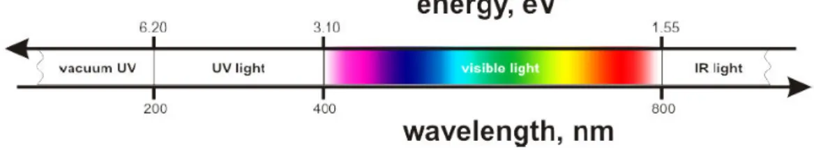
I.1.1.1 Light Light is an electromagnetic radiation described in terms of a “wave‐particle duality” concept. Precisely, light is characterized as a particle (photon) by its energy (E, eV) and as a wave by its wavelength (λ, nm) or wavenumber (�, cm−1) as shown in eq. 1, � = ℎ� = ℎ � � ��� � = 1 �Equation 1
16
respectively.
The range of electromagnetic radiations suitable for optical studies and photochemistry is shown in fig. 1 and is characterized by its E and λ.[2,3] Figure 1. Range of electromagnetic radiation in optical studies and photochemistry[5] I.1.1.2 Absorption An organic molecule resides in its ground state (S0, singlet by multiplicity) in the absence of external sources of perturbation. In the ground state, two electrons rest in the highest occupied molecular orbital (HOMO) while the lowest unoccupied molecular orbital (LUMO) is free. Exposing the molecule to a vacuum extreme‐ultraviolet radiation (λ < 200 nm) can provide enough energy to ionize it by the expulsion of one of its electrons. However, exposition of the molecule to UV/visible radiation (200‐800 nm) induces an electron transition from HOMO to LUMO causing an excited electronic state (S1, singlet by multiplicity). In the excited state, an
intersystem crossing to a triplet multiplicity (T1) can take place (fig. 2).[2,4]
Figure 2. Light‐induced phenomena I.1.1.3 The Beer‐Lambert law
the dye concentration (C, mol/L) and the light path length in a sample (l, cm). The molar absorption coefficient (Ɛ) is the proportionality coefficient in this relation (eq. 2).
� = ℰ. �. �
Equation 2
Many instrumentation and intrinsic factors can cause the fluorophore to absorb light non‐ linearly as a deviation from this law. Examples are: inner filter effect, broadband illumination, turbidity of biological samples, and molecular aggregation. The latter is the case of self‐ association that increases the particle size causing more light scattering or shift of its absorption maximum for instance. Aggregation is mainly observed if the fluorophore is concentrated or insoluble.[3]
I.1.1.4 Molar absorptivity or absorption coefficient
Molar absorptivity or extinction coefficient (Ɛ) signifies the probability of an electronic transition from the ground to excited state at a given wavelength. • If Ɛ > 105 M‐1.cm‐1, the transition is fully allowed. • If Ɛ < 102 M‐1.cm‐1, the transition is forbidden. • If 102 < Ɛ <105 M‐1.cm‐1, the transition is partially allowed. In general, Ɛ increases as the size of the fluorophore increases.[6] I.1.1.5 Franck – Condon principle
Since the electronic transitions (ex: S0‐S1) take place relatively faster (10–15 s) than the molecular vibrations (10–10‐10–12 s), “an electronic transition is most likely to occur without
changes in the positions of the nuclei in the molecular entity and its environment”.[7] This principle can be applied to both absorption and fluorescence emission that will be further discussed using the Jablonski diagram (fig. 3).[2]
I.1.2 Stage 2: Excited state and fluorescence lifetime
Fluorescence lifetime (τ) is the average “finite” time a fluorophore stays in the excited state prior to its return to the ground state. Generally, the fluorescence lifetime is in the range of 10−8_ 10−11 s. It depends on all decay forms of the fluorophore as shown in eq. 3, � =� 1 !+ �!" Equation 318
where kr is the radiative decay rate and knr is the sum of non‐radiative decay rates. Fluorescence
lifetime spectroscopy (FLS) can provide insights into changes of the microenvironment of the fluorophore. These changes impose direct influence on the non‐radiative decay rate by processes like collisional quenching, resonance energy transfer, and vibrational relaxation. This time‐resolved measurement involves the irradiation of the sample with a short pulse of light and recording the decay of fluorescence intensity with time.[3,8] Figure 3. The Jablonski diagram[9]
I.1.3 Stage 3‐Fluorescence emission
The Jablonski diagram[10] (fig. 3) shows the horizontal lines as the vibrational levels of the electronic states versus energy as the vertical axis. The electronic transition takes place from the lowest vibrational level of the ground state to different vibrational levels of the excited state. The arrows represent the different possible transition processes. An excited molecule undergoes vibrational relaxation (10–10‐10–12 s) to the relaxed excited state. Fluorescence may occur if the system returns to S0 emitting a photon (10–7‐10–10 s). The molecule might also undergo an intersystem crossing changing its electron spin multiplicity from a singlet (S1) to a triplet state (T1); from which the transition to So emits light as phosphorescence (10–1‐10–4 s).[4] According to
Kasha’s rule, luminescence emission occurs in an appreciable yield only from the lowest
transition processes related to light absorption and return to the ground state is much more complex and can involve solvent relaxation, delayed fluorescence, and many non radiative processes and phenomena.[3]
I.1.3.1 Steady‐state measurement
Steady‐state measurements as the major focus of this thesis, are carried out by exposing the sample to a continuous beam of light (fig. 4). The key optical properties that can be characterized are: the “Stokes shift” in homage to the British physicist Stokes and the fluorescence quantum yield.[3] Figure 4. Steady‐state measurement I.1.3.1.1 “Stokes shift” A fluorophore is mainly identified by its absorption maximum (λabs) and emission maximum (λem). Except for upconversion materials, fluorescence emission is generally shifted to the lower energy side compared to absorption (λem > λabs). This is mainly due to the dissipated part of absorbed energy in nonradiative relaxation. Stokes shift (ΔλSS) is the difference between the band maxima positions of the absorption and emission spectra of the same electronic transition. ΔλSS can be calculated as shown in eq. 4 and is usually expressed in wavenumber � units. Wavelength l units are also used for convenience.[3]
Δλ!! �� = �!"− �!"# �� Δλ!! ��!! = �!"#− �!"
20
I.1.3.1.2 Fluorescence quantum yield
The quantum yield (�) is the ratio of the number of photons emitted to the number of photons absorbed (eq. 5). It identifies the efficiency of the fluorescence process versus the other decay pathways of the excited species.[12] � = ������� �ℎ����� �������� �ℎ�����= � ! � !+ �!"
Equation 5 I.1.3.1.3 Relative brightness
Relative brightness can be calculated as the product of Ɛ and �. Thus, it takes into account both the absorption and the fluorescence efficiency.
II. Fluorophores
Fluorophores constitute a platform of functional dyes and active molecules with marvelous applications in many fields from material to medicinal sciences.[13,14] Some examples of the vast diversity of uses are enlisted in the “section III.2” related to fluorene molecules. However, since this work focuses on the design of fluorene push‐pull dyes for labeling nucleic acids and imaging biomembrane lipids as target applications, the bibliographical review will be mostly guided towards these dimensions.
The design of novel fluorophores often aims to achieve the maximum number of the following features:[1]
• High extinction coefficient: The higher Ɛ implies a more efficient excitation. However, there should be a compromise in designing a dye that is relatively small in size but yet, possesses a high extinction coefficient.
• High quantum yield, and hence higher brightness: This detects the absolute sensitivity of fluorescence detection.
• Optimal excitation wavelengths: Excitation is optimized to obtain the highest brightness at wavelengths close to the absorption maximum. This feature should be chosen based on the application and the availability of the light source.
• Large Stokes shift: It allows to reduce the light‐scattering effects and to collect more conveniently the emitted light.
emission.
• Optimal fluorescence lifetime, • High photostability,
• Optimal solubility – penetration – reactivity in the used system.
The plethora of dyes is extremely rich and diverse in structures, chemistries, and optical properties. Thousands of fluorophores are well described in literature. Due to this large variety, there is no ultimate universal criterion for classification. Here, we try to distinguish some classes, features, and applications in order to discuss few examples.
Fluorophores can be divided into two main classes based on their origins, namely intrinsic and extrinsic fluorophores.[3]
II.1 Intrinsic fluorophores
Intrinsic fluorophores are those that occur naturally with an autofluorescence such as some amino acids (tryptophan and phenylalanine), nicotinamide, flavin, heme, chlorophyll, fluorescent protein (e.g. hemoglobin, green fluorescent protein (GFP)).[3]
II.1.1 Fluorescent proteins
(GFP) isolated from jellyfish is one example (fig. 5). The folded structure of this fluorophore is encapsulated into a rigid hydrophobic cage increasing its fluorescence efficiency.[15] The encoded GFP and variants were used as extrinsic fluorescent reporters for numerous biological processes (vide infra).
II.1.2 Chlorophyll
Several types of chlorophyll exist. Their main component is the chlorin magnesium ligand (1, fig. 5). Light absorbed by chlorophyll is used to drive photosynthesis, dissipated as heat, or Figure 5. (A) Chlorin magnesium ligand, (B) X‐ray structure of GFP and the chemical structure of22
emitted as fluorescence (1‐2%). Despite this small proportion, fluorescence is easy to measure even in full sunlight. It is employed as an indicator of the photosynthetic energy conversion. This fact had resulted in devising special chloprophyll fluorometers that revolutionized the plant and algae research.[16]II.2 Extrinsic fluorophores
Extrinsic fluorophores are synthetic molecules or modified biochemicals that can be added to a specimen to provide fluorescence or to change its spectral properties. Encoded fluorescent proteins, fluorescent nanocrystals and small‐molecule fluorophores are three extrinsic classes.[8,17]II.2.1 Genetically encoded proteins
These fluorophores are often produced within cells.[18,19] Advances in this field had led to achieve artificial, endogenous proteins with infrared emissions. The major limitation of these proteins is their relatively large sizes (30‐50 kDa) compared to small‐molecule fluorophores (0.2‐2kDa). Their size restricts their applications as target‐injectable probes and may perturb imaging of biological processes.[8] (GFP) is a brilliant example that is used as a gene expression reporter and probe for previously invisible cellular processes (fig.5). GFP technology was the subject of the 2008 Nobel Prize in chemistry.
II.2.2 Fluorescent nanocrystals
The modern field of fluorescent nanocrystals is developing extremely fast for its prospective applications. Two examples are Quantum dots (Qdots) and upconversion nanocrystals (fig. 6). Figure 6. Fluorescent nanocrystals Qdots are photostable nanoparticles with ultrahigh brightness, broad excitation ranges, and narrow emission peaks.[20] To date, the relatively large size of Qdots and the toxicity of theirheavy metal constituents limit their applications.[8]
Upconverting nanocrystals are unclassical fluorophores that produce anti‐Stokes emission. They are excited at longer wavelength (NIR) and emit light at shorter wavelengths (NIR or visible). Endogenous fluorophores are not excited in this range. Hence, autofluorescence is reduced.[8]
II.2.3 Small‐molecule fluorophores
An array of small‐molecule fluorophores is described spanning the emission spectrum from blue to NIR. These organic compounds typically contain extended conjugated π‐ bonds.[8] There exists no benchmark to fairly classify this diverse class. We will categorize them generally into unsubstituted aromatic, classical, and environment‐sensitive dyes. This classification mainly depends on their structure dictating their spectroscopic properties and hence, their applications.
II.2.3.1 Unsubstituted aromatic dyes
Nonpolar molecules, such as aromatic hydrocarbons, constitute one part of classical dyes. They are barely sensitive to polarity so they are rarely used in probing environmental interactions. Their low solubility in common organic solvents and the difficulties of their purifications are other limitations. Unsubstituted aromatics have been used in labeling nucleic acids (Section VI.5.2.2). Further, they are imminent scaffolds for single molecule spectroscopy (SMS) where the main objective is to detect the optical signal of exactly one molecule hidden deep within a condensed phase. In this dimension, the minimum number of functionalities in the design and the higher photostability of unsaturated aromatic dyes are prospective properties. The 3‐D “pseudo‐image” of single molecules of pentacene (2) in p‐terphenyl (3) is shown as an example (fig. 7(A)).[21,22] Figure 7. (A) SMS of pentacene in p‐terphenyl,55 (B) Pyrene‐anthracene turn–on process
24
Another interesting pattern of this class, is the one‐way (irreversible) conversion of a non‐ luminous precursor into highly fluorescent pyrene‐anthracene (4) molecule by light or heat (fig. 7(B)).[23] This kind of turn‐on processes is very attractive to imaging and read‐only memory devices. Moreover, naked pyrene, as well as its derivatives, has found many applications in biological detections since its spectroscopic properties exhibit a ratiometric response due to excimer formation.[24,25] Excimers will be discussed in section III.1.3 in context of fluorene dyes. Finally, the luminescent polymers of these aromatic families (e.g. polyfluorenes, Section III.1.6) remain superb designs for many uses even in the absence of polar substituents.
The given examples highlight the importance of understanding the properties of aromatic molecules to architect innovative dyes. Fluorene as our major topic and a representative example of this category will be discussed in details in section III.
II.2.3.2 Classical dyes
A second category of classical fluorophores comprises some heteroaryl dyes such as xanthene‐based dyes (fluoresceins (5), rhodamines (6)), cyanines (7), and BODIPY (8)) that have been employed as fluorescent labels for biomolecules (fig. 8).[17,26] The spectral sensitivity of these dyes to environmental changes is small. The reason is that their electronic density is delocalized over the whole molecule as a typical feature of these resonant or mesomeric dyes. Two examples are briefly described here.[17] • Fluoresceins Fluorescein (5) belongs to the xanthene family with absorption and emission maxima in the visible region (490 nm and 512 nm in water). Fluorescein is quite water‐soluble and has appreciable extinction coefficient and high quantum yields. As a result, this family of dyes is one of the most used molecular probes in biolabeling. The drawbacks of these probes are the high rate of photobleaching and the self‐quenching problems after bioconjugation.[1,27] O O R COO -O N COO -N N B N F F R3 R3 R2 R1 R2 X N N X R n R' X = S, O, C(CH3)2 5 6 7 8 Figure 8. Examples of classical dyes
• Cyanines
The group of cyanine dyes (7) is a representative family of long‐wavelength fluorophores with high extinction coefficient (> 100000 M‐1.cm‐1) applied in bioconjugations.[28] The basic structure of cyanines is made up of two aryl or heteroaryl groups linked by a polymethine chain with carbon‐carbon double bonds. Classical cyanines have some disadvantages like low quantum yield and tendency to aggregate in aqueous media.[26] Advanced derivatives of cyanine have been also reported with improved optical properties for more sophisticated applications.[29]
II.2.3.3 Environment‐sensitive dyes
Environment‐sensitive dyes constitute a smart class of fluorophores that can sense biomolecular interactions. Unlike “classical” dyes, they possess the property of polarization of their electronic structures that changes upon optical excitation. This dynamic behavior imposes changes in their fluorescence properties (τ, λem, and � ) in response to molecular
environment.[1,17] Hence, they are suitable not only to label biological molecules but also to study their interactions (DNA/proteins, biophysical properties of membrane lipids).[30‐32] These dyes might also act as pH and ion chemosensors that respond by changes in their chemical structure by protonation/deprotonation or complexation reactions.[33] We will focus on their basic sensitivity that is guided by excited state reactions (conformational change, charge, electron and proton transfer, etc.), non‐covalent interactions with the surrounding (van der Waals, dipole– dipole, etc.), and hydrogen bonding.[17] Clearly, many different interactions and dynamical processes govern the sensitivity of these dyes. It is hard to identify which factor is dominant in a particular environment. Other factors involve viscosity and rate of solvent relaxation, probe‐ probe interaction, changes in decay rates, etc. Nevertheless, this complexity rendered these dyes indispensable tools in understanding the dogma of vital biological processes.
Molecular rotors and solvatochromic fluorescent dyes are two classes of environment‐ sensitive dyes. Solvatochromism will be discussed in details in the next section. In essence, molecular rotors respond to changes in viscosity. The conjugated system of these dyes features high rotational flexibility in non‐viscous environments (such as water) that quenches the fluorescence efficiency. On the other hand, viscous media (glycol solvents, biomembranes, biomacromolecules) restrict their rotation mobility increasing dramatically the intensity of their emission. Typical examples of molecular rotors are DCVJ probe (9) and the prodan analogue (10) depicted in fig. 9(A).[34,35]
26
Figure 9. (A) Molecular rotors, (B) Examples of negative solvatochromism
II.3
Solvatochromic fluorophores.
These dyes detect polarity and hydration of their surrounding microenvironments. They have found tremendous applications.[36]II.3.1 What
is
“solvatochromism”
in
fluorescence
context
(solvatofluorochromism)?
Solvatofluorochromism can be simply defined as the effect of solvent upon the fluorescence emission of the dye and hence its color. Absorption is generally less sensitive to polarity compared to emission, with few exceptions like molecular rotors. This is because absorption of light is too fast (10–15 s) relative to the motions of the fluorophore or solvent (10–9 ‐10–12 s) that have the key effect on emission.[31,37] For simplicity, “solvatochromism” term will be used instead of “solvatofluorochromism” in the next sections.
II.3.2 Negative solvatochromism:
Relative to thousands of dyes that exhibit positive solvatochromism, this type is rare. Negative solvatochromism or hypsochromic shift signifies a displacement in emission band to a shorter wavelength (blue shift) with increasing solvent polarity. A hypsochrmoic shift is observed when the first excited state of the dye is less polar than the ground state. Some examples are shown in fig. 9(B) such as the CI Basic Yellow (11) with delocalized charge and certain merocyanine dyes (12) with polar ground state due to a large contribution from its canonical form.[38]
II.3.3 Positive solvatochromism:
The great majority of dyes encounter a change in the emission band to a longer wavelength (red shift) with increasing solvent polarity. This is stated as “bathochromic shift”. In the sections that follow, discussions of solvatochromism will refer to this major category of dyes with bathochromic shifts.
Figure 10. The Jablonski diagram illustrating the effect of solvent
To explain the general effect of solvent polarity, the Jablonski diagram (fig. 10) is considered. In its ground state (S0), the fluorophore (in black) is surrounded by a sphere of solvent molecules (white). The system is promoted to an excited state (S1) upon absorbing a photon of the appropriate energy. The dipole moment of the dye in its excited state (Franck–Condon state) increases dramatically due to an intramolecular charge transfer (ICT). The solvent molecules reorient (or relax) fast to accommodate the now larger dipole of the fluorophore. This solvent relaxation lowers the energy of the excited singlet state resulting in solvent‐relaxed state (S1). Simultaneously, the ground state is destabilized into a transient state (S0) decreasing the gap of energy between the two states. Hence, the emitted photon will be of lower energy and the fluorescence band will be at a longer wavelength. As the polarity of the solvent increases, this
28
effect becomes larger and relaxation increases red‐shifting the emission spectra. These effects are guided by the electronic polarizability of the solvent (described by its refractive index n) and the molecular polarizability (which results from reorientation of solvent dipoles as a function of the static dielectric constant, ε). In a similar manner to dipole‐dipole interaction, protic solvents can interact with the fluorophore dipole through H‐bonding decreasing the energetic band and red‐shifting the emission. Water as an example causes a drastic red‐shifted emission since it is a strong H‐bond donor with a strong dipole.[17,30] As described earlier, other interactions might also occur in the excited states as represented in fig. 11 increasing the sensitivity of these dyes.[3]
Figure 11. Possible interactions that can influence fluorescence emissions
Another important property of most environment‐sensitive dyes is their poor fluorescence efficiency (quenching effect) in water and polar protic solvents. This is due to the sink of energy
via H‐bonds and formation of electron traps in bulk water in addition to the increased
competition of the non‐radiative decays since the energy gap becomes smaller. Manipulating this property has yielded predominant applications. Accordingly, when these dyes are incorporated into proteins or membrane lipids, they are shielded from the bulk water resulting in strong increase in fluorescence intensity (turn‐on).[17]
We will distinguish two types of solvatochromic dyes, two‐band dyes based on excited‐state intramolecular proton transfer ESIPT and single‐band dyes based on excited‐state charge transfer (CT or ICT). In addition, specific dyes show a special case of twisted internal charge transfer (TICT) that will be exemplified in the case of fluorenone (section III.1.4).
II.3.3.1 Two‐band dyes
3‐hydroxychromones are particularly interesting ESIPT dyes. The origin of their dual emission is the normal excited state (N*) and the ESIPT tautomer (T*)(fig. 12).[39,40] The intramolecular H‐bond occurs through a five‐membered cycle much weaker than a six‐ membered one. As a result, the interactions are easily perturbed sensing any change in the environment (polarity, hydration, electric field, etc.). The response is translated as a change in the relative intensities of the two characteristic emission bands. Hence, these molecules are efficiently used to interrogate the biophysics of membranes, to probe the hydration of the major groove of DNA, and to discriminate the conformational changes of DNA/DNA and DNA/RNA duplexes; to name few investigations.[41‐43] Figure 12. Dual emission by ESIPT mechanism