HAL Id: hal-01961259
https://hal.sorbonne-universite.fr/hal-01961259
Submitted on 14 Jan 2019
HAL is a multi-disciplinary open access
archive for the deposit and dissemination of
sci-entific research documents, whether they are
pub-lished or not. The documents may come from
teaching and research institutions in France or
abroad, or from public or private research centers.
L’archive ouverte pluridisciplinaire HAL, est
destinée au dépôt et à la diffusion de documents
scientifiques de niveau recherche, publiés ou non,
émanant des établissements d’enseignement et de
recherche français ou étrangers, des laboratoires
publics ou privés.
Muscle Shear Wave Elastography in Inclusion Body
Myositis: Feasibility, Reliability and Relationships with
Muscle Impairments
Damien Bachasson, Guillaume J.R. Dubois, Yves Allenbach, Olivier
Benveniste, Jean-Yves Hogrel
To cite this version:
Damien Bachasson, Guillaume J.R. Dubois, Yves Allenbach, Olivier Benveniste, Jean-Yves Hogrel.
Muscle Shear Wave Elastography in Inclusion Body Myositis: Feasibility, Reliability and Relationships
with Muscle Impairments. Ultrasound in Medicine & Biology, Elsevier, 2018, 44 (7), pp.1423-1432.
�10.1016/j.ultrasmedbio.2018.03.026�. �hal-01961259�
MUSCLE SHEAR WAVE ELASTOGRAPHY IN INCLUSION BODY MYOSITIS:
FEASIBILITY, RELIABILITY AND RELATIONSHIPS WITH MUSCLE IMPAIRMENTS
Damien Bachasson,
*
Guillaume J.R. Dubois,
*
Yves Allenbach,
*
,†Olivier Benveniste,
*
,†and Jean-Yves Hogrel
*
* Institute of Myology, Paris, France; and†Inflammatory Muscle and Innovative Targeted Therapies, Department of Internal Medicine and Clinical Immunology, University Pierre et Marie Curie, AP-HP, Hôpital Universitaire Pitié-Salpêtrière, Paris, France
Abstract—Degenerative muscle changes may be associated with changes in muscle mechanical properties. Shear wave elastography (SWE) allows direct quantification of muscle shear modulus (MSM). The aim of this study was to evaluate the feasibility and reliability of SWE in the severely disordered muscle as observed in inclusion body myositis. To explore the clinical relevance of SWE, potential relationships between MSM values and level muscle impairments (weakness and ultrasound-derived muscle thickness and echo intensity) were investigated. SWE was performed in the biceps brachii at 100°, 90°, 70° and 10° elbow flexion in 34 patients with inclusion body myositis. MSM was assessed before and after five passive stretch-shortening cycles at 4°/s from 70° to 10° elbow angle and after three maximal voluntary contractions to evaluate potential effects of muscle pre-conditioning. Intra-class correlation coefficients and standard errors of measurements were >0.83 and <1.74 kPa and >0.64 and <1.89 kPa for within- and between-day values, respectively. No significant effect of passive loading– unloading and maximal voluntary contractions was found (all p values >0.18). MSM correlated to predicted muscle strength (all Spearman correlation coefficients (ρ) > 0.36; all p values < 0.05). A significant correlation was found between muscle echo intensity and muscle shear modulus at 70° only (ρ = 0.38, p < 0.05). No correlation was found between muscle thickness and MSM (all ρ values > 0.23 and all p values > 0.25, respectively). Within- and between-day reliability of muscle SWE was satisfactory and moderate, respectively. SWE shows promise for assessing changes in mechanical properties of the severely disordered muscle. Further investigations are required to clarify these findings and to refine their clinical value. (E-mail: d.bachasson@institut-myologie.org)
Key Words: Skeletal muscle, Shear wave elastography, Quantitative muscle ultrasound imaging, Myositis,
Neu-romuscular disorders, Muscle stiffness, Muscle elasticity, Passive muscle mechanics.
INTRODUCTION
In the skeletal muscle, passive and active mechanical prop-erties may be affected by structural alterations induced by disuse and pathological processes (Wisdom et al. 2015). Measuring these properties may help to assess and monitor disease-induced muscle changes (Bilston and Tan 2015; Brandenburg et al. 2014).
Ultrasound elastography techniques provide an op-portunity for direct quantification of passive and active
muscle elasticity in real time (Dubois et al. 2015; Eby et al. 2013; Gennisson et al. 2013). In comparison with previ-ous ultrasound-based techniques for elastography, shear wave elastography (SWE) has been found to exhibit su-perior reliability (Bavu et al. 2011; Brandenburg et al. 2014). Assessments of muscle stiffness using SWE have been reported to be particularly relevant for investigat-ing mechanisms underlyinvestigat-ing limitations in range of motion in conditions involving muscle/tendon retraction and/or spasticity, such as cerebral palsy (Brandenburg et al. 2016), stroke (Lee et al. 2015) and Duchenne muscular dystro-phy (Lacourpaille et al. 2015). Good agreement between fibrosis staging using biopsy and elasticity assessed from SWE has been repeatedly reported in liver (Deffieux et al. 2015), highlighting the great potential of this technique to characterize tissue-level changes. However, studies that have investigated relationships between local muscle Address correspondence to: Damien Bachasson, Institute of Myology,
Hôpital Universitaire Pitié-Salpêtrière, Paris, 75651 Cedex 13, France. E-mail: d.bachasson@institut-myologie.org
Conflicts of Interest: Olivier Benveniste has received personal com-pensation for activities with Shire, LFB, Novartis, CSL Behring and Neovacs; he has received research support from Novartis and Neovacs. Other authors have no conflict of interest to disclose.
elasticity and the severity of degenerative muscle changes are particularly scarce (Bilston and Tan 2015). For in-stance, the muscle pathology in inclusion body myositis (IBM, i.e., the most common acquired inflammatory my-opathy after 50 y of age) combines inflammation and myofiber degeneration, leading to severe muscle atrophy, fatty infiltration, fibrosis and edema. It is unclear whether these changes affect passive mechanical muscle proper-ties when they co-occur (Virgilio et al. 2015; Wisdom et al. 2015). In addition, data regarding within- and between-day reliability of measurements using SWE in the severely disordered muscle are scarce.
Therefore, this study was aimed at assessing the fea-sibility and reliability (within- and between-day) of use of SWE in the severely disordered muscle that may be ob-served in patients with neuromuscular diseases such as IBM. The acute effect of stretching and muscle contrac-tion was also evaluated. To explore the clinical relevance of SWE, potential relationships between MSM values and level muscle impairment (weakness and ultrasound-derived muscle thickness and echo intensity) were investigated.
METHODS
Participants
A total of 34 patients diagnosed with IBM volun-teered to participate in this study (18 men: age= 67.5 ± 7.6 y, height= 172 ± 6 cm, weight = 78 ± 12 kg; 16 women: age= 61.9 ± 8.5 y, height = 161 ± 6 cm, weight = 63 ± 11 kg). All patients had definite IBM; that is, pathologi-cal examination of their biopsies revealed fibers invaded by lymphocytes, vacuoles and amyloid deposits (Benveniste and Hilton-Jones 2010). Symptom onset was 7.6± 4.5 y, and time since diagnosis was 2.6± 2.6 y. The mean IBM weakness composite index (Benveniste et al. 2011) was 60± 18 (maximal score = 100). Patients had no history of traumatic event in their right upper limb. This study con-formed to the Declaration of Helsinki and was approved by the local ethics committee. All patients gave written informed consent.
Muscle shear modulus assessment
Patient setup. Participants sat (85° hip flexion) on an ergometer (Biodex, Biodex Medical, Shirley, NY, USA) with the right upper limb positioned as follows: 90° shoul-der flexion, 20° shoulshoul-der abduction, 0° shoulshoul-der rotation, 90° elbow flexion, 0° supination. The upper body was sta-bilized with straps across the thorax and the abdomen.
Muscle SWE. Measurements were performed in the short head of the right biceps brachii because of its lon-gitudinal architecture (Lieber and Ward 2011) and because it is variably affected in patients with IBM (Cox et al. 2011).
SWE measurements in biceps brachii have also been found to be feasible and reliable in healthy subjects (Lacourpaille et al. 2012). Muscle shear modulus (MSM) was assessed at different elbow joint angles. Measurements were per-formed using an Aixplorer Ultrasound scanner (V9.2, Supersonic Imagine, Aix-en-Provence, France) driving a 4- to 15-MHz linear transducer array (SL15-4, 256 ele-ments, pitch= 0.2 mm). Settings were defined as follows: supersonic shear imaging mode enabled; musculoskel-etal pre-set, penetration mode enabled; tissue tuner at 1540 m/s; gain at 40%; dynamic range at 80 dB. MSM was calculated assuming a linear elastic behavior in muscle tissue (Bercoff et al. 2004) as:
μ ρ= Vs
2 (1)
whereρ is the density of muscle (1000 kg/m3), and V
sis
the shear wave speed.
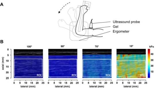
A generous amount of water-soluble transmission gel was used during scanning for optimal acoustic coupling, and minimal pressure was applied to the transducer to limit tissue deformation. The belly of the short head of the biceps brachii was identified during transverse scanning in B-mode at two-thirds of the distance between the acromion and antecubital fossa. Then the probe was rotated and care-fully aligned with the direction of muscle fascicles as recommended by Gennisson et al. (2010). Appropriate transducer alignment was achieved when several fas-cicles were continuously visible. A> 5-s delay was used before capturing all clips to obtain stabilized acquisition. During all measurements, participants were asked to keep their whole upper limb as relaxed as possible, and elastograms and B-mode images were carefully moni-tored. Clips were discarded if subtle movement and/or contraction was detected. Typical recordings for one patient are illustrated inFigure 1.
Post-processing of SWE data. Each frame of the 10-s clips was processed using a custom software developed in MATLAB (The MathWorks, Natick, MA, USA) (Dubois et al. 2015; Vergari et al. 2014). A rectangular region of interest (ROI) was manually defined on the first frame as large as possible between the superficial and the deep apo-neurosis in the muscle belly, and focal penetration defects or fibrous septa were carefully avoided. The ROI was tracked over other frames to evaluate the same region all over the measurement. MSM was computed as the mean of shear modulus values within whole ROIs. A normal-ized shear modulus was computed for each individual by dividing values at all tested joint angles by the value ob-tained at 100°.
Within- and between-day reliability
For all measurements, two clips were consecutively acquired after re-positioning the probe to assess
within-day reliability for all elbow flexion positions (100°, 90°, 70° and 10°). A sub-sample of 15 patients came on a second visit 1 week apart to evaluate between-day reliability of MSM measurements. To evaluate the between-day relia-bility, the first of the two measurements was used. Acute effect of muscle pre-conditioning and assessment of maximal voluntary strength
Passive muscle stiffness has been reported to be af-fected by the muscle’s previous history of length changes and contractions (Lacourpaille et al. 2014; Whitehead et al. 2001). Therefore, MSM at 100°, 90°, 70° and 10° elbow flexion was re-assessed immediately after five passive stretch-shortening cycles at 4°/s from 70° to 10° elbow angle using the Biodex ergometer in 19 patients. In 8 pa-tients, MSM was reassessed immediately after a set of voluntary contractions of the elbow flexors at 90° elbow flexion. Clips were acquired after probe re-positioning for both stretch-shortening cycles and voluntary contrac-tions. The set of contractions consisted of three increasing 5-s submaximal contractions followed by three 5-s maximal voluntary contractions with 1 min of rest between ma-neuvers. This set of voluntary contractions was performed in all subjects to assess maximal voluntary strength. Maximal peak torque over 500 ms was expressed as a per-centage of predicted value using previously published predictive equations (Harbo et al. 2012).
Quantification of muscle echo intensity and muscle thickness
To evaluate muscle echo intensity, a gray-scale index (GSI) ranging from 0 to 1 was defined as follows for B-mode
images (8-bit resolution, resulting in a number between 0 and 255, where black= 0 and white = 255) acquired transversally to muscle fascicles at 90° elbow flexion (2):
gray scale index− = −
(
⋅)
= ⋅
∑
1 1 255 1 n m i Ii n m (2)with Ii the intensity of the pixel i in a ROI of n× m. A
lower GSI value corresponds to greater muscle echo in-tensity (Dubois et al. 2018). As mentioned above, gain and all other ultrasound settings were kept constant for all sub-jects (Caresio et al. 2015). Thickness of the biceps brachii was measured with electronic calipers as previously de-scribed (Arts et al. 2010).
Statistical analysis
Data within text and tables are expressed as the mean± standard deviation (SD) or mean (95% confi-dence interval [lower 95% CI, upper 95% CI]). The assumptions of normality and sphericity were confirmed using the D’Agostino K-squared and Mauchly tests, re-spectively. To assess within- and between day reliability, change in mean and paired t-tests were used for detec-tion of systematic bias. Standard error of measurement (SEM) was used to study absolute reliability. Relative re-liability was assessed with the intra-class correlation coefficient (ICC) with 95% confidence interval. Regres-sion analysis and Bland–Altman plots were also performed. When available, data from the second visit were ana-lyzed jointly with data from the first visit to evaluate within-day reliability. A two-way analysis of variance (ANOVA,
0
Ultrasound probe Gel
Ergometer
Fig. 1. (a) Lateral view of the experimental setup. (b) Typical recordings of shear modulus measurements using shear wave elastography in the short head of the biceps brachii at 100°, 90°, 70° and 10° elbow joint angles in one patient with inclusion
trial× condition) were conducted to test main and inter-action effects on changes in MSM induced by passive conditioning. A one-way ANOVA was used to assess the effect of maximal voluntary contractions. Tukey’s honest significant difference (HSD) post hoc tests were con-ducted when a significant main and/or interaction effect was found. We hypothesized monotonic relationships between variables, but we did not assume that these re-lationships would be linear. Therefore, Spearman’s rank-order correlation coefficients (ρ) were computed to assess relationships between variables. All analyses were per-formed in the computing environment R Version 3.2.3. Statistical significance was set at p< 0.05 for all tests.
RESULTS
Within- and between-day reliability of muscle SWE values
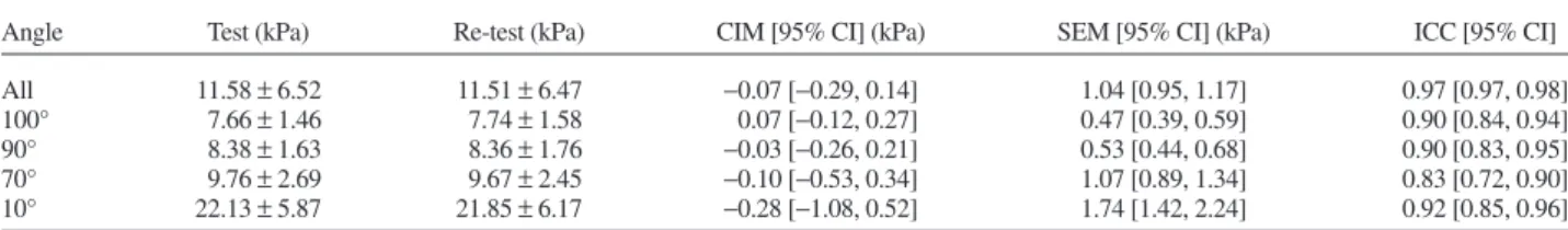
Within- and between-day reliability data of measure-ments performed at each tested angle are summarized in
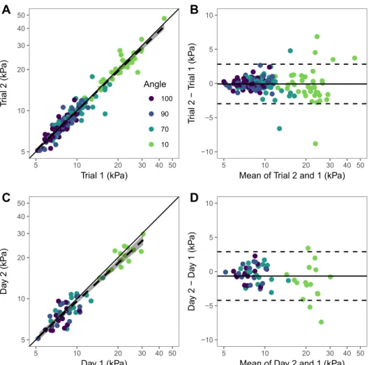
Tables 1 and 2, respectively. No systematic bias was de-tected for both within-day (all p> 0.44) and between-day (all p> 0.30) measurements. For within-day values, the ICC and SEM were>0.83 and <1.74 kPa, respective-ly. Regarding between-day values, the ICC and SEM were >0.64 and <1.89 kPa, respectively. Regression analysis and Bland–Altman plots are displayed inFigure 2. Lower and upper limits of agreements were−2.97 and 2.82 kPa for within-day measurements and −4.23 and 2.89 kPa for between-day measurements, respectively.
Effect of elbow joint angle and passive loading– unloading cycles on muscle shear modulus
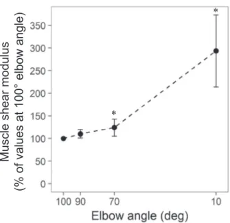
Normalized shear moduli at each tested angle during the first measurement are illustrated inFigure 3.
Normal-ized MSM at 10° was significantly greater than shear moduli at all other angle (all p values< 0.05). There were no significant differences between 100° and 90° (p= 0.77) or between 90° and 70° (p= 0.57).
Absolute values of muscle shear moduli at all elbow joint angles measured before and after passive loading/ unloading cycles are illustrated inFigure 4A. MSM at 10° was significantly greater than values at all other angles (p< 0.001). There were no significant differences between 100° and 90° (p= 0.96) or between 90° and 70° p = 0.97). No significant effect of trial or trial× angle interaction was found (p= 0.54 and p = 0.68, respectively).
Effect of maximal voluntary contractions on muscle shear modulus
Maximal elbow flexor isometric strength was 25.7± 11.1 N · m, corresponding to 70.1 ± 25.7% of pre-dicted values. Measurements of MSM at 90° before and after maximal voluntary contractions are illustrated in
Figure 4B. No significant effect of maximal voluntary con-tractions was found (p= 0.18).
Relationships between muscle shear modulus, muscle strength, muscle echo intensity and muscle thickness
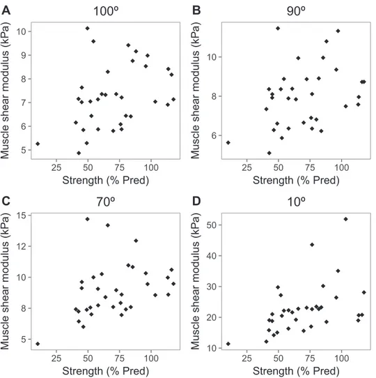
Muscle shear moduli at all angles significantly cor-related with muscle strength expressed as a percentage of predicted values (at 100°: ρ = 0.39, p < 0.05; at 90°: ρ = 0.36, p < 0.05; at 70°: ρ = 0.46, p < 0.01; at 10°: ρ = 0.53, p < 0.01). Individual data points are illustrated inFigure 5. No significant correlation was found between normalized MSM and muscle strength expressed as a per-centage of predicted values (at 90°:ρ = −0.03, p = 0.87; at 70°: ρ = 0.13, p = 0.47; at 10°: ρ = 0.32, p = 0.07). Muscle thickness was 1.37± 0.37 cm and correlated with Table 1. Within-day reliability of muscle shear modulus in patients with inclusion body myositis (n= 49)
Angle Test (kPa) Re-test (kPa) CIM [95% CI] (kPa) SEM [95% CI] (kPa) ICC [95% CI]
All 11.58± 6.52 11.51± 6.47 −0.07 [−0.29, 0.14] 1.04 [0.95, 1.17] 0.97 [0.97, 0.98]
100° 7.66± 1.46 7.74± 1.58 0.07 [−0.12, 0.27] 0.47 [0.39, 0.59] 0.90 [0.84, 0.94]
90° 8.38± 1.63 8.36± 1.76 −0.03 [−0.26, 0.21] 0.53 [0.44, 0.68] 0.90 [0.83, 0.95]
70° 9.76± 2.69 9.67± 2.45 −0.10 [−0.53, 0.34] 1.07 [0.89, 1.34] 0.83 [0.72, 0.90]
10° 22.13± 5.87 21.85± 6.17 −0.28 [−1.08, 0.52] 1.74 [1.42, 2.24] 0.92 [0.85, 0.96]
CIM= change in mean; SEM = standard error of measurement; ICC = intra-class correlation coefficient.
Table 2. Between-day reliability of muscle shear modulus in patients with inclusion body myositis
Angle Day 1 (kPa) Day 2 (kPa) CIM [95% CI] (kPa) SEM [95% CI] (kPa) ICC [95% CI]
All 12.07± 6.72 11.41± 6.09 −0.67 [−1.13, −0.20] 1.28 [1.09, 1.56] 0.96 [0.93, 0.97]
100 7.65± 1.43 7.32± 1.61 −0.33 [−100, 0.33] 0.85 [0.62, 1.34] 0.69 [0.30, 0.88]
90 8.11± 1.63 7.78± 1.41 −0.34 [−1.12, 0.45] 0.92 [0.66, 1.51] 0.64 [0.18, 0.87]
70 9.38± 2.26 9.10± 1.97 −0.28 [−0.97, 0.42] 0.96 [0.71, 1.46] 0.80 [0.54, 0.92]
10 22.30± 4.29 20.64± 3.87 −1.66 [−3.08, 0.24] 1.89 [1.39, 2.92] 0.73 [0.39, 0.89]
absolute strength (ρ = −0.43, p < 0.01). GSI was 0.70 ± 0.07 and correlated with predicted muscle strength (ρ = 0.46, p< 0.01). GSI also correlated with MSM at all angles except 10° (at 100°:ρ = 0.49, p < 0.05; at 90°: ρ = 0.54, p< 0.05; at 70°: ρ = 0.62, p < 0.01; at 10°: ρ = 0.17, p= 0.23).
DISCUSSION
The aim of the present study was to investigate the feasibility and relevance of muscle shear modulus assess-ment using ultrasound shear wave elastography in the biceps brachii of patients with IBM. Main results are as follows: (i) Within-day reliability of MSM measurements was sat-isfactory. (ii) Agreement of between-day MSM measurements was moderate. (iii) Passive loading/unloading cycles and maximal voluntary contractions did not
sig-nificantly affect MSM values. (4) Lower muscle stiffness was associated with more severe muscle weakness. Muscle stiffness and stretch-stiffening behavior
As repeatedly demonstrated using SWE (Lacourpaille et al. 2014) and conventional stress-strain tests (Eby et al. 2013), our data revealed an increase in MSM with in-creasing tensile load, illustrating the typical non-linear length–tension behavior of muscle tissue when sub-jected to tensile strain. Our data indicated substantial dispersion at the longest muscle length tested (i.e., 10° elbow angle) suggesting inter-individual differences in the passive force–length relationship (as estimated here using SWE) that may result from muscle remodeling and/or an-atomic variations (Fig. 2). In healthy subjects, the elbow joint angle corresponding to the slack length of the biceps brachii has been reported to occur at ~85°–95° (Lacourpaille 5 10 20 30 40 50 5 10 20 30 40 50 Trial 1 (kPa) T rial 2 (kP a) Angle 100 90 70 10 −10 −5 0 5 10 5 10 20 30 40 50
Mean of Trial 2 and 1 (kPa)
T rial 2 − T rial 1 (kP a) 5 10 20 30 40 50 5 10 20 30 40 50 Day 1 (kPa) Da y 2 (kP a) −10 −5 0 5 10 5 10 20 30 40 50
Mean of Day 2 and 1 (kPa)
Da
y 2 − Da
y 1 (kP
a)
Fig. 2. Regression analysis (a, c) and Bland–Altman plots (b, d) for within- and between day reliability of measurements. (a, b) Regression analysis and Bland–Altman plot for within-day measurements. (c, d) Regression analysis and Bland–Altman plot for between-day measurements. In (a) and (c), the solid line represents the identity line, and the dashed line indicates the linear regression line with shaded 95% confidence region. In (b) and (d), the solid line indicates the mean difference between
and Hug 2013; Lacourpaille et al. 2014). This may thus explain the absence of significant differences between MSM at 100° and 90° for both normalized and absolute MSM values. Consequently, it is reasonable to consider that mea-surements performed at 100° elbow joint angle reflects intrinsic shear modulus of the muscle tissue without passive tension.
Within-day and between-day reliability of muscle SWE values
Mean SEMs expressed as coefficients of variation were<8.6% and ICCs were >0.92, indicating satisfacto-ry agreement between within-day measurements of MSM. These data are in line with previous studies that reported similar results in biceps brachii in healthy subjects (i.e., ICC= 0.87 and SEM = 0.17 kPa (Lacourpaille et al. 2012)), patients with stroke (i.e., ICC= 0.93, coefficient of vari-ation= 4.5% (Lee et al. 2015)) and Duchenne muscular dystrophy (ICCs ranging from 0.67 to 0.80 (Brandenburg et al. 2015)). Prior to the current study, within- and between-day reliability of muscle mean shear modulus using an identical experimental setup were assessed in 12 un-matched healthy subjects. The ICC and SEM were 0.98 and 0.90 kPa, respectively (unpublished data). In IBM pa-tients, In the current work, within-day reliability was comparable between all tested angles; that is, when ex-pressed as coefficients of variation, SEMs were 6.0%, 6.4%, 8.6% and 7.2% at 100°, 90°, 70° and 10° elbow angles, respectively. Collectively, these results support that SWE may be used to assess acute changes in passive muscle stiff-ness induced by various factors (i.e., stretching, passive loading/unloading cycles, maximal voluntary contrac-tions) and to investigate relationships between variables as performed in the present study.
Our data indicated moderate agreement on between-day MSM measurements as indicated by ICCs<0.80 and substantial SEMs (i.e., SEMs expressed as coefficients of variation were 10.4, 10.6, 9.8 and 7.8% at 100°, 90°, 70° and 10°, respectively). There are several potential expla-nations for these findings. In the present work, probe Fig. 3. Muscle shear modulus at different muscle lengths. Muscle
shear modulus assessed with shear wave elastography in the short head of the biceps brachii at 100°, 90°, 70° and 10° elbow joint angles (n= 33). Values are normalized to values at 100°. *Significantly
dif-ferent from value at 100°.
Fig. 4. Effect of passive loading–unloading and maximal voluntary contractions on muscle shear modulus. Muscle shear modulus was assessed with shear wave elastography in the short head of the biceps brachii at elbow joint angles of 100°, 90°, 70° and 10° before (Pre 1, Pre 2) and after (Post) five passive stretch-shortening cycles at 4°/s from 70° to 10° elbow angle (n= 19) (A) or three maximal voluntary isometric contractions of the elbow flexors (n= 8) (B). *Significantly different from value at
positioning was based on anatomical landmarks and the skin was deliberately not marked between days to fit a clin-ical setting. Furthermore, patients studied in the present work exhibited severe muscle atrophy and heteroge-neously distributed degenerative changes (i.e., fatty infiltration, fibrosis, disrupted fascicles (Cox et al. 2011)) so that small positioning changes may result in more dra-matic variations in MSM in comparison to healthy muscle. Probe pressure is also a critical factor (Kot et al. 2012). Experimental apparatus (e.g., probe mounting jig, immer-sion of the probe in a silicon pool) have been proposed to bypass the confounding effect of probe pressure (Andonian et al. 2016; Koo et al. 2014). However, such experimental procedures question the feasibility of high-quality muscle SWE measurements within a clinical setting. The alignment of the probe with the direction of the fas-cicles is also critical factor (Gennisson et al. 2010;Maisetti
et al. 2012). In a recent work,Miyamoto et al. (2015) re-ported a significant effect of the probe angle relative to the fascicle on the shear modulus in the biceps brachii. Although the difference was small (<1.3% of the mea-sured values), for instance, as compared with the inter-observer reliability (around 9% according toDubois et al. 2015), this may contribute to explaining the variability of the measurements observed in the current study.
Another important factor to consider is that exer-cise, particularly eccentric bout, may cause a long-lasting increase in muscle stiffness (Green et al. 2012; Lacourpaille et al. 2014). High relative intensity of muscle contraction level during daily-life activities because of weakness associated with high sensitivity to muscle damage may thus lead to substantial fluctuations of muscle stiff-ness in patients. These exercise-induced increases in muscle stiffness have also been reported to be more prominent at
5 6 7 8 9 10 25 50 75 100 Strength (% Pred)
Muscle shear modulus (kP
a)
100º
6 8 10 25 50 75 100 Strength (% Pred)Muscle shear modulus (kP
a)
90º
5 8 10 12 15 25 50 75 100 Strength (% Pred)Muscle shear modulus (kP
a)
70º
10 20 30 40 50 25 50 75 100 Strength (% Pred)Muscle shear modulus (kP
a)
10º
Fig. 5. Relationship between muscle shear modulus at different joint angles and maximal voluntary strengths expressed as a percentage of predicted value in patients with inclusion body myositis. Muscle shear modulus was assessed with shear wave elastography in the short head of the biceps brachii at elbow joint angles of 100° (A), 90° (B), 70° (C) and 10° (D) (n= 34). % Pred= percentage of predicted value. A moderate correlation was found between predicted muscle strength and biceps brachii
longer muscle length, mainly because sensitivity to Ca2+ increases as muscle is elongated (Stephenson and Williams 1982). Although between-day variability of MSM was not particularly greater at longer muscle length, this may con-tribute to explaining the limited agreement of MSM measurements in patients with IBM. Patient relaxation is also a critical aspect that can substantially reduce the reliability of measurements and may also vary between days.
Effect of passive loading/passive cycles and maximal voluntary muscle activation on muscle SWE values
Our data indicated no significant effect of the passive conditioning on MSM measurements. In a recent work in-vestigating the time course effect of exercise-induced damage on muscle stiffness,Lacourpaille et al. (2014) re-ported no effect of passive loading/unloading cycles on the area under the loading curve before exercise. Con-versely, 48 h after intense exercise, significant changes were seen for the first cycle only. In the current work, repeat-ed maximal voluntary activation did not lead to significant acute changes in muscle stiffness. As mentioned above, intense exercise may lead to substantial changes in passive mechanical muscle properties (Green et al. 2012; Lacourpaille et al. 2014). These effects are observed mainly at muscle length greater than the slack length and appear to rely on calcium-dependent processes (Chen et al. 2007). Slack elasticity measurements may therefore be more rel-evant in characterizing the local elasticity of the muscle tissue while limiting the confounding effects of this phe-nomenon as well as those related to the inter-individual differences in the passive muscle length–tension relationship.
Relationships between muscle shear modulus, muscle strength, muscle echo intensity and muscle thickness
As expected and as reported previously, smaller muscle thickness was associated with smaller muscle strength observed, emphasizing the strong relationship between muscle size and muscle strength (Akagi et al. 2018; Jansen et al. 2012; Strasser et al. 2013). In line with previous work in neuromuscular disorders or in the elderly, greater muscle echogenicity was associated with more severe muscle weakness (Fukumoto et al. 2012; Jansen et al. 2012; Zaidman et al. 2010). Interestingly, our data also revealed moderate positive correlations between predicted muscle strength and muscle shear moduli at both slack and stretched lengths (Fig. 5). These find-ings are in line with a previous work that reported lower muscle stiffness in both upper- and lower-limb muscles of patients assessed with acoustic radiation force impulse ultrasound elastography (Botar Jid et al. 2012). Further-more, significant correlations were found between MSM and echo intensity. Fibrous and particularly adipose tissue
content within muscle has been reported as key explan-atory factors of increased muscle echo intensity (Caresio et al. 2015; Reimers et al. 1993). Fatty infiltration has been reported to be a prominent feature of muscle alter-ation in IBM as assessed with nuclear magnetic resonance imaging (Cox et al. 2011; Morrow et al. 2016). Together, these data suggest that reduce lower muscle stiffness might be related to greater structural muscle impair-ments, that is, . greater muscle fat content. Therefore, further investigations allowing the comparison of muscle mechanical properties and muscle degenerative pro-cesses assessed with nuclear magnetic resonance imaging/ spectroscopy must be conducted to clarify these findings and the clinical relevance of muscle SWE for diagnosis and follow-up.
Limitations
The absence of data for healthy controls in the present work limits the interpretation of findings. Post-processing of SWE data is an important factor for the reliability of measurements. In the present work, we used an ad-vanced method to post-process SWE clips (Vergari et al. 2014). However, minor changes (size, location) when de-fining the ROIs may have occurred between days. Mechanical anisotropy is also a critical factor in the char-acterization of mechanical tissue properties (Green et al. 2013; Virgilio et al. 2015) and has been reported to be altered in muscle degeneration (Qin et al. 2014). Mechan-ical anisotropy estimation using SWE (Chino et al. 2017) should therefore be assessed in future studies. In addi-tion, the dispersion of the shear waves may be studied from deeper exploitation of local tissue velocity maps ac-quired using SWE, allowing the quantification of muscle viscosity (Deffieux et al. 2009; Gennisson et al. 2010). The measurement of these complex viscoelastic and anisotro-pic mechanical properties will provide additional opportunities to characterize the diseased muscle.
CONCLUSIONS
Muscle SWE shows promise for the investigation of muscle degeneration in patients with muscle disorders such as those observed in IBM. Significant challenges remain to improve the applicability of muscle SWE in the se-verely disordered muscle and to clarify the complex interplay between biomechanical, structural and function-al muscle changes in IBM and in a broader scope of muscle diseases. Further investigations will be aimed at compar-ing muscle mechanical properties (i.e., shear modulus, shear viscosity and anisotropy) obtained using SWE and muscle changes quantified using nuclear magnetic resonance imaging/spectroscopy (e.g., fatty infiltration, inflamma-tion) in these populations.
Acknowledgments—We gratefully thank all the patients who partici-pated in this study.
This study was supported by the Association Française contre les My-opathies (AFM).
REFERENCES
Akagi R, Suzuki M, Kawaguchi E, Miyamoto N, Yamada Y, Ema R. Muscle size–strength relationship including ultrasonographic echo intensity and voluntary activation level of a muscle group. Arch Gerontol Geriatr 2018;75:185–190.
Andonian P, Viallon M, Le Goff C, de Bourguignon C, Tourel C, Morel J, Giardini G, Gergele L, Millet GP, Croisille P. Shear-wave elastography assessments of quadriceps stiffness changes prior to, during and after prolonged exercise: A longitudinal study during an extreme mountain ultra-Marathon. PLoS ONE 2016;11:e0161855. Arts IM, Pillen S, Schelhaas HJ, Overeem S, Zwarts MJ. Normal values for quantitative muscle ultrasonography in adults. Muscle Nerve 2010; 41:32–41.
Bavu E, Gennisson JL, Couade M, Bercoff J, Mallet V, Fink M, Badel A, Vallet-Pichard A, Nalpas B, Tanter M, Pol S. Noninvasive in vivo liver fibrosis evaluation using supersonic shear imaging: A clinical study on 113 hepatitis C virus patients. Ultrasound Med Biol 2011; 37:1361–1373.
Benveniste O, Hilton-Jones D. International Workshop on Inclusion Body Myositis held at the Institute of Myology, Paris, on 29 2009. Neuromuscul Disord 2010;20:414–421.
Benveniste O, Guiguet M, Freebody J, Dubourg O, Squier W, Maisonobe T, Stojkovic T, Leite MI, Allenbach Y, Herson S, Brady S, Eymard B, Hilton-Jones D. Long-term observational study of sporadic in-clusion body myositis. Brain 2011;134:3176–3184.
Bercoff J, Tanter M, Fink M. Supersonic shear imaging: A new tech-nique for soft tissue elasticity mapping. IEEE Trans Ultrason Ferroelectr Freq Control 2004;51:396–409.
Bilston LE, Tan K. Measurement of passive skeletal muscle mechani-cal properties in vivo: recent progress, clinimechani-cal applications, and remaining challenges. Ann Biomed Eng 2015;43:261–273. Botar Jid C, Vasilescu D, Damian L, Dumitriu D, Ciurea A, Dudea SM.
Musculoskeletal sonoelastography: Pictorial essay. Med Ultrason 2012; 14:239–245.
Brandenburg JE, Eby SF, Song P, Zhao H, Brault JS, Chen S, An KN. Ultrasound elastography: The new frontier in direct measurement of muscle stiffness. Arch Phys Med Rehabil 2014;95:2207–2219. Brandenburg JE, Eby SF, Song P, Zhao H, Landry BW, Kingsley-Berg
S, Bamlet WR, Chen S, Sieck GC, An KN. Feasibility and reliabil-ity of quantifying passive muscle stiffness in young children by using shear wave ultrasound elastography. J Ultrasound Med 2015;34: 663–670.
Brandenburg JE, Eby SF, Song P, Kingsley-Berg S, Bamlet W, Sieck GC, An KN. Quantifying passive muscle stiffness in children with and without cerebral palsy using ultrasound shear wave elastography. Dev Med Child Neurol 2016;58:1288–1289.
Caresio C, Molinari F, Emanuel G, Minetto MA. Muscle echo intensity: Reliability and conditioning factors. Clin Physiol Funct Imaging 2015; 35:393–403.
Chen W, Ruell PA, Ghoddusi M, Kee A, Hardeman EC, Hoffman KM, Thompson MW. Ultrastructural changes and sarcoplasmic reticu-lum Ca2+regulation in red vastus muscle following eccentric exercise in the rat. Exp Physiol 2007;92:437–447.
Chino K, Kawakami Y, Takahashi H. Tissue elasticity of in vivo skel-etal muscles measured in the transverse and longitudinal planes using shear wave elastography. Clin Physiol Funct Imaging 2017;37:394– 399.
Cox FM, Reijnierse M, van Rijswijk CS, Wintzen AR, Verschuuren JJ, Badrising UA. Magnetic resonance imaging of skeletal muscles in sporadic inclusion body myositis. Rheumatology (Oxford) 2011;50: 1153–1161.
Deffieux T, Montaldo G, Tanter M, Fink M. Shear wave spectroscopy for in vivo quantification of human soft tissues visco-elasticity. IEEE Trans Med Imaging 2009;28:313–322.
Deffieux T, Gennisson JL, Bousquet L, Corouge M, Cosconea S, Amroun D, Tripon S, Terris B, Mallet V, Sogni P, Tanter M, Pol S. Investi-gating liver stiffness and viscosity for fibrosis, steatosis and activity staging using shear wave elastography. J Hepatol 2015;62:317– 324.
Dubois G, Kheireddine W, Vergari C, Bonneau D, Thoreux P, Rouch P, Tanter M, Gennisson JL, Skalli W. Reliable protocol for shear wave elastography of lower limb muscles at rest and during passive stretching. Ultrasound Med Biol 2015;41:2284–2291.
Dubois GJR, Bachasson D, Lacourpaille L, Benveniste O, Hogrel JY. Local texture anisotropy as an estimate of muscle quality in ultra-sound imaging. Ultraultra-sound Med Biol 2018;44:1133–1140. Eby SF, Song P, Chen S, Chen Q, Greenleaf JF, An KN. Validation of
shear wave elastography in skeletal muscle. J Biomech 2013;46: 2381–2387.
Fukumoto Y, Ikezoe T, Yamada Y, Tsukagoshi R, Nakamura M, Mori N, Kimura M, Ichihashi N. Skeletal muscle quality assessed from echo intensity is associated with muscle strength of middle-aged and elderly persons. Eur J Appl Physiol 2012;112:1519–1525. Gennisson JL, Deffieux T, Mace E, Montaldo G, Fink M, Tanter M.
Vis-coelastic and anisotropic mechanical properties of in vivo muscle tissue assessed by supersonic shear imaging. Ultrasound Med Biol 2010;36:789–801.
Gennisson JL, Deffieux T, Fink M, Tanter M. Ultrasound elastography: Principles and techniques. Diagn Interv Imaging 2013;94:487– 495.
Green MA, Sinkus R, Gandevia SC, Herbert RD, Bilston LE. Measur-ing changes in muscle stiffness after eccentric exercise usMeasur-ing elastography. NMR Biomed 2012;25:852–858.
Green MA, Geng G, Qin E, Sinkus R, Gandevia SC, Bilston LE. Mea-suring anisotropic muscle stiffness properties using elastography. NMR Biomed 2013;26:1387–1394.
Harbo T, Brincks J, Andersen H. Maximal isokinetic and isometric muscle strength of major muscle groups related to age, body mass, height, and sex in 178 healthy subjects. Eur J Appl Physiol 2012;112:267– 275.
Jansen M, van Alfen N, Nijhuis van der Sanden MW, van Dijk JP, Pillen S, de Groot IJ. Quantitative muscle ultrasound is a promising lon-gitudinal follow-up tool in Duchenne muscular dystrophy. Neuromuscul Disord 2012;22:306–317.
Koo TK, Guo JY, Cohen JH, Parker KJ. Quantifying the passive stretch-ing response of human tibialis anterior muscle usstretch-ing shear wave elastography. Clin Biomech (Bristol, Avon) 2014;29:33–39. Kot BC, Zhang ZJ, Lee AW, Leung VY, Fu SN. Elastic modulus of
muscle and tendon with shear wave ultrasound elastography: Varia-tions with different technical settings. PLoS ONE 2012;7:e44348. Lacourpaille L, Hug F. Nordez A. Influence of passive muscle tension on electromechanical delay in humans. PLoS ONE 2013;8:e53159. Lacourpaille L, Hug F, Bouillard K, Hogrel JY Nordez A. Supersonic shear imaging provides a reliable measurement of resting muscle shear elastic modulus. Physiol Meas 2012;33:N19–N28.
Lacourpaille L, Nordez A, Hug F, Couturier A, Dibie C, Guilhem G. Time-course effect of exercise-induced muscle damage on local-ized muscle mechanical properties assessed using elastography. Acta Physiol (Oxf) 2014;211:135–146.
Lacourpaille L, Hug F, Guevel A, Pereon Y, Magot A, Hogrel JY Nordez A. Non-invasive assessment of muscle stiffness in patients with Duchenne muscular dystrophy. Muscle Nerve 2015;51:284–286. Lee SS, Spear S, Rymer WZ. Quantifying changes in material
proper-ties of stroke-impaired muscle. Clin Biomech (Bristol, Avon) 2015; 30:269–275.
Lieber RL, Ward SR. Skeletal muscle design to meet functional demands. Philos Trans R Soc Lond B Biol Sci 2011;366:1466–1476. Maisetti O, Hug F, Bouillard K, Nordez A. Characterization of passive
elastic properties of the human medial gastrocnemius muscle belly using supersonic shear imaging. J Biomech 2012;45:978–984. Miyamoto N, Hirata K, Kanehisa H, Yoshitake Y. Validity of
measure-ment of shear modulus by ultrasound shear wave elastography in human pennate muscle. PLoS One 2015;10:e0124311.
Morrow JM, Sinclair CD, Fischmann A, Machado PM, Reilly MM, Yousry TA, Thornton JS, Hanna MG. MRI biomarker assessment
of neuromuscular disease progression: A prospective observational cohort study. Lancet Neurol 2016;15:65–77.
Qin EC, Juge L, Lambert SA, Paradis V, Sinkus R, Bilston LE. In vivo anisotropic mechanical properties of dystrophic skeletal muscles measured by anisotropic MR elastographic imaging: The mdx mouse model of muscular dystrophy. Radiology 2014;273:726– 735.
Reimers K, Reimers CD, Wagner S, Paetzke I, Pongratz DE. Skeletal muscle sonography: A correlative study of echogenicity and mor-phology. J Ultrasound Med 1993;12:73–77.
Stephenson DG, Williams DA. Effects of sarcomere length on the force– pCa relation in fast- and slow-twitch skinned muscle fibres from the rat. J Physiol 1982;333:637–653.
Strasser EM, Draskovits T, Praschak M, Quittan M, Graf A. Associa-tion between ultrasound measurements of muscle thickness, pennaAssocia-tion angle, echogenicity and skeletal muscle strength in the elderly. Age (Dordr) 2013;35:2377–2388.
Vergari C, Rouch P, Dubois G, Bonneau D, Dubousset J, Tanter M, Gennisson JL, Skalli W. Non-invasive biomechanical characteriza-tion of intervertebral discs by shear wave ultrasound elastography: A feasibility study. Eur Radiol 2014;24:3210–3216.
Virgilio KM, Martin KS, Peirce SM, Blemker SS. Multiscale models of skeletal muscle reveal the complex effects of muscular dystro-phy on tissue mechanics and damage susceptibility. Interface Focus 2015;5:20140080.
Whitehead NP, Gregory JE, Morgan DL, Proske U. Passive mechani-cal properties of the medial gastrocnemius muscle of the cat. J Physiol 2001;536:893–903.
Wisdom KM, Delp SL, Kuhl E. Use it or lose it: Multiscale skeletal muscle adaptation to mechanical stimuli. Biomech Model Mechanobiol 2015; 14:195–215.
Zaidman CM, Connolly AM, Malkus EC, Florence JM, Pestronk A. Quan-titative ultrasound using backscatter analysis in Duchenne and Becker muscular dystrophy. Neuromuscul Disord 2010;20:805–809.