DOCUNENT ROOM,iU NMT ROOM 36-41. RESEARCH LABORATORY OF ELECTONICS NASSACHUSETS INSTITUTE OF TECHNOLOGY CAMBRIDGE 39, MASSACHUSETTS, U S.A.
AN ANALYTICAL STUDY OF ELECTRIC RESPONSES
AT THE PERIPHERY OF THE AUDITORY SYSTEM
WILLIAM T. PEAKE
TECHNICAL REPORT 365 MARCH 17, 1960
/
/
.
/
MASSACHUSETTS INSTITUTE OF TECHNOLOGY
RESEARCH LABORATORY OF ELECTRONICSCAMBRIDGE, MASSACHUSETTS
i5)i
/IiL-The Research Laboratory of Electronics is an interdepartmental laboratory of the Department of Electrical Engineering and the Department of Physics.
The research reported in this document was made possible in part by support extended the Massachusetts Institute of Technology, Research Laboratory of Electronics, jointly by the U. S. Army (Sig-nal Corps), the U.S. Navy (Office of Naval Research), and the U.S. Air Force (Office of Scientific Research, Air Research and Develop-ment Command), under Signal Corps Contract DA36-039-sc-78108, Department of the Army Task 3-99-20-001 and Project 3-99-00-000.
J
MASSACHUSETTS INSTITUTE OF TECHNOLOGY
RESEARCH LABORATORY OF ELECTRONICS
Technical Report 365 March 17, 1960
AN ANALYTICAL STUDY OF ELECTRIC RESPONSES AT THE PERIPHERY OF THE AUDITORY SYSTEM
William T. Peake
Submitted to the Department of Electrical Engineering, M.I.T., January 11, 1960, in partial fulfillment of the requirements for the degree of Doctor of Science.
Abstract
Acoustic stimuli, especially if they are impulsive in nature, give rise to well-defined neuroelectric events in the auditory nerve of many animal species. In this research, the summated action potentials of the cat's auditory nerve were recorded with gross electrodes from locations in the vicinity of the cochlea. This report is primarily concerned with the analytic study of the behavior of these neural potentials in relation to changes in stimulus parameters.
In order to investigate possible relations between the neural and the cochlear microphonic potentials, electrical activity was recorded in cats whose auditory nerve had degenerated. Characteristics of the microphonic response to clicks were deter-mined. A "slow" potential that does not reverse with stimulus polarity as the micro-phonic does was discovered.
Neural responses to condensation and rarefaction clicks were observed over a wide range of stimulus intensity; it was found that the differences between the responses to the two-click polarities depend on the intensity. These differences can be interpreted in terms of two excitatory processes, one of which can be related to the microphonic potential, and the other seems to be related to the "slow" potential.
Neural responses to impulsive stimuli were studied as a function of stimulus repe-tition rate. For moderate intensities, the amplitude of the neural response begins to decrease for rates higher than 10/sec. Stimulus-locked neural activity can be detected in averaged responses up to rates of nearly 3000/sec. The effect of overlapping of response waveforms is described in terms of a mathematical model.
TABLE OF CONTENTS
I. Introduction 1
1.1 General Plan of this Study 1
1. 2 General Anatomy and Physiology of the Auditory System 1
1.3 Anatomy of the Inner Ear 3
1.4 Mechanical Properties of the Inner Ear 3
1.5 Electrophysiology of the Cochlea 4
a. Endocochlear Potential 4
b. Cochlear Microphonic Potential 5
c. Summating Potential 6
1.6 Auditory Nerve Potentials 8
1.7 Experimental Procedures 11
II. Electric Responses from Denervated Cochleas 13
2.1 Special Technique 13
2.2 Impulse Response 14
2.3 Changes with Stimulus Intensity 17
2.4 Reversal of Response with Stimulus Polarity Change:
The Slow Potential 17
2.5 Responses to Noise Bursts 20
III. Changes in the Auditory-Nerve Responses as a Function of
Stimulus Intensity 23
3. 1 Introduction 23
3.2 Results 23
3.3 Interpretation 26
3.4 Discussion 30
IV. Changes in Auditory-Nerve Responses as a Function of
Stimulus Repetition Rate 32
4. 1 Introduction 32
4.2 Techniques 32
4.3 Results 37
a. Neural Responses at the Onset of Stimulation 37
b. Neural Responses in the Steady State 39
c. Effect of Anesthesia 41
d. Effect of Burst Length and Burst Pattern on
Neural Responses 43
e. Changes in Microphonic Potential with Rate 46
4.4 A Model Dealing with Overlapping of Responses 48
4.5 Discussion 54
V. Concluding Remarks 57
Acknowledgment 58
Bibliography 59
I. INTRODUCTION
1.1 GENERAL PLAN OF THIS STUDY
In studying a communication system, one can take the point of view that he is inter-ested only in input signals and output signals and the relations between them. This "black-box" approach may be fruitful in the study of systems in which the transfer func-tion can be simply defined in mathematical terms. But often it is not advantageous when it is applied to biological communication systems. For most of these systems the stimulus-response relations are so complex that it is difficult to determine appropriate mathematical descriptions that apply over a wide range of conditions. In studying such a system, it is desirable to maintain close connection between the description of input-output relations and the physiological processes involved, so that knowledge of one can help to organize the study of the other. In the work described here, we are concerned with both input-output relations and physiological mechanisms.
In Section IV a mathematical model has been developed which merely describes changes in response waveforms. However, the usefulness of the model lies not only in this description, but also in its implications concerning the physiological processes. In Section III, on the other hand, a model of a physiological process is proposed that leads to an interpretation of some observed input-output relations.
The research reported here deals with the periphery of the auditory system. Input signals are acoustic stimuli; output signals are summated action potentials of the audi-tory nerve recorded with gross electrodes. Sections III and IV present the results of studies of the relationship of the neural output to the input for changes in two stimulus parameters. Emphasis is put upon the study of impulsive stimuli. In Section II, input-output relations are studied for electrical responses of the cochlea that are not neural. The results of Section II assist us in interpreting the results of Sections III and IV. Section I contains: (a) descriptions of those features of the anatomy and physiology of the ear that are pertinent to signal-transmission processes; (b) a discussion of results and interpretations of previous studies of electrophysiological responses; and (c) a description of the experimental procedures employed.
1.2 GENERAL ANATOMY AND PHYSIOLOGY OF THE AUDITORY SYSTEM If the auditory system is thought of as a communication channel, the signal-transmission path can be quite well defined at the input end of the system (see Fig. 1). The acoustic signal produces a vibration of the ear drum, which is transmitted by the
small bones of the middle ear to the oval window of the cochlea. At this point the vibra-tion is transduced into a pressure variavibra-tion in the cochlear fluid, which bends the
cochlear partition near the oval window. This disturbance is then propagated along the cochlea in a wave motion on the cochlear partition. This motion, somehow, leads to the excitation of the nerve endings in the organ of Corti. The neural signals are then
Perilymph,
Scala
ExteHelicotrema
Scala tympani
tubeFig. 1. Schematic drawing of the ear. (From von Bekesy and Rosenblith (11).)
SCALA VESTIBUL
avow_%-!
IoNlE
/,. I
Ole VASCUL LaS
;e
S~~CALA,,:WAAHAIR CELLS
rtouas KITIFtuAL TECTONAL JEOLmt
bITES CELLS RODS' AN TUNNEL
or COTI SPIAL LGAMENT:
SPIRAL CANGLON
(Ptrft.P)
Fig. 2. Cross-section drawing Davis and Associates (1
of second turn of guinea pig cochlea.
2 (From ----* I I 7 -.. .. I ·.
propagated along the nerve fibers of the auditory nerve into the medulla. From that
point onward, the flow of the neural signals is not so well defined. Certain pathways
have been identified, but they will not be discussed here because we are primarily inter-ested in the peripheral part of the system. A fairly recent review discusses the "higher"
centers of the auditory system (30). The signal transmission through the peripheral
part of the auditory system may be affected by higher nervous centers. Two different
processes have been studied: (a) Neural signals can produce changes in the tension of the muscles of the middle ear which alter the signal transmission through the middle
ear (34, 79). (b) Efferent neuronal pathways may affect the transmission in the inner
ear (31, 61). There also is some evidence that auditory nerve responses can be altered
by stimulation of other sense modalities (60). 1.3 ANATOMY OF THE INNER EAR
The anatomy of the cochlea is rather well known. Figure 2 shows the structure of
the cochlea in a cross section of one turn. The paths followed by the nerve endings to
the hair cells have been determined in some detail (25), and the size and number of
fibers in the nerve have been studied (52, 29). The dimensions of the cochlea have been
determined accurately (26).
1.4 MECHANICAL PROPERTIES OF THE INNER EAR
Our knowledge of the mechanical properties of the cochlear membranes, and of the way in which they respond to various stimuli, results almost entirely from a long series
of experiments performed by von Be/kesy (10, 11). Stated briefly, the mechanics of the
cochlea is as follows: Vibration of the stapes produces a pressure difference in the
cochlear fluid and a resulting vibration of the cochlear partition. The vibration can be
described as a traveling wave moving from base to apex. For sinusoidal vibration of
the stapes, the partition has a maximum amplitude of vibration at one position. The
position depends on frequency, moving from the apex toward the base as the frequency
is increased. The tuning is not sharp, however. Low-frequency sinusoids produce a
point of maximum vibration near the apex, but the whole basilar membrane vibrates,
and the change in amplitude with position is very gradual. High-frequency tones produce
a maximum amplitude of vibration near the stapes, and the amplitude decreases rather
rapidly to zero on the apical side of the maximum. Hence, the tuning curves (amplitude
versus frequency) for a given point along the cochlea drop off faster above the peak fre-quency than below it.
This picture of the mechanical behavior of the cochlea is greatly simplified. Some
of the complications that have been observed are: (a) the wavelength for the traveling wave is not uniform along the cochlea and depends on the stimulus intensity at high inten-sity (1); and (b) if the motion of the structures along the organ of Corti is observed during sinusoidal stimulation, the direction of motion changes from one position to another (8).
1.5 ELECTROPHYSIOLOGY OF THE COCHLEA
The physiology of the cochlea has been studied extensively by recording electric potentials from electrodes placed in or near the cochlea. These potentials have been classified in four classes by Davis (14): the endocochlear potential, the cochlear micro-phonic potential, the summating potential, and the action potentials of the auditory nerve. It is this neural response with which this report is primarily concerned. It is the signal that carries "information" about the acoustic stimulus into the central nervous system. The other potentials are thought to play a role in the excitation of the action potentials, but their significance in the signal-transmission process is not completely determined. Since these other potentials will be referred to later, we shall discuss them now in some detail.
a. Endocochlear Potential
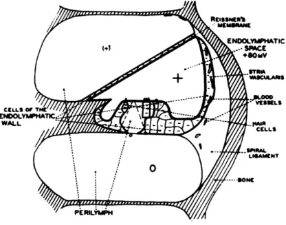
If potential differences are measured between various points in the cochlea, in the absence of acoustic stimuli, various dc potential differences are observed (6, 73). The potential within each scala is nearly constant, but a voltage of 70-90 mv can be measured between the scala media and either the scala tympani or the scala vestibuli. The region within which this positive potential exists is shown in Fig. 3. Three studies, in each of which a different method was used, have indicated that the stria vascularis plays an important role in the production and maintenance of this dc potential (18, 56, 76). The functional significance of this dc potential is a matter of speculation. Under some
Fig. 3. Diagram of the distribution of the dc potentials: intracellular, negative; endocochlear, positive. Reference potential is perilymph of scala tym-pani. (From Tasaki, Davis, and Eldredge (73).)
conditions it appears to change in parallel with the microphonic potential (7), and to be
distributed in space similarly to the microphonic potential (73). The endocochlear
potential can also be increased or decreased by a steady displacement of the basilar membrane (6, 73). These observations have led to the suggestion that the endocochlear potential represents a source which is amplitude-modulated to produce the cochlear
microphonic potential (6, 7, 14). However, Davis has also stated that "the D-C
endo-cochlear potential is not necessary for the generation of CM. (It may, however, serve
to make the CM response larger or more sensitive.)" (18). It has also been shown that
the microphonic can be small or absent when the dc potential is normal (18, 76).
b. Cochlear Microphonic Potential
The electric response that has been studied most extensively is called by Davis (14)
the cochlear microphonic (CM). The most striking characteristic of CM is its apparent
linear dependence on the acoustic stimulus over a considerable range. For sinusoidal
stimuli, CM is sinusoidal and its amplitude varies linearly with the amplitude of the stimulus up to moderate intensities (74).
The source of the cochlear microphonic is thought to lie in the organ of Corti, prob-ably in the hair cells. The evidence for this is the observation that the polarity of the microphonic reverses when the electrode is moved through the organ of Corti (7, 73). It has also been shown that in cases in which the organ of Corti is damaged or missing,
CM is reduced or missing (18, 76). The difference of polarity of CM in different
loca-tions has been used to produce records of auditory responses in which either the neural or microphonic component is emphasized (74).
It has been shown that the cochlear microphonic recorded by this technique is
pro-duced by small sections of the organ of Corti. The response recorded at one site is not
altered when the response at another position is varied by obstructing the motion or by addition of chemicals (75). It has also been demonstrated (74) that the responses recorded
from different turns of the cochlea may have quite different waveforms. By moving the
basilar membrane with a fine vibrating needle, von Bekesy showed that CM is
propor-tional to the displacement of the membrane (4). Also, the variation of the microphonic
with position along the cochlea has been observed to be covariant with the amplitude of
vibration (74). When the stimulus intensity is increased sufficiently, the amplitude of
CM increases more and more slowly until it levels off and even decreases with
increasing intensity (74). An interesting feature of the departure from linearity is that
the waveform of the response to sinusoidal stimulation remains nearly sinusoidal even in the nonlinear region for medium and high frequencies; some clipping, which has been ascribed to nonlinear transmission in the middle ear (14), can be observed at low fre-quencies.
When von Bekesy (9) moved the organ of Corti with a vibrating electrode, he found that the greatest microphonic potential was produced by radial vibrations when the elec-trode was near the outer hair cells, and by longitudinal vibration when the elecelec-trode was
5
-near the inner hair cells. (In both forms of vibration the electrode moves' parallel to the basilar membrane: in the longitudinal mode it moves along the direction from oval window to helicotrema, and in the radial mode it moves perpendicularly to this direction,
that is, transversely.) Von Bekesy's observations (8, 9) suggest that, for sinusoidal
stimuli, the inner and outer hair cells produce microphonics in response to different directions of motion at different positions along the cochlea.
Von Bekesy has shown that the power dissipated by the microphonic potential is greater than that delivered by the acoustic stimulus (5). Hence, it seems that the micro-phonic potential represents the output of a "biological power amplifier" rather than just a transduction of the acoustic energy into electric energy. Possibly the dc endocochlear potential represents the source that is modulated by the acoustic signal (6).
Although the role of the microphonic in the signal-transmission process is not defi-nitely established, it is often considered to act as a direct electrical stimulus on the
nerve endings (14). It has also been suggested that some sensory nerve endings are not
sensitive to electrical stimulation (39) and that, therefore, chemical excitation seems more reasonable (40).
c. Summating Potential
Less is known about the summating potential (SP) than about the endocochlear or the
cochlear microphonic (CM) potentials. It was first described (20) in 1950, but it proved
difficult to give a consistent description of its behavior. Recently, in several papers,
Davis and his coworkers suggest a possible mechanism for the production of SP, and assign to it a role in the excitation of the nerve endings (15, 17, 18).
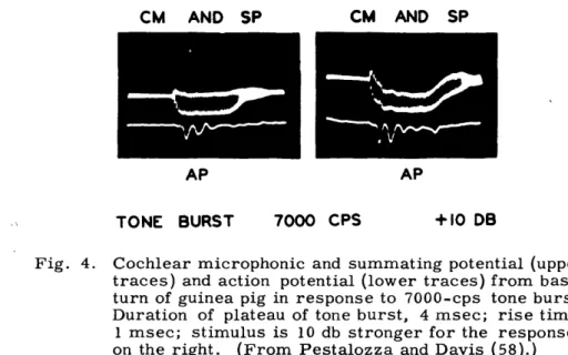
When cochlear responses to fairly long bursts of high-frequency tones are observed, the microphonic potential often appears to shift its base line during the burst (Fig. 4). This shift has been called the "summating potential." It can be thought of as resulting
CM AND SP CM AND SP
AP AP
TONE BURST 7000 CPS +10 DB
Fig. 4. Cochlear microphonic and summating potential (upper traces) and action potential (lower traces) from basal turn of guinea pig in response to 7000-cps tone burst. Duration of plateau of tone burst, 4 msec; rise time, 1 msec; stimulus is 10 db stronger for the responses on the right. (From Pestalozza and Davis (58).)
from a kind of detection or demodulation process that produces a signal having the wave-form of the signal envelope if the carrier has a sufficiently high frequency. The process performing the detection can be thought of as rectification followed by integration (sum-mation).
The difficulties connected with the study of this response arise largely from its var-iability from one preparation to another, and from time to time during an experiment. It sometimes has one polarity, and sometimes another, and it seems to be increased by anoxia and surgical trauma (17), which may indicate that it is not significant in a healthy preparation. Davis has dealt with these "vagaries of the summating potential" (15) by postulating two summating potentials of opposite polarity. However, the negative summating potential, SP , is the one that is most often observed, and has been placed in prominence by the theory that has been proposed (15). Davis also states that "the usual polarity is negative, ... under certain circumstances, particularly in fresh prep-arations and with weak stimuli, the polarity is positive" (17).
The amplitude of SP increases with intensity, but it is also difficult to obtain con-sistent measurements. In some cases the polarity of SP changes from plus to minus as the intensity of the stimulus is increased (17), so that one is not quite sure what he is measuring. It does seem to be quite well established that the amplitude of SP continues to increase for the high intensity levels at which CM is decreasing (17).
The summating potential seems to have its largest value when it is measured between an electrode in scala media and an indifferent electrode (neck, scala tympani). Its spatial distribution across the cochlea differs from that of the microphonic potential. However, this does not necessarily indicate that SP and CM arise from different structures, since the impedance of the membrane and fluids differ for the CM and the SP that have dif-ferent frequency compositions (17). The variation of the amplitude of the summating potential along the cochlea is also quite different from the microphonic. Although the actual data have not been published in detail, it is stated that "SP response is associated more closely with the position of maximum amplitude of displacement ... apparently the SP response is associated particularly with the part of the resonance pattern where amplitude is large but the wave length of the traveling wave is becoming short" (17). That is, SP seems to have a maximum in the region of the cochlea in which the amplitude of vibration is decreasing from its maximum (2).
Although the' source of the summating potential is not well established, evidence has been cited which suggests that the internal hair cells may be the source of the negative component, SP_ (18). This suggestion, together with several others, has been woven into a comprehensive theory by Davis (15) who assumes that SP_ is produced by the internal hair-cells in much the same way as the microphonic potential is produced by the external hair cells. The rectification and integration are presumed to result from mechanical
action, in which a constant shift of the tectorial membrane relative to the hair cells introduces a constant bending with resulting dc potential. This shift is assumed to occur in the longitudinal direction (along the cochlear duct). The possibility of having this kind
7
-of motion is indicated by von Bekesy's observation -of eddies (2) in the perilymph in the
same region where SP, presumably, is largest. Also, von Bekesy has shown that
longi-tudinal vibration of the tectorial membrane is most efficient in producing CM when the vibration occurs near the internal hair cells (7). In Davis's theory, the microphonic and summating potentials are then assumed to act as electric stimuli on the nerve endings
that terminate on the external and internal hair cells, respectively. The microphonic,
since it follows the fast vibration of the membrane, tends to excite the nerve fibers in synchrony with the stimulus frequency; this provides a mechanism that codes the stimulus
frequency into frequency of neural response. The summating potential will tend to excite
neurons in a more localized area because it is more closely associated with the point of
maximum deflection. Hence, SP seems suited to code stimulus frequency in terms of
place along the cochlea. The nerve fibers that end on the internal hair cells are more
discrete along the cochlea (25), and hence seem to be better adapted to efficient "place" coding. This theory thus offers two possibilities for frequency coding, since "place" and "period" (50, 21) are represented in the cochlea by separate mechanisms. Davis also suggests that SP is the more important process at high intensities because it is
still increasing when CM is decreasing. Hence, SP extends the dynamic range of the
cochlea. These are the important points of a theory that encompasses many known
phe-nomena, although many of the specific mechanisms are still to be verified. 1.6 AUDITORY NERVE POTENTIALS
The summated action potentials of the auditory nerve fibers can be recorded from
wire electrodes placed almost anywhere near the cochlea (67). Certain electrode
arrangements provide action potentials ("neurals") with a minimum of microphonic poten-tial (23, 67, 74). The potential recorded by these large ("gross") electrodes represents some kind of summation of the action spikes of the individual axons in the auditory nerve.
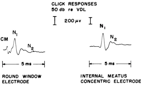
The shape of the neural in response to an acoustic click, as recorded from a cat, is
shown in Fig. 5. Click responses of similar waveform have been recorded from several
other animals (43, 45, 46, 60, 64, 66, 71). The two negative deflections have been
desig-nated (63) by the symbols N1 and N2.
When one looks for neural responses with gross electrodes, it is easiest to find
well-defined responses when the stimuli are impulsive. A great deal of work has been done
on determining characteristics of the "click response." (Generally, the click has been generated by applying a short (0. l-msec) rectangular voltage pulse to an earphone.) A neural response of similar shape is produced at the onset of noise or high-frequency
(above 5 kc) tone, if the stimulus is turned on rapidly enough. If the tone or noise is
turned on gradually, no neural response of this type (N1) is observed (37). This fact
can be explained if we consider the gross electrode response as an indication of synchro-nous "firing" of a large number of neural units. When the stimulus changes rapidly, many units are stimulated at the same time, and their responses are summated at the
electrode to give a large deflection. During maintained stimulation, however, the units
CLICK RESPONSES 50 db re VDL NI 200pv
I
NNN
I L- 5ms-
-
5 msROUND WINDOW INTERNAL MEATUS
ELECTRODE CONCENTRIC ELECTRODE
Fig. 5. Click responses recorded simultaneously between an electrode near the round window (left trace) and reference electrode, and between concentric electrodes in the internal auditory meatus (right). In all records, upward deflection indicates negativity of the round-window electrode with respect to the reference electrode, or negativity of the core electrode with respect to the sleeve for the concentric electrode. Note that the microphonic poten-tial is not visible in the response from the concentric electrode. (C-504.)
do not necessarily fire synchronously, and no synchronized response of the N1 type is
observed with the gross electrode.
For low-frequency stimuli (lower than 2000 cps), a neural response can be detected for each cycle of the stimulus, in addition to a larger response at the stimulus onset (23). If the frequency of the stimulus is sufficiently low that the responses in each cycle do not overlap, the shape of the neural is similar to that of the click response (68). After a low-frequency tone has been turned on, the amplitude of the neural response decreases steadily for several minutes (23). In order to avoid this adaptation phenomenon when studying the effect of other stimulus parameters, many workers have preferred to use clicks and short bursts of tone at a repetition rate that is low enough (/sec) to avoid adaptation. Under these conditions, the amplitudes of the click responses seem to be independent of each other (27). Changes in response with repetition rate will be described in Section IV.
By using stimuli of this type, the changes in the response with stimulus intensity have been studied (28, 18). In general, the amplitudes of N1 and N increase with intensity.
This is interpreted as an indication that more and more neural units respond as the stim-ulus strength is increased. Frishkopf describes this behavior mathematically in terms of a probability model (27, 28). He found that, for low intensities, the data can be repre-sented in terms of a uniform population of identical neural units with randomly varying thresholds. The question of how many populations are needed to represent the dynamic range has not been studied in detail, but it has been suggested that there are three populations (18).
9
It is also observed that the latency of the "neurals" decreases as the stimulus
inten-sity is increased. The mechanisms involved in this latency change have not been
inves-tigated, but a similar change is found when peripheral nerve is excited electrically (24). It has been suggested that this decrease in latency is attributable to the fact that with stronger excitation the "triggering level" is reached earlier (22).
One of the most striking properties of the neural response is observed when impul-sive stimuli are presented against a background of noise. A very low level of noise
(that is, one that cannot be detected in the microphonic component) reduces the neurals
appreciably (63). This phenomenon can also be interpreted in terms of a
desynchro-nizing of the action potentials. The noise stimulates the neural units continuously, and
some of them respond at any given instant. Hence, when the click occurs, some of the
recent responders are refractory and unable to respond. This interpretation is supported by the observation that acoustic noise can mask responses to electric stimulation of the
auditory nerve (49). The masking phenomenon was also treated by Frishkopf in terms
of a probabilistic model (27). The results supported the single-population model for
low-intensity stimuli.
Observations have also been made of the effects of previous stimuli on neural
responses to impulsive stimuli. McGill and Rosenblith (53, 54) found that the response
to the second of two clicks was reduced if the two clicks were separated by less than
approximately 100 msec. The response to the second click decreases if the interval
between clicks decreases, and if the intensity of the first click is increased. The
response to the second click is never supernormal. Neural responses to clicks can be
affected for longer periods by bursts of high-intensity sound (48, 42, 65). Following
exposure to noise and high-frequency tones, the amplitude of the neural response
recovers monotonically to its pre-exposure level. For exposures at low frequencies
(200-500 cps) there is a period of supernormal response (65, 44). It is interesting to note that for most reversible recoveries after exposure to intense sound, CM remains nearly unchanged (42, 48, 65).
In this section we have emphasized the main characteristics of the neural response
for various stimulus patterns. We did not attempt to present all of the work that has
been done; studies of the effects of drugs, temperature, surgical interference, and so
forth, have not been mentioned. An extensive bibliography of this work has been given
by Davis (14).
Some important observations of the response of single auditory nerve fibers have been made by means of microelectrodes (71, 72, 47). In general, it can be stated that
the results are compatible with the model which considers the N1 deflection of the gross
electrode response to be a summation of single-unit action-potential spikes from the
auditory nerve fibers. Tasaki's results indicate that the auditory nerve fibers quite
often respond with two spikes to an impulsive stimulus, and he suggested that the second
neural component (N2) may be a result of this (71). Other data (67) indicate, however,
that this interpretation may be only partially correct. In the presence of continued
10
stimulation by high-frequency tones single units exhibit a decrease in the rate of firing after an initial burst (32, 33, 71). They continue, however, to fire at a rate that exceeds the rate of spontaneous activity. The decrease in number of firing units together with a desynchronization of those that fire makes it more difficult to detect the existing neural activity with gross electrodes.
Although it is desirable to draw parallels between electrophysiological and behavioral data, very little work has been done on collecting both kinds of data from the same ani-mals. In one study it was found that the threshold for observation of the click response in pigeons was very close to the behavioral threshold (43).
1.7 EXPERIMENTAL PROCEDURES
In general, the techniques used in all of the work reported here are similar to those used by other workers (e.g., Frishkopf (27), and Goldstein, Kiang, and Brown (38)). A cat is anesthetized with Dial (75 mg/kg) injected into the peritoneal cavity. After a tracheal canula has been inserted, the bulla of one ear is exposed and the bone opened so that the round window of the cochlea becomes accessible. A wire electrode is placed in contact with the bone near the round window. Responses are recorded between this electrode and a reference lead attached to the headholder. In some experiments, a section of the skull and a portion of the cerebellum were removed to expose the eighth nerve at the point where it enters the medulla from the internal auditory meatus. Responses were then recorded from a concentric electrode placed in the nerve.
In all experiments, the stimuli were generated by a Permoflux PDR-10 earphone connected to a plastic tube that was tied into the external auditory meatus.
The animal was placed in a soundproof, electrically shielded room. Various stim-ulus waveforms were generated electronically (38) and fed into the room to the earphone. The potentials obtained from the electrodes were amplified (amplifier passband, 8-7000 cps) and observed on an oscilloscope, recorded on magnetic tape, and/or processed by an average response computer (13).
During the experiment, the condition of the animal was maintained as constant as possible. The temperature of the room was regulated at approximately 25°C. Rectal temperature was monitored and maintained between 35-37°C by using a heating pad. If there were indications that the effect of anesthesia was wearing off, an additional dose of 0.25-0.5 cc was given.
At the beginning of each experiment the animal's threshold for neural responses was determined by reducing the stimulus intensity to a level that produced responses that
were visually detectable on the oscilloscope screen approximately 50 per cent of the time.
This "tthreshold" is called VDL (visual detection level). This level was determined at the beginning of the experiment and the measurement was repeated several times during the experiment, in order to check the stability of the preparation. Furthermore, the size of an averaged response to a moderately intense stimulus (20-30 db re VDL) was measured at least once every hour, and after periods of intense stimulation, which might
11
be expected to cause long-lasting aftereffects. If the response amplitude differed by more than 10 per cent from the "standard" value, all acoustic stimulation ceased until the response had returned to the 10 per cent limit. For all of the animals studied, the VDL for "clicks" produced by applying 0.1-msec rectangular pulses to the earphone at a rate of 1/sec was between -95 db and -105 db re 4 volts.
12
II. ELECTRIC RESPONSES FROM DENERVATED COCHLEAS
We now present results from experiments performed on cats with unilateral degener-ation of the eighth nerve. The purpose of these experiments was to study the behavior of non-neural electric responses that can be recorded from electrodes near the round window. The results of these experiments are involved in the interpretation of the behav-ior of neural responses (Secs. III and IV).
2.1 SPECIAL TECHNIQUE
The surgery required to obtain the denervated preparations was performed by Dr. N. Y-S. Kiang at the Eaton Peabody Laboratory of Auditory Physiology, of the Massachusetts Eye and Ear Infirmary. The eighth cranial nerve was sectioned at the point where it enters the medulla from the internal auditory meatus. In order to expose the nerve at this point, a portion of the skull was removed and the cerebellum was pushed aside above the nerve. After the nerve had been sectioned, the wound was closed and the animal allowed to recover from the surgery. After sectioning, a time of approximately five weeks was allowed for degeneration of the nerve cells. After this period, the animals were prepared as described in Section I, and the remaining electric responses were observed with electrodes near the round window. Histological preparation of the studied cochleas is still being carried out at the Eaton Peabody Laboratory. Since it is difficult to section the nerve completely without injuring the blood supply to the cochlea, the extent of the degeneration will not be known completely until the histology has been completed.
This technique has been used previously to observe CM responses to clicks (62, 69). The shapes of the CM responses to short rectangular pulses which we obtained (Fig. 6)
are reasonably similar to those obtained by other workers who used similar acoustic systems (69, 27). (Another technique (74) for observing microphonic potentials that are relatively free from "neurals," without damaging the nerve, has been used extensively in guinea pigs.)
The CM responses to clicks as observed with a round-window electrode are "con-taminated" in several ways:
(a) The shape of the microphonic response is determined by the entire mechanical system, consisting of the earphone, connecting tube, outer and middle ear cavities (including the eardrum and ossicles), and the relevant portions of the cochlea. Since we did not monitor the signal at any point between the earphone input and the round-window
electrode, we do not know in what way the various parts of the system contribute to the CM response. The best indicator of the acoustic pressure response to the rectangular pulse is that measured by Frishkopf (27) with the same type of earphone operating into a 1-cc cavity.
(b) The CM recorded at the round window is a weighted sum of the microphonic potentials produced all along the basilar membrane, and hence will probably not repre-sent the motion of any one region. Probably, only the basal turn contributes significantly
13
____I
I
__ I L L ---I-C-510
ImO
c-usI
C-St5
Fig. 6. Microphonic potentials in response to rarefaction clicks from 4 animals with denervated cochleas. Clicks were produced by applying rectangular pulses of 0.1-msec duration to PDR-10 earphone. Intensity, -60 db re 4 volts. Traces were obtained by averaging 64 responses recorded from electrode near round window.
to CM at the round window (3, 75), and there is evidence that a large part of the basal turn vibrates in phase (1, 74). Hence, as a first approximation, the shape of the CM
recorded from the round window can be assumed to represent the motion of the membrane in the basal turn. The validity of this approximation depends upon the frequency compo-sition of the acoustic stimulus.
(c) The extent to which the potentials recorded at the round window contain compo-nents other than CM may depend upon the success of the nerve-sectioning procedure, the physiological condition of the animal, and so on.
2.2 IMPULSE RESPONSE
In Section I evidence indicating that the microphonic potential can be considered as the response of a linear system for low and medium intensity stimuli was cited. If this
is true, the system can be characterized by its "impulse response," h(t). In particular, the response e(t) to any stimulus waveform ei(t) (within the linear range) can be found by convolving the stimulus with the impulse response. Thus
eo(t) = ei(T) h(t-T) dT (1)
Also, the Fourier or Laplace transform of the impulse response is the transfer function of the system (35). In order to determine the impulse response of the system (earphone, tube, ear), a rectangular pulse was applied to the earphone and the microphonic response
14
I T
I msec
10
/v50- sec PULSE (-54 db) ... 25 - sec PULSE (-48 db)
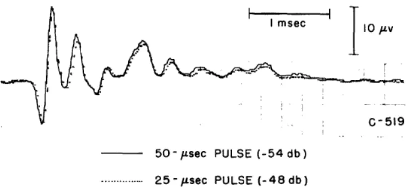
Fig. 7. Averaged microphonic responses to rarefaction clicks produced by 25-psec and 50-Fsec pulses of equal area.
II 0I a 0. M2 2
2
PULSE DURATION (ps)Fig. 8. Peak-to-peak amplitude of the microphonic in response
to rarefaction clicks as a function of pulse duration. The
area under the pulse is kept constant. Inset indicates the
amplitude that was measured. Intensity of 100-1Lsec pulse,
-60 db re 4 volts.
was observed. The duration of the pulse was then decreased and its amplitude increased
so that the area under the pulse remained constant. If the pulse is short compared
with the impulse response, it will effectively be an impulse for the system. For
this condition, changing the pulse duration and keeping the area constant should not
change the response. In Fig. 7, CM responses for 25- and 50-1Fsec pulse durations are
superimposed to show that the difference is negligible in that range. In Fig. 8,
peak-to-peak amplitude of the CM is plotted against pulse duration; a pulse of approximately
50 sec, or less, is effectively an impulse. Since 100-Lsec pulses had been used
extensively by previous workers and the departure from the impulse response is slight, we have continued to use this length. The data indicate that the CM responses to the "clicks" can indeed be considered to be the "impulse response" of the system
at moderate intensities.
15
YI··-·II_-IL_·.-·._._·
·-I·II--^
____
__·--__I
·II
- ·RAREFACTION CLICKS GAIN X40 X40 CONDENSATION CLICKS a.< X 2.5 -40 X *I --30 - , . ." -20 --10 I' 5 me 5 ms XI X i - , - , X I , t I250p
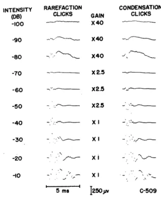
Fig. 9. Averaged responses to condensation and rarefaction clicks (0. 1 msec) from a denervated cochlea. Threshold for visual detection of the microphonic in single traces lies at approximately -70 db! Click repetition rate 10/sec up to -30 db, 1/sec above -30 db. Click reference level, 3.8 volts. Number of responses averaged: 512 at -100 and -90 db; 256 at -80 and -70 db; 128 from -60 to -30 db; 32 at -20 db; 16 at -10 db. 100 50 30 20 I-:E E I0 2 3 2 - 4--//
f /I I
I I l . I -100 -90 -80 -70 -60 -50 -40 -30 -20 -10 INTENSITY (DB) 0Fig. 10. Amplitudes of CM and "slow" potentials versus intensity from data of Figs. 9 and 12. The amplitudes measured are indicated in the insets. The "slow" potential cannot be measured at high stimulus intensities because a faster component overrides it in the records. The straight line indicates the linear growth of CM in the low-intensity range; that is, the amplitude increases by a factor of 10 for a 20-db rise in stimulus intensity.
16 INTENSITY (0S) -100 -80 -70 -50 - --- X2.5 C-509 rllA~> * * I -90 , S2 X40 -60 ---- X2.5 · ---O,. cv X
2.3 CHANGES WITH STIMULUS INTENSITY
Figure 9 shows averages of cochlear responses of a denervated ear to short pulses of both polarities over a range of 90 db. It appears that the waveform of the "early" CM response does not change appreciably over the low-intensity range (-100 to -50 db). The amplitude of this CM response is plotted against intensity in Fig. 10. The linear increase of amplitude with waveform over the low-intensity range supports the conclusions of others (for example, Tasaki, Davis, and Legouix (74)) that the CM can be considered as the response of a linear system over this range. The departure from linearity at high intensities (Fig. 10) is similar to that in CM curves obtained for pure tones in normal preparations (70, 74, 79). In the high-intensity range, the most striking change in the waveform of the response is the appearance of large potentials at relatively long latencies.
2.4 REVERSAL OF RESPONSE WITH STIMULUS POLARITY CHANGE: THE SLOW POTENTIAL
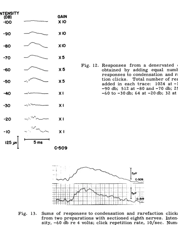
One of the characteristics of CM that has been used to distinguish it from other potentials is its reversal of polarity with a change in stimulus polarity (63). Inspection of the records of Fig. 9, however, shows that the electric response from the denervated ear does not reverse completely when the polarity of the stimulus pulse is changed. In Fig. 11 averaged responses to condensation and rarefaction pulses are superimposed to demonstrate this lack of total reversal. Figure 12 shows the waveforms that are obtained when we add an equal number of responses to condensation and rarefaction clicks. The resulting waveforms represent mathematically the component in the two types of responses that does not reverse. This "common" component appears to have a relatively constant shape in the low-intensity range. Figure 13 shows this for two different animals. This common component starts at a significantly longer latency than the CM. This indi-cates that it is not merely the result of imperfect cancellation resulting from variability in the response or the preparation. For low intensities, cancellation is complete during the first quarter or half millisecond after the onset of CM. The amplitude of the slow wave is plotted against intensity in Fig. 10. At intensities above -50 db the common component changes in character because it then includes faster waves. This may indicate that when the process that gives rise to CM becomes nonlinear, it becomes asym-metrical, so that the CM responses cease to cancel perfectly. These faster potentials might also be partly summating potential, since SP becomes more prominent at high intensities (17). Because SP has only been studied for tone bursts, it is difficult to determine what it might look like in a click response.
Since the slow common potential at low intensities is primarily negative and has about the same latency as N1, it might represent the response of some nerve fibers that have
remained functional after the surgery. In order to determine whether this response has the properties of a neural response, a series of responses to condensation and
17
CONDENSATION
I
,
-
Tov
RAREFACTION
Fig. 11. Superimposed averaged responses to condensation and rarefaction clicks. (Same data as for Fig. 9.) Note asymmetry with respect to base line. (C-509.)
GAIN ---. X 10 X10 X5 X5 X5 Xl
Fig. 12. Responses from a denervated cochlea obtained by adding equal numbers of responses to condensation and rarefac-tion clicks. Total number of responses added in each trace: 1024 at -100 and -90 db; 512 at -80 and -70 db; 256 from -60 to -30db; 64 at -20db; 32 at -10 db. Xl XI XI C-509 5 ms C509
Fig. 13. Sums of responses to condensation and rarefaction clicks from two preparations with sectioned eighth nerves. Inten-sity, -60 db re 4 volts; click repetition rate, 10/sec. Num-ber of responses averaged, 128.
18 INTENSITY (DB) -100 -90 -80 -70 -60 -50 -40 -30 -20 -10 125I
ii
+ AND - BURSTS CLICKS OF NOISE _ ,'
~
L, ., !'---N--I 200 .,\'. 500 . C-519 IOPv I - 20 ms-Fig. 14. Averaged responses from a denervated cochlea as a function
of stimulus repetition rate. Left-hand column was computed
by adding equal numbers of condensation (-) and rarefaction (+)
click responses. Right-hand column is averaged responses to
0.1-msec bursts of wideband noise. Number of responses
aver-aged: Left column, /sec, 64; other rates, 512. Right column,
1/sec, 128; other rates, 256. Click intensity, -60 db re 4 volts; noise-burst intensity, -60 db re 1 volt rms.
x
x .
x
l
* 100% = 1.8 p.V FOR LEFT EAR x 100% = 2 0 0. V FOR RIGHT EAR
I I I I I 111 I I I I I L L 10 100 R * . *o. X xx .. XX X X I I I I I II Xx-, 1000 STIMULUS REPETITION RATE IN BURSTS PER SEC
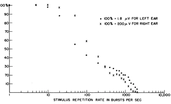
Fig. 15. Normalized response amplitude versus repetition rate. were 0. 1-msec noise bursts with an intensity of -65 db The left eighth nerve was sectioned; right ear, normal.
19 STIMULI PER SEC I 10 20 50 100 -- N x I00%j 90 80 70 60 50 40 30 70 10 I i I 1I l 10,000 Stimuli re 1 volt. (C-510.)
_
I_____
_ ____
CII
_ _ _ l _ l Crarefaction clicks was recorded at several repetition rates. The CM response does not change in shape or amplitude for click rates up to 800/sec (see Fig. 48). At this rate the responses overlap each other, so that it is impossible to measure the individual responses directly. The common potential computed from the sum of responses to con-densation and rarefaction clicks also remains constant for repetition rates below those
at which overlapping occurs (see Fig. 14). This constancy with repetition rate is not observed with neural responses from normal ears at moderate stimulus intensities (see Sec. IV). In this sense, this "slow" response appears to be non-neural. In one of the sectioned preparations (C-510) a small component potential was observed that decreased with rate in nearly the same way as the neural potential in a normal ear. Figure 15 indicates that in this preparation some of the nerve cells remained responsive after the surgery.
2.5 RESPONSES TO NOISE BURSTS
If short (0. 1 msec) noise bursts are used as stimuli, the microphonic potentials have waveforms similar to that obtained with rectangular pulses, but the amplitude of CM varies; sometimes it has the polarity that results from a rarefaction click, sometimes the polarity that results from a condensation click (Fig. 16). This result can be pre-dicted from the linear system model for CM. If the noise burst has a duration, 6, then the superposition integral can be written.
eo(t) = i(T) h(t-T) dT (2)
If the noise burst is very brief compared with the variations in the impulse response,
RAREFACTION CONDENSATION NOISE
CLICKS CLICKS BURSTS
I 25v i 2.5 ms C-519
Fig. 16. Microphonic responses to clicks (0. 1-msec), and 0. l-msec noise bursts. Click inten-sity, -60 db re 4 volts; noise-burst inteninten-sity, -40 db re 1 volt rms.
the integral can be simplified:
e(t) = = h(t) h(t) ei(T) dT (3)
This indicates that the CM response to a short burst of noise, has the wave shape of the impulse response and an amplitude that is a random variable equal to the integral of the input voltage during the burst. If the noise is symmetrical about zero voltage, then the mean amplitude of the CM response to noise bursts will be zero. Hence, if a large number of responses to noise bursts is averaged, CM should tend to be very small (13). However, when responses to noise bursts are averaged a slow potential remains, which resembles that obtained when responses to condensation and rarefaction clicks are summed. This potential changes with noise-burst intensity as shown in Fig. 17, and with repetition rate as shown in Fig. 15. This potential seems to be the same as the previously discussed "slow" potential. The presence of this potential in both averaged responses to noise bursts and in sums of responses to condensation and rarefaction clicks is compatible with a model in which the response is the sum of a linear-system response plus a "slow" potential of invariant polarity.
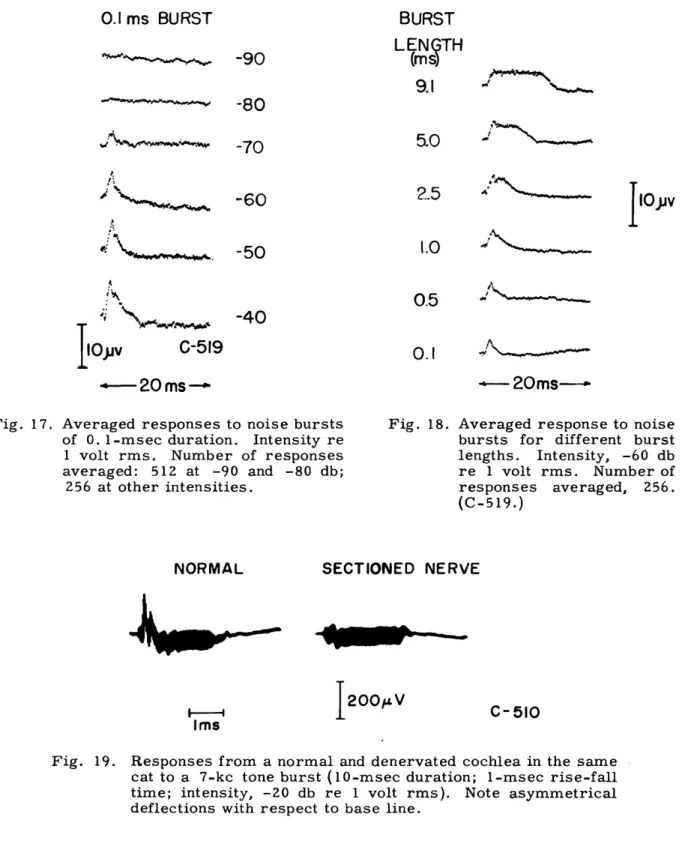
The invariant polarity of the "slow" potential might suggest that we are dealing with the summating potential. Most summating potentials have been recorded in responses to high-frequency tone pips whose duration is several milliseconds. For click stimuli, the presence of SP is difficult to establish. However, noise bursts can be increased in length and if our "slow" potential is SP, its duration should be equal to the duration of the stimulus. Figure 18 shows averaged responses to noise bursts of different length. The slow potential increases in duration with increasing burst length. In this respect it is similar to SP. However, there are several characteristics of this slow potential that distinguish it from SP_ as Davis and his associates (17) describe it: (a) The slow potential is more prominent at low intensities, whereas the threshold for detection of the SP is normally at least 20 db above the "threshold" for CM. Our electrode place-ments differ from those used by Davis' s group, so that such comparisons may not be meaningful. (b) The growth of the slow potential with intensity seems to level off in the middle-intensity range (Fig. 10), whereas SP_ continues to increase with intensity up to levels of cochlear injury (15). (c) The onset of the "slow" potential is approximately
0.4 msec later than the onset of CM to clicks, whereas for tone pips SP_ starts essen-tially simultaneously with CM (58).
We have also found that the slow potential may have a different polarity from SP when the two are observed in the same ear. Figure 19 shows responses to tone bursts from a denervated and a normal ear in the same cat. In both instances the
asymmetri-cal displacement of the base line (SP) is positive. However, the "slow" potential for this same ear was, as in all our preparations, negative. The positive polarity for SP
in Fig. 19 (as observed at the round window) is the same polarity reported by other workers (58), and corresponds to negative summating potential SP.
21
0.1 ms BURST
-90
-80
-70
t,-60
-50
A.
:i-,9;i
-40
C-519
--
20
ms--0.1
20ms--Fig. 17. Averaged responses to noise bursts of 0. l1-msec duration. Intensity re 1 volt rms. Number of responses averaged: 512 at -90 and -80 db;
256 at other intensities.
NORMAL
Fig. 18. Averaged response to noise bursts for different burst lengths. Intensity, -60 db re 1 volt rms. Number of responses averaged, 256. (C-519.)
SECTIONED NERVE
I200V
Ims
C-510
Fig. 19. Responses from a normal and denervated cochlea in the same cat to a 7-kc tone burst (10-msec duration; 1-msec rise-fall time; intensity, -20 db re 1 volt rms). Note asymmetrical deflections with respect to base line.
It would be desirable to determine the distribution of the "slow" potential inside the cochlea, in order to obtain some knowledge of its origin. We would also like to know
how it varies with distance along the basilar membrane.
22
BURST
LENGTH
(ms
9.1
5.0
2.5
^'
1.0
.
ilOjv
TIiOv
0.5
I
-40100000MEMEMbft-m-_III. CHANGES IN AUDITORY-NERVE RESPONSES AS A FUNCTION OF STIMULUS INTENSITY
3.2 INTRODUCTION
The dependence of the auditory-nerve response on stimulus intensity has been
described by many workers (28, 18). Frishkopf (27) has presented click-response data covering a wide intensity range. Davis's (18) "input-output curves" plot the amplitude of neural responses against the intensity for high-frequency tone pips. In all the pub-lished data, the amplitude of the neural response increases rather smoothly with increasing intensity (although there is sometimes a plateau) (27), while both onset and peak latency decrease with increasing intensity. The latency decreases fairly rapidly (20 sec/db) near threshold, and is almost constant for high intensities (27).
3.2 RESULTS
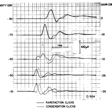
In studying click responses recorded from the vicinity of the round window, several workers have noted differences between the responses to rarefaction and condensation clicks at certain intensities (19, 43, 63). However, it is difficult to be sure that these differences involve just the neurals because of the possible contribution of the CM com-ponent in records obtained from electrodes near the round window. It has been shown that auditory-nerve responses relatively free from CM can be obtained from a concen-tric electrode placed in the internal auditory meatus (23). Figure 5 illustrates the rela-tion between the two types of record.
Figure 20 shows single responses to condensation and rarefaction clicks recorded by such a concentric-electrode configuration for an intensity range of 100 db. In Fig. 21 average peak-to-peak amplitude and N1-peak latency are plotted against intensity for the
data of Fig. 20. These results can be described as follows: (a) At low intensities the neural responses to the two polarities are nearly equal in amplitude and latency. (b) At high intensities the amplitudes are nearly equal, but the latency of the response to the rarefaction click is consistently shorter by approximately 0. 2 msec. (c) At -60 db and -50 db the neurals differ strikingly; for condensation clicks the amplitude increases monotonically with intensity, while the response to rarefaction clicks changes shape and becomes smaller (in some sense) in this range. At -60 db (rarefaction click) a new "bump" (arrow) appears at the front end of N1; this bump grows, and at -50 db is larger
than the second deflection. Since we measure latency to the largest negative peak, a large change in latency occurs between -60 db and -50 db.
Figure 22 shows superimposed averaged responses to condensation and rarefaction clicks in another preparation. This display shows clearly that the two response wave-forms are nearly the same for both low and high intensities, but a definite latency dif-ference appears at high intensities. Amplitude and latency of the averaged responses are plotted in Fig. 23 as a function of intensity.
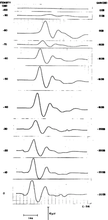
Figure 24 shows averaged responses to noise bursts of short (0. l-msec) duration
(SaNO3SI1 I W) 1 'AON31V 0 C" O a CD _ 0 D 0 N c Nc N - - 0 0 0 C In I 0~~
4 1:
0 t : 0 0 -4.-~~z
z "k -3 0 O~~ < Z I~~~LL
u W 0 crz a' o2 -J A ' -Q / ' I I I I I I 0 00 00000 0 00 00 00 o o o o c I I I I o O O' o O ) 3d , N -(AT/) V ' 3anildWV a) A+q 0 0 U 0 0) - Q o c o o Q 0 Cte U) u ' 4.-4 o 0)0 0 r.( ad D td r -2 * Cd ' 9,a C.)t~~ o * U) '' rl U)ku $.4C) o - > M o O O r Q 0 0 o . In - 0 - w 3 t a Cd CIS U ' 1) C.) ).) o 41C d o Cd 4.o C -0 ) 00+ 0) *e ,4 o0 .. d a uO E -o,- s 0 ) b0 cd C x 0c,0C 0 CI od -4! ·fD Or( J.,l 0 0)0)0) 'v ) .) 04 .4 0 0 4., Q)~
0 OIm0 0.
-4 N zo0
o } c rr
f
z a x t(D z 0 zS c O I-z 0o x x Ui) to 10 * X X I X X X X X Xf
o o 4 o 4 o o O I T 0~~.: # Vq a P _- _ . 4) oU O 4) o ) a. o C , 0) C c 0 , o ) U) a 0) 0d ;q 4. > ) 0) 24 a; .E 0 0 b0 C. O 0 $A 0.1 laS
01 0 ID q 4.0 En 2s M I l 2 1 f -IINTENSITY
- RAREFACTION CLICKS
--- CONDENSATION CLICKS
Fig. 22. Averaged responses to condensation and rarefaction clicks (C-504). Conditions are the same as for Fig. 20, except that clicks were presented at a rate of 5/sec. Click reference level, 2. 8 volts. Number of responses averaged: at -90 db, 128; at -70 db, 64; at -50 db, 64; at -30 db, 16; at -10 db, 16.
-o
I AnT 1.6 1.4 1.2 .-0.6 0.2 0.2 I I_
-
/_ I
x RAREFACTION - CONDENSATION C-504 i/ , I , I , I I Z,4 2.2 2.0 1.8 °z 0 1.6 , Co 1.4 J -J 1.2 E 1.0 >-0.8 W 0.6 -0.4 0.2 n v -100 -80 -60 -40 -20 0 INTENSITY (DB)Fig. 23. Amplitude and latency versus intensity for
C-504. Measurements made in the same
way as those plotted in Fig. 21. At -50 db, the latencies of both negative peaks in the rarefaction click response are indicated.
ITENSITY GAIN(08) (De) *-00 008 -90 008 , ~ oo -70 -1608 -60 -816 -50 . -1608 -40 -16DB -1 , -2088 -20 -2000 -10 -20018 0 -200 I f I I ; . C-516 I I % V Imr
Fig. 24. Averaged neural responses to noise bursts as a function of intensity. (0 db = 1 volt rms
to earphone.) Number of responses
aver-aged: 256 at -100 and -90 db; 128 at -80 db; 64 from -70 to 0 db.
* TAKEN AS INTENSITY INCREASED .4 .2 .0 a .6 i-.4 U 2 J 4 4 2 -100-90 -80 -70 -60 -50 -40 -30-20 -10 0 INTENSITY (DB re I VOLT RMS)
Fig. 25. Amplitude and latency of averaged responses to noise bursts (Fig. 24) versus
intensity. Stimulus intensity was raised from -100 db to 0 db, and decreased
to -100 db. The dip in the amplitude curve at -30 db might be the result of
putting a large number of rarefaction click responses similar to those at
-50 db in Figs. 20 and 22 into the average. (Note that intensity levels for
noise bursts and clicks cannot be compared directly.) A rather sudden change in latency appears after the dip in the amplitude curve.
recorded from electrodes near the round window. Figure 25 is a plot of amplitude and
latency as a function of intensity for these neurals. We have seen that these short noise
bursts yield CM responses that resemble CM responses to clicks (see Fig. 16); we might therefore expect that the averaged neural responses to noise bursts would be
com-parable to neural responses to clicks. Figure 25 shows in some detail that the data on
neural responses to noise bursts appear to be consistent with our data on condensation and rarefaction clicks.
3.3 INTERPRETATION
In order to interpret the differences in the behavior of neural responses to conden-sation and rarefaction clicks, we would like to know the mechanisms that underlie the
neural responses. Although these mechanisms are not known, many experimental
results are helpful in thinking about the process of excitation of neurals. In particular, we can think in terms of the excitation of neural units by a wave that travels along the cochlear partition from base to apex (11).. The form of this wave for our stimuli is not known, but it has been shown that CM recorded inside the cochlea is proportional to the
displacement of the membrane (4, 74). There is also evidence that displacement in one
direction only excites the neural response (20, 68). We would like to interpret the
con-densation and rarefaction click responses in terms of the waveform of the CM response to the clicks. Since we do not have data on the form of the CM along the whole cochlea, we must make the approximation that the CM observed near the round window is an adequate representation of the motion of a large part of the basal turn.
Davis has stated that rarefaction at the eardrum leads to neural excitation (20). This
27