HAL Id: hal-01975570
https://hal-univ-rennes1.archives-ouvertes.fr/hal-01975570
Submitted on 22 Mar 2019HAL is a multi-disciplinary open access archive for the deposit and dissemination of sci-entific research documents, whether they are pub-lished or not. The documents may come from teaching and research institutions in France or abroad, or from public or private research centers.
L’archive ouverte pluridisciplinaire HAL, est destinée au dépôt et à la diffusion de documents scientifiques de niveau recherche, publiés ou non, émanant des établissements d’enseignement et de recherche français ou étrangers, des laboratoires publics ou privés.
ventricular fibrosis
Elena Galli, Emilie Vitel, Frédéric Schnell, Virginie Le Rolle, Arnaud Hubert,
Mathieu Lederlin, Erwan Donal
To cite this version:
Elena Galli, Emilie Vitel, Frédéric Schnell, Virginie Le Rolle, Arnaud Hubert, et al.. Myocardial constructive work is impaired in hypertrophic cardiomyopathy and predicts left ventricular fibrosis. Echocardiography, Wiley, 2019, 36 (1), pp.74-82. �10.1111/echo.14210�. �hal-01975570�
Myocardial constructive work is impaired in hypertrophic cardiomyopathy and
predicts left ventricular fibrosis
Running title: myocardial work estimation in hypertrophic cardiomyopathy
Elena Galli, MD, PhD1; Emilie Vitel E, MD1; Frédéric Schnell 1, MD, PhD; Virginie Le Rolle, PhD1; Arnaud Hubert, MD1; Mathieu Lederlin, MD, PhD1; Erwan Donal, MD, PhD1.
1
Univ Rennes, CHU Rennes, Inserm, LTSI – UMR 1099, F-35000 Rennes, France
Declarations of interest: none
Corresponding author:
Dr Elena GALLI, MD, PhD
University Hospital of Rennes, Cardiology Department 2, Rue Henri Le Guillou
35000 – Rennes (FRANCE) Tel.: +33 2 99 28 78 96 Fax: +33 2 99 28 25 29
Email: elena.galli@chu-rennes.fr; gallelena@gmail.com
ABSTRACT
Background: The estimation of myocardial work by pressure strain loops (PSLs) is a totally new
non-invasive approach to assess myocardial performance, and its role in patients with hypertrophic cardiomyopathy is unknown. Aims of the present study are therefore: 1) to compare myocardial work in patients with non-obstructive hypertrophic cardiomyopathy (HCM) and in a subset of age-matched healthy controls; 2) to assess the correlation between myocardial work and left ventricular (LV) fibrosis
Design: 82 patients with non-obstructive HCM (58±14 years) and 20 age-matched healthy subjects
(58±7 years, p=0.99) underwent standard and speckle tracking echocardiography to assess myocardial dimensions and deformation parameters. PSLs analysis was used to estimate global myocardial constructive work (GCW) and wasted work (GWW). LV fibrosis was estimated at cardiac magnetic resonance (CMR) by qualitative assessment of late gadolinium enhancement (LGE), and significant fibrosis was defined as LGE in≥2 LV segments.
Results: GCW (1599±423 vs 2248±249 mmHg%, p<0.0001) was significantly reduced in HCM
compared to the control group. No difference was observed in GWW (141±125 vs 101±88 mmHg%, p=0.18) and LV ejection fraction (63±13 vs 66±4% p=0.17) between the two groups. In HCM, GCW was the only predictor of LV fibrosis at multivariable analysis (OR 1.01, 95%CI: 0.99-1.08, p=0.04). A cut-off value of 1623 mmHg% (AUC 0.80, 95% CI: 0.66-0.93, p<0.0001) was able to predict myocardial fibrosis with a good sensitivity and fair specificity (82% and 67%, respectively).
Conclusions: GCW is significantly reduced in HCM despite normal LVEF and is associated with
the LV fibrosis as assessed by LGE.
KEYWORDS: hypertrophic cardiomyopathy, cardiac magnetic resonance imaging, myocardial
strain
INTRODUCTION
Hypertrophic cardiomyopathy (HCM) is the most common heritable cardiomyopathy, and is characterized by heterogeneous patterns of left ventricular (LV) hypertrophy, and increased risk of sudden cardiac death (SCD)(1). The histological features of HCM include myocytes’ hypertrophy and disarray as well as interstitial fibrosis(2). Previous studies have shown that the existence of late gadolinium enhancement (LGE) on cardiac magnetic resonance (CMR), an in-vivo-marker of fibrosis, is a predictor of SCD risk in patients with HCM(3). LV deformation is often impaired in patients with HCM, even in the presence of a preserved LV ejection fraction (LVEF), and is associated with the amount of LV fibrosis(4), symptoms’ onset and long-term outcomes(5).
The assessment of myocardial work by pressure-strain loops (PSLs) is a recently introduced tool that allows to estimate the myocardial performance. Recent studies have shown that the amount of myocardial constructive work is a predictor of cardiac resynchronization therapy response(6)(7). The amount of myocardial work assessed by PSLs is also correlated with the uptake of fluro-desoxy-glucose at myocardial positron emission tomography (PET) scan(8), which implies a relationship between the non-invasive estimation of myocardial work and myocardial metabolism. No previous study has assessed myocardial work in patients with HCM. Thus, the aims of this survey were 1) to compare myocardial work in patients with non-obstructive HCM vs normal subjects; 2) to evaluate if myocardial work indices could be relevant parameters for the assessment of LV fibrosis.
MATERIAL AND METHODS
Population
82 patients with non-obstructive HCM were consecutively recruited from our Regional Competence Center of Genetic Disease. The diagnosis of HCM was established according to current guidelines(1). Patients with obstructive cardiomyopathy, concomitant moderate or severe
valvular heart disease, myocardial storage disease, uncontrolled hypertension, and coronary artery disease were excluded from the study. All HCM patients underwent clinical examination, standard and speckle tracking 2D-transthoracic echocardiography, 48h-Holter monitoring, and cardiopulmonary exercise test (CPET). A subset of patients was investigated with cardiac magnetic resonance (CMR). All patients were screened for mutation in myosin-binding protein C (MYBPC3), β-myosin heavy chain (MYH7), regulatory and essential light chain of myosin (MYL2 and MYL3), and cardiac troponin T (TNN2) and I (TNNI3).
20 age-matched healthy individuals were also included in the study as a control group. Controls were recruited from healthy hospital staff, and people screened for working license. The study was conducted in accordance with the “Good Clinical Practice” Guidelines as laid down in the Declaration of Helsinki and reviewed by an independent ethics committee. All patients gave their written informed consent to participate in the study.
Echocardiography
All patients underwent standard transthoracic echocardiography using a Vivid 7, Vivid E9, or E95 ultrasound system (GE Healthcare, Horten, Norway) equipped with a 3S or M5S 3.5-mHz transducer. 2D, color Doppler, pulsed-wave and continuous-wave Doppler data were stored on a dedicated workstation for the off-line analysis (EchoPAC, GE Healthcare, Horten, Norway). LV volumes and function were measured by the biplane method as recommended(9). LA volume, LA antero-posterior diameter, and LV diastolic function were assessed as recommended(9)(10).
To calculate LV global longitudinal strain (GLS), two-dimensional greyscale images were acquired in the apical 4-, 3-, and 2-chamber views at a frame rate ≥60 frames/s. The recordings were processed using an acoustic-tracking dedicated software (EchoPAC version 112.99, Research Release, GE Healthcare, Horten Norway), to estimate LV global longitudinal strain (GLS)(11). Mechanical dispersion was calculated as the standard deviation of the time to maximal myocardial
shortening, measured from the electrocardiographic onset Q/onset R wave in the 17 LV segments(12)(13).
Myocardial work quantification
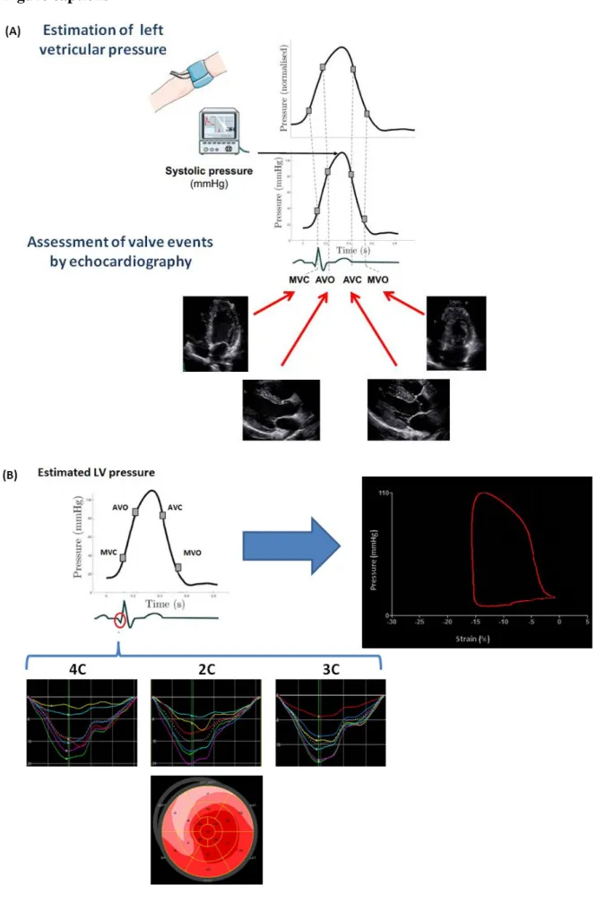
Myocardial work and related indices were estimated using custom software. Myocardial work was estimated as a function of time throughout the cardiac cycle by the combination of LV strain data obtained by speckle-tracking echocardiography and a non-invasively estimated LV pressure curve, as described in previous works (6)(7)(14). A 17-segment model was used for the estimation of segmental myocardial work. LV global longitudinal strain (GLS) was calculated as recommended(11). Peak arterial pressure measured with a cuff-manometer was assumed to be equal to peak systolic LV pressure and to be uniform throughout the ventricle. The resulting non-invasive LV pressure curve was obtained using an empiric, normalized reference curve that was adjusted according to the duration of the isovolumetric and ejection phases of the LV, defined by the timing of aortic and mitral valve events by echocardiography (Figure 1 A). The reliability of this non-invasively estimated LV pressure curve was previously validated in a dog model and in patients with dilated cardiomyopathy and LV dyssynchrony(8)(14). Strain and pressure data were synchronized using the R wave on ECG as a common time reference. Myocardial work was then quantified by calculating the rate of segmental shortening by differentiating the strain curve and multiplying the resulting value by the instantaneous LV-pressure (Figure 1B). The result is a measure of instantaneous power, which was integrated over time to obtain myocardial work as a function of time.
Work was calculated from mitral valve closure until mitral valve opening. During the isovolumic contraction and LV ejection period, segmental shortening contributes to the final LV ejection, whereas segmental stretch or lengthening do not contribute to LV ejection. As a result, the work performed by the myocardium during segmental shortening represents constructive work, whereas
the work performed by the myocardium during stretch or segmental lengthening represents energy loss, which is defined as wasted work. During isovolumic relaxation, segmental lengthening contributes to LV relaxation, whereas segmental shortening doesn’t. As a result, the work performed by the myocardium during segmental shortening, which doesn’t promote LV relaxation, was considered wasted work, whereas the work performed by the myocardium during segmental lengthening was considered segmental constructive work (Figure A, Supplementary material). By averaging segmental constructive and wasted work for each segment, global constructive work (GCW) and global wasted work (GWW) were estimated for the entire LV. The interobserver and intra-observer concordance for the estimation of constructive and wasted work myocardial work have already been evaluated(6)(7).
Cardiopulmonary exercise test
All subjects underwent a progressive exercise test on an ergocycle (ERG 900; Jaeger, Hochberg, Germany) according to the recommendations(15). The initial workload of 30 Watts was progressively increased by 15–25 W every 2-minutes until symptoms’ onset or maximal exertion was reached. Breath-by-breath gas exchanges were analyzed using an Oxycon device (Jaeger), and the electrocardiogram (CardioSys; Marquette-Hellige, Freiburg, Germany) was continuously monitored to detect eventual arrhythmias and/or repolarization alterations. The maximal oxygen uptake peak was (VO2peak) expressed as a percentage of the predicted value.
Cardiac Magnetic Resonance Imaging
CMR was performed on a 3-T clinical magnetic resonance system (Ingenia, Philips Medical Systems, Best, The Netherlands) with a 32-channel cardiovascular array coil. LGE images were acquired 10-15 minutes after intravenous administration of 0.2 mmol/kg of gadolinium (Gadoterate meglubine, Dotarem, Guerbet, Aulnay-sous-bois, France), using 2D breath-hold inversion-recovery and phase-sensitive inversion-recovery sequences in short-axis plane (spoiled gradient- echo, slice thickness 8 mm, repetition time 6.1 ms, echo time 2.9 ms, flip angle 25°, inversion time adjusted to null normal
myocardium, typical breath-hold 11 seconds). The regional LGE extent was semi-quantitatively assessed on a per-segment basis (AHA 17-segment model, leaving out the apex). The presence of ≥2 segments with LGE was chosen to define significant fibrosis.
Statistical analysis
Continuous variables are expressed by their mean and standard deviation and compared using the Student’s t-test. Categorical data are expressed in terms of frequencies and percentage and compared by the χ² test. Receiver operator characteristics curve (ROC) analysis was used to determine the GCW cut-off able to predict myocardial fibrosis by CMR. To identify correlates of GCW, univariable linear regression analysis was carried out. After excluding variables showing collinearity (Pearson’s coefficient ≥0.6), all the variables that were significant at univariable analysis were entered into a stepwise multivariate regression analysis.
Univariable logistic regression analysis was performed in order to find the variables associated with a significant LV fibrosis. Multivariable logistic regression analysis was then performed (forward stepwise method, entry and removal value 0.05 to 0.10) providing that covariates were not too highly correlated (Pearson’s coefficient <0.6), and hazard ratios (ORs) were estimated. A p-value ≤0.05 indicated statistical significance. All statistical analysis was performed using a standard statistical software program (SPSS Version 20.0, IBM, Chicago - IL, USA).
Results
Main characteristics of patients and controls.
We included in the study 82 patients and 20 healthy controls.
Compared to controls, patients had significant LV hypertrophy (p<0.0001). No difference in LV ejection fraction (LVEF) was observed between patients and controls. HCM was associated with increased LA volume and diastolic dysfunction. GLS, mechanical dispersion and GCW were significant impaired in HCM (all p<0.0001) (Figure 2A). GWW was slightly increased in HCM compared to controls, but this difference did not reach statistical significance (Table 1). All patients
underwent genetic testing and 22 (27%) had confirmed HCM-related pathogenic mutations. The presence of a sarcomere myofilament mutation was not associated with significant difference in GCW (ANOVA p=0.66).
Following the Maron's classification, the most frequent hypertrophy patterns were Type II (n=55, 67%) and III (n=23, 28%). Type I (n=2, 2,4%) and Type IV (n=2, 2,4%) hypertrophy were rare. No differences in GCW were observed between these groups (ANOVA p=0.43).
Figure 3 depicts an example of constructive work estimation in a healthy subject and in a patient with HCM.
Cardiac magnetic resonance imaging findings
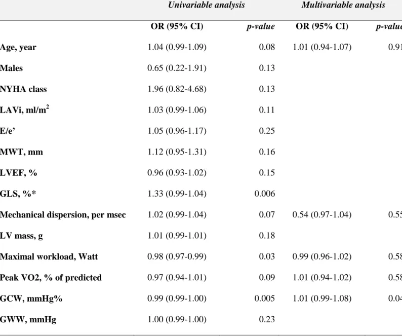
Among patients with HCM, 70 (85%) underwent CMR. By visual assessment, 20 (32%) patients had LGE in at least 2 myocardial segments. With respect to patients with no or mild LV fibrosis, HCM patients with significant fibrosis by CMR had increased wall thickness (15±4 vs 18±4 mm, p=0.002). GLS (-13±3 vs -15±3, p=0.003), mechanical dispersion (84±46 vs 65±22, p=0.03), and GCW (1343±411 vs 1735±419 mmHg%) (Figure 2B), were significantly impaired in patients with LV fibrosis, whereas a slight but not significant difference was observed in GWW (167±96 vs 120±66 mmHg%, p=0.22) between the two groups. At univariable analysis, GCW and exercise capacity were the only predictors of LV fibrosis. At multivariable analysis (entry p value <0.10) including age, mechanical dispersion, and the main functional parameters obtained at the CPET, GCW remained a significant predictor of LV fibrosis (OR 0.99, 95% CI: 0.99-1.00, p=0.04) (Table 2). At ROC curve analysis, a cut-off of GCW of 1623 mmHg% (AUC 0.80, 95% CI: 0.66-0.93, p<0.0001) was able to predict myocardial fibrosis with good sensitivity and fair specificity (82% and 67%, respectively (FIGURE 4).
Parameters related with myocardial constructive work.
The main correlates of GCW at linear regression analyses are depicted in Table 3. GCW was significantly associated with LV mass, LV diastolic and systolic function parameters, and GWW.
Functional parameters such as NYHA, VO2Peak and maximal exercise capacity were also correlated
with GCW. Interestingly, at multivariable analysis VO2peak (β=0.65, p=0.007) was the only
parameter associated with GCW.
Discussion
This pilot study is the first to explore new pressure-strain loops indices measured by transthoracic echocardiography in non-obstructive hypertrophic cardiomyopathy. Myocardial constructive work was significantly reduced in HCM compared to healthy controls, and emerged as a significant predictor of myocardial fibrosis.
Assessment of myocardial performance in hypertrophic cardiomyopathy.
In patients with HCM, myocardial fiber disarray and interstitial fibrosis are responsible for the progressive alteration of LV deformation parameters, which precedes overt LV dysfunction(5) (13). Another important feature of HCM is coronary microvascular dysfunction, which contributes to myocardial ischemia, LV remodeling, and adverse outcomes(16)(17).
Previous studies have shown that LV deformation is significantly impaired in patients with HCM despite the presence of a preserved LVEF(13)(12), and that LV mechanical dispersion is associated with myocardial fibrosis and arrhythmic events(13).
In our study GCW, GLS and mechanical dispersion were significantly impaired in HCM patients compared to controls, despite the presence of a similar LVEF.
The presence of LV fibrosis was associated with a further reduction of these functional parameters in patients with HCM, and GCW emerged as the main predictor of LV fibrosis at multivariable regression analysis. Previous studies by Russel et al. (14) and Delhaas et al.(18), have shown that the estimation of myocardial work by pressure-strain loops is not a mere measure of myocardial deformation, but it is an index of regional and global myocardial oxygen consumption and
metabolism. In patients with HCM, different degrees of microvascular dysfunction have been described(16)(17), which are associated with the localization (19) and global amount of myocardial fibrosis assessed by CMR(17).
According to these observations, the alteration of GCW observed in HCM patients might reflect both fibers disarray and metabolic impairment, becoming a sensitive marker of LV fibrosis. As a matter of fact, in our population a cut-off of 1623 mmHg% for GCW was able to predict myocardial fibrosis with a good sensitivity (82%) and fair specificity (67%). Correlates of GCW In patients with HCM, LV systolic and diastolic function parameters assessed by transthoracic echocardiography, LV mass, NYHA class and functional parameters assessed by CPET were all associated with GCW at univariable linear regression analysis. GCW estimation allows the assessment of LV function during the systolic and isovolumic relaxation phase. This might be of particular interest in patients with HCM, who experience a simultaneous impairment of LV deformation and diastolic properties.
In HCM, the alterations in LV geometry and mechanics are strictly associated with the development of LA dilatation and diastolic dysfunction (1)(2). Previous studies have shown that the development of diastolic dysfunction precedes overt LV systolic dysfunction in patients with non obstructive HCM, and is associated with the progression of functional limitation and poor outcome(20)(21). Interestingly, in the present study VO2peak was the main correlate of GCW at
multivariable analysis. Previous studies have shown that peak exercise parameters obtained by CPET are altered in patients with HCM(22)(23)(24). The reduction in VO2peak is ascribed to a
limited increase in cardiac output during exertion (22) (23), which is attributable to the increased LV wall thickness, LV fibrosis, and microvascular dysfunction observed in HCM patients (22).
Through its relationship to VO2 peak, GCW might provide an indirect estimation of functional
capacity in HCM patients. Compared to CPET, which require resources, expertise and training, the estimation of myocardial work can be easily performed by echocardiography and can be also applied to patients who are not able to exercise.
Clinical perspectives
In patients with HCM, LV systolic function assessed by LVEF does not reflect the entity of LV functional impairment. Myocardial constructive work by pressure strain loop is a simple and useful tool that is correlated with functional capacity and is a predictor of LV fibrosis.
Limitations
This is a retrospective study realized on a limited cohort of patients with non-obstructive HCM. The study population was heterogeneous in functional level, symptoms, and disease stage. CMR was performed only in a subset of patients, and myocardial fibrosis was estimated qualitatively. Further studies might explore the relationship between GCW and LV fibrosis as assessed quantitatively by LGE (25) or T1 mapping(26). Because of the limited number of patient included, the relationship between myocardial work and prognosis was not assessed in our survey. Follow-up studies to evaluate the relationship between GCW and events in HCM should be specifically designed.
Conclusion
GCW assessed non-invasively by pressure-strain loops was significantly reduced in HCM despite the presence of a normal LVEF. GCW emerged as a predictor of LV fibrosis, and was also correlated with VO2peak measured by CPET. These data support the utility of the estimation of
GCW in HCM in order to estimate LV performance and overall functional capacity.
Disclosures
The other authors have no relationship with industry and other relevant entities that may represent a conflict of interest in connection with the submitted article.
Acknowledgments
We deeply thank Mrs Dominique Fargeaud and the research team working at the CIC-IT1414, CHU Rennes for their skillful assistance.
Author contributions
EG: concept/design of the study, data analysis and interpretation, drafting article EV: data collection and analysis
AH: data collection
FS: data analysis, drafting article CL: critical revision of the article ML: data collection and analysis
ED: data collection, critical revision of the article, approval of the article
REFERENCES
1. Elliot PM, Anastas A, Borger MA et al: 2014 ESC Guidelines on diagnosis and management of hypertrophic cardiomyopathy: The Task Force for the Diagnosis and Management of Hypertrophic Cardiomyopathy of the European Society of Cardiology (ESC). Eur Heart J 2014;35:2733–2779. 2. Marian AJ., Braunwald E. Hypertrophic Cardiomyopathy: Genetics, Pathogenesis, Clinical
Manifestations, Diagnosis, and Therapy. Circ Res 2017;121(7):749–70. Doi: 10.1161/CIRCRESAHA.117.311059.
3. Chan RH, Maron BJ, Olivotto I, et al: Prognostic value of quantitative contrast-enhanced cardiovascular magnetic resonance for the evaluation of sudden death risk in patients with hypertrophic
cardiomyopathy. Circulation 2014;130:484–495.
4. Hinojar R, Fernández-Golfín C, González-Gómez A, et al: Prognostic implications of global myocardial mechanics in hypertrophic cardiomyopathy by cardiovascular magnetic resonance feature tracking. Relations to left ventricular hypertrophy and fibrosis. Int J Cardiol 2017;249:467–472.
5. Reant P, Mirabel M, Lloyd G, et al: Global longitudinal strain is associated with heart failure outcomes in hypertrophic cardiomyopathy. Heart Br Card Soc 2016;102:741–747.
6. Galli E, Leclercq C, Fournet M, et al: Value of Myocardial Work Estimation in the Prediction of Response to Cardiac Resynchronization Therapy. J Am Soc Echocardiogr 2018;31:220-230. 7. Galli E, Leclercq C, Hubert A, et al: Role of myocardial constructive work in the identification of
responders to CRT. Eur Heart J Cardiovasc Imaging 2017. Doi: 10.1093/ehjci/jex191.
8. Russell K, Eriksen M, Aaberge L, et al:Assessment of wasted myocardial work: a novel method to quantify energy loss due to uncoordinated left ventricular contractions. Am J Physiol Heart Circ Physiol 2013;305:H996-1003.
9. Lang RM, Badano LP, Mor-Avi V, et al: Recommendations for cardiac chamber quantification by echocardiography in adults: an update from the American Society of Echocardiography and the European Association of Cardiovascular Imaging. J Am Soc Echocardiogr 2015;28:1–39.
10. Nagueh SF, Smiseth OA, Appleton CP, et al: Recommendations for the Evaluation of Left Ventricular Diastolic Function by Echocardiography: An Update from the American Society of Echocardiography and the European Association of Cardiovascular Imaging. Eur Heart J Cardiovasc Imaging
2016;17:1321–1360.
11. Voigt J-U, Pedrizzetti G, Lysyansky P, et al: Definitions for a common standard for 2D speckle tracking echocardiography: consensus document of the EACVI/ASE/Industry Task Force to standardize
deformation imaging. J Am Soc Echocardiogr 2015;28:183–193.
12. Schnell F, Matelot D, Daudin M, et al: Mechanical dispersion by Strain Echocardiography: A Novel Tool to Diagnose Hypertrophic Cardiomyopathy in Athletes. J Am Soc Echocardiogr 2017;30:251–261. 13. Haland TF, Almaas VM, Hasselberg NE, et al: Strain echocardiography is related to fibrosis and
ventricular arrhythmias in hypertrophic cardiomyopathy. Eur Heart J – Cardiovasc Imaging 2016;17:613–621.
14. Russell K, Eriksen M, Aaberge L, et al: A novel clinical method for quantification of regional left ventricular pressure-strain loop area: a non-invasive index of myocardial work. Eur Heart J 2012;33:724–733.
15. Guazzi M, Adams V, Conraads V, et al: EACPR/AHA Scientific Statement. Clinical recommendations for cardiopulmonary exercise testing data assessment in specific patient populations. Circulation 2012;126:2261–2274.
16. Bravo PE, Pinheiro A, Higuchi T, et al: PET/CT assessment of symptomatic individuals with obstructive and nonobstructive hypertrophic cardiomyopathy. J Nucl Med 2012;53:407–414.
17. Olivotto I, Girolami F, Sciagrà R, et al: Microvascular function is selectively impaired in patients with hypertrophic cardiomyopathy and sarcomere myofilament gene mutations. J Am Coll Cardiol
2011;58:839–848.
18. Delhaas T, Arts T, Prinzen FW, et al: Regional fibre stress-fibre strain area as an estimate of regional blood flow and oxygen demand in the canine heart. J Physiol 1994;477:481–496.
19. Kong E-J, Lee S-H, Cho I-H. Myocardial Fibrosis in Hypertrophic Cardiomyopathy Demonstrated by Integrated Cardiac F-18 FDG PET/MR. Nucl Med Mol Imaging 2013;47:196–200.
20. Harris KM, Spirito P, Maron MS, et al: Prevalence, clinical profile, and significance of left ventricular remodeling in the end-stage phase of hypertrophic cardiomyopathy. Circulation 2006;114:216–225. 21. Maron MS, Rowin EJ, Olivotto I, et al: Contemporary Natural History and Management of
Nonobstructive Hypertrophic Cardiomyopathy. J Am Coll Cardiol 2016;67:1399–1409.
22. Lele SS, Thomson HL, Seo H, et al: Exercise capacity in hypertrophic cardiomyopathy. Role of stroke volume limitation, heart rate, and diastolic filling characteristics. Circulation 1995;92:2886–2894. 23. Sharma S, Elliott P, Whyte G, et al: Utility of cardiopulmonary exercise in the assessment of clinical
determinants of functional capacity in hypertrophic cardiomyopathy. Am J Cardiol 2000;86:162–168. 24. Saberi S, Wheeler M, Bragg-Gresham J, et al: Effect of Moderate-Intensity Exercise Training on Peak
Oxygen Consumption in Patients With Hypertrophic Cardiomyopathy: A Randomized Clinical Trial. JAMA 2017;317:1349-1357.
25. Aquaro G, Positano V, Pingitore A, et al: Quantitative analysis of late gadolinium enhancement in hypertrophic cardiomyopathy. J Cardiovasc Magn Reson 2010;12:21.
26. Ellims AH, Iles LM, Ling L, et al: Diffuse myocardial fibrosis in hypertrophic cardiomyopathy can be identified by cardiovascular magnetic resonance, and is associated with left ventricular diastolic dysfunction. J Cardiovasc Magn Reson 2012;14:76.
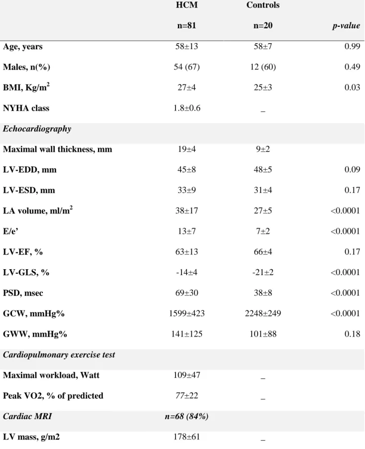
Table 1 Characteristics of patients with HCM and healthy controls. HCM n=81 Controls n=20 p-value Age, years 58±13 58±7 0.99 Males, n(%) 54 (67) 12 (60) 0.49 BMI, Kg/m2 27±4 25±3 0.03 NYHA class 1.8±0.6 _ Echocardiography
Maximal wall thickness, mm 19±4 9±2
LV-EDD, mm 45±8 48±5 0.09 LV-ESD, mm 33±9 31±4 0.17 LA volume, ml/m2 38±17 27±5 <0.0001 E/e’ 13±7 7±2 <0.0001 LV-EF, % 63±13 66±4 0.17 LV-GLS, % -14±4 -21±2 <0.0001 PSD, msec 69±30 38±8 <0.0001 GCW, mmHg% 1599±423 2248±249 <0.0001 GWW, mmHg% 141±125 101±88 0.18
Cardiopulmonary exercise test
Maximal workload, Watt 109±47 _
Peak VO2, % of predicted 77±22 _
Cardiac MRI n=68 (84%)
LV mass, g/m2 178±61 _
LV-EDV, ml 147±46 _
LV-ESV, ml 51±33 _
LGE≥2 segments, n(%) 21 (31)
BMI, body mass index; EDD, end-diastolic diameter; EDV, end-diastolic volume; EF, ejection fraction; ESD, end-systolic diameter; ESV; end-systolic volume; GCW, global constructive work; GWE, global work efficiency; GWW, global wasted work; IVSd, diastolic inter-ventricular septum thickness; LA, Left atrium; LV, left ventricle; LGE, late gadolinium enhancement; NYHA, New York Heart Association functional class.
Table 2. Predictor of significant myocardial fibrosis at late gadolinium enhancement
Univariable analysis Multivariable analysis
OR (95% CI) p-value OR (95% CI) p-value
Age, year 1.04 (0.99-1.09) 0.08 1.01 (0.94-1.07) 0.91 Males 0.65 (0.22-1.91) 0.13 NYHA class 1.96 (0.82-4.68) 0.13 LAVi, ml/m2 1.03 (0.99-1.06) 0.11 E/e’ 1.05 (0.96-1.17) 0.25 MWT, mm 1.12 (0.95-1.31) 0.16 LVEF, % 0.96 (0.93-1.02) 0.15 GLS, %* 1.33 (0.99-1.04) 0.006
Mechanical dispersion, per msec 1.02 (0.99-1.04) 0.07 0.54 (0.97-1.04) 0.55
LV mass, g 1.01 (0.99-1.01) 0.18
Maximal workload, Watt 0.98 (0.97-0.99) 0.03 0.99 (0.96-1.02) 0.58
Peak VO2, % of predicted 0.97 (0.94-1.01) 0.09 1.01 (0.94-1.02) 0.58
GCW, mmHg% 0.99 (0.99-1.00) 0.005 1.01 (0.99-1.08) 0.04
GWW, mmHg 1.00 (0.99-1.00) 0.23
*This variable was not inserted in the multivariable model to avoid collinearity
Table 3. Relationship between global constructive work and different morphological and functional parameters in HCM
Univariable analysis Multivariable analysis
Variables β p-value β p-value
Age, years -0.31 0.001 -0.18 0.36
NYHA class (I to IV) -0.34 0.005 -0.27 0.19
LAVi, ml/m2 -0.39 <0.0001 -0.16 0.38
E/e’ -0.55 <0.0001 -0.14 0.43
LV-EF, % 0.31 0.001 -0.04 0.82
GLS, %* -0.92 <0.0001 -
Mechanical dispersion, msec -0.51 <0.0001 -0.27 0.14
LV Mass, g -0.35 0.007 -0.04 0.82
GWW, mmHg% -0.21 0.03 -0.26 0.19
VO2peak, % 0.36 0.01 0.64 0.007
Maximal workload, Watt 0.27 0.04 -0.39 0.12
*This variable was not inserted in the multivariable model to avoid collinearity
Figure captions
Figure 1.
The noninvasive LV pressure curve was obtained using an empiric, normalized reference curve. The latter was adjusted according to the duration of the isovolumetric and ejection phases of the left ventricle, which were defined by the assessment of valvular events by echocardiography (A). Pressure data were then combined with left ventricular global longitudinal strain data using the R-wave onset in the electrocardiogram as a common time reference (B, left panel). These data, obtained in a patient with septal hypertrophic cardiomyopathy, were then used for the estimation of the pressure-strain loop (B, right panel).
Figure 2.
Box plots of global constructive work (GCW) in 82 patients with hypertrophic cardiomyopathy (HCM) and 20 healthy controls (A). Box plots comparing GCW in HCM patients with (LGE+) and without (LGE-) significant left ventricular fibrosis at late gadolinium enhancement cardiac resonance imaging (B).
Figure 3.
(A) Example of global constructive work (GCW) estimation in a control patient. No left ventricular hypertrophy is evident at transthoracic echocardiography (upper panels). The pressure strain loop (PLS) in the lower panel represent the GCW performed by the left ventricle (LV). In the bull’s eye representing regional constructive work, work is homogenously distributed through the different LV segments (lower panel, right side).
(B) Example of myocardial constructive work estimation in a patient with hypertrophic cardiomyopathy (HCM). Myocardial hypertrophy is localized in the LV septum (yellow arrows) and is associated with myocardial fibrosis detected by late gadolinium enhancement (green arrows, upper panel).
In the lower panel, the pressure strain loop represents the constructive work performed globally by the left ventricle (red curve) and by the antero-septal segment (green curve). The bull’s eye shows a significant reduction of myocardial constructive work in the septal segment, where myocardial fibrosis has been observed (asterisks).
Figure 4.
ROC curve analysis for global constructive work as a predictor of left ventricular fibrosis
Supplementary material Figure A.
Example of the calculation of myocardial work for a left ventricular segment.
For each segment, myocardial work was calculated from left ventricular pressure (upper panel) and strain recordings (middle panel). Work was calculated as a function of time through the cardiac cycle, from mitral valve opening to mitral valve closure.
The myocardial work curve and the method of calculation of myocardial constructive work (CWsegm) and wasted work (WWsegm) for a LV segment are depicted in the lower panel.
Constructive and wasted work are marked respectively as grey and red segments.
AVC, aortic valve closure; AVO, aortic valve opening; CW, constructive work; IVC, isovolumic contraction; IVR, isovolumic relaxation; E, left ventricular ejection; MVC, mitral valve closure; MVO, mitral valve opening; WW, wasted work.