HAL Id: inserm-00709339
https://www.hal.inserm.fr/inserm-00709339
Submitted on 18 Jun 2012
HAL is a multi-disciplinary open access
archive for the deposit and dissemination of
sci-entific research documents, whether they are
pub-lished or not. The documents may come from
teaching and research institutions in France or
abroad, or from public or private research centers.
L’archive ouverte pluridisciplinaire HAL, est
destinée au dépôt et à la diffusion de documents
scientifiques de niveau recherche, publiés ou non,
émanant des établissements d’enseignement et de
recherche français ou étrangers, des laboratoires
publics ou privés.
subpopulations reveals genetic pathways that regulate
tangential migration in the vertebrate hindbrain.
Karsten Benzing, Stefanie Flunkert, Andreas Schedl, Dieter Engelkamp
To cite this version:
Karsten Benzing, Stefanie Flunkert, Andreas Schedl, Dieter Engelkamp. A novel approach to
se-lectively target neuronal subpopulations reveals genetic pathways that regulate tangential migration
in the vertebrate hindbrain.. PLoS Genetics, Public Library of Science, 2011, 7 (6), pp.e1002099.
�10.1371/journal.pgen.1002099�. �inserm-00709339�
Subpopulations Reveals Genetic Pathways That Regulate
Tangential Migration in the Vertebrate Hindbrain
Karsten Benzing1, Stefanie Flunkert1, Andreas Schedl2,3, Dieter Engelkamp1,4*
1 Max Planck Institute for Brain Research, Department of Neuroanatomy, Frankfurt, Germany, 2 INSERM UMR636, Centre de Biochimie, Nice, France, 3 University of Nice Sophia Antipolis, Nice, France,4 Department of Biology, Friedrich-Alexander-University of Erlangen-Nu¨rnberg, Erlangen, Germany
Abstract
Vertebrate genes often play functionally distinct roles in different subsets of cells; however, tools to study the cell-specific function of gene products are poorly developed. Therefore, we have established a novel mouse model that enables the visualization and manipulation of defined subpopulations of neurons. To demonstrate the power of our system, we dissected genetic cascades in which Pax6 is central to control tangentially migrating neurons of the mouse brainstem. Several Pax6 downstream genes were identified and their function was analyzed by over-expression and knock-down experiments. One of these, Pou4f2, induces a prolonged midline arrest of growth cones to influence the proportion of ipsilaterally versus contralaterally settling neurons. These results demonstrate that our approach serves as a versatile tool to study the function of genes involved in cell migration, axonal pathfinding, and patterning processes. Our model will also serve as a general tool to specifically over-express any gene in a defined subpopulation of neurons and should easily be adapted to a wide range of applications.
Citation: Benzing K, Flunkert S, Schedl A, Engelkamp D (2011) A Novel Approach to Selectively Target Neuronal Subpopulations Reveals Genetic Pathways That Regulate Tangential Migration in the Vertebrate Hindbrain. PLoS Genet 7(6): e1002099. doi:10.1371/journal.pgen.1002099
Editor: Sabine Cordes, University of Toronto and Samuel Lunenfeld Research Institute at Mount Sinai Hospital, Canada Received August 9, 2010; Accepted April 8, 2011; Published June 16, 2011
Copyright: ß 2011 Benzing et al. This is an open-access article distributed under the terms of the Creative Commons Attribution License, which permits unrestricted use, distribution, and reproduction in any medium, provided the original author and source are credited.
Funding: This work was supported by the Max Planck Society. KB was supported by the Graduiertenkolleg Neuronale Plastizita¨t. The funders had no role in study design, data collection and analysis, decision to publish, or preparation of the manuscript.
Competing Interests: The authors have declared that no competing interests exist. * E-mail: dengelka@biologie.uni-erlangen.de
Introduction
Understanding cell-specific regulatory mechanisms is a major challenge in the post-genome era. Particularly in mammals, the reiterated usage of the same transcription factor in distinct subsets of cells or during distinct developmental time points provides the basis to generate thousands of individual cell types with a relatively small number of genes. A single transcription factor may therefore elicit variable downstream effects depending on the context of its expression. Tissue-specific knockout strategies based e.g. on the Cre-lox-system, or promoter-driven transgenic models allow a cell-specific manipulation of genes. However, as these techniques rely on the generation of new transgenic animals for each gene-combination analyzed they are laborious and time-consuming. Here, we combined a transgenic model with tissue-specific transfection protocols and organotypic cultures to enable the quick analysis of numerous genes in a cell-specific manner. As a proof of principle we applied our system to decode molecular pathways initiated by the transcription factor Pax6 which is involved in neuronal cell migration and axonal pathfinding processes.
Pax6, a homeodomain and paired domain containing transcrip-tion factor, is a major determinant of visual and olfactory sensory structures and is essential for a variety of patterning and pathfinding processes throughout the nervous system [1–3]. Depending on the context and area of expression Pax6 initiates varying downstream effects. Homozygous small eye (Pax6Sey/Sey)
mouse and rat embryos, which lack functional Pax6, do neither generate eye nor nasal structures and are deficient in ventral diencephalic structures [4–9]. In the ventral hindbrain and spinal cord, Pax6 controls the dorso-ventral patterning of motorneurons and of interneurons [5,10]. In the cerebral cortex Pax6 determines the neurogenic potential of radial glial cells [11,12]. Throughout the developing nervous system, with the exception of the midbrain, Pax6 is expressed in a ventral and a dorsal pool of progenitor cells. Although the dorsal Pax6 expression domain has achieved much less attention than the ventral domain there is evidence that Pax6 plays a pivotal role in the specification and migration of neurons derived from this domain [13–16].
The dorsal domain of Pax6 positive neuronal precursors of the hindbrain includes the rhombic lip (RL) [14,16] which comprises the interface between the dorsal neuroepithelium and the roof plate. The RL is the source of several tangentially migrating neurons (see also Figure 1A) [14,17–24]. The most notable are the neurons of the marginal migratory stream (mms; also pes) which migrate from the rhombic lip circumferentially around the medulla towards their contralateral destinations to settle in the ECN (external cuneate nuclei) and the LRN (lateral reticular nuclei) [17,25]. Owing to the superficial nature of the mms migration these neurons serve as paradigm to study neuronal migration and axonal pathfinding processes.
The highly complex neuronal circuits of the vertebrate nervous system are established during development when growing axons travel considerable distances towards their targets to generate the
appropriate connections. This wiring process depends on attrac-tive and repulsive factors which emanate from final or interme-diate cellular targets and which are interpreted by cell surface receptors located on axonal growth cones [26,27]. Although the general principles were uncovered during the past years our understanding of axonal pathfinding processes is far from being complete. Current methods to analyze candidates regulating neuronal migration and axonal navigation processes are laborious and often involve the generation of transgenic animals for each gene analyzed. Non-transgenic methods, as DiI labeling of neurons or vector-driven mis-expression of gene constructs, are suitable for use with certain applications, however, they are neither cell specific nor can they be targeted to distinct neuronal subpopulations.
Here we describe a novel transgenic mouse model, which allows the specific and exclusive visualization and manipulation of subsets of neurons in the developing brain. To demonstrate the power of this system we have analyzed the role of Pax6 in migrating neurons of the brainstem. In Pax6 mutant mice migration of these neurons is distorted and some neurons differentiate at ectopic positions. Using transplantation, knock-down and over-expression experi-ments we show that distinct migratory features are controlled by discrete sets of Pax6 downstream genes. These results demonstrate the potential of our transgenic mouse model as a tool to study the role of Pax6 in individual neurons. Moreover, our system should be widely applicable to study virtually any gene that acts during cell determination, axonal pathfinding and/ or cell migration processes.
Results
A novel mouse model to visualize and manipulate subsets of neurons
The functional analysis of genes in restricted tissues often involves the generation of inducible knockout mice or mice over expressing transgenic constructs. To simplify this time-consuming process we developed an in vitro model that enables the visualization and manipulation of defined populations of neurons. To label neurons in a largely unlabelled background we searched for genes that were expressed in only a subset of neuronal precursors and in migrating neurons. Pax6 meets these criteria ideally. Pax6 is expressed in several groups of tangentially migrating neurons and their precursors as well as in a small population of radially migrating neurons and their precursors (Figure 1A–1C) [5,10,12,14,16,28].
We adopted the Tet binary system [29] and generated YAC (yeast artificial chromosome) transgenic mice which expressed the tetracycline dependent transactivator (tTA) in all Pax6 positive cells. A 420 kb YAC spanning the human PAX6 locus (Y593) [30] was modified such that the PAX6 coding region was replaced with a cassette containing an IRES (internal ribosomal entry site) and the tTA (Figure 1D). Previously, we and others had shown that the unmodified YAC Y593 contains all elements driving full functional PAX6 expression [30–32] and, in agreement with this, Tg(PAX6-tTA) mice showed a wide overlap of tTA and endogenous murine Pax6 expression (Figure S1). Tg(PAX6-tTA)mice were entirely normal and control experiments insured that neuronal patterning and migration was unaltered.
tTA is a transcriptional activator that at moderate levels of expression is completely inert in vertebrates, yet, enables the activation of artificial constructs containing a tTA-DNA-binding element (TRE = tetracyline responsive element). To examine whether our transgenic model specifically allows the labeling of only Pax6 positive cells we introduced by electroporation reporter constructs driving the green fluorescent protein into transgenic embryos. In all instances only Pax6 positive cells, e.g. retinal precursor cells, cortical precursors, or cerebellar granule cells, expressed the reporter genes (Figure S1). Non-transgenic embryos or Pax6 negative tissues did not induce reporter gene expression (Figure 1H, Figure S1). Together these results demonstrate that Tg(PAX6-tTA)mice enable the targeting of reporter gene constructs specifically to Pax6 positive cells and tissues during development. As Tg(PAX6-tTA) mice allow any gene to be targeted to Pax6 expressing cells, they are of potential value to study neuronal migration and axonal pathfinding processes and for the analysis of Pax6 downstream effects. As a proof of principle, we chose to focus on the marginal migration stream (mms). Like other tangentially migrating neurons, mms neurons use the same or similar navigational cues as do growing axons, and migration of mms neurons is severely disturbed in Pax6 mutant Pax6Sey/Seymice (see below). Neurons of the mms are generated at the rhombic lip and migrate circumferentially around the embryonic brainstem to generate the contralateral lateral reticular (LRN) and the external cuneate (ECN) nuclei (Figure 1A) [16,21–23,25]. Migration starts at E13.0 and is completed by E16.5. Pax6 is expressed in precursors at the rhombic lip, in all migrating neurons of the mms and during the initial period of settling in the target nuclei (Figure 1B, 1C and data not shown). Antibody staining and in situ hybridization (not shown) of Tg(PAX6-tTA) mice confirmed a complete overlap of Pax6 and tTA expression in these neurons (Figure 1I).
To visualize migrating mms neurons in Tg(PAX6-tTA) mice, reporter constructs were introduced into neuronal precursor cells in the left rhombic lip by electroporation at E12.5 before migration had begun (Figure 1E). Whole brainstems including the cerebellar primordium were then sustained in organotypic filter cultures for up to 14 days as an open book preparation which allowed the observation of migrating neurons with a fluorescence microscope from above (Figure 1F, 1G). Our approach to use a binary system ensured that only Pax6 positive neurons containing tTA and a TRE reporter construct expressed the desired reporter genes. This procedure resulted in the specific labeling of mms neurons originating only from one rhombic lip. Pax6 positive neurons originating from the opposite rhombic lip remained unlabelled as were Pax6 negative (and therefore tTA negative) neurons originating from regions close to the rhombic lip. Unlabelled neurons included neurons of the submarginal migra-tion stream (sms) which generate the inferior olive (IO) thus demonstrating the specificity of our model. To allow the
Author Summary
In mammals, many genes execute a unique set of distinctive and common functions in different cell types. Strategies to address these individual roles often involve the generation of series of transgenic animals. Here, we present a novel approach that combines a single transgenic mouse line with tissue-specific transfection protocols and organotypic cultures to enable the quick analysis of numerous genes in a cell-specific manner. As a proof of principle, we analyzed the function of transcrip-tion factors in tangentially migrating neurons of the developing vertebrate hindbrain. We identified a tempo-rary halt in migration as a novel mechanism for neurons to decide whether to cross or not cross the midline. Our model may serve as a general tool to quickly study axonal pathfinding, neuronal cell migration, or patterning pro-cesses in a well-defined population of neurons.
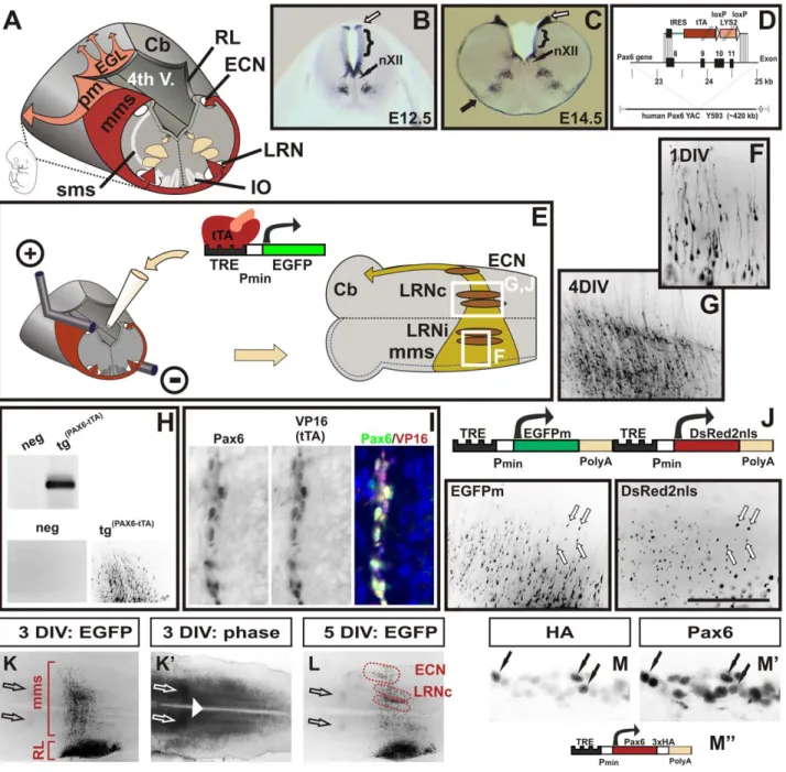
Figure 1. A novel mouse model to visualize and manipulate Pax6 positive neurons. (A) Schematic diagram of tangentially migrating neurons in the mouse brainstem. Pax6 positive neurons of the marginal migration stream (mms) and of the pontine migration (pm) are derived from the rhombic lip (RL) and generate the lateral reticular (LRN), the external cuneate nuclei (ECN), the pontine nuclei and the pontine reticulotegmental nuclei (not shown), respectively. Pax6 positive external granule cells (EGL) initially migrate rostrally on the surface of the cerebellar primordium (Cb). Pax6 negative neurons of the submarginal migration stream (sms) generate the inferior olive (IO). Distribution of Pax6 positive cells in the wt hindbrain at E12.5 (B) and at E14.5 (C) as shown by in situ hybridization on coronal vibratome sections. Pax6 is expressed in precursor cells at the caudal RL (open arrows in B,C), in ventral precursor cells at the basal plate, in groups of ventrally migrating neurons and in the mms (filled arrow in C). The dorsal alar plate (brackets in B,C), the hypoglossal nuclei (nXII), and the floor plate are negative for Pax6. (D) Diagram of the targeting construct to replace the coding sequence of human PAX6 in YAC Y593 with an IRES-tau-tTA-LYS2 cassette. For a more detailed description of the construct see the Materials and Methods section. (E) Schematic drawing of the electroporation and brainstem culture procedures. Responder constructs consisting of tetracycline responsive elements (TRE), a minimal promoter (pmin), and a reporter gene (e.g. EGFP) are injected into the fourth ventricle of E12.5
Tg(PAX6-tTA)embryos and then transfected into one RL by electroporation. Subsequently, the hindbrain is dissected and cultured with the ventricular face onto MilliporeCM filters. After prolonged cultures mms neurons settle either in the ipsi- or the contralateral LRN (LRNi, LRNc), or in the ECN. All neurons project to the contralateral cerebellar hemisphere (Cb). (F and G) Migrating mms neurons visualized by EGFP fluorescence in transfected brainstems. The enlarged areas shown are indicated by white boxes in (E). After 1 day in vitro culture (1 DIV) migrating neurons with long leading and short trailing processes emerge from the transfected RL (F). After 4DIV the majority of mms neurons have settled in contralateral LRN (LRNc) (G). Genotyping of transfected cultures confirms that responder constructs are exclusively activated in Tg(PAX6-tTA)and not in non-transgenic embryos (H).
Double-immunolabeling of mms neurons with Pax6 and VP16 (to recognize tTA) antibodies confirm a complete overlap of Pax6 and tTA expression (I). A responder vector containing two TRE elements drives expression of two genes simultaneously: a cytoplasmic green fluorescent protein (EGFPm) and a nuclear red fluorescent protein (DsRed2nls) (J). The left and right images are green and red fluorescent images, respectively, of migrating mms neurons after 1.5DIV. K, K9, and L are low magnification fluorescence (K, L) and phase contrast (K9) images of cultures transfected with an EGFP
simultaneous visualization and manipulation of neurons we designed reporter constructs containing two TRE elements (Figure 1J). Control constructs co-expressed a cytoplasmic green fluorescent protein (EGFPm) and a nuclear red fluorescent protein (DsRed2nls) in 99% (61; n = 10) of labeled neurons demonstrating that our reporter constructs enable the co-expression of two genes in the same neurons (Figure 1J). To enable statistical analysis of the cultures, the territories of the LRN and the ECN were delineated using visible landmarks (Figure 1K, 1K9, 1L; see also the Materials and Methods section). Immunolabeling of cultures expressing a HA-tagged Pax6 construct demonstrate that over-expression of TRE constructs in Tg(PAX6-tTA) transgenic cultures result in moderate levels of protein expression that are in the range of physiological Pax6 concentrations (Figure 1M, 1M9, 1M’’).
Pax6 plays multiple roles in patterning and guiding migrating ECN and LRN neurons
Pax6 mutant Pax6Sey/Seymice display multiple neuronal pattern-ing and migration defects. We therefore wished to determine whether Pax6 also regulates the mms. At the anatomical level, several features of the mms are severely disturbed in Pax6 mutant Pax6Sey/Sey embryos. Most noticeable, the initiation of migration and the midline crossing was delayed by 0.5 days (asterisks in Figure 2A, 2A9 and data not shown; see also Figure S2 and Figure 4A, 4A9 The expression patterns of Pax2, Dcx, NK1R, and DopH was unaltered indicating that there is no general developmental delay in the mutant brainstem (data not shown). In Pax6Sey/Seyembryos some migrating mms neurons used a sub-marginal instead of a sub-marginal migration path (black arrowhead in Figure 2A9; see also Figure S2 and Figure 4A9) and at E14.5 a large number of mutant neurons accumulated around the midline suggesting a reduced pace in midline crossing (black arrow in Figure 2B, 2B9). Furthermore, a subset of Pax6 positive neurons migrated along the midline into the parenchyma of the hindbrain (white arrowheads in Figure 2C, 2C9). We used a variety of markers, e.g. antibodies against the potassium channel Kcnj6, and DiI tracing of mossy fiber projections to discriminate between mms neurons (generating the ECN and LRN) and neurons of the sms (generating the IO). These experiments all indicated a complete loss of neurons in the ECN and a disorganized settling of neurons in the LRN (Figure 2D, 2D9, 2E, 2E9; Figure S2, and data not shown). Many Kcnj6 positive neurons were even observed within the inferior olivary territory (black arrows in Figure 2E9) and dorsally to the IO at the midline (open arrow in Figure 2E9). In agreement with previous reports [16], we found a slight enlargement of the IO at E14.5 when we used the Ets transcription factor Etv1 as a IO specific marker [33] (Figure S3 and data not shown). However, by labeling for the axon-guidance-molecule B (RgmB) no alterations in the general architecture of the IO were seen [34] (Figure S3). This is consistent with our observation that misguided Pax6Sey/Sey mms neurons make only a negligible contribution to the IO or settle in the periphery of the IO. In summary, these data demonstrate that migration of Pax6Sey/Seymms neurons is severely disrupted. Mutant neurons of the mms are
initially delayed. Later, a number of neurons use a sub-marginal migration path, migration is disturbed at the midline and several neurons migrate to ectopic positions along the midline. Lastly, the normal structure of the LRN is lost, the ECN is completely missing, and the IO is enlarged.
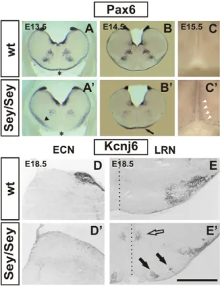
Figure 2. Migration defects ofPax6Sey/Sey pre-cerebellar
neu-rons: histology. Coronal vibratome Pax6 in situ hybridizations of wt (A - C) and mutant (A9–C9) embryos. The Pax6Sey/Seymutation is a result of
single base pair substitution in the Pax6 gene that leads to a shortened non-functional protein product [4]. Pax6 transcript levels, however, are largely unaffected in mutants therefore allowing the detection of Pax6 expressing cells in Pax6Sey/Seyembryos. At E12.5 the mms has not yet
formed (see Figure. 1B), but, at E13.5 first neurons of the mms have already crossed the midline (asterisk in A). In the mutant, migration of mms neurons is delayed, some neurons use a sub-marginal migration (arrowhead in A9), and by E13.5 neurons have not reached the midline (asterisk in A9). At E14.5 Pax6 positive neurons accumulate around the ventral midline (arrow in B9) and at E15.5 some neurons migrate dorsally along the ventral midline (white arrowheads in C9). In wt embryos all mms neurons migrate along the marginal migration route (A,B) and no Pax6 positive cells are seen at the ventral midline (C). Immunohisto-chemical labeling with an a-Kcnj6 antibody on coronal E18.5 wt (D,E) and Pax6Sey/Sey(D9,E9) sections. The a-Kcnj6 antibody strongly labels the
wt ECN (D) and the LRN (E). a-Kcnj6 is completely absent from the ECN territory in Pax6Sey/Seyembryos (D9) and mutant LRN neurons (E9) are
scattered at the LRN territory, within the inferior olivary complex (black arrows in E9), and at both sides of the midline (open arrow in E9). The dotted line in E and E9 indicates the midline. (Scale bar is 1 mm in [A, A9]; 1.2 mm in [B, B9]; 380 mm in [C]; 0.55 mm in [D, D9, E, E9].) doi:10.1371/journal.pgen.1002099.g002
construct as indicated in E. After 3DIV several hundred neurons have left the transfected rhombic lip (RL) to migrate along the mms. At 5DIV all neurons have reached their target positions. 25 similar cultures were used to outline the territories of the LRNc and ECN in respect to surrounding ‘‘landmarks’’: the superior olivary nuclei (open arrows), the inferior olivary nuclei, the midline (white arrowhead), and the rhombic lip (RL). Double-immunolabeling of cultures transfected with a hemagglutinin (HA)-tagged Pax6 construct (M, M9, M’’) confirm that over-expression results in Pax6 levels comparable to endogenous Pax6 levels. Immunolabeling with HA-tag antibodies (M) mark transfected cells (black arrows) which express endogenous and exogenous Pax6. Note, that Pax6 levels in some untransfected cells (only endogenous Pax6) are as high as Pax6 levels in transfected cells (endogenous plus exogenous Pax6). (The scale bar represents 0.8 mm in [B]; 1 mm in [C]; 150 mm in [F]; 0.44mm in [G, J]; 0.9 mm in [H]; 70 mm in [I]; 1.8 mm in [K, K9, L]; 50 mm in [M, M9]).
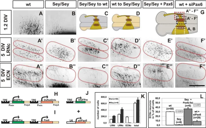
In order to dissect the complex neuronal cell migration defects observed in Pax6Sey/Seymice, Pax6Sey/Sey mice were crossed to the Tg(PAX6-tTA)transgenic line. Comparison of cultures obtained from wt and from Pax6Sey/Sey embryos confirmed the anatomical observations described above. Pax6Sey/Seymms cells showed an initial delay in the onset of the migration (Figure 3A, 3B) and a disturbance at midline crossing later on. After 5 DIV (5 days in vitro) Pax6Sey/Sey mms neurons settled randomly in the LRN (Figure 3B9) but failed to form any ECN structures (Figure 3B’’). In contrast, wt cultures formed a well organized LRN in which cells settled in a dorsal and ventral sub-nucleus of the LRN (Figure 3A9) and in a distinguished ECN (Figure 3A’’). To quantify the effect we counted labeled cells in cultures from wt (LRNi = ipsilateral LRN = 11267; LRNc = contralateral LRN = 266616; ECN = 86614; total number of cells = 610634; n = 50) and Pax6Sey/Seyembryos (LRNi = 313623; LRNc = 426646; ECN = 862; total number of cells = 670671; n = 8) (Figure 3K). These data suggested that there were no alterations in the gross number of migrating neurons between wt and Pax6Sey/Seyembryos and confirmed the complete absence of an ECN in Pax6Sey/Sey embryos. In both, mutant and wt tissues, a proportion of LRN neurons settled ipsilaterally (Figure 3K). All LRN neurons, however, projected to the contralateral cerebellum in
respect to their origin from one rhombic lip, explaining the observations by Bourrat and Sotelo of an ipsilateral and contralateral contribution of mossy fibers [17].
The Pax6Sey/Sey migration defect could be caused either by a direct cell autonomous action of Pax6 in migrating RL precursors or via an indirect non-autonomous effect, for example in the ventral domain of Pax6expression (e.g. by altering migration cues at the midline). We performed three types of experiments to discriminate between these alternatives. First, we transplanted transfected Pax6Sey/Sey rhombic lips onto wt brainstems and vice versa (Figure 3C, 3D). Unexpectedly, migrating Pax6Sey/Seyneurons (in a wt host) formed a well organized LRN (Figure 3C9), but no ECN (Figure 3C’’). In contrast, wt neurons (in a mutant host) failed to form a correctly organized LRN (Figure 3D9), but were able to generate a normal ECN (Figure 3D’’). These data suggested, that Pax6 may act cell autonomously in generating ECN neurons, but non-autonomously in specifying the correct sub-organization of LRN neurons. To further validate this assumption we rescued the Pax6Sey/Sey migration defect by re-expression of Pax6. We tested the two major splice variants and of these, the expression of the Pax6(-5a) isoform in PaxSey/Sey6 rhombic lips resulted in a full recovery of the ECN (Figure 3E’’),
Figure 3. Migration defects ofPax6Sey/Seypre-cerebellar neurons: cultures. (A–F) Fluorescence images of transfected brainstem cultures. The
enlargements shown are indicated by dotted boxes in (G). Dotted lines in A9–F9 and A’’–F’’ indicate the LRN and ECN territories, respectively. The respective positions of the LRN and ECN are placed according to landmarks derived from wt cultures (see Figure 1). 1.2DIV wt mms neurons have left the rhombic lip (RL) and have almost reached the midline (A). Neurons settle after 5DIV in two sub-nuclei of the contralateral LRN (A9, LRNc) and in the ECN (A’’). In contrast, Pax6Sey/Seymms neurons are initially delayed (B), and then settle in a scattered LRNc (B9) but not in the ECN (B’’). After
transplantation of a Pax6Sey/SeyRL (beige area in C) onto a wt brainstem (grey area in C), two LRN sub-nuclei are formed (C9), but no ECN (C’’), whereas, transplantation of a wt RL (grey area in D) onto a Pax6Sey/Seybrainstem (beige area in D), results in a scattered LRNc (D9) and a normal ECN (D’’).
Transfection of the Pax6Sey/SeyRL with Pax6(-5a) results in a rescue of the ECN (E’’), but not of the LRNc phenotype (E9). shRNA down regulation of
Pax6 causes a loss of the ECN (F’’), however, the LRNc remains unaffected (F9). Responder constructs used are: (H) for (A–D), (I) for (E), and (J) for (F), respectively. A9, B9, and E9 are red fluorescence images of the nuclear DsRednls protein; all other images are green fluorescence images of the cytoplasmic EGFP. (K) Quantification of settled LRN and ECN cells after 5DIV. There are no significant differences between the total number of migrating cells in wt and Pax6Sey/Seycultures. (L) Quantification of neurons settling in the ECN territory after 5DIV in cultures. Standard Student’s t test
was used to assess the significance of changes as indicated by a p-value of ,0.001; values are 6SEM. (Scale bar is 0.44 mm in [D–I’’].) doi:10.1371/journal.pgen.1002099.g003
but a disorganized LRN (Figure 3E9), whereas, the Pax6(+5a) variant was ineffective (not shown). Thus, in the RL Pax6 splice variants differ in their biological activity, similar to the embryonic cortex [35]. Lastly, we diminished the endogenous Pax6 mRNA by using siRNAs. Transfection of siRNAs or over expression of shRNA constructs directed against Pax6 (Figure S4) resulted in a massive reduction of ECN cells in wt explants (Figure 3F’’), whereas, control constructs had no effect (not shown). The effect was quantified by counting labeled cells that had settled in the ECN (Figure 3L). Taken together, these experiments demonstrate that our model system enables the simultaneous visualization and manipulation of tangentially migrating cells in the mouse brainstem. In addition, we have shown that Pax6 plays numerous distinct roles in the formation and migration of mossy fiber producing neurons. Moreover, the combination of a binary model and organotypic culture assays facilitates a quick discrimination between cell-autonomous and non-autonomous effects.
Axonal pathfinding receptors guide mms neurons
We identified several genes whose expression was altered in Pax6Sey/Seymms neurons (Figure S5; see also Materials and Methods). To gain more insights into the function of these putative Pax6 downstream targets all genes were over-expressed or their
expression level was diminished with shRNAs. Those genes which showed the most noticeable effects are summarized in Table 1.
The altered migration and settling behavior of Pax6Sey/SeyECN/ LRN neurons suggested that migration cues were changed in Pax6Sey/
Sey
embryos. The most prominent candidates are ligand/ receptor couples of the Slit/ Robo- and Netrin/ Dcc- pathways [36,37]. Expression of Netrin1, Dcc, and Robo1,2 and 3 was unaltered in migrating Pax6Sey/Sey mms neurons (Figure 4A, 4A9, Figure S2, and data not shown). However, Slit1 and Slit2 which were expressed in the hypoglossal nuclei were both lost in Pax6Sey/Seyembryos (Figure 4B, 4B9 and Figure S2) [5,10]. Motorneurons of the hypoglossal nuclei are in close proximity to the LRN settling territories suggesting that Slit1 and Slit2 expression provided from these neurons may determine the place of LRN settlement. To test this hypothesis we performed transplantation experiments and shRNA driven knock-down of the Slit-receptor Robo3 in migrating mms neurons. Both types of experiments resulted in a disorganized LRN similar to the phenotype observed in Pax6Sey/Seymice (Figure 4C-4H). The above results indicate that factors provided from the hypoglossal nucleus, (most likely Slit1 and Slit2) determine the place of LRN settlement. These data also explain the cell non-autonomous role of Pax6 during this process. Hypoglossal neurons are Pax6 negative, but are completely lost in Pax6Sey/Seyembryos (Figure S2) [5,10]; hence, Slit1 Table 1. Summary of phenotypes obtained in organotypic cultures.
gene
Pax6Sey/Sey
expression phenotype Pax6 (Q) : unaltered migration
: (shRNA or siRNA or EnR) migration delay, ECN loss, midline arrest Slit1,2 (nXII:Q) : unaltered migration
: (ablation of the Slit1-positive hypoglossal nuclei): sub-marginal migration, LRN disruption Robo3 < : (shRNA) sub-marginal migration, LRN disruption
Dcc < : enlarged ECN
: (shRNA or siRNA) migration delay, ECN loss, altered migration path Unc5h1 Q(RL only) : reduced cell migration and cell death
Unc5h3 < : migration delayed, altered migration path : (shRNA) unaltered migration
Neurod1 < : migration delay, altered migration path (EnR): no migration
Neurod2 < : migration delay, altered migration path (EnR): no migration
Pou4f2 Q : growth cone stop at midline, altered ipsi-/contra-lateral settlement : (shRNA) midline arrest of cells
Pou4f1 q : unaltered migration : (shRNA) unaltered migration
Math1 < : growth cone stop at midline, altered ipsi-/contra-lateral settlement Gap43 Q : normal migration
: (shRNA) altered ipsi-/contra-lateral settlement Shh < : no migration
Chordin Q : unaltered migration Mafb Q : unaltered migration Q: gene downregulated in the Pax6Sey/Sey
mms; <: gene expressed similarly in the wt and the Pax6Sey/Sey
mms;q: gene upregulated in the Pax6Sey/Sey
mms; ?: overexpression via TRE responder constructs; : knockdown via siRNAs or shRNA constructs, ablation of expression domain, or functional knockdown by fusion to the repressor domain of engrailed (EnR); nXII: hypoglossal nucleus;
Genes showing no or only minor mms migration phenotypes after over-expression:
Apbb2, Calpactin LC, Diras2, Gata3, Gli1, Hermes, Hip1, Internexin, Irx2, Irx6, Isl2, L1Cam, Math5, Nell2, Neurod6, Nfl, Ngn1, Ngn2, Nrp1, Olf1, Persyn, Pou6f1, RgmA, RgmB, Sema 7a, Stathmin, Syt4, Syt13.
and Slit2 are most likely not direct targets of Pax6. Additional experiments suggest that Slit1 and Slit2 may also act as repellent to push mms neurons to the marginal migration route during the initial phase of migration (data not shown).
Pou4f2 controls ipsilateral versus contralateral settling of LRN neurons and causes a migrational arrest at the midline
Two POU transcription factors were among the genes whose expression pattern was altered in the mms of Pax6Sey/Seyembryos. Pou4f2 (also: Brn3b) was strongly expressed in about 18.6%
(64.6%, n = 3) of E14.5 and 23.3% (66.2%, n = 3) of E15.5 wt mms neurons but was completely lost in the Pax6Sey/Sey mms (Figure 5A, 5A9, 5B, 5B9). Pou4f1 (also: Brn3a) was expressed between E13.5 and E15.5 in a subset of mms neurons, but was up-regulated in the E14.5 and E15.5 Pax6Sey/Seymms (Figure 5C, 5C9). Expression of Pou4f1 and Pou4f2 in Pax6Sey/Sey IO neurons was unaltered (Figure 5A, 5A9, 5C, 5C9). Pou4f2 plays several roles in specifying and guiding retinal ganglion cells and their axons. We therefore asked whether Pou4f2 may accomplish similar tasks in rhombic lip derived neurons. Pou4f2 was only expressed in a subset of wt mms neurons. We therefore over-expressed Pou4f2 in all migrating mms neurons. Remarkably, growth cones of all Pou4f2 over-expressing neurons were arrested at the midline for about 1.5 days (60.5 days, n = 17), whereas the majority axons in control cultures crossed the midline instantly (Figure 5D, 5F). Interesting-ly, in control cultures the growth cones of some neurons also appeared to be arrested at the midline: 5% (63%) at 1DIV, 15% (66%) at 2DIV, 25% (65%) at 3DIV, and 6% (63%) at 4DIV (n = 11). This correlates well to the peak of Pou4f2 expression at E14.4 and E15.5 (in cultures: 2DIV and 3DIV). Over-expression of Pou4f2 had also a noticeable effect on the settling behavior of LRN neurons. Quantification of LRN neurons at 5DIV revealed that Pou4f2 expressing LRN neurons preferably settled at the ispilateral side (LRNc/LRNi = 0.8 6 0.1, n = 17; Figure 5G, 5M) compared to control cultures in which the majority of LRN neurons settled at the contralateral side (LRNc/LRNi = 2.5 60.1, n = 50; Figure 5E, 5M). Similar relations were obtained at 6DIV and 8DIV suggesting that Pou4f2 over-expression altered the migration behavior of mms neurons and did not cause a delayed settlement of these neurons. The effect was specific to Pou4f2 and could not be mimicked by over-expression of Pou4f1, Pou4f3 or Pou6f1 (Figure 5M and data not shown). Together these data suggest that Pou4f2 acts through a novel mechanism which induces an arrest of growth cones at the midline to regulate the ratio of ipsilaterally versus contralaterally settling neurons.
We altered expression levels of about 25 potential Pou4f2 retinal target genes [38–40] and of these two showed an effect on the migration behavior of mms neurons. Over-expression of Gfi1, a zinc finger transcription factor, reduced the contra-/ipsi-lateral ratio of LRN neurons (Figure 5M). In contrast, the down-regulation of Gap43 by shRNA constructs caused a higher contra-/ ipsi-lateral ratio of LRN neurons (Figure 5M and Figure S4). Gap43 is slightly reduced in the Pax6Sey/Sey mms (Figure S5). In addition, mis-expression of Pou4f2 resulted in a massive down-regulation of Pou4f1 in transfected, but not in control, rhombic lips (Figure 5N), suggesting that the loss of Pou4f2 in Pax6Sey/Seymms neurons leads to an up-regulation of Pou4f1 (Figure 5C, 5C9). Pou4f1 over-expression or down-regulation, however, did not alter migration behavior of mms neurons (Figure 5M).
Pou4f2 is expressed only in a subset of Pax6 positive mms neurons suggesting that other factors together with Pax6 may co-regulate Pou4f2. In the developing retina Pou4f2 expression depends on two transcription factors: the bHLH protein Math5 and the zinc finger gene Wt1 [41–44]. Wt1 was found to be expressed in the rhombic lip, though, in a region just dorsally to the Pax6 positive domain (Figure 5J). Math5 was neither expressed in the rhombic lip nor in migrating mms neurons, however, a close homologue, Math1, was expressed in neuronal precursors at the rhombic lip and in a subset of early migrating mms neurons [22,23] (Figure 5K). Thus, Math1, but neither Math5 nor Wt1, was the most likely candidate to regulate Pou4f2 or Pou4f1 expression in mms neurons. Consistent with this, mis-expression of Math1, but not of Wt1 (+ and – KTS splice variants) or Math5, led to a midline arrest of migrating mms neurons and a reversed settling behavior of LRN neurons Figure 4. The Slit-Robo pathway controls the settlement of LRN
neurons. (A,A9,B,B9) In situ hybridization of E13.5 coronal wt (A, B) and Pax6Sey/Sey (A9,B9) vibratome sections. The Slit receptor Robo3 is
expressed in neurons at the basal and alar plate and in the mms (A). In Pax6Sey/Sey embryos basal and alar plate expression of Robo3 is
identical to the wt expression (A9). However, Robo3 expression in the Pax6Sey/Seymms again demonstrates that mutant cells are delayed in midline crossing (white arrow in A9). In wt embryos Slit2 is expressed in a small group of cells at the midline, in the hypoglossal nuclei (arrowheads in B), in two small groups of cells at the site of the future LRN (black arrow in B), and at the ventricular zone of the rhombic lip. In Pax6Sey/Sey embryos the hypoglossal nuclei and therefore also the
hypoglossal Slit2 expression are lost, whereas, other sites of Slit2 expression are unchanged (B9). shRNA driven knockdown of Robo3 results in a scattered LRN, suggesting that the Slit-Robo pathway controls the settling of LRN neurons (C). Control constructs had no effect on the LRNc (C9). To determine whether a local or a longitudinal distribution of guidance cues regulates the settling of LRN neurons into two sub-nuclei transplantation experiments were performed. After electroporation brainstem cultures were cut along the midline and the contralateral side was shifted to a posterior direction (D). The positions of the inferior olive (IO) and the hypoglossal nuclei (nXII) are indicated. The extent of the shift was determined by Etv1 in situ hybridization which specifically labels IO neurons (open arrows in F). (G) In a typical experiment in which the shift was larger than 500 mm neurons settled randomly in the contralateral LRN, suggesting, that cues close to the inferior olive, e.g. the hypoglossal nuclei, serve for the correct guidance of LRNc neurons. In control experiments in which there was no shift (E), LRNc neurons settled in a wt pattern and formed two groups of neurons (H). (Scale bar is 1.3 mm in [A,A9,B,B9]; 0,7 mm in [C,C9]; 0.6 mm in [F]; 0.44 mm in [G,H].)
(Figure 5H, 5I, 5M). Mis-expression of Math1 resulted in an up-regulation of Pou4f2 and a down-up-regulation of Pou4f1 (Figure 5O, 5P). Together, these data suggest, that Pou4f2 expression in rhombic lip derived mms neurons depends on Pax6 and Math1 and that Pou4f2 may regulate Pou4f1 and Gap43 in mms neurons.
In summary, our work led to the identification of a gene cascade acting in tangentially migrating neurons of the brainstem, in which Pou4f2 plays a central role to induce a previously unknown mechanism that controls midline crossing behavior. Furthermore, our results imply that our model system is applicable to quickly analyze genetic hierarchies in Pax6 positive cells and may therefore serve as a general tool.
Discussion
Tg(PAX6-tTA)mice as a model for Pax6 function, cell migration, and axonal pathfinding processes
The extraordinary complexity of cell determination, migration and wiring processes in the developing mammalian brain creates a
major challenge for developmental neurobiologists. Here, we introduced a simple yet powerful technology to quickly analyze any gene potentially involved in these processes. Our model is of threefold use: first to study the function of Pax6 and of Pax6 downstream genes in their genuine environment, second to investigate genes involved in general patterning, axonal pathfind-ing and cell migration processes, and third to enable the analysis of tissue-specific gene functions. The Tg(PAX6-tTA)model complements and improves existing approaches and has certain benefits: it combines cell specific transfection protocols and organotypic culture assays, thus, facilitating the quick analysis of genes in a natural tissue environment. The experimental design and the binary nature of the Tg(PAX6-tTA) model is fundamentally simple and has several advantages over systems that are based purely on transgenic animals. First, the electroporation and subsequent culture of embryonic tissues allows the screening of large number of genes without the need of generating new transgenic animals for each construct. In fact, less than 10% of the constructs we have tested revealed phenotypes. Thus, only those genes showing Figure 5. Pou4f2 induces a midline arrest of neurons in themms. Immunohistochemical labeling of Pou4f2 on coronal wt (A) and Pax6Sey/Sey (A9) E14.5 sections counterstained with DAPI (B and B9, respectively); high magnifications of the ventral mms are shown as indicated in (L). Pou4f2 is expressed in E14.5 wt embryos in neurons of the inferior olive (IO) and in a fraction of migrating mms neurons (white arrows in A and B). In Pax6Sey/Sey embryos Pou4f2 is expressed in the IO but not in neurons of the mms (A9,B9). In situ hybridization of coronal wt (C) and Pax6Sey/Sey(C9) E15.5 sections
reveals that Pou4f1 expression is massively up-regulated in Pax6Sey/Seyembryos (arrow in C, C9). Over-expression of Pou4f2 (F) and of Math1 (H), but not of control constructs (D), result in a complete arrest of growth cones at the midline (black arrows in F and H) after 2DIV. In control cultures after 5DIV few cells settle in the ipsilateral LRN (LRNi) (E), whereas, in cultures over-expressing Pou4f2 or Math1 a significant higher proportion of cells settles in the LRNi (G, I). (J, K) Distribution of Wt1, Pax6 and Math1 on coronal wt E14.5 sections as indicated in (L). (J) Immunohistochemical labeling of Pax6 (green) and Wt1 (red) indicates that Wt1 is expressed in the roof plate dorsally to the rhombic lip and that Pax6 and Wt1 are non-overlapping. (K) In situ hybridization with Math1 indicates that Math1 is expressed at the rhombic lip and in a subset of cells emanating from the rhombic lip (arrowhead in K). (M) Quantification of mms neurons settling in the LRN; values are given for the proportion of cells settling in the LRNc or LRNi. The significance of changes is indicated by a p-value of ,0.001; values are 6SEM. Over-expression of Pou4f2, Math1, and Gfi1 resulted in a higher ratio of LRNi cells and down-regulation of Gap43 led to a lower ratio of LRNi cells. (N-P) In situ hybridization of sectioned cultures after over-expression of Pou4f2 or Math1. Pou4f1 expression is reduced in Pou4f2 and in Math1 transfected rhombic lips (open arrows in N,P), but not in the control rhombic lip (open arrowheads in N,P). Pou4f2 is up-regulated in Math1 transfected (open arrow in O) but not in control (open arrowhead in O) rhombic lips. (Scale bar is 110 mm in [A, A9, B, B9, J, K]; 1,4mm in [C, C9, N, O, P].); 440 mm in [D–I].)
positive results in culture assays may be used subsequently to generate transgenic lines. Of note however, some of the phenotypes reported here, for example, the midline arrest or the altered ipsi- to contra-lateral ratio, would have been missed in purely transgenic systems. Second, variation of the electroporation protocol allows transfections ranging from just a few cells to a complete Pax6 expression domain with thousands of cells. Hence, our approach allows adjustment according to the needs: either to monitor single migrating cells or to determine global patterning effects. In addition, neighboring cells and non-electroporated contra-lateral sides serve as internal controls. The usefulness of our system critically depends on the tightness of the TRE based promoter and on the ability of the constructs to express two genes simultaneously. To ascertain the tightness of our system we used repeated electroporations and high DNA concentrations (up to 5mg/ml). Even under these extreme conditions we were never able to detect any reporter gene expression in Pax6 negative cells at any developmental stage. Thus, under the conditions used in this report the combination of Tg(PAX6-tTA) mice and TRE based promoters allow expression of reporter gene constructs only in Pax6 positive cells. It is also important to note, that our strategy to use a YAC based technology combined with an internal ribosomal entry site (IRES) resulted in moderate levels of reporter gene expression which were in the range of physiological concentra-tions. To ensure the simultaneous expression of two reporter genes we tested several types of TRE constructs. Only our approach, to use two consecutive TRE based promoters led to the activation of nearly equal amounts of two genes at the same time in the same cell. A bidirectional TRE element that previously had been shown to work in transgenic animals failed in our system [45]. One obvious difference is that in transgenic animals typically multiple copies of constructs are stably integrated into the genome, whereas, in our assay transfections were transient.
Pax6 loss of function phenotypes are often highly complex involving massive malformations in the affected organs. Pax6 is expressed in neuronal precursors of the telencephalon, commis-sural neurons in the dorsal spinal cord, in adult neuronal stem cells, the early eye cup, in the pancreas, in precursors and in migrating cells of several tangential and radial migration streams of the rhombencephalon and of the forebrain [5,6,10,11,14, 28,46,47]. In addition to its technical advances, the Tg(PAX6-tTA) model represents a novel, highly versatile technology to study the function of Pax6 or any other gene in these tissues. As a paradigm, we have dissected the role of Pax6 in tangentially migrating cells of the brainstem. In principle, however, this system shall be applicable to any Pax6 positive tissue and we have initial evidence that our model allows to specifically target Pax6 positive telencephalic precursor cells, cerebellar granule cells, the devel-oping retina, the rostral migratory stream, the pontine migration and ventral precursor cells of the brainstem and spinal cord (Figure S1 and data not shown). With the help of this model it should therefore be possible to systematically analyze cell fate decisions and the migratory behavior of Pax6 expressing cells at any developmental stage.
Several studies have revealed that Pax6 is required for hindbrain and spinal cord development [5,7,10,14,15]. Our work adds that Pax6 also controls the determination and migration of rhombic lip derived neurons (for a summary see Table 1 and Figure 6). Pax6 functions twofold: first, Pax6 controls guidance cues which push migrating mms neurons to the marginal path and which control the settling pattern of LRN neurons. The most likely sources of these cues are the hypoglossal nuclei which are located close to the midline and in proximity to the LRN. Slit1 and Slit2 are expressed in the hypoglossal nuclei and the Slit receptor Robo3 is expressed in
migrating mms neurons [48]. Slit expression provided by the hypoglossal nuclei may therefore act as repellent to push mms neurons to a marginal migration route and may also specify the settlement of neurons in the LRN. The loss of Slit-expressing hypoglossal nuclei in Pax6Sey/Sey embryos [5,10] causes a major reduction of the repellent (a minor source of Slit is still present in midline cells). Consequently, migrating mms neurons would use a more sub-marginal migration route and settle less organized in Pax6Sey/Seyembryos. Furthermore, Slit expression at the RL may be involved during the initial phase of mms migration. Secondly, Pax6 functions cell-autonomously in migrating mms neurons to control the determination, the timing of migration, and midline crossing. Several genes show altered expression in Pax6Sey/Seymms neurons (Table 1: Pou4f1, Pou4f2, Unc5h1, Mafb, Chordin) and may convey individual aspects of migration.
We and others find that several transcription factors relay Pax6 downstream effects in dorsal brainstem neurons: Ngn1 in precursor cells ventral to the RL [16], and Pou4f1, and Pou4f2 in migrating neurons (this report). Mis-expression of Ngn1 or Ngn2 in Pax6Sey/Sey embryos failed to rescue the migration defects observed in the Pax6 mutant (Table 1). Neither did the mis-expression or down-regulation of these genes generate small eye - like migration defects in wt embryos (Table 1). On the other hand, Pou4f2, which is lost in the mms of Pax6Sey/Sey embryos (Figure 5B and also in the pontine migration and in the cerebellum, data not shown), alters migration behavior of mms neurons. Together these data suggest that Pou4f2 may regulate genes involved in pathfinding processes, whereas, Ngn1 acts earlier in the cell determination process.
There are striking similarities in gene expression pattern between sensory neurons and RL derived neurons. We found that at least two thirds of the genes which are co-expressed with Pax6 and Pou4f2 in retinal ganglion cells are also co-expressed with these genes in mms neurons. Furthermore, genetic hierarchies seem to be analogous: in the retina Math5 controls Pou4f2, which then acts upstream of Pou4f1 [38,41–43], whereas, in RL derived neurons Math1, a close homologue of Math5, initiates related pathways. General genetic pathways are conserved between retinal and RL derived neurons and our model may therefore help to elucidate some of the phenotypes observed in Pou4f1-/-and Pou4f2-/-mice. Both mouse models have revealed distinct axonal pathfinding errors [39,49–52]. Mis-expression of Pou4f2 (or Math1) in RL derived neurons stalls growth cones at the midline for several hours. To our knowledge, this is the first report of such a midline arrest and it may thereby be a paradigm for a novel mechanism controlling midline crossing. The arrest does neither induce a growth cone collapse nor does it inhibit midline crossing per se as all neurons generate axons that cross the midline after a ‘‘waiting period’’. These axons all migrated into the cerebellum like those of control cultures. Gfi1 mis-expression and Gap43 knockdown were able to partially mimic the Pou4f2 induced phenotype, however, additional unknown targets or a combinatory code may be needed to elicit the full phenotype.
As Pou4f2 was only expressed in about 1/4 of wt mms neurons, the loss of Pou4f2 in Pax6Sey/Seyembryos mimics only minor aspects of the Pax6Sey/Sey phenotype. The down regulation of Pou4f2 by shRNA constructs resulted in a severe midline disturbance of neuronal processes at similar to the phenotype observed in Pax6Sey/Sey cultures, whereas, in control cultures neuronal processes crossed the midline instantly (data not shown). Comparable phenotypes were also observed in Pax6Sey/Seycultures, in cultures transfected with Pax6 shRNA constructs, and in Pax6Sey/Seybrainstem sections.
Tangentially migrating neurons follow similar navigational cues as developing axons [14,19,48,53–58]. Hence, tangentially migrating neurons of the mms provide an excellent system to
study axonal pathfinding and neuronal cell migration processes. Migrating mms neurons are easily accessible as they navigate along the pial surface. Our model takes advantage of the superficial migration of these neurons and provides a straightforward assay to specifically label and manipulate these cells without affecting their surroundings. Members of most families of guidance receptors (Netrin receptors, Slit receptors, Semaphorins, Eph receptors, and Ephrins) are expressed in migrating mms neurons [48,53,54] (Engelkamp, unpublished) and at least two of these pathways are essential for the correct guidance of mms neurons: the Slit/ Robo -[48] and the Netrin/ Dcc- pathways [53,56,57] (see also Table 1). Our system should therefore also have important implications for the study of the signal cascades entailed in these pathways.
In summary, we have established a novel model system which allows the simultaneous visualization and manipulation of neuronal subpopulations. As a prototypical model we have focused on the role of Pax6 in migrating brainstem neurons. Yet, our results imply that our model system is applicable to a range of other cells in the developing brain and may therefore serve as a general tool to quickly study axonal pathfinding, neuronal cell migration or patterning processes.
Materials and Methods Animals
The Small Eye allele [4] was maintained on a CD1 background. Embryos were obtained from matings of heterozygote (Pax6Sey/+) mice. 0.5 denotes the morning when the vaginal plug was found. Experiments were always performed on matching pairs of control (wt) and Pax6Sey/Sey embryos that were carefully staged. All phenotypes described were confirmed on at least six individual Pax6Sey/Seyembryos obtained from different crossings. There was no noticeable phenotypic difference between Pax6Sey/+and wt embryos and therefore, in our experiments wt designates wt and Pax6Sey/+ embryos. For brainstem cultures, matings between heterozygote
Tg(PAX6-tTA)and wt CD1 mice or between heterozygote Pax6Sey/+/ Tg(PAX6-tTA)and heterozygote Pax6Sey/+mice (to generate Pax6Sey/Sey cultures) were used. Genotyping was performed by PCR with primers directed against the Tet repressor (upper: GCG-CTGTGGGGCATTTTACTTTAG; lower: CCGCCAGCCCC-GCCTCTTC). All animal procedures were carried out in accordance to the guideline approved by institutional protocols.
Generation of Tg(PAX6-tTA) mice
YAC Y593 [30] was modified such that exons 8 to 11 of the Pax6 gene were replaced by homologous recombination with a construct containing the following elements in 59 to 39 order: Pax6k30 – IRES – tTA – loxP – LYS2 – loxP – Pax6k32. Pax6k30 and Pax6k32 corresponded to the sequences 29.792 to 30.296 and 31.587 to 32.095 of the Pax6 cosmid cFAT5 (NCBI accession no. Z95332), respectively, and were generated via PCR. The IRES (internal ribosomal entry site) was derived from pIRES-EGFP (Invitrogen), however, the original ATG-11 start codon was reconstituted to enhance translational initiation. tTA (Tet-On-system), a fusion of the tetracycline repressor and the activation domain of VP16, was derived from pUHD15-1neo (Clontech). The LYS2 gene from S. cerevisiae was derived from pAF107, which was obtained from B. Dujon, Institute Pasteur, Paris, France [59]. LoxP sequences were generated via PCR. All constructs were sequence verified. Homologous recombination in yeast was performed using standard techniques. The integrity of the recombined YAC was then verified by PCR and southern blotting. Preparation of the YAC DNA and the generation of transgenic mice were as described [30].
In situ hybridisation
In situ hybridization was performed on free floating vibratome sections as previously described [14]. Probes for Math1 [60], Neurod1 and Neurod2 [61], Pax6 [47], Unc5h3 [62] and Slit1, Slit2 [63] were Figure 6. Summary of phenotypes observed inPax6Sey/SeyRL derived neurons. (A) In Pax6Sey/Sey
embryos the mms migration is delayed, some neurons migrate sub-marginally, neurons are arrested at the midline, the ECN is lost, the LRN is disrupted, and the IO is enlarged [16]. Additionally, the aes is reduced resulting in an absence of the pontine nuclei and granule cells of the cerebellum migrate ectopically into the inferior culliculus [14]. To search for genes expressed in mms neurons a large scale in situ hybridization screen was performed and a summary of the expression data of selected genes is shown in (B). The color code indicates expression of individual genes during migration of mms neurons: dark grey marks expression at the RL, beige before midline crossing, red during midline crossing, blue after crossing the midline and light grey marks expression during settlement in the ECN and LRN. Genes, which show altered expression levels in Pax6Sey/Seyembryos are marked in red. (C) A gene
cascade in which Pax6 and Math1 positively regulate Pou4f2 expression leads to a repression of Pou4f1 expression. mms: marginal migration stream forming the external cuneate (ECN) and lateral reticular (LRN) nuclei; sms: sub-marginal migration stream forming the inferior olive (IO); aes: anterior extramural migration stream forming the pontine and pontine reticulotegmental nuclei; egl: external granule cell layer of the cerebellum; RL: rhombic lip; FP: floor plate.
obtained from H. Zoghbi, A. Bartholoma¨, R. Hill, S. Ackerman, and M. Little, respectively. Probes for Dcc and RgmB were as published [34,64]; other probes were obtained by RT-PCR. The PCR products were subcloned and their identities were confirmed by sequencing. The general staining patterns of all probes matched published expression patterns. Probes were as follows: Etv1 (bp 853– 1820 of NM_007960); Fgfr2 (bp 343–1192 of NM_201601); Pou4f1 (bp 1321–2199 of NM_011143), Pou4f2 (bp 216 – 1762 of S68377); Robo3 (bp 3648–4673 of NM_011248). Several genes which are down- or up-regulated in Pax6Sey/Seyembryos were identified with
the help of a large scale in situ screen using .300 putative candidates. Individual probes are available on request.
Recombinant constructs
The vector for the co-expression of two constructs in Tg(PAX6-tTA) mice contained the following elements in 59 to 39 order: MCSI – TRE – PminCMV – IntronA BGHPolyA – MCSII – TRE
-SV40PolyA; MCS = multiple cloning sites; TRE = 7 repeats of the tetracycline responsive element, PminCMV = minimal CMV
promoter, and IntronA were from ptetOi-MCS (obtained from Martin Spiegel, Tu¨bingen); SV40polyA and BGHPolyA = polyadenylation signals (derived from pTetOi-MCS and pRc/ CMV, Invitrogen, respectively). Fluorescent markers to label migrating cells were a modified EGFP or DsRed2 (Clontech). Full length clones for Gfi1, Math1, Math5, Ngn1, and Ngn2 were obtained from the German Resource Center for Genome Research (RZPD) and sequence verified; clones for all other genes were obtained by RT-PCR and confirmed by sequencing. Fusions with a triple HA-tag or the engrailed repressor domain (EnR) were generated by PCR. shRNA constructs were generated in the psiSTRIKE vector (Promega) using the Promega Web tool for designing the hairpin oligonucleotides. In the psiSTRIKE vector shRNAs are expressed under control of the U6 RNA polymerase promoter. Efficiency of shRNA knockdown was demonstrated in HEK293 cells using the psiCHECK/ Dual Luciferase system according to the manufactur-ers protocol (Promega). All constructs were sequence verified.
Brainstem cultures
Responder constructs (2-4ml at 0.5mg/ml in GBSS/ 0.01% Methyl Fast, Sigma) were injected into the fourth ventricle of E12.5 wt and Tg(PAX6-tTA)mouse embryos by using glass needles. Electroporation was then performed with forceps-like electrodes with platinum ending (Ø = 0.5 mm) (one Electrode above the right RL and the other under the left jaw). Conditions were 8 pulses at 50V, 50msec with a pulse interval of 1sec. We used the square pulse generator EPI2500 (L. Fischer, Heidelberg). After electro-poration, the hindbrain (rhombomeres 1–8 including the cerebel-lar anlage) was dissected, opened at the roof plate and cultivated with the ventricular site onto MillicellCM filters (Millipore) in culture medium (DMEM/F12 (1:1); 0.6% Glucose; 0.02 mM Glutamine; 5 mM HEPES; 5% Fetal Calf Serum; 5% Horse Serum) at 37uC and 5%CO2.
Immunohistochemistry
Depending on the antibodies used, brainstem preparations were fixed with 4% or 0.2% PFA in PBS for 12 hours at 4uC. Cryosections were cut at 14mm. Primary and secondary antibodies used for staining were as follows: mouse monoclonal (mAb) a-Pax6 ([10], 1:1000, DSHB); rabbit pAb a-Pou4f2 (also Brn3b, 1:300, Covance); rabbit pAb a-Kcnj6 (also Girk2, 1:300, Chemicon); rabbit a-Wt1 (Santa Cruz); rabbit a-VP16 (Clontech); a-HA-tag (1:100, Roche) and a-mouse and a-rabbit secondary antibodies conjugated with Alexa488 or Alexa596 (Molecular
Probes). Quantification of Pou4f2 positive mms neurons was done on every 3rdof serial sections double stained for Pax6 and Pou4f2.
Image analysis
Images were taken at a Zeiss Axiophot microscope equipped with a Spot camera, at a confocal Zeiss LSM microscope, or at a Leica MZ12 equipped with a camera device. Images were processed using the MetaView software (Universal Imaging Corporation) and Adobe Photoshop. To perform statistical analysis the position of the ECN and the LRN were determined in wt un-manipulated cultures by in situ RNA staining of Pax6 and Kcnj6. The resulting territories were then overlaid onto the electroporated cultures with the help of three landmarks: a) the position of the rhombic lip; b) the position of the floor plate; and c) the position of the superior and inferior olivary complexes, which both are visible in phase contrast images of the cultures. This procedure allowed classifying 97% of labeled neurons on the contralateral side and 90% on the ipsilateral side as either ECN or LRN neurons. The remaining 3% (or 10% for the ipsilateral side) of labeled cells were scattered neurons mainly in between the ECN and the LRN. Quantification of growth cones arrested at the midline in wt cultures was done by counting all growth cones in a 25mm wide territory at the midline. Continuous observations of cultures implied that mms growth cones traveled at an average speed of at least 500mm/day, suggesting that within any 25mm interval only 5% of growth cones should be detected if migration would not pause. Quantification of the volume of the inferior olive was done with AxioVision (Zeiss).
Supporting Information
Figure S1 Pax6, tTA and EGFP expression in tg(PAX6-tTA) embryos. (A,B,E,F,I,J) In situ hybridizations of coronal vibratome sections of tg(PAX6-tTA) transgenic embryos. Alternating sections were stained for either Pax6 (A,E,I) or tTA (B,F,J). In the developing eye, Pax6 and tTA are co-expressed in the neural retina (nr), the lens, and the surface ectoderm (s.e.) which generates the future cornea. In the forebrain, both genes are co-expressed in the cortex (ctx), the ventral diencephalon (di), and the epithalamus (epi). In the cerebellum, Pax6 and tTA are co-expressed in granule cells of the external granule cell layer (egl). (C,D,G,H,K,L) Electroporation of EGFP reporter constructs into tg(PAX6-tTA) transgenic embryos. After electroporation tissues were cultured for one or two days on MilliporeCM filters. Reporter gene expression is activated in the developing retina, but not in the surrounding tissue (C); (D) Higher magnification of retinal precursor cells. (G) Electroporation of the telencephalon at E13.5 results in a specific labeling of the Pax6 positive cortex (ctx, blue dotted line), but not of the Pax6 negative striatum (red dots) or other surrounding tissues. (H) Electroporation of the E18.5 anterior telencephalon reveals precursor cells with the typical appearance of radial glial cells; vz, svz, iz and cp denote the ventricular, subventricular, and intermediate zones, and the cortical plate, respectively. (K) In the developing cerebellum EGFP expression is seen in granule cells which leave the transfected region and start to migrate parallel to the cerebellar surface. (L) Higher magnification of (K). Scale bar: 0.6 mm in [A,B]; 1.9 mm in [E,F]; 0.8 mm in [I,J]; 400mm in [C,H, K]; 1.7 mm in [G]; and 150mm in [D,L].
(TIF)
Figure S2 Pax6Sey/SeyBrainstem defects. The hypoglossal nucleus (black arrows) is lost in Pax6Sey/Seyembryos as shown by Slit2, Isl1, L1Cam, and Unc5h3 labelling (A–D). Migrating mms neurons (white arrows) take a submarginal route in Pax6Sey/Seyembryos as shown by Robo3, Dcc, and Cntn2 (also: Tag1) staining (E–I). Zic1 marks mms neurons which ectopically settle in the IO territory of
Pax6Sey/Seyembryos (J). Red arrows indicate positions of the ECN and LRN and red arrowheads indicate ectopic settlement of mms neurons in Pax6Sey/Sey embryos. (Scale bar is 0.3mm in [A, A9]; 0.2 mm in [B, B9, C, C9, D, D9, F, F9, H, H9]; 0.9 mm in [E, E9]; 1 mm in [G, G9]; 1.2 mm in [I, I9]; 2 mm in [J, J9].)
(TIF)
Figure S3 Increased size of the Pax6Sey/Sey inferior olive. (A) Complete series of coronal E14.5 wt and Pax6Sey/Sey brainstem vibratome sections stained with Etv1 by in situ hybridization. (B) Examples of the size determination of the inferior olivary nuclei. Shown are two individual pairs of E14.5 embryos stained with Etv1 and one pair of E14.5 embryos stained with Pou4f2. Note that Etv1 is a specific marker for inferior olivary cells, whereas, Pou4f2 stains mms and inferior olivary neurons and may therefore also include mms neurons that have ectopically migrated into the inferior olivary territory. Values are given inmm2for the area taken by the left plus right inferior olivary sub-nuclei. (C,D,E,F) In situ hybridization of coronal wt (C,D,E) and Pax6Sey/Sey (F) E14.5 (C,D) and E18.5 (E,F) vibratome sections. Fgfr2a (C) and Unc5h3 (D) label both: migrating mms neurons and IO neurons. RgmB labels equally the dorsal and principal sub-nuclei of the wt and the Pax6Sey/Sey inferior olive suggesting a normal patterning of the mutant inferior olivary nucleus. Scale bar: 0.44 mm in [A]; 0.7 mm in [C,D]; 0.55 mm in [E,F].
(TIF)
Figure S4 Efficiency of shRNA and siRNA gene knockdown. (A–D) Efficiency of RNA knockdown. The fold repression was determined by the degree of silencing of Renilla luciferase-targeting construct relative to the firefly luciferase control. Averages of two to four individual experiments are shown as indicated. (E) The target sequences of shRNA constructs as
indicated by their positions in Pax6 (accession # NM_123627), Robo3 (AF060570), Pou4f2 (S68377), and Gap43 (NM_008083) cDNAs. Dharmacon siRNA SMART pools consist of a pool of 3 individual siRNAs; the sequence is not provided.
(TIF)
Figure S5 Altered gene expression in the Pax6Sey/Sey hindbrain. Differential expression of putative Pax6 downstream genes in the RL (A,B), the mms (C–H) and the cerebellum (I–K). The dotted line in B, B9 demarcates the boundary between the rhombic lip (open arrowhead) and the remaining alar plate. White arrows in I– K indicate the external granule cell layer (egl). (Scale bar is 0.4 mm in [A, A9]; 0.2 mm in [B, B9]; 0.7 mm in [C, C9, D, D9, E, E9, F, F9, I, I9, J, J9, K, K9]; 0.3 mm in [G, G9, H, H9].) (TIF)
Acknowledgments
We are grateful to G. Heib-Herzberger, U. Arbogast, and D. Landrock for excellent technical assistance; to U. Schirmer and V. Bihrer for the Pou4f2 and Robo3 shRNA constructs; and to D. Schulte, H. Rohrer, and S. Haverkamp for critically reading and improving the manuscript. We thank H. Wa¨ssle for his support and his constant encouragement; S. Ackerman, A. Bartholoma¨, R. Hill, M. Little, and H. Zoghbi for the gift of in situ probes; E. Bloch-Gallego for the DiI protocol; D.-J. Kleinjan, M. Spiegel, and B. Dujon for constructs; the German Recource Centrer for Genome Research (RZPD, Berlin) for cDNAs; and the Developmental Studies Hybridoma Bank for antibodies.
Author Contributions
Conceived and designed the experiments: KB AS DE. Performed the experiments: KB SF DE. Analyzed the data: KB SF DE. Wrote the manuscript: DE.
References
1. van Heyningen V, Williamson KA (2002) PAX6 in sensory development. Human Molecular Genetics 11: 1161–1167.
2. Pichaud F, Desplan C (2002) Pax genes and eye organogenesis. Current Opinion in Genetics & Development 12: 430–434.
3. Simpson TI, Price DJ (2002) Pax6; a pleiotropic player in development. Bioessays 24: 1041–1051.
4. Hill RE, Favor J, Hogan BL, Ton CC, Saunders GF, et al. (1991) Mouse small eye results from mutations in a paired-like homeobox-containing gene [published erratum appears in Nature 1992 Feb 20;355(6362):750]. Nature 354: 522–525. 5. Osumi N, Hirota A, Ohuchi H, Nakafuku M, Iimura T, et al. (1997) Pax-6 is
involved in the specification of hindbrain motor neuron subtype. Development 124: 2961–2972.
6. Stoykova A, Fritsch R, Walther C, Gruss P (1996) Forebrain patterning defects in Small eye mutant mice. Development 122: 3453–3465.
7. Takahashi M, Osumi N (2002) Pax6 regulates specification of ventral neurone subtypes in the hindbrain by establishing progenitor domains. Development 129: 1327–1338.
8. Warren N, Caric D, Pratt T, Clausen JA, Asavaritikrai P, et al. (1999) The transcription factor, Pax6, is required for cell proliferation and differentiation in the developing cerebral cortex. Cerebral Cortex 9: 627–635.
9. Vitalis T, Cases O, Engelkamp D, Verney C, Price DJ (2000) Defects of tyrosine hydroxylase-immunoreactive neurons in the brains of mice lacking the transcription factor Pax6. Journal of Neuroscience 20: 6501–6516.
10. Ericson J, Rashbass P, Schedl A, Brenner-Morton S, Kawakami A, et al. (1997) Pax6 controls progenitor cell identity and neuronal fate in response to graded Shh signaling. Cell 90: 169–180.
11. Heins N, Malatesta P, Cecconi F, Nakafuku M, Tucker KL, et al. (2002) Glial cells generate neurons: the role of the transcription factor Pax6. Nature Neuroscience 5: 308–315.
12. Go¨tz M, Stoykova A, Gruss P (1998) Pax6 controls radial glia differentiation in the cerebral cortex. Neuron 21: 1031.
13. Estivill-Torru´s G, Vitalis T, Ferna´ndez-Llebrez P, Price DJ (2001) The transcription factor Pax6 is required for development of the diencephalic dorsal midline secretory radial glia that form the subcommissural organ. Mechanisms of Development 109: 215–224.
14. Engelkamp D, Rashbass P, Seawright A, van Heyningen V (1999) Role of Pax6 in development of the cerebellar system. Development 126: 3585–3596.
15. Yamasaki T, Kawaji K, Ono K, Bito H, Hirano T, et al. (2001) Pax6 regulates granule cell polarization during parallel fiber formation in the developing cerebellum. Development 128: 3133–3144.
16. Landsberg RL, Awatramani RB, Hunter NL, Farago AF, DiPietrantonio HJ, et al. (2005) Hindbrain rhombic lip is comprised of discrete progenitor cell populations allocated by Pax6. Neuron 48: 933–947.
17. Bourrat F, Sotelo C (1990) Migratory pathways and selective aggregation of the lateral reticular neurons in the rat embryo: a horseradish peroxidase in vitro study, with special reference to migration patterns of the precerebellar nuclei. Journal of Comparative Neurology 294: 1–13.
18. Rodriguez CI, Dymecki SM (2000) Origin of the precerebellar system. Neuron 27: 475–486.
19. Yee KT, Simon HH, Tessier-Lavigne M, O’Leary DDM (1999) Extension of long leading processes and neuronal migration in the mammalian brain directed by the chemoattractant netrin-1. Neuron 24: 607–622.
20. Wingate RJT (2001) The rhombic lip and early cerebellar development. Current Opinion in Neurobiology 11: 82–88.
21. Kyriakopoulou K, de Diego I, Wassef M, Karagogeos D (2002) A combination of chain and neurophilic migration involving the adhesion molecule TAG-1 in the caudal medulla. Development 129: 287–296.
22. Machold R, Fishell G (2005) Math1 is expressed in temporally discrete pools of cerebellar rhombic-lip neural progenitors. Neuron 48: 17–24.
23. Wang VY, Rose MF, Zoghbi HY (2005) Math1 Expression Redefines the Rhombic Lip Derivatives and Reveals Novel Lineages within the Brainstem and Cerebellum. Neuron 48: 31–43.
24. Ray RS, Dymecki SM (2009) Rautenlippe Redux -- toward a unified view of the precerebellar rhombic lip. Curr Opin Cell Biol 21: 741–747.
25. Altman J, Bayer SA (1987) Development of the precerebellar nuclei in the rat: III. The posterior precerebellar extramural migratory stream and the lateral reticular and external cuneate nuclei. Journal of Comparative Neurology 257: 513–528.
26. Dickson BJ (2002) Molecular mechanisms of axon guidance. Science 298: 1959–1964.
27. Tessier-Lavigne M, Goodman CS (1996) The molecular biology of axon guidance. Science 274: 1123–1133.
28. Hack MA, Saghatelyan A, de Chevigny A, Pfeifer A, Ashery-Padan R, et al. (2005) Neuronal fate determinants of adult olfactory bulb neurogenesis. Nat Neurosci 8: 865–872.