Kluge et aZ.: PTPS-cDNAs from human brain Pteridines
Vol. 7, 1996, pp. 91- 93
Isolation of 6-Pyruvoyl-tetrahydropterin Synthase cDNAs from Human
Brain
- :audia Kluge, Walter Leimbacher, Claus W. Heizmann, Nenad Blau and Beat Thony§
~)l\'ision of Clinical Chemistry, Department of Pediatrics, University of Zurich, Steinwiesstrasse 75, CH-8032 Zurich, Switzerland
Received July 15, 1996)
Introduction
The recent update summary of tetrahydrobio-pterin deficiency due to autosomal recessive mu-tations in 6-pyruvoyl-tetrahydropterin synthase (PTPS) compiled 186 cases in total, i.e. 156 cases with the more common severe form and 30 cases with the rare mild or peripheral form (1). Pa-tients with the mild type of deficiency, as opposed to the severe form, do not show any ab-normalities of monoamine neurotransmitter meta-bolites in CSF and thus do not require re-placement therapy with L-DOPA and 5-0H-typ-tophan. Genetic as well as biochemical properties of the normal human PTPS in comparison with mutant alleles might be a key to the understanding of these phenotypical differences. As an example, tissue-specific splicing was uncovered for the Dro-sophila purple gene that expresses the fly PTPS ac-tivity (2). The single purple gene was shown to ex-press two transcripts with the same putative cod-ing region from two different promoters; i.e. a weak constitutive body promoter and a strong transient head-specific promoter, expressing the alternatively spliced mRNAs with exon structures a-bl-c-d-e and b2-c-d-e, respectively. In-terestingly, the molecular characterization of the first patient with the mild form of PTPS de-ficiency revealed one allele, RI6C, with the cor-responding mutation located in exon 1 in the sin-gle human gene (3,4). In the case of a similar
tis-§ Author to whom correspondence should be addressed.
sue-specific splicing as observed for the Dro-sophila purple gene, a brain-specific, non-mutated 5 I exon might be present in the human neuronal
PTPS-cDNA.
In this work we addressed the question wheth-er the brain-specific form (s) of the human PTPS-cDNA is identical to the liver-specific form. We first isolated PTPS-cDNAs from human brain li-braries by using the liver-specific cDNA as a probe. As we did not find any clones with 5 '-ends comparable to the known liver cDNA, we applied RACE technology to examine 5 I -stretches from
freshly prepared RNA of neuronal cells expressing the active PTPS enzyme. Analysis of these am-plified cDNAs revealed that the neuron-specific PTPS was identical to the liver-specific form. These findings argue against e.g. tissue-specific splicing as a molecular mechanism to develp the rare peripheral form of PTPS deficiency as op-posed to the more common central form.
Materials and Methods
Screening of Agtll-vector derived human brain cDNA libraries (hypothalamus 5 '-stretch, Clon-tech HL1l72b and Alzheimer patient, ClonClon-tech HLI 028) was carried out following conventional protocols. As a probe, the human liver PTPS-eDNA was hybridized at a temperature of 62°C.
To isolate 5' ends of the PTPS eDNA, a stan-dard protocol describing the 'rapid amplification of cDNA ends' (5'-R..-\CE) by Frohman et al. was ap-plied (5). The two PTPS-specific primers PTPS7B
92
(5'-GGCACATCCATATCCAG-3') and PTPS9
( 5 ' -C G G GAT C C G G G G C T G CAT
AA-TCGCCTCC-3') were used for the first and
second round, respectively, of PCR amplification.
As a template, we used reverse transcribed total
RNA (2 j..Lg) prepared from the cultured human
neuroblastoma cells SK-N-BE. Restriction
frag-ments from positive A-phage clones or from
5'-RACE products were cloned into pUC18/ 19
plasmids and sequenced using the AutoRead
sequencing kit and an Automated Laser
Flu-orescent (A.L.F.) DNA sequencer from Pharmacia
Biotech.
Results and Discussion
Previous Western blot analysis of
PTPS-cross-reactive material in extracts from human brain
tis-sues (hypothalamus, mesencephalon, hypophysis)
and the neuroblastoma cell line SK-N-BE
sug-gested the presence of the same PTPS in
neu-ronal tissue as in human liver cells or fibroblasts.
Similarly, Northern blot analysis of human
SK-N-BE (neuroblastoma cells), Hep G2 (hepatoma
cells) and fibroblasts showed no difference in
PTPS transcript size between the different RNA
extracts (unpublished observations). Nevertheless,
we performed extensive screening of two
comm-ercially available human brain cDNA libraries,
Al-zheimer patient and hypothalamus, with the liver
PTPS-cDNA as a probe and isolated three clones
with different 5'-and 3'-ends (Fig. 1). Apart from
the fact that none of these isolated had a poly-A
tail, each exhibited a different length of its 3 '-end.
While the longest clone had an extension of 415
bp downstream from the TAG stop codon, the
two other clones matched either the previous
cDNA isolates from liver (6) or leukemia cells (7)
with an extension of 198 bp and 249 bp,
respec-tively, of untranslated DNA. Comparison of the 3'
untranslated sequence of the PTS gene revealed
that the entire 415 bp are part of exon 6.
Moreov-er, polyadenylation signals, a classical (AATAAA)
and a non-classical (ATTAAA), were present 15
bp and 16 bp upstream of the 3' end of the
clones with the 415 bp and 249 bp non-coding
DNA, respectively. These findings suggest that up
to 3 different 3' -ends of the PTPS transcript may
be present in neuronal and other human tissues.
Regarding the 5'-ends of the cDNA library
iso-lates, the two longer clones were deleted for exon
3, and only one extended into exon 1. A deletion
of the 23 bp in the PTPS-cDNA, comprising
exon 3 on genomic DNA, leads to a frame shift
Pteridines / Vol. 7 / No. 3
Kluge et aZ.: PTPS-cDNAs from human brain
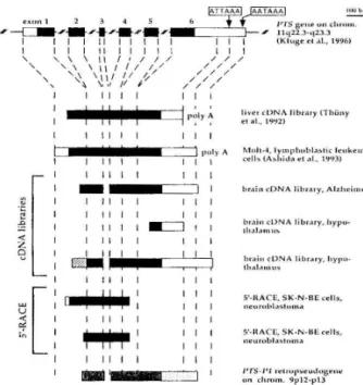
I
II
I I
, ptllyA liver eDNA libr.uy tThunyel OIL, 1992)
I II I I I I I
I puly A i\1nlt-4, IYl11phuhl.,.!>tic leukemia I II I I I cells (A.!>hida et ill.. 1'JlJ3)
I I II I I I
I
-
I I br,\ill eDNA IihriHy. Alzheimer~[
I I II I I I I II I II I I I I I
I II I -==:J I I brain Ihal,HulIeD!. NA libr.uy, h ypo-Z I II I
'2 I I II I I
brain eON A libr.uy. hypu.
I ~ thal;tmllS I I II I I
~[
II
II
I
I S'-HACE. SK-N-BE ,"'ells, I I I neufuhl.lslun,,, I I II I I~
I S'·It.ACE, SK-N-BE cells,I I I Ilcurol>laSlom.l
I I II I I I I
-'
I , un ""1'5c·1'1 rclrupseuJ.vsenc hromo 9p12-p13 (Kluge el .. I., 1996)Figure 1. Organization of the human PTS gene and
ex-tension of corresponding cDNA clones isolated from
various sources. The PTS gene located on chromosome
11q 22.3-q23.3 is composed of 6 exons asincticated by
the boxes. The coding regions are in black, whereas the
5'-and 3'-untranslated stretches are depicted as open
bars. The position of the two putative polyadenylation
signals (ATTAAA and AATAAA) are incticated by arrows.
The different sources of inctividual cDNA isolates are
given on the right. Polyadenin stretches found at the
3'-ends of cDNAs are incticated by 'polyA'. The hatched
box is a spliced fragment which originates from intron 1 (see text for details). For comparison, the extension of
the 74%-identiy to the PTS-Pl retropseudogene is given
at the bottom.
and a subsequent stop codon. A similar mutant
cDNA allele has been isolated previously from a
PTPS patient (K54X allele; 8). The cause for
ex-pressing such a mutant RNA was a genomic T to
A transversion at position-7 in the 3 '-acceptor
splice site of the patient's intron 2, which leads to
exon 3 skipping (4). Interestingly, a sequence
alignment between the liver PTPS cDNA and the
PTS-Pl retropseudogene revealed a significant
homology gap of 23 bp extending over the exon 3
region. This implicates that exon 3 skipping does
not occur exclusively due to 3'-acceptor splice
mu-tations, but might also be caused by a splicing
po-lymorphism.
In addition to a deletion of exon 3, we found
in one clone from the hypothalamus cDNA
li-brary a DNA fragment which was fused at the 5
,-end of exon 2. This fragment turned out to be an
Kluge et al.: PTPS-cDNAs from human brain
poslt:J.on 1451-1676 in the deposited GenBank
se-quence L76259). This cDNA clone, which is a
splice product consisting of the 226 bp 'exonic'
fragment plus exons 2, 4, 5 and 6, did also not
yield any open reading frame in its 5 I -portion of the cDNA.
Although at this stage we could not exclude
any functional role for the isolated brain cDNA
clones, we assumed that the exon 3 deletion in
two clones and the spliced fragment from intron
1 in one clone were due to some artifacts
present in the neuronal cDNA libraries analyzed.
Furthermore, as we detected PTPS enzyme
ac-tivity (9) and cross-reactive material in human
hypothalamus extract (not shown), we expected
to find a cDNA that encodes a functional PTPS.
To discriminate between erroneously spliced
cDNAs present in the brain cDNA libraries and
potentially alternative splicing in neuronal cells,
we carried out 5 ' -RACE experiments with
fresh-ly prepared RNA from cultured ·
neurobla-stoma cells that expressed the human PTPS
en-dogenously (SK-N-BE cells, unpublished
ob-servation). The PTPS-specific downstream prim-ers were selected to amplifY the cDNA upstream
from exon 5 (PTPS7B and PTPS9; for details
see Materials and Methods). The 5 '-RACE
pro-ducts were gel purified, subcloned into plasmid
vectors, and sequenced. The two clones that we analyzed did neither show any alternatively
splic-ed forms of PTPS-cDNA, nor a deletion of exon
3 (Fig. 1). Furthermore, one clone had the
com-plete coding sequence of exon 1 plus 18 bp of
the non-coding 5 ' -upstream sequence.
Un-fortunately, we did not isolate any clones that were extended up to the previously described 68
bp upstream from the translational start site
(Ashida et ai., 1993).
In summary, our findings argue against
alt-ernative 5 '-exons in the human PTS gene for
ex-pression of a functional enzyme, since no such
tis-sue- or cell-specific transcripts for the human
PTPS were isolated. Whether the occurrence of a
deletion of exon 3 in processed transcipts is a
po-lymorphism or has some functional meaning
ne-eds to be clarified. Furthermore, to unravel the
molecular basis of clinical and biochemical
diff-erences of the various forms of PTPS insufficiency,
more data about the molecular defects in patients
especially with the peripheral type of PTPS de-ficiency are required.
93
Acknowledgments
We thank M. Killen for help with the
pre-paration of the mansucript. This work was
sup-ported by grants from the Swiss National Science
Foundation (project No. 31-43380.95) and the
Hartmann-Muller Stiftung.
References
1. Blau N. Barnes 1. Dhondt, 1.L. (1996). International
database of tetrahydrobiopterin deficiencies. 1. Inher.
Metab. Dis., 1996; 19: 8-14.
2. Kim N. Kim, J. Park D. Rosen C. Dorsett D. Yim 1.
Structure and expression of wild-type and
suppres-sible alleles of the Drosophila purple gene. Genetics,
1995; 142: 1157-1168.
3. Thony B. Leimbacher W. Blau N. Harvie A.
Heizmann C.W. Hyperphenylalaninemia due to de
-fects in tetrahydrobiopterin metabolism: molecular
characterization of mutations in
6-pyruvoyl-tetrahy-dropterin synthase. 1994: Am. 1. Hun. Genet, 54:
782-792.
4. Kluge C. Brecevic L. Heizmann C.W. Blau N. Thony
B. Chromosomal localization, genomic structure and
characterization of the human gene and a
retropseu-dogene for 6-pyruvoyltetrahydropterin synthase.
1996: Eur. 1. Biochem. 240,477-484.
5. Frohman M.A. Rapid amplification of eDNA ends
(RACE): user-friendly eDNA cloning. Perkin Elmer
Cetus amplifications, a forum for PCR users: 1990:
11-15.
6. Thony B. Leimbacher W. Burgisser D. Heizmann C.
W. Human 6-pyruvoyl-tetrahydropterin synthase:
eDNA cloning and heterologous expression of the
re-combinant enzyme. Biochem. Biophys. Res. Comm.
1992; 189: 1437-1443.
7. Ashida A. Hatakeyama K Kagamiyama H. cDNA
clon-ing, expression in Escherichia coli and purification of
human 6-pyruvoyl-tetrahydropterin synthase. 1993:
Biochem. Biophys. Res. Comm.: 195: 1386-1393.
8. Oppliger T. Thony B. Nar H. Burgisser D. Huber R.
Heizmann C.W. Blau N. Structural and functional
consequences of mutations in 6-pyruvoyl-tetrahy
-dropterin synthase causing hyperphenylalaninemia in
humans: phosphorylation is a requirement for in vivo
activity. 1995; 1. BioI. Chern., 270: 29498-29506.
9. Heizmarm C.W. Leimbacher W. Kierat L. Blau N.
Measurement of enzymes involved in the
biosyn-thesis of tetrahydrobiopterin, pterins and
nu-urotransmitter metabolites in various regions of the
human brain. In: Blau N. Curtius H.C. Le"ine R.A.
Cotton R.GH. eds. Pteridines and Biogenic Amines
in Neurology, Pediatrics and Immunology.
Lak-eshore Publishing 1991; 95 -99.