Carcmogenesis vol.14 no.3 pp.457-462, 1993
Effects of sodium butyrate on DNA content, glutathione
S-transferase activities, cell morphology and growth characteristics
of rat liver nonparenchymal epithelial cells in vitro
Dietmar Utesch1, Margarete Traiser2, Ingolf Gath, Adriaan W.C.Dorresteyn3, Peter Maier4 and Franz Oesch
Institute of Toxicology, University of Mainz, Obere Zahlbacher Str. 67, D-6500 Mainz, 3Institute of Zoology, University of Mainz, D-6500 Mainz, Germany and 4Institute of Toxicology, ETH and University of Zurich, CH-8603 Schwerzenbach, Switzerland
'Present address: Institute of Toxicology, E.Merck, D-6100 Darmstadt, Germany
2To whom correspondence should be addressed
The effects of sodium butyrate, which has been shown to act as a differentiation promoting agent in several different tumor cell lines, were studied in a rat liver nonparenchymal epithelial cell line. Exposure of these cells to 3.75 mM butyrate resulted in an inhibition of cell proliferation and, at the same time, an increase in cell diameter (2- to 6-fold) and size of the nuclei (~ 2-fold) after 3 days in culture. Binucleated cells arose, comprising ~ 12% of the cells investigated, and the number of cells with an abnormal set of chromosomes was increased. Intercellular communication, measured by dye transfer of Lucifer Yellow, was unchanged. From the various xenobiotic metabolizing enzyme activities measured, only those of glutathione S-transferases were significantly altered (increases of 4- to 9-fold) by butyrate treatment. These increases were mainly due to the predominant rise in the x class isoenzyme which is a well-known tumour marker in rat hepatocarcino-genesis. Thus, our results cannot be interpreted as being either due to promotion of differentiation or due to transformation. The state and type of cell under study has to be considered and investigations of further differentiation parameters are needed to obtain a deeper insight into the biological activity and the underlying mechanisms of cell state modifying agents like butyrate.
Introduction
Various effects of sodium butyrate on different types of cells
in vivo and in vitro have been described. Inhibition of histone
deacetylation (1,2) and arrest of cells in Gl phase of the cell cycle (3,4) are reported effects on the DNA level. Other possible targets are cellular membranes and the cytoskeleton (2,5). Furthermore, alteration of enzyme activities, such as alkaline phosphatase (6) and tyrosine aminotransferase (7), as well as changes in a-fetoprotein and albumin production (7,8) have been measured. In effect, transformed cells differentiated into more mature or 'normal' cells after butyrate treatment (9). For these reasons, butyrate, retinoic acid and polar solvents such as dimethyl-sulfoxide or dimethylformamide, are intensively investigated as differentiation promoting factors in transformed proliferating cells
in vitro and during carcinogenesis in vivo (10,11).
•Abbreviations: NEC, rat liver nonparenchymal epithelial cells; GST, glutathione S-transferase; mEH, microsomal epoxide hydrolase; cEH, cytosolic epoxide hydrolase; ST, phenol sulfotransferase; UDPGT, UDP-glucuronosyl transferase.
On the other hand, compounds like sodium butyrate (2) and dimethylsulfoxide (12,13) have been shown to stabilize stationary rat liver parenchymal cells in primary culture. In these systems, the progressive decline of one toxicologicaly relevant differen-tiation characteristic of liver parenchymal cells, i.e. expression of cytochrome P450, was at least partially inhibited by addition of these agents to the culture medium.
In the present investigation the effects of sodium butyrate on another type of rat liver cell, namely liver nonparenchymal epithelial cells (NEC*), were studied. Though these cells are of epithelial origin, like the liver parenchymal cells, they differ in that the NEC can proliferate in culture (14). Germain et al. (7) postulated that such cells are bipotential progenator cells that are capable of differentiating along either the hepatocytic or biliary epithelial cell lineage. The initial aim of this study was to investigate if NEC could be induced to form a more differentiated cell type by the addition of butyrate to the culture medium. The concentration of 3.75 mM butyrate was chosen because this dose has proved to be effective in a similar cell system described previously (7). To characterize the differentiated status of NEC, the activities of several xenobiotic metabolizing enzymes were measured in the absence and presence of butyrate. If a more differentiated character were induced, an increase of one or more enzyme activities would be detected. Throughout these studies further effects of butyrate on the morphology and the DNA level of NEC were observed and are duly described.
Materials and methods
Chemicals
Williams' medium E and colcemid were purchased from Biochrom (Berlin, Germany) and 2-naphthol from Fluka (Neu-Ulm, Germany). Sodium butyrate, Lucifer Yellow CH, lithium chloride, l-chloro-2,4-dinitrobenzene and ethacrynic acid were obtained from Sigma (Deisenhofen, Germany). rram-4-Phenyl-3-buten-2-one was purchased from EGA (Steinheim, Germany).
4-Hydroxynon-2-trrms-emi was kindly provided by H.Esterbauer (Graz, Austria). [7-3H]Styrene oxide was supplied by Amersham Buchler (Baunschweig, Germany), and 1-naphthol and unlabelled styrene oxide by Merck (Darmstadt, Germany). fra/u-[a,/3-3H]Stilbene oxide was synthesized as described before (15). Rabbit polyclonal antisera against glutathione S-transferase (GST) subunits 1, 3, 4, 7 and 8 were purchased from Bioprep (Dublin, Ireland), horseradish peroxidase-conjugated anti-rabbit immunoglobulin antibodies were from Dakopatts (Hamburg, Germany).
NEC establishment and culture
NEC were initiated as described before (16). In brief, thin liver slices from an adult, male Sprague-Dawley rat were repeatedly digested with trypsin. Purified cells from the collected digests were cultured, cloned and further maintained in Williams' medium E including 10% fetal bovine serum, gentamicin (50 mg/1) and insulin (10 mg/1). In our study NEC were used between passage 82 and 129. NEC grown in the continuous presence of 3.75 mM butyrate (for at least five passages) were initiated from the same batch of cells used for control experiments and experiments with short-term exposure (3 days) to butyrate. Determination of the cell number per plate was carried out by harvesting cells after trypsin treatment and subsequent counting in a haematocytometer.
Morphometric determinations
For determination of cell and nuclei areas, cultured cells were recorded on VHS video tape digitalized with a soft imaging system (SIS Software, Munster, Germany). After calibration of the system 20 randomly chosen cells from each group were analysed. Additionally, the number of nuclei per cell was determined in at least 100 cells per group. The cell diameters of the whole-population of
trypsinized NEC were recorded with a Coulter counter, model ZM, plus chan-nalizer, C-1000. Calibration of this system was done using a latex standard of 18.3 /tm in diameter.
Determination of the number of chromosomes
Subconfiuent cultures were treated with 20 /il colcemid/ml cell culture medium, and trypsinized cells were incubated in a hypo-osmotic solution of sodium chloride (5.67 g/1 H2O). The cells were fixed on microscopic slides with a solution of ice cold methanol—acetic acid, and the chromosomes finally stained with Giemsa.
Intercellular communication
NEC were injected with 556 Lucifer Yellow CH dissolved in 0.1 M lithium chloride using a micromanipulator 5170 and a pneumatic microinjector 5242 (Eppendorf, Hamburg, Germany). The injection was followed with an Axiovert 35 microscope (Zeiss, Oberkochen, Germany, with filter block number 487907). A minimum of 10 cells per group were injected and the dye transfer was checked 2 mm after injection. The dye coupling index is defined as the number of injected cells, expressed as a percentage, which show dye transfer via gap junctions to at least one neighbouring cell.
Protein and enzyme determinations
NEC were trypsinized, centrifuged and resuspended to yield a concentration of 20x 10* cells/ml of appropriate buffer used for the different enzyme assays. They were then homogenized by Bonification and directly frozen at -80°C.
Protein determination was carried out according to Lowry et al. (17) with bovine serum albumin as a standard. Cytochrome P450 was measured according to Omura and Sato (18). Microsomal epoxide hydrolase (mEHj activity was determined with styrene 7,8-oxide according to Oesch et al. (19) in the absence of Tween 80 as described by Oesch (20). Cytosolic epoxide hydrolase (cEH) activity was measured using jraro-stilbene oxide as substrate according to the method of Schladt
et al. (21). The enzymatic activities of phenol sulfotransferase (ST) and
UDP-glucuronosyl transferase (UDPGT) were determined using 2-naphthol (22) and 1-naphthol (23) as substrates respectively. GST activities were measured accord-ing to Habig et al. (24) with cUorodinhroberizene, phenylbutenone and emacrynic acid as substrates. The GST-catalysed turnover of hydroxynonenal with glutathione was determined according to Alin et al. (25).
For Western blot analysis cytosolic fractions were prepared from NEC homogenates by centrifugation at 100 000 g for 60 min. Ten microliters of each sample were electrophoresed in a 10% SDS-polyacrylamide resolving gel accord-ing to SchSgger and Von Jagow (26). The transfer of proteins to nitrocellulose was performed as described (27). After blotting overnight, nitrocellulose sheets were blocked with 3% (w/v) bovine serum albumin and 0.05% (v/v) Tween 20 in a Tris-buffered saline solution (10 mM Tris—HC1 buffer, pH 7.4, containing 0.9% NaCl) and incubated with the corresponding antiserum dilution in Tris-buffered saline containing 0.5% (w/v) gelatine and 0.05% Tween 20. The blots were washed with Tris-buffered saline and the immunoreactive proteins were located with 4-chloro- 1-naphthol after incubation of the sheets with horseradish peroxidase-conjugated anti-rabbit immunoglobulin antibodies. Molecular weight marker proteins were visualized after transfer to nitrocellulose by staining with Ponceau 5.
Results
Cell growth and morphology
The growth curve of NEC is shown in Figure 1. NEC grew with an approximate doubling time of 21 h to a density of ~ 10X106
call number/dish (X106)
"* i 0 1 I I 4
culture time (days)
Fig. 1. Growth of rat liver NEC in the absence (solid line) or presence (dotted line) of 3.75 mM sodium butyrate. The arrow indicates the time of butyrate addition to one part of the cultures. Values are means ± SD of three independent experiments.
cells/10 cm-0-dish. Addition of butyrate to the cultures on Day 1 stopped the increase in cell number immediately. A few cells died, floated on the surface of the culture medium, and were removed by daily medium changes. A few mitoses were observed in these cultures. After the presence of butyrate for 3 days, the surviving NEC formed a near-confluent layer which was more the result of an increase in cell size than an increase in cell number. The analysis of cell morphometry of these 4 day old cultures is shown in Table I and Figure 2.
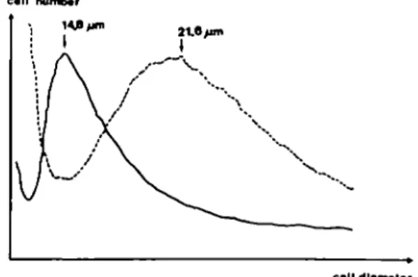
Table I presents the results of the single cell analysis of cell morphology in a representative experiment. It shows that both the total cell area and the nuclear area were significantly increased by butyrate, the former by a factor of about six and the latter by a factor of only two. This resulted in a change of the cytoplasm to nucleus ratio. Figure 2 represents the size distribution of the whole population of trypsinized cells from the same experiment. In the control population the maximum diameter of cells was 14.8 /im. Addition of butyrate for 3 days increased the cell size and the maximum diameter reached was 21.6 /xm in diameter. Furthermore, the variation in cell size within the population was more extended as shown by an increase in width of the peak after butyrate treatment in comparison to the relatively narrow peak of the control cells. The increase in diameter of these cells in suspension, after trypsinization from the culture dishes, was less (— 1.5-fold, Figure 2) than that measured in the cultured cells (~ 2.5-fold, Table I). This means that the butyrate-treated cells were spread more broadly and flatter on the culture dishes than the untreated cells.
The long-term effects of butyrate on NEC was studied by establishing a cell line which proliferated in the presence of
TaWe I. Cell morphometry and intercellular communication in cultures of rat liver N E C
NEC NEC + butyrate
Cell diameter (/an) Cell area (jisn2) Nuclear area (/un2) Cell:nucleus ratio Binucleated cells (%) Dye coupling index (%)
26.7 501 162 3.12 100 ± ± ± ± 0 ± 2.7 98 28 0.58 0 67.6 ± 3690 ± 325 ± 11.6 ± 12 100 ± 13.3** 1050** 61** 2.5** 0 •NEC were grown for 3 days in the absence or presence of 3.75 mM sodium butyrate (cf. Figure 1). Values are means ± SD of a typical experiment in which at least 20 single cells have been analysed. "Significantly different to control using Student's Mest; P s 0.01. The number of binucleated cells was determined in 100 cells per group.
call diameter
Fig. 2. Cell size distribution of suspensions of rat liver NEC trypsinized from 4 day old cultures (cf. Figure 1) grown in the absence (solid line) or presence (dotted line) of 3.75 mM sodium butyrate.
Sodium butyrate in rat liver epithelial cells
butyrate. After 2 to 3 weeks in the continuous presence of butyrate, cells recovered and started to grow slowly. After five passages with low splitting ratios (1:2), cells had a doubling time of ~ 4 days and formed a confluent layer with a mean density of 1.4X10⁶ cells/10 cm-<£-dish. The cell size distribution of
these cells (not shown) was similar to that of NEC treated for 3 days with butyrate (cf. Table I and Figure 2).
Number of binucleated cells and chromosome number
The untreated control cultures consisted exclusively of mononucleated cells. Cultures treated with butyrate contained a considerable amount of binucleated cells (Table I) which was similar for short-term (3 days; 12%) and for long-term exposure ( > 5 passages, 14%, data not shown in the table).
The number of chromosomes per nucleus was already abnormal in the control cultures which showed a maximum of cells with 54 chromosomes per nucleus (Figure 3) in comparison to 42 chromosomes per nucleus in the rat in vivo. This shift in the chromosome number was certainly due to the age of the cultures indicated by the relatively high passage number of the investigated cells. However, no growth of NEC in soft agar occurred, either in the absence or in the continuous presence of butyrate. Due to the few mitotic events immediately after butyrate addition to the cultures, the chromosome number of NEC could only be determined in proliferating cultures after long-term exposure to butyrate. In that model the peak of chromosome distribution was widened, showing maxima of nuclei with 54, 61 and 66 chromosomes. Cells which had been exposed to butyrate for 3 days were analysed by two-parameter flow cytometry (28). Using this technique, similar effects were observed as in the long-term treatment model (data not shown). The variation in DNA content of GQ/G! phase cells was enhanced, reflecting an increase in the number of cells with an abnormal chromosome set. Furthermore, the contribution of cells in the S phase and in the G2/M phase was increased due to delayed cell cycling. The cells did not accumulate in the GQIG\ phase and the cellular protein content was not significantly altered in these experiments.
Intercellular communication
Dye transfer of Lucifer Yellow from the injected cells to their primary and secondary adjacent neighbouring cells occurred rapidly (within less than 30 s). All primary and many secondary recipients were stained by every injection. Dye transfer was not inhibited in the cells treated with butyrate for 3 days (Table I). Neither the number of primary adjacent recipients nor the number
of secondary fluorescing cells were measurably reduced. Further-more, NEC grown in the continuous presence of butyrate did not show an altered dye coupling index when compared to the control NEC.
Protein content and xenobiotic metabolizing enzymes
Total cellular protein was slightly higher after butyrate treatment (Table II). However, the effect was weak and a statistically significant increase was detected in only one set of experiments (Table HI).
Cytochrome P450, mEH, cEH and ST were below the level of detection in NEC (Table II); GST and UDPGT were, however, easily measurable. Both enzyme activities were measured in preconfluent and confluent cultures with chlorodinitrobenzene and 1-naphthol as substrates respectively. No statistically significant dependence of enzyme activities on the growth state was detected (data not shown). Furthermore, no relevant differences in enzyme activities existed between cells from low or high passage numbers. However, GST activity was clearly higher in NEC which were cultured in medium containing butyrate for at least five passages (Table II). UDPGT activity was not measurably affected by butyrate treatment.
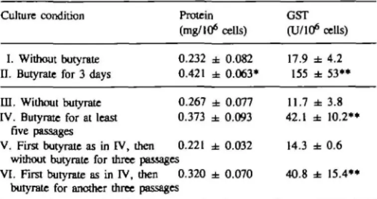
The increase in GST activity by butyrate was further investigated (Table HI). The results showed that the exposure of NEC to butyrate for only 3 days already resulted in a signifi-cant increase in GST activity. In addition, when cells which were
Table II. Protein content and activities of xenobiotic metabolizing enzymes
in subconfluent cultures of rat liver N E C
NEC NEC + butyrate
Protein (mg/106 cells) P450 (nmoL/106 cells) mEH (U/106 cells)b cEH (U/106 cells)b GST (U/106 cells)b ST (U/106 cells)" UDPGT (U/106 cells)b
0.267 ± 0.077 <0.02 <0.08 <0.02 11.7 ± 3.8 <0.01 2.63 ± 0.19 0.373 ± 0.093 <0.02 <0.08 <0.02 42.1 ± 1 0 . 2 " <0.01 2.90 ± 0.40 *NEC were grown for at least five passages in the absence or presence of 3.75 mM sodium butyrate. Values are means ± SD of at least three independent cell preparations.
••Significantly different to control using Student's r-test; P s 0.01. bEnzyme units are defined as nmol product formed/min for mEH, GST (substrate: l-chloro-2,4-dinitrobenzene), ST and UDPGT, or pmol product formed/min for cEH.
Number of Nuclei
40 (0 M M M 70
Chromosome* per Nucleus
Fig. 3. Number of chromosomes per nucleus in cultures of rat liver NEC
grown in the absence (black bars) or presence of 3.75 mM butyrate for more than five passages (striped bars).
Table ID. Protein content and GST activity in cultures
under different culture conditions Culture condition
I. Without butyrate n. Butyrate for 3 days in. Without butyrate IV. Butyrate for at least
five passages
V. First butyrate as in IV, then
Protein (mg/106 0.232 ± 0.421 ± 0.267 ± 0.373 ± 0.221 ± without butyrate for three passages VI. First butyrate as in IV, then 0.320 ±
butyrate for another three passages
cells) 0.082 0.063* 0.077 0.093 0.032 0.070
of rat liver NEC
GST (U/106 17.9 ± 155 ± 11.7 ± 42.1 ± 14.3 ± 40.8 ± cells) 4.2 53** 3.8 10.2** 0.6 15.4**
Values are means ± SD of at least three independent cell preparations. Significantly different to the corresponding control using Student's r-test;
*P <; 0.05; **P <; 0.01.
Table IV. Protein content and activities of GST isoenzymes in cultures of
rat liver NEC*
NEC NEC + butyrate Protein (mg/106 cells) 1 -Chloro-2,4-dinitrobenzene 4-Hydroxynon-2-enal mzJis-4-Phenyl-3-buten-2-one Ethacrynic acid 0.253 7.50 5.00 0.180 4.75 0.448 66.3 20.0 0.749 22.0 •NEC were grown for 3 days in the absence or presence of 3.75 mM sodium butyrate (cf. Figure 1). Enzyme activities of a typical experiment are expressed in nmol product formed/min/106 cells.
cultured in butyrate-containing medium for more than five passages were released from this treatment and then cultured in control medium, GST activity dropped again to the control levels. The effect of butyrate on GST activity was further characterized by using more specific substrates for single GST isoenzymes (Table TV). The increase in turnover of the broad spectrum substrate chlorodinitrobenzene was in the same range as in the former experiments (i.e. —9-fold, cf. Table HI). Turnover of the other substrates, i.e. hydroxynonenal, phenylbutenone and ethacrynic acid, was increased 4- to 5-fold after butyrate treat-ment. Catalytic activities with further substrates, i.e. 1,2-dichloro-4-nitrobenzene, 1,2-epoxy-3-(para-nitrophenoxy)-propane, menaphthyl sulfate, and A5-androstene-3,17-dione were below
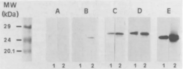
the detection level in the absence and presence of butyrate (data not shown). The immunological characterization, based on the reactivity of polyclonal antisera generated against purified GST isoenzymes, confirmed the results obtained with the different isoenzyme substrates. Besides moderate increases of a and y. class isoenzymes a remarkable elevation of the GST 7 - 7 level was detected in butyrate-treated NEC (Figure 4).
Discussion
The most obvious effect of sodium butyrate on NEC presented in this investigation was the inhibition of cell proliferation (Figure 1) which is, however, not simply a toxic effect. Under toxic conditions NEC detach from the dishes and the dead cells float on the surface of the culture medium. Only a few cells died after butyrate addition. The majority of cells survived the treat-ment and started to divide again after several weeks. The fact that NEC regain their original characteristics very soon after removal of butyrate from the medium, further implies that the changes were not due to selection of a resistant subpopulation of cells. Restoration of the original characteristics of the cells after removal of the differentiation inducing agent from the culture medium has also been described for other cell systems (10,29). The observed inhibition of cell proliferation after butyrate exposure is, however, contradictory to other studies where an increase in the doubling time of the cells under investigation was observed after treatment with other differentiation promoting factors (30).
The second microscopically visible effect of butyrate on NEC was the increase in cell volume (Table I and Figure 2). The increase in protein content, if significant at all, was much less abundant (Tables II and HI). Similar effects were observed after short-term exposure (3 days) in stationary cultures and long-term exposure (more than five passages, i.e. at least 2 months) in proliferating cultures. An examination of the literature showed similarities and dissimilarities in these parameters in other cells.
MW (kDa) 29 - ^ 24 - « * 20.1 — « B 1 2 1
Fig. 4. Western blot analysis of GST subunit content of rat liver NEC
grown in the absence (1) or presence of 3.75 mM sodium butyrate for 3 days (2). Incubations of polyclonal antisera against GST subunits were carried out with anti-a class GST 1-1 (A) and 8 - 8 (B), anti-ji class GST 3 - 3 ( Q and 4 - 4 (D) as well as anti-T class GST 7 - 7 (E).
For example butyrate treatment resulted in an increase in cell size, cell volume and, additionally, in the protein content of transformed rat hepatic tumour cells in culture (5). In that model, a more normal appearing hepatocytic phenotype was achieved with respect to intermediate filament and microfilament organiza-tion. When comparing NEC after butyrate treatment with normal liver parenchymal cells it is obvious that the increased cell size and the low proliferation rate of the NEC reflect typical characteristics of parenchymal cells; the protein content of NEC was, however, ~ 5-fold lower (14).
The observed changes at the DNA level after butyrate treat-ment were significant as well. Increases in the volume of the nuclei (Table I), the number of binucleated cells (Table I) and the number of chromosomes per nucleus (Figure 3) occurred. A high number of binucleated cells is typical for parenchymal cells of the liver in vivo. The frequency of these cells is depen-dent on the animal species and the age of the individual. Never-theless, the described effects cannot be interpreted as being specific for a differentiation inducing process because the varia-tion in the number of chromosomes per nucleus was increased, which is more characteristic for transformed cells. However, the nature of the untreated cells has to be taken into account. This means that for the present investigation untransformed NEC (growth in soft agar did not occur) which already had an abnormal chromosome number (In = 54) were used. These cells were chosen for our experiments to ensure that both of the effects of butyrate on the chromosome number, i.e. decrease to a more normal or increase to a more transformed genotype, could be measured. The same cell clone showed a normal chromosome set (In = 42) at low passage numbers ( < 25) and an increased set (2/i = 57) at very high passage numbers ( > 186) in the absence of butyrate. In contrast to the changes induced by butyrate described in Figure 3, the variation in chromosome number of the spontaneously transformed NEC was much lower (data not shown).
A direct mutagenic or transforming effect of butyrate has, to our knowledge, not so far been published. However, butyrate can act as a co-mutagen. For example, pretreatment of V79 Chinese hamster fibroblasts with butyrate enhanced the cytotoxic and mutagenic response of these cells to daunorubicin (31). Furthermore, in vitro pretreatment of human peripheral blood lymphocytes with butyrate led to a significant increase in the frequencies of dimeric and ring chromosomes induced by X-irradiation (32). In both cases, butyrate-induced primary changes might have sensitized the chromosomes to the effects of the actual mutagens.
The change of normal cells to a more transformed phenotype is often correlated with either an inhibition of die heterotypic
Sodium butyrate in rat liver epithelial cells
intercellular communication between normal and transformed cells or a reduction of the homotypic communication within the same cell type (33,34). In the presented cell culture system butyrate did not inhibit the homotypic intercellular communica-tion of NEC as measured by dye transfer of Lucifer Yellow. Sodium butyrate (2) and dimethylsulfoxide (13) were shown to retard the progressive decline of cytochrome P450 in stationary primary cultures of rat liver parenchymal cells. Furthermore, a massive albumin production was induced by butyrate in proliferating rat liver oval cells under certain culture conditions (35). The studies on the activities of xenobiotic metabolizing enzymes in the present investigation showed different results for NEC as compared to the other investigated cell types and, thus, may be cell specific. Xenobiotic metabolizing enzyme activities which were below the detection level in untreated NEC, e.g. cytochrome P450 were also not measurable in NEC after butyrate treatment.
From the enzymes investigated, only UDPGT and GST were clearly measurable. GST activity has been shown to be depend-ent on the proliferation status of normal NEC (36,37) but not so for tumour cells which originate from normal NEC after transformation in vivo (36). No such correlation of GST activity and the proliferation status of the NEC cultures used in this investigation was found. GST-catalysed conjugation of chlorodinitrobenzene with glutathione was significantly increased after butyrate treatment of NEC (Tables II and HI). The absolute activity after butyrate exposure was still ~ 10-fold lower than in freshly isolated rat liver parenchymal cells when enzyme activities were related to the cell number (14). Induction of differentiation of NEC towards a more parenchymal phenotype by butyrate was thus not revealed by these experiments. Interestingly, no such increase in chlorodinitrobenzene conjuga-tion occurred after exposure of primary cultured, staconjuga-tionary rat liver parenchymal cells to the same dose of butyrate for 3 days (data not shown). Even inhibition of purified GST by sodium butyrate in vitro has been reported (38). The concentrations used in those experiments were higher (5—50 mM) compared with that in the present investigation (3.75 mM). These results, together with those described above, imply that butyrate affects the cell as a whole and, furthermore, may be cell specific for NEC.
GST represents a family of dimeric isoenzymes in which the subunits belong to a supergene family of four classes, alpha, mu, pi and theta (39,40). The activity of most isoenzymes is more or less satisfactorily detected by the broad spectrum substrate chlorodinitrobenzene. During hepatocarcinogenesis in vivo (41) and differentiation of liver parenchymal cells in vitro (42), changes in gene expression of single GST subunits occur. Differences in the regulation of different GST subunits in normal and transformed NEC have also been described (36). The present investigation has shown that, in addition to the broad spectrum substrate chlorodinitrobenzene, the GST-dependent turnover of hydroxynonenal, phenylbutenone and ethacrynic acid could be easily measured. The metabolism of these substrates was similarly increased after butyrate treatment of NEC (Table IV).
These results are supported by the immunological data which showed slightly higher protein levels for a and \L class isoenzymes in butyrate-treated NEC, but a strong increase of the irclass GST subunit 7, a well-known tumour marker in rat hepatocarcino-genesis (43). Obviously, this isoenzyme contributed predominantly to the activities found with chlorodinitrobenzene and ethacrynic acid as substrates. The level of subunit 7, which is hardly detectable in freshly isolated liver parenchymal cells, was
significantly elevated when these cells were kept in primary culture (44). In contrast to these findings, subunits of the a and
ft class are down-regulated in primary cultures of liver
paren-chymal cells (45). On the other hand, there is immunological evidence for the presence of subunit 7 in bile duct cells (41). In the present investigation the additional increase in GST 7—7 levels caused by treatment with butyrate, accompanied by a tar less dramatic rise in a and n class isoenzymes, resembled the processes which are observed during hepatocarcinogenesis (46). Similarly, Castro et al. (47) showed that IT class GST was the predominant form in a large number of tumour cell lines and that a and /i class GST isoenzymes were present in significantly lower concentrations. Moreover, they concluded that most of the chlorodinitrobenzene metabolizing activity was due to the presence of -w class isoenzyme. For the present study these results indicate that butyrate induces NEC to a more fetal or differentiated status when GST isoenzyme activities are con-sidered as a differentiation marker.
In conclusion, butyrate showed diverse effects upon NEC which only partly correspond to those reported in the literature for other cell types. A general differentiation promoting activity of butyrate could not be detected. Furthermore, no indication of a general mechanism of the action of butyrate upon these cells could be detected. There are many open questions, e.g. why were GST activities increased and not those of UDPGT, or how the changes on the DNA level are linked to the changes in enzyme activities or cell size. Experience has shown (14,16) that single physiological parameters that are crucial for the differentiated status of a given cell type in vitro may be regulated independently and, thus, may respond differently to external factors. Further-more, different types of cells react differently to agents such as sodium butyrate. For these reasons, we recommend the very careful use of such in vitro models when the mechanisms of action of apparent differentiation-inducing agents are being studied. And, finally, the question of whether the described effects of sodium butyrate have any relevance to in vivo should be clarified in further experiments using additional cell types and dose levels comparable to those found in vivo.
Acknowledgements
We thank N.Beer and H.P.Schawalder for their excellent technical assistance, H.Martus for performing the soft agar test, and H.DGrk for patiently taking photographs. This work was supported by the Deutsche Forschungsgemeinschaft, SFB 3O2/A80.
References
l.StaeckerJ.L., Sawada.N. and Pitot,H.C. (1987) Stimulation of DNA synthesis in primary cultures of adult rat hepatocytes by sodium butyrate. Biochem.
Biophys. Res. Commun., 147, 7 8 - 8 5 .
2. Engelmarm.G.L., StaeckerJ.L. and Richardson,A.G. (1987) Effect of sodium butyrate on primary cultures of adult rat hepatocytes. In Vitro CelL Dev. BioL, 23, 8 6 - 9 2 .
3. Gladhaug.I.P., Refsnes.M., Sand.T.E. and ChristoffersenJ. (1988) Effect of butyrate on epidermal growth factor receptor binding, morphology, and DNA synthesis in cultured rat hepatocytes. Cancer Res., 48, 6560—6564. 4. Niles.R.M. (1987) Retinoic acid-induced arrest of mouse melanoma cells in Gl without inhibition of protein synthesis. In Vitro Cell. Dev. BioL, 23, 803-804.
5. Ryan.M.P., Borenfreud.E. and Higgins.P.J. (1987) Butyrate-induced cyto-architectural reorganization of Mallory body-containing rat hepatic tumor cells.
J. Natl. Cancer Inst., 79, 555-567.
6. Koyama,H. and Ono.T. (1975) Induction of short-chain fatty acids of alkaline phosphatase activity in cultured mammalian cells. J. CeU. PhysioL, 88,49—56. 7. Germain,L., Blouin.M.J. and Marceau.N. (1988) Biliary epithelial and
hepatocytic cell lineage relationships in embryonic rat liver as determined by the differential expression of cytokeratins, or-fetoprotein, albumin, and cell surface-exposed components. Cancer Res., 483 4909-4918,
8. Tokiwa,T.,Endo,A. and SatoJ. (1990) Butyrate decreases AFP and increases albumin in combination with laminin in a human hepatoma cell line. In Vitro
Cell. Dev. Bioi, 26, 220-221.
9. Prasad.K.N. and Sinha.P.K. (1976) Effect of sodium butyrate on mammalian cells in culture. In Vitro, 12, 125-132.
10. Spremulli.E.N. and Dexter.D.L. (1984) Polar solvents: a novel class of antineoplastic agents. J. Clin. Oncol., 2, 227-241.
11. Sporn.M.B. and Roberts.A.B. (1983) Role of retinoids in differentiation and carcinogenesis. Cancer Res., 43, 3034-3039.
12. Isom,H.C, Secott.T., Georgoff.I., Woodworth.C. and Mummaw.J. (1985) Maintenance of differentiated rat hepatocytes in primary culture. Proc. NatL
Acad. Sci. USA, 82, 3252-3256.
13. Muakkassah-KeUy,S.F., Bieri.F., Waechter,F., BenUey.P. and Sulubly.W. (1987) Long-term maintenance of hepatocytes in primary culture in the presence of DMSO: further characterization and effect of nafenopin, a peroxisome proliferator. Exp. Cell Res., 171, 3 7 - 5 1 .
14. Utesch.D. and Oesch.F. (1992) Dependency of the in vitro stabilization of differentiated functions in liver parenchymal cells on the type of cell line used for co-culture. In Vitro Cell. Dev. Biol, 28A, 193-198.
15.Oesch,F., Sparrow.A.J. and Platt.K.L.. (1980) Radioactively labelled epoxides, part II: tritium labelled cyclohexene oxide, /ra/u-stilbene oxide and phenanthrene 9,10-oxide. /. Labelled Compd. Radiophamt, 17,'93-102. 16. Utesch.D., Molitor.E., Platt.K.L. and Oesch.F. (1991) Differential stabiliza-tion of cytochrome P-450 isoenzymes in primary cultures of adult rat liver parenchymal cells. In Vitro Cell. Dev. Biol., 27, 858-863.
17. Lowry.O.H., Rosebrough.W.J., Farr,A. and Randall.R.L. (1951) Protein measurement with me Folin phenol reagent. J. Biol. Chem., 193, 265 - 2 7 5 . 18. Omura.T. and Sato.R. (1964) The carbon monoxide-binding pigment of liver
microsomes. J. Biol. Chem., 239, 2370-2385.
19. Oesch.F., Jerina,D. and Daly J. A. (1971) A radiometric assay for hepatic epoxide hydrase activity with 7-3H-styrene oxide. Biochim. Biophys. Acta, 227, 685-691.
20. Oesch.F. (1974) Purification and specificity of a mkrosomal human epoxide hydrase. Biochem. J., 139, 7 7 - 8 8 .
21. Schladt.L., W6rner,W., Setiabudi.F. and Oesch.F. (1986) Distribution and inducibility of cytosolic epoxide hydrolase in male Sprague-Dawley rats.
Biochem. Pharmacol., 35, 3309-3316.
22. Arand.M., Robertson.L.W. and Oesch.F. (1987) A fluorometric assay for quantitating phenol sulfotransferase activities in homogenates of cells and tissues. Anal. Biochem., 163, 546-551.
23.Bock,K.W. and White.I.N.H. (1974) UDP-Glucuronosyltransferase in perfused rat liver and in microsomes: influence of phenobarbital and 3-methylcholanthrene. Eur. J. Biochem., 46, 451.
24. Habig.W.H., Pabst.M.J. and Jakoby.W.B. (1974) Glutathione S-transferases. The first enzymatic step in mercapturic acid formation. J. Biol. Chan., 249, 7130-7139.
25. Alin.P., Danielson.U.H. and Mannervik.B. (1985) 4-Hydroxyalk-2-enals are substrates for glutathione transferases. FEBSLett., 179, 267-270. 26. Schagger.H. and Von Jagow.G. (1987) Tricine-sodium dodecyl
sulfate-polyacrylamide gel electrophoresis for the separation of proteins in the range from 1 to 100 kDa. Anal. Biochem., 166, 368-379.
27. Towbin.H., Staehelin.T. and GordonJ. (1979) Electrophoretic transfer of proteins from polyacrylamide gels to nitroceUulose sheets: procedures and some applications. Proc. NatL Acad. Sci. USA, 76, 4350-4354. 28. Maier.P. and Schawalder.H.P. (1988) Alteration in the cellular DNA and
protein content determined by flow cytometry as indicators for chemically induced structural and numerical chromosome aberrations. Mwagenesis, 3, 219-226.
29. Cordeiro.R.F. and Savarese.T.M. (1986) Role of glutathione depletion in the mechanism of action of A'-methylformamide and A'.A'-dimethylformamide in a cultured human colon carcinoma cell line. Cancer Res., 46, 1297-1305. 30. Dexter.D.L., BarbosaJ.A. and Calabresi.P. (1979) A'.A'-Dimethylforma-mide-induced alteration of cell culture characteristics and loss of tumorigenicity in cultured human colon carcinoma cells. Cancer Res., 39, 1020-1025. 31. Pani.B., Babudrin.N., Giancotti.V. and Russo.E. (1984) Sodium butyrate
affects the cytotoxic and mutagenic response of V79 Chinese hamster cells to the genotoxk agents daunorubicin and U.V. radiation. Mutat. Res., 140, 175-179.
32. Sankaranarayanan.K., NatarajanAT., Mullenders.L.H.F. and vanRipU.L.S. (1985) Effects of pretreatment with sodium butyrate on the frequency of X-ray-induced chromosomal aberrations in human peripheral blood lymphocytes. Musat. Res., 151, 269-274.
33. Yamasalri.H. (1990) Gap junctional intercellular communication and carcinogenesis. Carcinogenesis, 11, 1051 — 1058.
34. Metha.P.P., Bertram^.S. and Loewenstein.W.R. (1989) The actions of retinoids on cellular growth correlate with their actions on gap junctional communication. J. Cell BioL, 108, 1053-1065.
35. Germain.L., Noei.M., Gourdeau.H. and Marceau.N. (1988) Promotion of growth and differentiation of rat ductular oval cells in primary culture. Cancer
Res., 48, 368-378.
36. Batist.G., Woo.A. and Tsao.M.S. (1991) Effect of proliferative state on glutathione S-transferase isoenzyme expression in cultured rat liver epithelial cells. Carcinogenesis, 12, 2031-2034.
37. Schrenk.D., Eisenmann-Tappe.I., Gebhardt.R., Mayer.D., FJ Mouelhi.M., Rohrdanz.E., MQnzel.P. and Bock.K.W. (1991) Drug metabolizing enzyme activities in rat liver epithelial cell lines, hepatocytes and bile duct cells.
Biochem. Pharmacol., 41, 1751-1757.
38. Dierickx.P.J. (1984) In vitro binding of butyric acid and crotonic acid by the soluble glutathione S-transferases from rat liver. Res. Comm. Chem. PathoL
Pharmacol., 45, 471-474.
39. Mannervik,B. (1985) The isoenzymes of glutathione transferase. Adv. EnzymoL
Rel. Areas Mol. Biol, 57, 357-417.
40. Meyer.D.J., Coles.B., Pemble.S.E., Gilmore.K.S., Fraser.G.M. and Ketterer.B. (1991) Theta, a new class of glutathione transferases purified from rat and man. Biochem. J., 274, 409-414.
41. Ketterer.B. (1988) Protective role of glutathione and glutathione transferases in mutagenesis and carcinogenesis. Mutat. Res., 202, 343-361. 42. Vandenbergbe.Y., Glaise.D., Meyer.D., Guillouzo.A. and Ketterer.B. (1988)
Glutathione transferase isoenzymes in cultured rat hepatocytes. Biochem.
Pharmacol, 37, 2482-2495.
43.Tatematsu,M., Tsuda.H., Shirai.T., Masui.T. and Ito.N. (1987) Placental glutathione S-transferase (GST-P) as a new marker for hepatocarcinogenesis: in vivo short-term screening for hepatocarcinogens. ToxicoL Pathol., 15, 6 0 - 6 8 .
44. Abramovitz.M., Ishigaki.S. and Listowsky.I. (1989) Differential regulation of glutathione S-transferases in cultured hepatocytes. Hepatology, 9, 235-239. 45. Vandenbergne.Y., Tee.L., Morel.F., Rogiers.V., Guillouzo.A. and Yeoh.G.
(1991) Regulation of glutathione S-transferase gene expression by phenobarbital in cultured adult rat hepatocyes. FEBS Lett., 284, 103-108.
46. Power.C, Sinha.S., Webber.C, Manson.M.M. and Neal.G.E. (1987) Transformation related expression of glutathione S-transferase P in rat liver cells. Carcinogenesis, 8, 797-801.
47. Castro.V.M., Soderstrom.M., Carlberg.I., Widersten.M, Platz.A. and Mannervik.B. (1990) Differences among human tumor cell lines in the expression of glutathione transferases and other glutathione-linked enzymes.
Carcinogenesis, 11, 1569-1576.
Received on August 3, 1992; revised on December 2, 1992; accepted on December 9, 1992