Impact of duration of chest tube drainage on pain after cardiac surgery
Xavier M. Mueller
a,*, Francine Tinguely
a, Hendrik T. Tevaearai
a, Patrick Ravussin
b,
Frank Stumpe
b, Ludwig K. von Segesser
aaClinic for Cardiovascular Surgery and Surgical Intensive Care Unit, CHUV (Centre Hospitalier Universitaire Vaudois), CH-1011 Lausanne, Switzerland bClinic for Cardiovascular Surgery and Surgical Intensive Care Unit, HoÃpital de Sion, CH-1951 Sion, Switzerland
Received 11 April 2000; received in revised form 31 May 2000; accepted 6 June 2000
Abstract
Objective: This study was designed to analyze the duration of chest tube drainage on pain intensity and distribution after cardiac surgery. Methods: Two groups of 80 cardiac surgery adult patients, operated on in two different hospitals, by the same group of cardiac surgeons, and with similar postoperative strategies, were compared. However, in one hospital (long drainage group), a conservative policy was adopted with the removal the chest tubes by postoperative day (POD) 2 or 3, while in the second hospital (short drainage group), all the drains were usually removed on POD 1. Results: There was a trend toward less pain in the short drainage group, with a statistically signi®cant difference on POD 2 (P 0:047). There were less patients without pain on POD 3 in the long drainage group (P 0:01). The areas corresponding to the tract of the pleural tube, namely the epigastric area, the left basis of the thorax, and the left shoulder were more often involved in the long drainage group. There were three pneumonias in each group and no patient required repeated drainage. Conclusions: A policy of early chest drain ablation limits pain sensation and simpli®es nursing care, without increasing the need for repeated pleural puncture. Therefore, a policy of short drainage after cardiac surgery should be recommended. q 2000 Elsevier Science B.V. All rights reserved.
Keywords: Pain; Postoperative; Cardiac surgery; Drainage
1. Introduction
Although coronary artery bypass operation has been considered `as the most completely studied procedure in the history of surgery' [1], postoperative pain pattern and management of this and other cardiac surgical procedures are still incompletely explored. Postoperative cardiac surgery patients are involved in mobilization, incentive spirometry and coughing in order to prevent mainly respira-tory tract infection which has been reported to occur in as many as 10.8% of cardiac operations [2]. All of these tasks can be hindered by postoperative pain. Postoperative pain for the adult cardiac surgery patients is a multidimensional phenomenon. Incision and intraoperative tissue retraction and dissection provide nociceptive stimuli which are common to all surgical procedures. However, patients undergoing cardiac surgery have in addition chest tubes inserted to drain surgically induced ¯uids and to reexpand lung segments. These tubes represent an additional activator of pain-sensing ®bers.
This study was designed to analyze the effect of the dura-tion of chest tube drainage on pain intensity and distribudura-tion
after cardiac surgery. Pain perception is subjective and is in¯uenced by internal and external factors as well as patient psychological and intellectual processes. Therefore, we performed a standardized topographical analysis in order to allow a more precise description of postoperative pain pattern.
2. Patients and methods
Two groups of 80 cardiac surgery adult patients operated on in two different hospitals were analyzed. The data were collected prospectively and patient inclusion started April 1997. Both hospitals used similar postoperative strategies, the same group of cardiac surgeons and similar analgesic options on the order form. The only difference laid in the duration of chest drainage. In one hospital (long drainage group), a conservative policy was adopted with the removal the mediastinal drains on postoperative day 2 (POD 2) and that of the pleural drains between PODs 2 and 3 with the idea to drain completely the pleural space once the patient has been mobilized. In the second hospital (short drainage group), all the drains were removed on POD 1 if the total drainage during the last 6 h did not exceed 200 ml.
All the patients underwent median sternotomy for open
1010-7940/00/$ - see front matter q 2000 Elsevier Science B.V. All rights reserved. PII: S1010-7940(00)00515-7
www.elsevier.com/locate/ejcts
* Corresponding author. Tel.: 141-21-314-2313; fax: 141-21-314-2278. E-mail address: [email protected] (X.M. Mueller).
heart surgery and ful®lled selection criteria chosen to mini-mize heterogeneity of the sample and to ensure a proper data collection. These criteria included the presence of at least one pleural tube, an extubation before the ®rst postoperative morn-ing, the absence of alterations in cognitive functioning at any time during the hospital stay as well as a ¯uent French speak-ing and readspeak-ing ability. Moreover, patients were excluded if they required a ventricular assist device, an intra-aortic balloon counterpulsation, or a second operative procedure (cardiac or non-cardiac) during the same hospital stay.
All of the patients underwent standard bypass procedures with membrane oxygenation and moderate hypothermia. Sternum was closed with ®ve peristernal wires. Mediastinal and thoracic drains were passed through the rectus abdomi-nis muscles just below the xyphoid area. Every patient had a pericardial and a retrosternal drain, while the pleural space(s) was (were) drained if they were opened, most frequently because of left internal mammary artery harvest. Polyvinylchloride drains were used.
Basically the analgesic regimen included, during the ®rst 24 h, intravenous morphine sulfate at a dose of 1 mg/h when the body weight was less than 90 kg and 2 mg/h for heavier patients. From the ®rst postoperative day until POD 2, 500 mg paracetamol was given four times a day per os, and 5±10 mg morphine was injected subcutaneously prn. Then para-cetamol was administered prn alternating with tramadol tablets of 50 mg up to four times a day.
Pain location and intensity, as well as the number and side of chest tubes were documented between 07:00 and 09:00 h on the ®rst, second, third and seventh POD. The nurse in charge was instructed to report exactly the painful areas of the thorax and its surroundings on a specially designed picture as shown in Fig. 1. There were 18 anatomical areas. Boundaries between areas were drawn at anatomical landmarks when possible. It was expected that these divi-sions would approximate those used by patients when asked to describe their pain location. A 0±10 numerical rating scale, with 0 representing `no pain' and 10 representing `the worst possible pain', was used to assess the subject's maximal pain intensity. Importantly, all of the nurses involved received uniform instructions before the beginning of the patient recruitment. All pain data were collected by the nurse in charge on a separate sheet every observational day. Each sheet included the drawing shown in Fig. 1.
Complications which are potentially related to the chest
tubes were recorded. They include repeated drainage for post-tube ablation pneumothorax or pleural effusion, pneu-monia and mediastinitis.
Data were expressed as mean value ^ 1 standard devia-tion (SD). Mean values were compared using a t-test, the chi-squared test or Fisher's test when appropriate. Values were considered to differ signi®cantly if P , 0:05. 3. Results
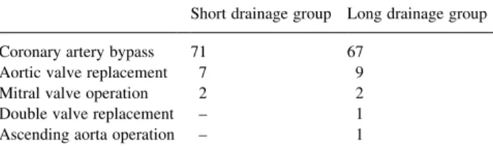
The patients characteristics are listed in Table 1. The operation indications for each group are shown in Table 2. The number of pleural drains and their location on every observational day are listed in Table 3.
Maximal pain intensity data are shown in Table 4. When both groups are compared, there is a trend toward less pain in the short drainage group, with a statistically signi®cant difference on POD 2 (P 0:047). When comparisons are made between different observations in each group, a statis-tically signi®cant difference is found between PODs 1 and 7 (P , 0:01), and between PODs 2 and 7 (P , 0:01) in both groups, and between PODs 2 and 3 in the long drainage group (P 0:02).
Table 5 reports the number of patients who do not mention any pain area. There was a trend towards more patients without pain in the short drainage group, and the difference was statistically signi®cant on POD 3.
Table 6 depicts the number of patients involved with pain in the different areas described on Fig. 1. The number of patients of each group involved at every observational day were compared. The following comparisons were statistically signi®cant. For the anterior part of the left shoulder (area 4), more patients of the long drainage group complained on POD 3 (P 0:003). For the left basis of the thorax (area 6), more patients of the long drainage group complained on POD 3 (P 0:008). For the epigastric area (area 8), more patients
Fig. 1. Picture of the body with the 18 anatomical areas as found on the observational sheet for pain localization analysis.
Table 1
Patient characteristics
Short drainage group Long drainage group Mean age (years) (^1 SD) 63 (8) 62 (9)
Age range (years) 29±83 28±79 Male/female 58/22 57/23 Emergency operation 5 6
Redo 5 5
Table 2
Operation indication
Short drainage group Long drainage group Coronary artery bypass 71 67
Aortic valve replacement 7 9 Mitral valve operation 2 2 Double valve replacement ± 1 Ascending aorta operation ± 1
of the long drainage group complained on PODs 2 and 3 (P 0:046 and 0.001, respectively).
Three patients in each group required antibiotics treat-ment for a pneumonia. No patient required a secondary pleural puncture after chest tube removal in either group during the observational period and no mediastinitis occurred. Lastly, no patient required pericardial drainage after mediastinal tube removal during the hospital stay. 4. Discussion
This comparison of two groups of cardiac surgery patients with different policies of chest tube removal shows that the pain intensity is signi®cantly higher in the group with prolonged drainage on POD 2. Moreover there was a signi®-cantly smaller number of patients with no pain on POD 3 in the same group. The topographic analysis of the pain demon-strates that the areas which are more often involved at these time points in the prolonged drainage group are the left shoulder, the left basis of the thorax and the epigastric area. The ®rst two areas are likely to correspond to the tip of the pleural drain which lies either at the apex or in the costo-diaphragmatic sinus according to the operator preference or to the self-positioning of the drain as it is slipped through the pleurotomy. The left hemithorax is more often involved with pain because of the high proportion of coronary artery bypass grafting with left internal mammary artery harvest and left pleural opening. The left thoracic pain could be related to the harvest procedure itself. However, the vertical forces applied to the sternum by the retraction device for the internal mammary artery harvest increases the incidence of sternal fracture [3], whereas it is the indirect force applied by the
retraction of the sternum during the cardiac operation itself which leads to fracture of the posterior and lateral aspects of the upper ribs [4±6]. These rib fractures are expected to occur at the upper part of the thorax and with equal frequency on both side of the chest. Therefore, it is unlikely that these fractures explain the increased frequency of pain sensation of the left basis of the thorax. The third area, the epigastric area, may be regarded as chest tube-related because it is the exit site of all the tubes: this is underscored not only by its more frequent involvement in the prolonged drainage group on PODs 2 and 3, but also by the fact that it is the second most frequently involved area after the sternotomy area in both groups.
The few studies which have dealt with chest tube-related pain have analyzed the sensations and the analgesic strategy during chest tube removal [7±9]. So far, pain directly related with the chest tubes themselves has not been formally addressed. Voigt et al. [10] analyzed the impact of a stan-dardized pain ¯owsheet to document pain assessment and pharmacologic management on cardiac surgical patient-reported pain intensity. The preimplementation group reported signi®cantly lower pain intensity rating. However, because the same group showed a signi®cant difference in the number of days the subject had chest tubes in place, the authors suggested that the earlier removal of chest tubes could have been an explanation for the decrease in pain perceived by this group. Paiement et al. [11] interviewed 100 cardiac surgery patients on PODs 5 and 6 about their worst memory in such an experience. Twenty-two patients mentioned the drain and 20 the endotracheal tube or any event related to intubation. Although suggestive, these studies are only indirect reports of the impact of chest tubes on postoperative pain.
An ideal study of the impact of chest tubes on postopera-tive pain would have been to compare a group with chest tubes against a group without chest tubes, which obviously is unethical in the setting of cardiac surgery. Therefore, we chose to compare two groups of patients with different poli-cies of chest tube removal, operated on in two different hospitals, during the same time period, with similar post-operative strategies, the same group of cardiac surgeons and similar analgesic options on the order form. The presence of at least one pleural tube, which is an important nociceptive stimulus, was a selection criteria in order to further mini-mize the heterogeneity of the samples. Although it is almost impossible that two such groups are strictly comparable,
Table 3
Number of pleural drains according to their locations and postoperative day (POD) in each groupa
Pleural drain POD 1 POD 2 POD 3 POD 7
Short Long Short Long Short Long Short Long
Left 68 69 7 69 0 44 0 0
Right 3 1 1 1 1 1 0 0
Bilateral 9 10 4 10 1 9 0 0
a Short, short drainage group; long, long drainage group.
Table 4
Data of maximal pain intensity
POD 1 POD 2 POD 3 POD 7 Short drainage group
Mean (^1 SD) 3.6 ^ 1.8 3.5 ^ 1.7 3.05 ^ 1.8 2.4 ^ 1.7 Minimal±maximal values 0±8 0±9 0±8 0±8 Long drainage group
Mean (^1 SD) 4 ^ 1.9 4.2 ^ 1.7 3.4 ^ 1.5 2.9 ^ 1.8 Minimal±maximal values 0±9 0±9 0±10 0±8 P-value 0.3 0.047 0.25 0.15
these settings minimize the differences as emphasized by the similarities in the patients' characteristics, the operation indications and the number and side of pleural drain inserted at the operation. Moreover, the nurses of both institutions were instructed by the same investigators.
Importantly, neither type of policy signi®cantly in¯u-enced the chest tube-related complications rate. On one side, the short drainage duration policy did not increase the incidence of repeated drainage after chest tube ablation; on the other side the long drainage duration policy did not increase the incidence of pneumonia and mediastinitis.
We conclude that a policy of early chest drain ablation limits pain sensation and simpli®es nursing care, without increasing the need for secondary pleural puncture. There-fore, a policy of short drainage after cardiac surgery should be recommended.
Acknowledgements
We wish to thank the nurses of the Surgical Intensive
Care Unit and the Clinic for Cardiovascular Surgery (CHUV ± Lausanne and HoÃpital de Sion) who recorded the data of the patients.
References
[1] American College of Cardiology/American Heart Association. Guidelines and indications for coronary artery bypass surgery: a report of the American College of Cardiology/American Heart Asso-ciation task force on assessment of diagnostic and therapeutic cardi-ovascular procedures (subcommittee on coronary artery bypass graft surgery). J Am Coll Cardiol 1991;17:543±589.
[2] Zickmann B, Sablotzki A, FuÈssle R, GoÈrlach G, Hempelmann G. Perioperative microbiologic monitoring of tracheal aspirates as a predictor of pulmonary complications after cardiac operations. J Thorac Cardiovasc Surg 1996;111:1213±1218.
[3] Moore R, Follette D, Berkoff H. Possternotomy fractures and pain management in open cardiac surgery. Chest 1994;106:1339±1342. [4] Greenwald LV, Baisden CE, Symbas PN. Rib fractures in coronary
bypass patients: radionuclide detection. Radiology 1983;148:553± 554.
[5] Woodring JH, Royer JM, Todd EP. Upper rib fractures following median sternotomy. Ann Thorac Surg 1985;39:355±357.
Table 5
Number of patients with no pain area
POD 1 POD 2 POD 3 POD 7
Short Long Short Long Short Long Short Long
Patients without pain 15 13 15 8 22 9 25 15
P-value 0:7 0:17 0:01 0:1
Table 6
Number of patients involved with pain according to the de®ned areasa
Pain area no. POD 1 POD 2 POD 3 POD 7
Short Long Short Long Short Long Short Long
1 11 6 3 2 2 4 3 3 2 0 4 3 5 1 7 4 9 3 49 50 48 47 46 45 39 48 4 1 5 4 3 0 9 (0.003) 4 10 5 3 5 5 4 4 6 2 6 6 12 17 11 17 3 14 (0.008) 5 11 7 1 2 0 3 1 3 0 3 8 16 25 15 26 (0.046) 6 22 (0.001) 6 10 9 1 6 4 8 1 4 0 4 10 0 1 1 1 0 0 1 7 11 3 1 4 3 1 6 3 8 12 2 4 4 8 3 5 7 7 13 3 0 2 4 1 4 5 12 14 3 2 4 2 1 7 1 5 15 0 0 0 1 0 2 0 4 16 0 0 0 0 1 0 1 2 17 4 3 4 4 1 5 2 3 18 0 0 0 1 0 1 0 0
[6] Curtis JA, Libshitz HI, Dalinka MK. Fracture of the ®rst rib as a complication of midline sternotomy. Radiology 1975;115:63± 65.
[7] Puntillo KA. Dimensions of procedural pain and its analgesic management in critically ill surgical patients. Am J Crit Care 1994;3:116±122.
[8] Puntillo K. Analgesics and chest tube removal pain. Am J Crit Care 1994;3:245.
[9] Gift A, Spearing Bolgiano C, Cunningham J. Sensations during chest tube removal. Heart Lung 1991;20:131±137.
[10] Voigt L, Paice JA, Pouliot JA. Standardized pain ¯owsheet: impact on patient-reported pain experiences after cardiovascular surgery. Am J Crit Care 1995;4:308±313.
[11] Paiement B, Boulanger M, Jones CW, Roy M. Intubation and other experiences in cardiac surgery: the consumer's views. Can Anaesth Soc J 1979;26:173±180.