Abscisic acid effect on the DNA microgradients of
decapped maize roots
Paul-Emile Pilet and Dorota Nocera-Przybecka
Institute of Plant Biology and Physiology of the University 1005 Lausanne, 6 PI. de la Riponne, Switzerland
(Received June 3, 1978)
Longitudinal and transversal total DNA gradients were analysed in 42-/*m thin sec-tions of maize root tips. The optimum DNA concentration was found in the apex (quiescent centre and meristem). It significantly increased after removal of the root cap, which can be considered an essential source of abscisic acid (ABA). When the caps were taken off and agar blocks containing ABA were immediately placed on the apical cut sections, the total DNA level in the root apex decreased gready. ABA regulation of cellular and nuclear behaviour is discussed.
Key words: ABA — Apex — Cap — DNA — Maize — Meristem — Nuclear behaviour — Nucleic acid — Root.
Geosensitivity in roots is lost after removal of the cap (9, 17, 18, 27, 28), but is regained after a few hours, which is considered the georecovery time (17, 27, 31). It has been assumed that the georecovery time is directly related to the formation of a new root cap (2, 12, 17). Decapping does indeed stimulate the cells of the quies-cent quies-centre to divide to form a new cap (3,13). However, it has been clearly demon-strated that georecovery precedes cap reformation by many hours (3, 4). Conse-quently, georecovery is not related to regeneration of the cap. During the ageotropic period of the decapped roots, some ultrastructural changes in the cells of the apex have been reported (5-7) and the formation of some amyloplasts has been observed (7). It is quite clear now that the cap imposes quiescence on cells at the pole of the root. As expected, removing the cap stimulates the cells of the quiescent centre and the meristem to grow and divide (3, 5). This was also clearly demonstrated by Clowes (11): he cut off the distal half of the cap and, 5 hr later, observed that the average rate of cell division had declined in the cap initials but increased in the quies-cent quies-centre and the apex cells.
On the other hand, the cap is the source of growth inhibitors (15, 29) which move basipetally (30, 41). For horizontally placed roots, downward transport of these inhibiting substances occurs inside the apex (33, 36) and their level is greater in the lower than the upper half of the cap (56", 37, 41, 47). These findings indicate clearly that the cap inhibitors control root geotropism (37, 45, 48, 49). One of these growth regulators could be abscisic acid (ABA), which strongly inhibits root elon-gation (23, 24, 26) and seems to have the same effects on root growth as the cap-inhibiting substances (32—34, 46). Kundu and Audus (19, 20) detected the presence of a cap inhibitor, the chromatographic properties of which were similar to those of
ABA. According to GC/MS data (40), the cap and the apex of maize roots (cv Kelvedon 33) contain 36.1 and 66.5 fig ABA kg"1 fresh weight, respectively.
Although the mechanisms controlled by ABA are still uncertain (23), physiolo-gical and biochemical analyses have led to a general proposal. Its primary effect could be the inhibition of DNA biosynthesis (43). Using Lemna in sterile cultures, Stewart and Smith (42) reported that the incorporation of 3H thymidine into DNA
was inhibited by 80 to 90% within 1 to 3 hr of ABA application. It is well establish-ed now (23) that ABA restablish-educes the content of DNA, inhibiting its biodegradation. But the decrease in the DNA level has been suggested to be only a reflection of earlier events (1). Time scale and the complexity of the response make it difficult to believe that DNA synthesis is the primary target of ABA (23).
The present study first analysed the longitudinal and transversal distribution of total DNA in the tip of intact maize roots. Second, the effect of the cap removal on the DNA content was analysed in the same tissues after a given time. Third, as the cap cells may be the source of ABA, the effect of this regulator on the DNA level of decapped roots was examined.
Materials and methods
The conditions for preparing maize roots and root segments have been previously described in detail (32, 35, 37). Selected caryopses of Zea mays L. cv Kelvedon 33 (Hurst Gunsons, London, UK) were soaked and germinated in darkness (22°C) and the roots were allowed to elongate vertically on moist paper towels. When the pri-mary roots reached 15 ± 3 mm, root segments of 10 ±0.2 mm were prepared and mounted vertically in plastic frames which were then placed in closed, humidified (90±5%) boxes (22±1°C) under white light (0.9±6x 10~2 J m"2 s~i) from a
fluo-rescent lamp (Osram L 13 W/25). At zero time, some of the segments were decapp-ed: their apical ends were removed at 450 ±50 [im from the tip (25). For some assays, the apical cut sections were covered with an agar block. The cylindrical agar block (diameter, 2.5 mm; thickness, 1 mm) contained ABA at 10~6 M. Buffered
(pH 6.1) agar (Difco purified) at 1.8% was used (33, 35). For each experiment, intact or decapped segments, with or without agar blocks containing ABA, were kept 6 hr in a vertical position.
The technique for preparing root inclusions and thin sections was previously described (21, 22). Segments were quickly embedded and frozen in liquid N2.
The inclusion medium employed was "Tissue Teck" (Ames Prod., Miles Seravac). Roots were then unmoulded and cut while frozen into thin sections (42 /jm) by using a Bright Cryostat (—25°C).
Of the many techniques for estimating nucleic acids the most suitable is that in which DNA and RNA are separated by hydrolysis with dilute alkali (14). The microgradient of total DNA was tested according to the method based on the me-asurement of the desoxyribose formed (39) and quantitatively estimated using a diphenylamine reagent (8) at 600 nm (SP-1800 Unicam Spectrophotometer). For standard determination, suitable amounts of hydrolysed calf thymus DNA were used. In some experiments using larger root sections, total DNA was analysed by a similar technique adapted to that previously described for lentil root extracts (38).
Results and discussion
In the first set of assays, the longitudinal gradient of total DNA was analysed in each 42-//m transversal thin cut section. Fig. 1 gives the data for the tips of apical maize root segments kept 6 hr in a vertical position. The DNA distribution is re-lated to the histological structure of the maize root end (16, 25). In the intact root segments (Fig. 1A), there was practically no DNA in the cap cells, while the apex (quiescent centre and meristem) contained the most DNA. The total DNA con-centration was lower in the differentiated part of the tip, but increased with distance from the end of the root. Such a distribution, closely related to the division pattern
0 210 (20 630 840 10S0 DISTANCE FROM THE TIP IN A i m
Fig. 1.
420 210 0 ' 210 £20 DISTANCE FROM THE CENTRE IN / U r n
Fig. 2.
Fig. 1. Longitudinal gradient of total DNA for intact (A) and decapped (B) apical root segments of maize cv
Kelvedon 33, kept 6 hr in a vertical position. Transversal sections of 42 fim were for analysed DNA in fig
per 4 roots from the tip (0 /im). For decapped segments, caps were removed at zero time.
Fig. 2. Transversal gradient of total DNA for intact (A) and decapped (B) apical root segments of maize cv
Kelvedon 33, kept 6 hr in a vertical position. Longitudinal sections of 42 fim were analyzed for DNA in fig per 4 roots (1050 fim from the tip) from the centre. For decapped segments, caps were removed
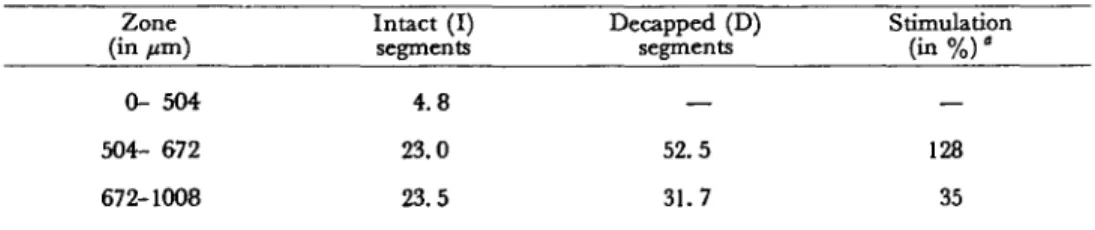
Table 1 Total DNA content in intact and decapped root segments of maize cv Kelvedon 33 Zone (in /an) 0- 504 504- 672 672-1008 Intact (I) segments 4.8 23.0 23.5 Decapped (D) segments — 52.5 31.7 Stimulation (in % ) ' — 128 35
DNA (in fig per 100 thin sections of 42 /on each) was analysed in three zones (counted from the tip) of apical segments kept 6 hr in a vertical position. For decapped segments, caps were removed (at 450 ±50 pm) at zero time.
of the cells, is very similar to that previously described (10, 11). When the root segments were decapped at zero time and their total DNA analysed 6 hr later (Fig. IB), a similar distribution of DNA was found, together with a strongly enhanced nucleic acid level.
Using these histochemical data, the total DNA obtained for the thin sections was calculated for the three essential zones of the root tips. As can be seen in Table 1, when the cap was removed, the DNA content greately increased. The relative values were significantly higher in the apex (the lSO-^m zone below the cap) than in the differentiated zones, as seen in Fig. 1.
In a second series of experiments, the transversal gradient of DNA was examined in 42 /an longitudinal thin cut sections of the apical 1050 /j,m segments. Fig. 2 gives the results for root segments kept 6 hr in a vertical position. In the intact segments (Fig. 2A), the highest level of DNA was found at the centre of the root. A similar distribution was obtained for decapped root segments (Fig. 2B), with the essential difference that the DNA concentration was much higher, as previously observed for the longitudinal gradient (see Table 1). Therefore, removal of the cap causes a significant increase in the DNA content of the apex cells.
From these data, we conclude that the cap forms some "regulators" which re-duce the concentration of the total DNA and regulate aspects of cellular and nuclear behaviour. Barlow (5, 6) supposed the presence, in the root apex, of a gradient of auxins and cytokinins, the concentration of which maintains nuclear DNA synthesis in cells of the meristem and elsewhere in the apex. But, as discussed above, ABA could surely be considered as one of the growth inhibitors produced or released by the cap. And it seems clear now that ABA is transported basipetally from the cap to the apex and the elongating zone of the growing root. We also stated in the intro-duction that ABA may control the DNA biosynthesis.
Analyses of the DNA accumulation after decapping (from 0 to 6 hr) are now in progress. A possible correlation seems to exist to between the rate of formation of nucleic acids in the root apex and the reformation (not easy to test) of the new cap cells from the quiescent centre.
To analyse the possible role of ABA—as an endoegnous regulator—on the DNA level of the apex cells, another experiment was done. Root caps were first removed and agar blocks, containing ABA or not, were immediately fixed on the apical cut sections. Decapped root segments were maintained vertically for 6 hr. The total
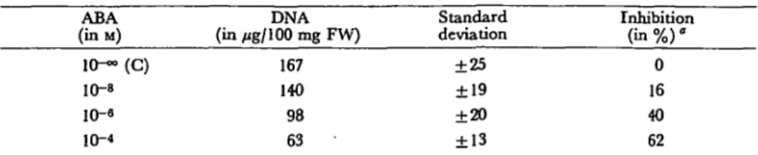
Table 2 Total DNA content in decapped root segments of maize cv Kelvedon 33 treated with ABA ABA (inM) 10—(C) 10-s 10-« 10-4 DNA (in /jg/100 mg FW) 167 140 98 63 Standard deviation ±25 ±19 ±20 ±13 Inhibition ( i n % ) ° 0 16 40 62
DNA (in ftg per 100 mg of fresch weight) was analysed in the 1.5 ±0.1 mm part below the apical end. For decapped segments, caps were removed (at 450 ± 5 0 pm) at zero time. Agar blocks (with or with-out ABA) were immediately fixed on the apical cut sections. Measurement was made after the segments kept were for 6 hr in a vertical position.
' o/o = _iQ2 (T-C)/C C: control T: treated
DNA was then analysed for the 1.5±0.1 mm region located just before the apical cut end. The results, reported in Table 2 indicate a significant decrease in the total DNA content: the lowest level of DNA was found with the highest ABA concentra-tions tested. The decapping causes major changes in the structure of the root apex
(7, 11), and ABA also significantly changes the ultrastructure of the quiescent centre
and the meristem of Lepidium roots (44).
Consequently, we can conclude that ABA, formed in the cap and moving in the basipetal direction from the root tip to the apex and then to the extending part of the elongating root, does decrease the total DNA. When the cap was removed, the concentration of endogenous ABA immediately decreased and that of total DNA greately increased. This increasing accumulation of DNA must be related to the division of the quiescent centre and the meristematic cells. Consequently, these successive processes may induce the regeneration of the root cap.
References
( / ) Addicott, F. T. and J. L. Lyon: Physiology of abscisic acid and related substances. Ann. Rev.
Plant Physiol. 20: 139-164 (1969).
(2) Audus, L. J . : Geotropism in root. In The Development and Function of Roots. Edited by J. G.
Torrey and D. T. Clarkson. p. 327-363. Academic Press, London, 1975.
( 3 ) Barlow, P. W.: Regeneration of the cap of primary roots of Zea mays. New Phylol. 73: 937—954 (1974).
( 4 ) Barlow, P. W.: Recovery of geotropism after removal of the root cap. J. Exp. Bot. 25: 1137-1146(1974).
( 5 ) Barlow, P. W.: Towards an understanding of the behaviour of root meristems. J. Theor. Biol. 57: 433-451 (1974).
( 6) Barlow, P. W.: The integrity and organization of nuclear DNA in cells of the root cap of Zea
mays probed by terminal deoxynucleotidyl transferase and microdensitometry. Z. Pflanzen-physiol. 80: 271-278 (1976).
( 7) Barlow, P. W. and M. Grundwag: The development of amyloplasts in cells of the quiescent centre of Zea roots in response to removal of the root cap. ibid. 73: 56-64 (1974).
( 8 ) Burton, K.: The relation between the synthesis of deoxyribonucleic acid and the synthesis of protein in the multiplication of bacteriophage T2. Biochem. J. 61: 473-483 (1955).
( 9) Cercek, L.: Effect of X-ray irradiation on regeneration and geotropic function of barley root caps. >Int. J. Radial. Biol. 17: 187-194 (1970).
(11) Clowes, F. A. L.: Regulation of mitosis in roots by their caps. Nature New Biol. 235: 143-144
(1972).
(12) Clowes, F. A. L. and B. E. Juniper: Plant Cells. Blackwell Sci. Publ. Oxford, 1968.
(13) Feldman, L. J.: The de novo origin of the quiescent center in regenerating root apices of Zea mays. Planta 128: 207-212 (1976).
(14) Fleck, A. and D. Begg: The estimation of ribonudeic acid using ultraviolet absorption
measure-ments. Biochim. Biophys. Acta 108: 333-339 (1965).
(15) Gibbons, G. S. B. and M. B. Wilkins: Growth inhibitor production by root caps in relation to
geotropic responses. Nature 226: 558-559 (1970).
(16) Juniper, B. E. and A. French: The fine structure of the cells that perceive gravity in the root tip
of maize. Planta 95: 314-329 (1970).
(17) Juniper, B. E., S. Groves, B. L. Schachar and L. J. Audus: Root cap and the perception of
gravity. Nature 209: 93-94 (1966).
(18) Konings, H.: The significance of the root cap for geotropism. Acta Bot. Neerl. 17: 203-211
(1968).
(19) Kundu, K. K. and L. J. Audus: Root growth inhibitors from root cap and root meristem of Zea mays L. J. Exp. Bot. 25: 479-489 (1974).
(20) Kundu, K. K. and L. J. Audus: Root growth inhibitors from root tips of Zea mays. Planta
117: 183-186(1974).
(21) Mayor, G. and P. E. Pilet: Gradients racinaires: une nouvelle microm£thode et son application
a l'analyse des phosphomonoestherases acides. Physiol. vig. 13: 479-487 (1975).
(22) Mayor, G. and P. E. Pilet: Longitudinal and transversal microgradient of acid
phosphomono-esterase in growing and geostimulated maize roots. Physiol. Plant. 39: 236-238 (1977).
(23) Milborrow, B. V.: The chemistry and physiology of abscisic acid. Ann. Rev. Plant Physiol. 25:
259-307 (1974).
(24) Newton, R. J.: Abscisic acid, effects on growth and metabolism in the roots of Lemna minor. Physiol. Plant. 30: 108-112 (1974).
(25) Nougarede, A. and P. E. Pilet: Inhibiteur racinaire et zone de courbure de segments verticaux
du Lens culinaris et du Zea mays. C. R. Acad. Sc. 279: 477-480 (1974).
(26") Pilet, P. E.: The effect of auxin and abscisic acid on the catabolism of RNA. J. Exp. Bot. 21: 446-451 (1970).
(27) Pilet, P. E.: Root cap and georeaction. Nature 233: 115-116 (1971).
(28) Pilet, P. E.: Role de l'apex radiculaire dans la croissance, le g£otropisme et le transport des
auxines. Bull. Soc. Bot. Swsse 81: 52-65 (1971).
(29) Pilet, P. E.: Root cap and root growth. Planta 106: 169-171 (1972).
(30) Pilet, P. E . : Growth inhibitor from the root cap of Zea mays. ibid. I l l : 275-278 (1973).
(37) P i l e t , P . E . : Georeaction of decapped roots. Plant Sci. Lett. 1: 137-140 (1973).
(32) Pilet, P. E.: Effects of light on the georeaction and growth inhibitor content of roots. Physiol. Plant. 33: 94-97 (1975).
(33) Pilet, P. E.: Abscisic acid as a root growth inhibitor: physiological analyses. Planta 122:
299-302 (1975).
(34) Pilet, P. E.: Action of fusicoccin and abscisic acid on root growth. Plant Sci. Lett. 5: 137-140
(1975).
(35) Pilet, P. E.: The light effect on the growth inhibitors produced by the root cap. Planta 130:
245-249 (1976).
(36") Pilet, P. E.: Effects of gravity on the growth inhibitors of geo-stimulated roots of Zea mays L. ibid. 131: 91-93 (1976).
(37) Pilet, P. E.: Growth inhibitors in growing and geostimulated maize roots. In Plant Growth Regulation. Edited by P. E. Pilet. p. 115-128. Springer Verlag, Berlin, 1977.
(38) Pilet, P. E. and R. Braun: The interrelation of RNA, auxin and auxin-oxidases in lentil roots. Physiol. Plant. 20: 870-878 (1967).
(39) Przybecka, D. and P. E. Pilet: DNA distribution in the tip of intact and decapped maize roots.
In Coll. abstr. of the pap.-demonstr. of the 9th Intern. Conf. on Plant Growth Substances. Edited by P. E. Pilet. p. 308-310. Lausanne, 1976.
(40) Rivier, L., H. Milon and P. E. Pilet: Gas chromatography-mass spectrometric determinations
of abscisic acid levels in the cap and the apex of maize roots. Planta 134: 23-27 (1977).
(41) Shaw, S. and M. B. Wilkins: The source and lateral transport of growth inhibitors in
geo-tropically stimulated roots of Zea mays and Pisum satiman. ibid. 109: 11-26 (1973).
(42) Stewart, G. R. and H. Smith: Effects of abscisic acid on nucleic acid synthesis and the induction
ofnitratereductaseinZwnna/io/j'rAizfl. J. Exp. Bot. 23: 875-885 (1972).
(43) Van Overbeck, J., J. E. Loeffler and M. I. R. Masson: Dormin, inhibitor of plant DNA
synthe-sis? Science 156: 1497-1500 (1967).
(44) Volkmann, D.: In cress roots, abscisic acid prevents the development of the central cap cells
into statocytes. Planta 130: 89-91 (1976).
(45) Wain, R. L.: Root growdi inhibitors. In Plant Growth Regulation. Edited by P. E. Pilet. p. 109-114. Springer Verlag, Berlin, 1977.
(46) Wilkins, H. and R. L. Wain: The role of the root cap in the response of the primary roots of Zea mays L. seedlings to white light and to gravity. Planta 123: 217-222 (1975).
(47) Wilkins, H. and R. L. Wain: Abscisic acid and the response of the roots of Zea mays to gravity.
ibid. 126: 19-23 (1975).
(48) Wilkins, M. B.: The role of the root cap in root geotropism. Curr. Adv. Plant Set. 13: 317-328
(1975).