Structural Biology at The Single Particle Level: Imaging Tobacco Mosaic Virus
by Low-Energy Electron Holography
Jean-Nicolas Longchamp1, Tatiana Latychevskaia1, Conrad Escher1 and Hans-Werner Fink1
1Physics Department, University of Zurich, Switzerland
A milestone for structural biology would definitely be attained if methods and tools were available, that do away with averaging over an ensemble of molecules and thereby enable structural biology on a truly single molecule level. To obtain atomic resolution information about the structure of any individual biological molecule, entirely new concepts and technologies are needed. One approach of this kind is associated with the recent X-ray free electron laser (XFEL) projects. Unfortunately, there are now strong indications that also for XFEL experiments averaging over a large number of molecules will be inevitable in order to obtain images with a sufficiently high signal-to-noise ratio to enable numerical reconstruction of the diffraction pattern with atomic resolution [1,2].
Our approach to structural biology at the single molecule level is motivated by the experimental evidences that electrons with a kinetic energy in the range of 50-250eV are harmless to biomolecules [3,4]. Even after exposing fragile molecules like DNA or proteins to a total electron dose of 106e-/Å2, more than six orders of magnitude higher than the critical dose in transmission electron microscopy, no radiation damage could be observed [3]. This, combined with the fact that the de Broglie wavelengths associated with this energy range are between 0.8 and 1.7Å, makes low-energy electron microscopy a candidate for structural biology at the single molecule level.
In our low-energy electron holographic setup (Figure 1), inspired by Gabor’s original idea of inline holography [5,6], a sharp tungsten tip acts as source for a divergent beam of highly coherent electrons [6]. The electron field emitter is brought as close as 100nm to the sample with the help of a 3-axis nanopositioner. Part of the emitted electron wave is elastically scattered off the object and hence is called the object wave, while the un-scattered part of the wave represents the reference wave. At a distant detector, the hologram, i.e. the pattern resulting from the interference of these two waves is recorded. A hologram contains the phase information of the object wave, and the object structure can thus be reconstructed unambiguously. In low-energy electron holography, a lens-less technique not suffering from lens aberrations, the resolution limit is given by the deBroglie wavelengths (λ) and by the numerical aperture (NA) of the detector system. With λ being as small as 0.7Å and NA=0.54 in our instrument, atomic resolution shall eventually be possible [7].
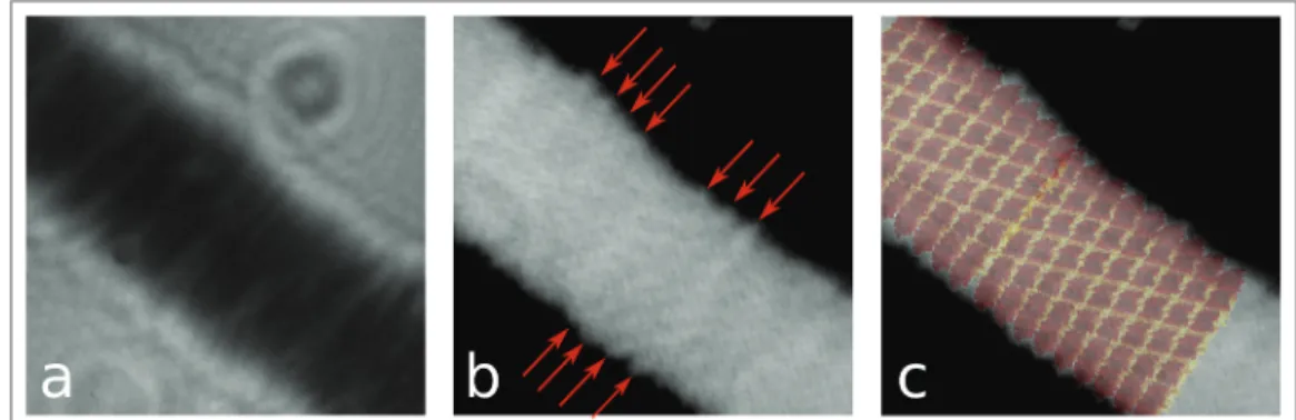
Here, we will report nanometer resolution imaging by means of low-energy electron holography of individual tobacco mosaic viruses (TMV) deposited onto ultraclean freestanding graphene [8]. We will show that structural details arising from the helical structure of the viruses can be revealed and that the agreement between our images and an atomic model of TMV available from the protein database is remarkable (Figure 2). We will also describe our on-going efforts towards 2Å resolution by means of low-energy electron coherent diffraction imaging (CDI). By implementing a CDI experimental scheme for low-energy electrons, we could already image a 210nm freestanding region of graphene at 2.3Å resolution, revealing more than half a million of graphene unit cells at once [9]. Finally, we will describe in details the procedure to prepare ultraclean freestanding graphene by the Pt-metal catalysis method; a method enabling the application of single-layer graphene as template for electron microscopy [10].
Paper No. 0255 509
doi:10.1017/S1431927615003347 © Microscopy Society of America 2015
Microsc. Microanal. 21 (Suppl 3), 2015
https://doi.org/10.1017/S1431927615003347
References:
[1] J.W. Miao, H.N. Chapman, J. Kirz, D. Sayre, K.O. Hodgson, Taking X-ray diffraction to the
limit: Macromolecular structures from femtosecond X-ray pulses and diffraction microscopy of cells with synchrotron radiation, Annu. Rev. Biophys. Biomol. Struct. 33 (2004) 157–176.
[2] H.N. Chapman, P. Fromme, A. Barty, T.A. White, R.A. Kirian, A. Aquila, et al., Femtosecond
X-ray protein nanocrystallography, Nature. 470 (2011) 73–77.
[3] M. Germann, T. Latychevskaia, C. Escher, H.-W. Fink, Nondestructive imaging of individual
biomolecules, Phys. Rev. Lett. 104 (2010) 95501.
[4] J.-N. Longchamp, T. Latychevskaia, C. Escher, H.-W. Fink, Non-destructive imaging of an
individual protein, Appl. Phys. Lett. 101 (2012) 93701.
[5] D. Gabor, A new microscopic principle, Nature. 161 (1948) 777–778.
[6] H.-W. Fink, W. Stocker, H. Schmid, Holography with low-energy electrons, Phys. Rev. Lett.
65 (1990) 1204–1206.
[7] T. Latychevskaia, J.-N. Longchamp, C. Escher, H.-W. Fink, Holography and coherent
diffraction with low-energy electrons: A route towards structural biology at the single molecule level, Ultramicroscopy. (2014).
[8] J.-N. Longchamp, T. Latychevskaia, C. Escher, H.-W. Fink, Structural biology at the single
particle level: imaging tobacco mosaic virus by low-energy electron holography, (2014).
[9] J.-N. Longchamp, T. Latychevskaia, C. Escher, H.-W. Fink, Graphene Unit Cell Imaging by
Holographic Coherent Diffraction, Phys. Rev. Lett. 110 (2013) 255501.
[10] J.-N. Longchamp, C. Escher, H.-W. Fink, Ultraclean freestanding graphene by platinum-metal catalysis, J. Vac. Sci. Technol. B Microelectron. Nanom. Struct. 31 (2013) 020605.
Figure 1: Scheme of the experimental setup of low-energy electron holography. The source-sample
distance amounts to typically 100-1000nm, which leads to kinetic electron energies in the range of 50-250eV.
Figure 2: a High-magnification hologram of TMV recorded at 80eV. b Reconstruction of the shape
of TMV from a the red arrows mark the presence of details arising from the helical structure of TMV. c An atomic model for TMV is superimposed on the image presented in b.
510 Microsc. Microanal. 21 (Suppl 3), 2015
https://doi.org/10.1017/S1431927615003347