THE
JOURNAL
OF
CELL
BIOLOGY
The Rockefeller University Press $30.00 J. Cell Biol. Vol. 183 No. 6 1101–1114
Correspondence to B é n é dicte Dargent: benedicte.dargent@univmed.fr Sandrine Pereira ’ s present address is Institut National de la Sant é et de la
Recherche M é dicale, Unit é Mixte de Recherche 910, Universit é de la
M é diterran é e, Facult é de M é decine secteur-Timone, Marseille 13385, France. Abbreviations used in this paper: AIS, axonal initial segment; CK2, protein ki-nase CK2; CNS, central nervous system; DIV, days in vitro; DMAT, 2-dimethyl-amino-4,5,6,7-tetrabromo1 H -benzimidazole; MAP2, microtubule-associated protein 2; MBD, membrane-binding domain; MBD-ank, MBD-ankyrin; PNS, pe-ripheral nervous system; SPR, surface plasmon resonance.
Introduction
In neurons, action potentials are generated at the axonal initial segment (AIS), and in myelinated axons, their saltatory conduc-tion occurs via the nodes of Ranvier ( Stuart et al., 1997 ; Kole et al., 2008 ). These processes require a precise distribution of volt-age-gated sodium channels that accumulate at high density in these two highly specialized axonal subdomains, defi ned by the segregation of the cytoskeletal adaptor complex ankyrin G/  IV spectrin ( Zhou et al., 1998 ; for review see Salzer, 2003 ). This scaffolding complex not only concentrates voltage-gated so-dium channels Na v 1.2 and Na v 1.6 ( Boiko et al., 2003 ), but also
potassium channels KCNQ2/KCNQ3 ( Devaux et al., 2004 ; Pan et al., 2006 ), cell adhesion molecules neurofascin-186 (NF-186), and neuronal cell adhesion molecule (NrCAM; Davis et al., 1996 ; Basak et al., 2007 ; Dzhashiashvili et al., 2007 ; Hedstrom et al., 2007 ). Recently, several additional new components have
been shown to accumulate at the AIS and nodes, such as mem-bers of the nuclear factor B signaling pathway ( Schultz et al., 2006 ; Politi et al., 2007 ), growth factors FHF2 and FHF4 ( Lou et al., 2005 ; Wittmack et al., 2004 ), and extracellular matrix components aggrecan and brevican ( Bruckner et al., 2006 ; John et al., 2006 ). The implications of these newly characterized components in AIS and node assembly, maintenance, or func-tion are still unknown.
Although they share a common subset of proteins, the as-sembly of AIS and nodes of Ranvier has been shown to differ ( Dzhashiashvili et al., 2007 ). The formation of the AIS depends on intrinsic properties of the neuron and occurs during the early steps in establishment of neuronal polarity in cultured neurons ( Alessandri-Haber et al., 1999 ; Boiko et al., 2007 ; Yang et al., 2007 ). Genetic, biochemical, and cell biology studies converge on the crucial role of ankyrin G as the key player in organizing the AIS ( Zhou et al., 1998 ; Jenkins and Bennett, 2001 ; Dzhashiashvili et al., 2007 ; Hedstrom et al., 2007 ). Unlike the AIS, the formation of the nodes of Ranvier requires exquisite communication with
I
n neurons, generation and propagation of actionpoten-tials requires the precise accumulation of sodium chan-nels at the axonal initial segment (AIS) and in the nodes of Ranvier through ankyrin G scaffolding. We found that the
ankyrin-binding motif of Na v 1.2 that determines channel
concentration at the AIS depends on a glutamate residue (E1111), but also on several serine residues (S1112, S1124, and S1126). We showed that phosphorylation of these
resi-dues by protein kinase CK2 (CK2) regulates Na v channel
interaction with ankyrins. Furthermore, we observed that CK2 is highly enriched at the AIS and the nodes of Ranvier
in vivo. An ion channel chimera containing the Na v 1.2
ankyrin-binding motif perturbed endogenous sodium channel accumulation at the AIS, whereas phosphorylation-defi cient chimeras did not. Finally, inhibition of CK2 activity reduced sodium channel accumulation at the AIS of neurons. In con-clusion, CK2 contributes to sodium channel organization by regulating their interaction with ankyrin G.
Protein kinase CK2 contributes to the organization
of sodium channels in axonal membranes by
regulating their interactions with ankyrin G
Aline Br é chet , 1,2 Marie-Pierre Fache , 1,2 Anna Brachet , 1,2 G é raldine Ferracci , 2,4 Agn é s Baude , 3 Marie Irondelle , 1,2
Sandrine Pereira , 1,2 Christophe Leterrier , 1,2 and B é n é dicte Dargent 1,2
1 Institut National de la Sant é et de la Recherche M é dicale, Unit é Mixte de Recherche 641, Marseille F-13916, France
2 Universit é de la M é diterran é e, Facult é de M é decine Secteur-Nord, Institut F é d é ratif de Recherche 11, Marseille F-13916, France
3 Universit é de la M é diterran é e, Centre National de la Recherche Scientifi que, Unit é Mixte de Recherche 6231, Marseille F-13916, France
4 Centre d ’ Analyse Prot é omique de Marseille, Marseille F-13916, France
© 2008 Bréchet et al. This article is distributed under the terms of an Attribution– Noncommercial–Share Alike–No Mirror Sites license for the fi rst six months after the publica-tion date (see http://www.jcb.org/misc/terms.shtml). After six months it is available under a Creative Commons License (Attribution–Noncommercial–Share Alike 3.0 Unported license, as described at http://creativecommons.org/licenses/by-nc-sa/3.0/).
on January 6, 2009
jcb.rupress.org
Therefore, we fi rst determined the critical residues of the ankyrin-binding motif responsible for K v 2.1-Na v 1.2 accumulation at the
AIS. We observed that segregation was driven by the joint ac-tion of two types of residue: a glutamate residue (Na v 1.2 E1111)
and several serine residues (Na v 1.2 S1112, S1123, S1124, and
S1126). Sequence analysis reveals that these serine residues are part of potential protein kinase CK2 (CK2) phosphorylation sites. Using the surface plasmon resonance (SPR) method ( Wil-son, 2002 ; Rich and Myszka, 2007 ), we directly demonstrated that CK2-mediated phosphorylation regulates the interaction between the Na v 1 ankyrin-binding motif and the MBD of
axo-nal ankyrins. Furthermore, a high concentration of CK2 in the AIS and the nodes of Ranvier was detected in vivo and in hippo-campal cultures, which is consistent with a possible role of CK2 in regulating the interaction between Na v and ankyrin.
More-over, a peculiar property of the K v 2.1-Na v 1.2 chimera allowed
us to unravel the functional signifi cance of this regulation: ex-pression of the K v 2.1-Na v 1.2 chimera containing the
ankyrin-binding motif of Na v 1.2-depleted endogenous sodium channels
in the AIS of cultured hippocampal neurons. In contrast, chime-ras containing a phosphorylation-defi cient ankyrin-binding mo-tif failed to perturb sodium channel accumulation at the AIS. Finally, inhibition of CK2 reduced sodium channel concentra-tion at the AIS of cultured neurons. Altogether, our fi ndings are consistent with the conclusion that CK2 contributes to the accu-mulation of sodium channels at the AIS and in the nodes of Ranvier by regulating their inter action with ankyrin G.
Results
Abrogation of the glutamate residue of the
Na v 1 ankyrin-binding motif is not suffi cient
to impair ion channel segregation at the AIS
When expressed by transfection in cultured hippocampal neu-rons, the potassium channel K v 2.1 was clustered at the cell surface
on soma and on proximal dendrites identifi ed by microtubule-associated protein 2 (MAP2) staining ( Fig. 1 A ), as described previously ( Lim et al., 2000 ). The deletion of the C terminus of K v 2.1 (K v 2.1- ⌬ Cter) induced a loss of compartmentalization
( Fig. 1 B ). The addition of a segment encompassing the AIS motif of sodium channel Na v 1.2 (amino acids 1,080 – 1,203) to
the C terminus of K v 2.1 (K v 2.1-Na v 1.2) was suffi cient to
redi-rect K v 2.1 to the AIS ( Fig. 1 C ). This was observed at the cell
surface of 70.9 ± 10.9% ( n = 609) of cells expressing K v
2.1-Na v 1.2, in line with our previous study ( Garrido et al., 2003 ).
To determine the critical residues involved in K v 2.1-Na v 1.2
seg-regation, we fi rst converted Na v 1.2 E1111 into alanine, a point
mutation that was suffi cient to impair CD4-Na v 1.2 II-III
com-partmentalization at the AIS ( Fache et al., 2004 ). Because a mutation of the corresponding E residue (Na v 1.5 E1053K) altered
the traffi cking of sodium channel Na v 1.5 in cardiomyocytes
( Mohler et al., 2004 ), we differentially stained the surface and total channel populations. Surface staining was observed in 58.3% ( n = 230) of the cells expressing the K v 2.1-Na v 1.2 E1111A mutant
and in 90.4% ( n = 240) of the cells expressing K v 2.1-Na v 1.2.
Strikingly, K v 2.1-Na v 1.2 E1111A was correctly localized at the
AIS in 52.7 ± 13.9% ( n = 178) of the cells displaying surface myelinating cells. In the nodes of the peripheral nervous system
(PNS), the aggregation of axonal components is dependent on ankyrin G, but the priming event is initiated by gliomedin, a membrane protein positioned in the microvilli of the Schwann cell facing the node of Ranvier, via a direct interaction with the extracellular domain of neurofascin-186 and neuronal cell adhe-sion molecule ( Eshed et al., 2005 , 2007 ; Dzhashiashvili et al., 2007 ). The nucleation of cell adhesion molecules in turn induces ankyrin G,  IV spectrin, and sodium channel recruitment ( Eshed et al., 2005 ; Dzhashiashvili et al., 2007 ).
Among the interactions responsible for AIS formation and maintenance, the Na v sodium channel interaction with ankyrin G
is of particular importance ( Lambert et al., 1997 ; Zhou et al., 1998 ; Garrido et al., 2003 ; Lemaillet et al., 2003 ; Fache et al., 2004 ). To circumvent diffi culties in expressing full-length Na v
in hippocampal neurons, we previously developed an approach based on CD4 chimera expression ( Garrido et al., 2001 ). This approach allowed us to identify a sequence of 27 residues, called the AIS motif, that determines sodium channel targeting and clustering at the AIS ( Garrido et al., 2003 ; Fache et al., 2004 ). This conserved determinant, located in the cytoplasmic linker between domains II and III of the Na v 1 pore-forming
␣ subunit (Na v 1 II-III), constitutes the ankyrin-binding motif of
Na v sodium channels. We also showed that the mutation of the
glutamate residue Na v 1.2 E1111 was suffi cient to impair the
compartmentalization at the AIS of CD4 – Na v 1.2 II-III, a
chi-mera in which the cytoplasmic region of CD4 was replaced by Na v 1.2 II-III ( Fache et al., 2004 ). In the meantime, an
indepen-dent study demonstrated that the PIALGESD sequence located within the AIS motif directly interacts with the membrane-binding domain (MBD; Lemaillet et al., 2003 ), a domain highly con-served in ankyrin G and ankyrin B ( Kordeli et al., 1995 ). Ankyrin G and B display a complementary subcellular distribu-tion in the axons of cultured hippocampal neurons ( Boiko et al., 2007 ) and in myelinated fi bers ( Kordeli et al., 1990 ; Bennett and Lambert, 1999 ; Ogawa et al., 2006 ). Strikingly, sodium channel accumulation is restricted to ankyrin G – positive subdo-mains both in vivo and in cultured neurons ( Boiko et al., 2001 , 2003 ). Because the ankyrin-binding motif is highly conserved in all neuronal sodium channels ( Garrido et al., 2003 ; Lemaillet et al., 2003 ; Pan et al., 2006 ) and binds to a domain highly conserved in ankyrin B and ankyrin G ( Kordeli et al., 1995 ), it is diffi cult to understand how sodium channels are specifi cally res tricted to ankyrin G – positive subdomains in the axon. We thus addressed the question as to whether an additional mechanism specifi es sodium channel accumulation at the AIS and in the nodes of Ranvier.
To further analyze the molecular mechanisms involved in the discrete localization of sodium channels in the axonal membrane, we chose to use K v 2.1-Na v 1.2, an ion channel chimera in which the
C terminus of the K v 2.1 potassium channel was replaced by the AIS
motif of Na v 1.2 instead of CD4 – Na v 1.2 II-III ( Garrido et al., 2003 ;
Fache et al., 2004 ). We have previously shown that endocytosis and domain-selective tethering confers CD4-Na v 1.2 II-III
segre-gation at the AIS ( Fache et al., 2004 ). However, the endocytotic signal identifi ed in the Na v 1.2 II-III linker ( Fache et al., 2004 ) is
not present in K v 2.1-Na v 1, which suggests that it could follow a
different traffi cking pathway and be directly sorted to the AIS.
on January 6, 2009
jcb.rupress.org
Downloaded from
http://jcb.rupress.org/cgi/content/full/jcb.200805169/DC1 Supplemental Material can be found at:
either S1126A or S1123-24A mutations ( Fig. 2, D and E ). Mu-tations E1111A-S1112A signifi cantly increased the number of cells with a nonpolarized distribution of K v 2.1-Na v 1.2 ( Fig. 2 E ).
These results indicated that K v 2.1-Na v 1.2 segregation is
depen-dent on the concerted action of E1111 with serine residues (S1112, S1123-24, or S1126).
The interaction between neuronal sodium channels and axonal ankyrins is regulated by CK2 phosphorylation
The ankyrin-binding motif of sodium channels directly inter-acts with a highly conserved domain in ankyrin G and B called the MBD ( Srinivasan et al., 1992 ; Lemaillet et al., 2003 ; Mohler et al., 2004 ). We next examined the possibility that CK2-mediated phosphorylation regulates this interaction. We fi rst determined whether the ankyrin-binding motif of Na v 1.2 is phosphorylated
by CK2 by conducting in vitro phosphorylation assays with GST proteins fused either to the linker II-III of Na v 1.2 (Na v 1.2
II-III) or truncated mutants ( Fig. 3 A ). In the presence of CK2, Na v 1.2 II-III was phosphorylated in vitro, as visualized by
auto-radiography ( Fig. 3 B ). A similar result was obtained when the C terminus of the Na v 1.2 II-III construct was truncated at residue
1133, whereas the deletion of the AIS motif (GST-Na v 1.2 989 –
1,079) resulted in a loss of signal. A positive signal was observed with a GST protein bearing the segment encompassing the AIS motif (GST-Na v 1.2 1,080 – 1,203). Several site-directed
muta-tions of GST-Na v 1.2 1,080 – 1,203 were generated. The
respec-tive abrogation of S1112, S1123, or S1124 did not affect in vitro phosphorylation of GST-Na v 1.2 1,080 – 1,203 by CK2 ( Fig. 3 C ).
In contrast, S1126A mutation and the double mutation S1123-24A resulted in a decrease in signal. The double mutation (S1124-26A) and the triple mutation (S1112-24-26A) impaired GST-Na v 1.2 1,080 – 1,203 phosphorylation ( Fig. 3 C ). All
to-gether, these results indicated that the ankyrin-binding motif of Na v 1.2 is phosphorylated in vitro by CK2. We next examined
staining ( Fig. 1, D – F , and Fig. 2 E ). After cell permeabilization, this mutant was visualized not only in the soma and throughout the dendrites but also in the ER identifi ed by calreticulin costaining (unpublished data). In contrast, K v 2.1-Na v 1.2 was only
occasion-ally detected in the ER (unpublished data). These fi ndings indi-cate that the abrogation of Na v 1.2 E1111 altered K v 2.1-Na v 1.2
traffi cking, presumably by favoring retention in the ER, but did not impair its ability to be targeted to the AIS. In view of these observations, we surmised that, in addition to E1111, other resi-dues of the ankyrin-binding motif contribute to K v 2.1-Na v 1.2
segregation at the AIS.
A combination of glutamate and
serine residues is required for channel segregation at the AIS
Sequence analysis of the targeting and clustering motif of Na v 1.2 ( Blom et al., 2004 ) revealed that S1112, S1124, and
S1126 contribute to three potential phosphorylation sites for CK2, defi ned by the sequence S/TXXD/E ( Meggio and Pinna, 2003 ). S1123 could also be phosphorylated by CK2 after phos-phorylation of S1126 ( Meggio and Pinna, 2003 ). All these ser-ine residues are highly conserved in the sodium channel types (Na v 1.1-1.3 and Na v 1.6) predominantly expressed in the central
nervous system (CNS; Fig. 2 A ). In an attempt to identify the additional residues involved in K v 2.1-Na v 1.2 segregation at the
AIS, we thus evaluated the impact of different serine mutations (S1112A; S1123-24A; S1126A; S1123-24-26A, designated 3SA; and S1112-23-24-26A, designated 4SA; Fig. 2 C ) on the steady-state surface distribution of K v 2.1-Na v 1.2 in
hippocam-pal neurons. All K v 2.1-Na v 1.2 serine mutants were
predomi-nantly compartmentalized at the AIS, whatever the number of serine mutations ( Fig. 2, B and C ). In contrast, serine mutations associated with the E1111A mutation profoundly perturbed K v 2.1-Na v 1.2 compartmentalization ( Fig. 2, D and E ). In fact,
segregation was totally impaired by combining E1111A with
Figure 1. Abrogation of E1111 of the Na v 1.2 ankyrin-binding motif did not impair ion channel segregation at the AIS of hippo-campal neurons. Cell surface distribution of
K v 2.1 (A), K v 2.1- ⌬ Cter (B), K v 2.1-Na v 1.2 (C), and K v 2.1-Na v 1.2 E1111A mutant (D – F) in cultured hippocampal neurons. The addition of the Na v 1.2 ankyrin-binding motif to the C terminus of K v 2.1- ⌬ Cter segregated K v 2.1 at the AIS (C, arrow). K v 2.1-Na v 1.2 compartmen-talization at the AIS was not impaired by the Na v 1.2 E1111A mutation (D and F, arrow). The indicated constructs were immunodetected with an antibody to myc (green) before cell permeabilization; the somatodendritic domain was subsequently identifi ed by MAP2 staining. Bars, 10 μ m.
on January 6, 2009
jcb.rupress.org
or MBD-ankB injections (see Materials and methods). Under these conditions, increasing concentrations of MBD-ank injec-tions produced a strongly increased binding signal. Kinetic analysis (see Materials and methods) of the interaction be-tween MBD-ankG and the CK2-phosphorylated GST-Na v 1.2
1,080 – 1,203 gave an association rate (k on ) of 42.4 ± 41.6 10 5
M ⫺ 1 s ⫺ 1 and a dissociation rate (k off ) of 2.1 ± 0.8 10 ⫺ 3 s ⫺ 1 ,
result-ing in a K D of 0.9 ± 0.7 10 ⫺ 9 M. Similar binding affi nities were
obtained with MBD-ankB ( Fig. 3 D and Table I ). When GST-Na v 1.2 989 – 1,133 was immobilized on the sensor surface,
inter-action with MBD-ankB was also strongly modulated by CK2 phosphorylation ( Fig. 3 E and Table I ). These affi nity constants were similar to those obtained with GST-Na v 1.2 1,080 – 1,203
( Table I ), which indicates that they are independent of the type of immobilized GST-Na v 1.2 II-III constructs. We further
exam-ined the impact of Na v 1.2 E1111 on the association between
whether CK2 phosphorylation regulates the association of the ankyrin-binding motif and MBD-ankyrin (MBD-ank). With this aim, we used SPR technology ( Wilson, 2002 ; Rich and Myszka, 2007 ). Purifi ed GST-Na v 1.2 1,080 – 1,203 was immobilized on
the sensor surface by immunoaffi nity. GST – MBD-ank were purifi ed, and the GST-tag was removed using PreScission Pro-tease (see Materials and methods). When an increasing concen-tration of MBD-ankB was injected over fl ow cells, a weak surface reactivity was observed ( Fig. 3 D ). Binding analysis (see Materials and methods) indicated that these two domains associate with an apparent affi nity constant (K D ) of 1.2 ± 0.4
10 ⫺ 6 M ( Table I ). When ankG was substituted for MBD-ankB, a similar affi nity constant was obtained (a K D of 1.7 ± 0.5
10 ⫺ 6 M; Table I ). To evaluate the impact of phosphorylation on the interaction, CK2 phosphorylation was performed on im-mobilized GST-Na v 1.2 1080 – 1203, before either MBD-ankG
Figure 2. Requirement for glutamate 1111 and serine residues for K v 2.1-Na v 1.2 compartmentalization at the AIS of hippocampal neurons. (A) Schematic
representation of the ankyrin-binding motif of sodium channels predominantly expressed in the CNS. Note the presence of four conserved serines (black boxes) and three potential serine phosphorylation sites for CK2 (Na v 1.2 S1112, S1124, and S1126). Numbers refer to the position of the amino acid residues in the corresponding Na v 1 types. (B – E) Cell surface distribution of K v 2.1-Na v 1.2 mutants. Cultured hippocampal neurons were transfected with the indicated constructs. 1 d after transfection, K v 2.1-Na v 1.2 was detected with an antibody to myc (green) before permeabilization, and the somatodendritic domain was subsequently identifi ed by MAP2 staining (red). Mutations of different serine residues in the ankyrin-binding motif did not perturb K v 2.1-Na v 1.2 segregation at the AIS (B and C). In contrast, co-mutation of E1111 with serine residues induced a loss of K v 2.1-Na v 1.2 compartmentalization (D and E). (C and E) Histograms of the cell surface distribution of K v 2.1-Na v 1.2 mutants. The percentage of myc-positive neurons were classifi ed into four categories: myc staining segregated at the AIS ([AIS]); distributed at the cell surface of the soma and proximal dendrites with an enrichment at the AIS (SD-[AIS]); en-riched at the AIS with a distribution in soma, proximal dendrites, and axons (SD-[AIS]-PA); and uniformly distributed at the cell surface (non polarized; SD-A). 100% represents the total population of transfected neurons. Data are means ± SD from two to three different experiments; n denotes the total number of cells analyzed for each construct. Bars, 10 μ m.
on January 6, 2009
jcb.rupress.org
Table II ). All together, these in vitro observations revealed that the association between the ankyrin-binding motif of neuronal sodium channels and the MBD of axonal ankyrins (MBD-ankG and MBD-ankB) is regulated by CK2 phosphorylation. These fi ndings strongly suggest that CK2 is able to strengthen the inter-action between ankyrin and sodium channels.
CK2 is highly enriched at the initial segment and in the nodes of Ranvier
If CK2 has a role in ankyrin – Na v interaction, one would expect
that it is present at the AIS in vivo. To test this possibility, we looked at the subcellular localization of CK2 in neurons using a polyclonal antibody directed against the catalytic ␣ subunit. In cultured hippocampal neurons, CK2 immunostaining was sodium channels and ankyrins by SPR. In the absence of
phos-phorylation, immobilized GST-Na v 1.2 1,080 – 1,203 E1111A
failed to recruit MBD-ank ( Fig. 3 F ). After on-chip CK2 phos-phorylation, GST Na v 1.2 1,080 – 1,203 E1111A associated with
MBD-ank with an affi nity constant (K D ) of 2.4 ± 0.8 10 ⫺ 8 M
( Fig. 3 F and Table I ). Finally, we evaluated the contribution of each of the serine residues of the ankyrin-binding motif on the phospho-dependent association between GST-Na v 1.2 1,080 –
1,203 and MBD-ank. The single serines to alanine mutation and double mutation S1123-24A did not modify the phospho-dependent association ( Table II ). The double mutation (S1124-26A) led to a decrease in affi nity (K D = 1.6 ± 1.3 10 ⫺ 7 M;
Table II ). The triple mutation robustly altered association, resulting in a 1,000-fold decrease in affi nity (K D = 2.6 ± 1.1 10 ⫺ 6 M;
Figure 3. CK2 phosphorylates the Na v 1.2 ankyrin-binding motif and regulates its inter-action with MBD-ank. (A – C) The Na v 1.2 an-kyrin-binding motif is phosphorylated by CK2 in vitro . (A) Schematic representation of GST-Na v 1.2 linker II-III constructs. The position of the ankyrin-binding motif is indicated by gray boxes. Numbers refer to the position of cor-responding amino acid residue in Na v 1.2. (B) The constructs were subjected to in vitro CK2 phosphorylation followed by SDS-PAGE. Coo-massie brilliant blue staining (CBB) and 32 P in-corporation revealed by autoradiography are shown. (C) The effect of site-directed serine mu-tations on CK2 phosphorylation of GST-Na v 1.2 II-III. CBB staining (bottom) and 32 P incorpora-tion revealed by autoradiography (top) are shown. Numbers to the right of the gel blots indicate the molecular mass of standard mark-ers (in kD). (D – F) SPR analysis of the interaction between the Na v 1.2 ankyrin-binding motif and MBD-ank. Typical sensorgrams are illustrated. Increasing concentrations of MBD-ankB rang-ing from 0.1 to 250 nM were injected over immobilized GST-Na v 1.2 1,080 – 1,203 (D), GST-Na v 1.2 989 – 1,133 (E), and GST-Na v 1.2 1,080 – 1,203 E1111A (F) before (top) and after in situ CK2 phosphorylation (bottom) of the immobilized constructs.
Table I. The effect of phosphorylation on the interaction between MBD-ank and the ankyrin-binding motif of sodium channels
Immobilized construct MBD CK2 phosphorylation k on k off K D
M ⫺ 1 s ⫺ 1 s ⫺ 1 M GST-Na v 1.2 1,080 – 1,203 ankG ⴚ NA NA 1.7 ± 0.5 × 10 ⫺ 6 a ankB ⴚ NA NA 1.2 ± 0.4 × 10 ⫺ 6 a ankG + 42.4 ± 41.6 × 10 5 2.1 ± 0.8 × 10 ⫺ 3 0.9 ± 0.7 × 10 ⫺ 9 ankB + 28.0 ± 11.0 × 10 5 3.0 ± 0.1 × 10 ⫺ 3 1.2 ± 0.4 × 10 ⫺ 9 GST-Na v 1.2 989 – 1,133 ankB ⴚ NA NA 1.3 ± 0.2 × 10 ⫺ 6 a + 60.1 ± 39.8 × 10 5 2.3 ± 0.1 × 10 ⫺ 3 0.5 ± 0.2 × 10 ⫺ 9
GST-Na v 1.2 1,080 – 1,203 E1111A ankB ⴚ ND ND ND + 1.4 ± 0.3 × 10 5 3.2 ± 0.6 × 10 ⫺ 3 2.4 ± 0.8 × 10 ⫺ 8
Mean values ± SD were obtained from three to six experiments. ankB and ankG, ankyrin B and G, respectively; NA, not applicable; ND, no binding detected.
a K
D values were calculated using steady-state analysis (see Materials and methods).
on January 6, 2009
jcb.rupress.org
Pan et al., 2006 ). We next addressed the question as to whether K v 2.1-Na v 1.2 acts as a dominant negative of sodium channel and
KCNQ2/KCNQ3 potassium channel accumulation at the AIS. We therefore quantifi ed immunostaining of endogenous sodium channels, KCNQ2/KCNQ3 channels, and ankyrin G in cells ex-pressing K v 2.1-Na v 1.2 (transfected cells) and in nontransfected
cells. The intensity of sodium channel staining was robustly re-duced in K v 2.1-Na v 1.2 – positive cells as compared with that
ob-served in nontransfected cells, whereas ankyrin G staining was unmodifi ed ( Fig. 6, A and B ). In contrast, KCNQ2/KCNQ3 staining intensity detected with an antibody directed against KCNQ3 was equivalent in cells expressing K v 2.1-Na v 1.2 and in
nontransfected cells ( Fig. 6, A and C ). This strongly suggested that the presence of K v 2.1-Na v 1.2 specifi cally disturbed the
accumulation of sodium channels but not KCNQ2/KCNQ3. Our SPR analysis demonstrated that the association between sodium channels and the MBD of axonal ankyrins is regulated by CK2 phosphorylation. Immunostaining, both in cultured neurons and in vivo, revealed that CK2 is highly enriched at the AIS and in the nodes of Ranvier. These observations led us to examine the possibility that CK2-mediated phosphorylation of sodium chan-nels occurs locally, at the AIS, contributing to channel clustering. We thus examined whether the perturbation of endogenous so-dium channel accumulation induced by K v 2.1-Na v 1.2 was
depen-dent on CK2 phosphorylation sites. Upon mutation of a single serine into alanine (S1112A and S1126A, respectively), mutated K v 2.1-Na v 1.2 induced a decrease in sodium channel staining
in-tensity, like K v 2.1-Na v 1.2 (unpublished data). When the three
ser-ine residues located in the C-terminal end of the AIS motif were all mutated into alanines, the effect of the corresponding mutant (3SA) was less pronounced than with K v 2.1-Na v 1.2 ( Fig. 6 B ). The
abrogation of four serine residues into alanines (4SA) impaired the dominant-negative effect of K v 2.1-Na v 1.2 on sodium channel
clustering ( Fig. 6, A and B ). These fi ndings revealed that expres-sion of phosphorylation-defi cient AIS motif constructs failed to perturb sodium channel accumulation in cultured neurons.
The effect of a CK2 inhibitor on the accumulation of sodium channels at the AIS of cultured hippocampal neurons
To further demonstrate that CK2 is involved in the accumula-tion of sodium channels at the AIS, we evaluated the effect of concentrated in the AIS but not in the distal part of the axon
( Fig. 4, A1 – A3 ). CK2 was also visualized in nuclei ( Filhol et al., 2003 ; Theis-Febvre et al., 2005 ), in soma, and in proximal dendrites. When hippocampal neurons were subjected to deter-gent extraction before cell fi xation, CK2 immunostaining at the AIS was still observed (unpublished data), which suggests teth-ering to the cytoskeleton ( Garrido et al., 2003 ; Fache et al., 2004 ). In vivo immunoreactivity for CK2 was further examined in the hippocampus ( Fig. 4, B and C ), cerebellum ( Fig. 4 D ), and cor-tex (Fig. S1, available at http://www.jcb.org/cgi/content/full/ jcb.200805169/DC1). Initial segments were intensely immuno-reactive for CK2 in all observed structures, as revealed by co-labeling with ankyrin G ( Figs. 4 and S1) and sodium channels (not depicted). Initial segments emanating from cell bodies of prin-cipal cells were observed in CA1 ( Fig. 4, B and C ), CA3, and dentate gyrus (Fig. S1, A and B). Initial segments positive for CK2 were also found in strati radiatum and oriens of CA1 and CA3, the molecular layer in the dentate gyrus, and in the hilus. Intense CK2 labeling appeared to underline the internal face of the plasma membrane of the initial segment, whereas the axoplasm displayed weak immunoreactivity, similar to ankyrin G localization ( Fig. 4 B ). In the hippocampus, 80% of ankyrin G – positive structures dis-played a positive labeling for CK2 in the white matter ( n = 637) and 88% in the gray matter ( n = 784). CK2 was also detected in nu-clei of neurons and putative glial cells ( Fig. 4 ). Punctate labeling for CK2, colocalizing with ankyrin G, was observed in the alveus and white matter of forebrain and cerebellum ( Fig. 5, A and B ). In the cerebellum, 77% of ankyrin G – positive structures displayed a costaining with CK2 in the white matter ( n = 64) and 88% in the gray matter ( n = 92). Finally, CK2 immuno reactivity was present in the nodes of Ranvier of sciatic nerves ( Fig. 5, C and D ): 100% of the nodes of Ranvier were costained for ankyrin G and CK2 ( n = 40). Collectively, these observations demonstrated that CK2 is a novel component of the AIS and of the nodes of Ranvier.
Evaluation of the role of the serine residues in the clustering of endogenous sodium channels
In addition to sodium channels, KCNQ2/KCNQ3 potassium channels are highly concentrated at the AIS both in cultured hippocampal neurons and in vivo, via an interaction with the MBD of ankyrin G ( Devaux et al., 2004 ; Chung et al., 2006 ;
Table II. The effect of serine point mutations on the interaction between MBD-ank and the ankyrin-binding motif of sodium channels
GST-Na v 1.2 1,080 – 1,203 mutants k on k off K D
M ⫺ 1 s ⫺ 1 s ⫺ 1 M S1112A 8.9 ± 0.2 × 10 5 3.3 ± 0.4 × 10 ⫺ 3 3.7 ± 0.3 × 10 ⫺ 9 S1123A 19.1 × 10 5 3.4 × 10 ⫺ 3 1.8 × 10 ⫺ 9 a S1124A 16.3 ± 3.2 × 10 5 3.9 ± 0.3 × 10 ⫺ 3 2.5 ± 0.7 × 10 ⫺ 9 S1126A 16.2 ± 14.4 × 10 5 3.1 ± 0.8 × 10 ⫺ 3 2.6 ± 1.3 × 10 ⫺ 9 S1123-24A 8.2 ± 2.6 × 10 5 3.4 ± 0.2 × 10 ⫺ 3 4.5 ± 1.3 × 10 ⫺ 9 S1124-26A 0.9 ± 1 × 10 5 4.2 ± 1.6 × 10 ⫺ 3 1.6 ± 1.3 × 10 ⫺ 7 S1112-24-26A NA NA 2.6 ± 1.1 × 10 ⫺ 6 b
Mean values ± SD are obtained from three to fi ve experiments. NA, not applicable.
a Value from one experiment.
b K
D values were calculated using steady-state analysis (see Materials and methods).
on January 6, 2009
jcb.rupress.org
or DMSO (control cells) at two stages of maturation. It has been shown previously that the percentage of cells with an AIS increases between 2 and 4 d in vitro (DIV; Yang et al., 2007 ; Martin et al., 2008 ). The inhibition of CK2 activity in immature 2-dimethylamino-4,5,6,7-tetrabromo1 H -benzimidazole (DMAT),
a CK2 inhibitor ( Pagano et al., 2004 ), in cultured hippocampal neurons ( Fig. 7 ). The immunostaining of ankyrin G and Na v .1
channels was quantifi ed in neurons treated with either DMAT
Figure 4. CK2 ␣ is concentrated at AISs. (A) A cultured hippocampal neuron was immunostained for CK2 ␣ (A1) and ankyrin G (A2). CK2 ␣ immunoreactiv-ity is highly expressed in the initial segment (box). Enlarged views of the initial segment are visualized in A1 – A3, bottom. (B and C) In the CA1 area of the hippocampus, CK2 ␣ immunoreactivity is visible within nuclei (B1, arrowheads) of putative pyramidal cells and interneurons. As demonstrated by colabeling with ankyrin G (B2, arrows), CK2 ␣ is present in initial segments all over the different strati (B1 and B3, arrows). CK2 ␣ shows light and diffuse additional staining within the neuropil. CK2 ␣ immunoreactivity (C1 and C3, arrows) and ankyrin G labeling (C2, arrows) are concentrated along the cytoplasmic membrane of an initial segment in the stratum pyramidale next to a CK2 ␣ -labeled nucleus (C1 and C3, asterisks). (D) In cerebellum, nuclei (D1, asterisk) of Purkinje cells and interneurons of the molecular layer (ml) are labeled for CK2. Coimmunolabeling for CK2 ␣ (D1) and for ankyrin G (D2) is visible within initial segments of a putative Purkinje cell (D1 – D3, arrows), granular cells (D1 – D3, double arrowheads), and interneurons (D1 – D3, single arrowheads). gl, granular layer; Pcl, Purkinje cell layer, so, stratum; sp, stratum pyramidale; sr, stratum radiatum. Bars: (A) 23 μ m; (B) 26.8 μ m; (C and D) 6.82 μ m.
on January 6, 2009
jcb.rupress.org
Discussion
CK2 phosphorylation regulates the interaction between neuronal sodium channels and ankyrin
We previously identifi ed a determinant of 27 residues ( Garrido et al., 2003 ), called the AIS motif, that includes the PIALGESD sequence defi ned by Lemaillet et al. (2003) . In close agreement with our previous work ( Garrido et al., 2003 ), we show here that the determinant required for high-affi nity ankyrin binding is not restricted to the PIALGESD sequence but extends further to-ward the C-terminal sequence, including two CK2 phosphorylation cells (from 2 to 4 DIV) induced an increase in the number of
cells negative for ankyrin G staining as compared with what was observed in control cells. In the other cell population (ankyrin G – positive), we observed a decrease in the staining in-tensity of ankyrin G and Na v .1 channels ( Fig. 7, A and B ). At 9
DIV, all the cells present an AIS ( Yang et al., 2007 ; Martin et al., 2008 ). The addition of DMAT in the culture medium of neurons at 9 DIV during 30 h induced a decrease in ankyrin G and Na v .1 channels staining ( Fig. 7, C and D ). These results showed
that inhibition of CK2 activity perturbs the accumulation of sodium channels and ankyrin G at two different stages of AIS organization.
Figure 5. CK2 ␣ is concentrated in CNS and PNS nodes of Ranvier. (A and B) CK2 ␣ immunoreactivity is visible as numerous punctiform structures (B1 and B3, arrows) colabeled for ankyrin G (B2 and B3, arrows) within the alveus and cortical white matter that probably correspond to nodes. B1 – B3 are enlarged views of the boxed areas in A1 – A3. (C and D) Ranvier nodes are labeled for CK2 ␣ and ankyrin G (C1 – C3, arrowheads) in teased fi bers of the sciatic nerve. CK2 ␣ labeling is concentrated at the membrane of the nodal region (D1, arrows). CK2 ␣ immunoreactivity is also visible in the nucleus of a Schwann cell (e.g., C1 and C3, arrows). al, alveus; wm, white matter. Bars: (A) 26.8 μ m; (B) 6. 71 μ m; (C) 23.2 μ m; (D) 3 μ m.
on January 6, 2009
jcb.rupress.org
Figure 6. Impact of K v 2.1-Na v 1.2 and of phosphorylation-defi cient K v 2.1-Na v 1.2 constructs on the accumulation of sodium channels (Na v 1) and KCNQ2/ KCNQ3 potassium channels at the AIS of cultured hippocampal neurons. (A, top and middle) K v 2.1-Na v 1.2 expression perturbed Na v 1 accumulation at the AIS, unlike the phosphorylation-defi cient K v 2.1-Na v 1.2 4SA mutant. Hippocampal neurons were transfected with either K v 2.1-Na v 1.2 or phosphoryla-tion-defi cient K v 2.1-Na v 1.2 mutants. Then cells were stained for myc (gray), ankyrin G (red), and sodium channels (green). (B) Quantifi cation of Na v 1 and ankyrin G staining intensity in untransfected cells (A, open arrowheads) and in transfected cells (closed arrowheads). (A, bottom) K v 2.1-Na v 1.2 expression did not perturb KCNQ3 accumulation at the AIS. Cells transfected with K v 2.1-Na v 1.2 were subsequently stained for myc (gray), ankyrin G (red), and KCNQ3 potassium channels (green). (C) Quantifi cation of KCNQ3 and ankyrin G staining intensity. Fluorescence intensity measured in transfected cells, identifi ed by myc staining, was normalized by taking as 100% the staining intensity measured in nontransfected cells (arrowheads). Numbers at the base of the bars denote the number of quantifi ed cells. Error bars indicate mean ± SEM. Mann-Whitney test: *, P < 0.05; **, P < 0.01; ***, P < 0.001. WT, wild type. Bars, 10 μ m.
on January 6, 2009
jcb.rupress.org
In addition, it was profoundly altered upon mutation of two to three serine residues. Co-mutation of S1124 with S1126 in-duced a robust decrease in binding affi nity from 1.2 10 ⫺ 9 M to 1.6 10 ⫺ 7 M. Third, we observed that K v 2.1-Na v 1.2 segregation
at the AIS required the cooperation of two motifs: Na v 1.2 E1111
and the serine residues S1112, S1124, and S1126. Co-mutation of E1111 with either S1124 or S1126 was found to be more ef-fective than E1111-S1112 mutations. Fourth, we observed that expression of phosphorylation-defi cient AIS motif constructs failed to perturb sodium channel accumulation in cultured neu-rons ( Fig. 6 ). At the steady-state surface distribution, all the K v 2.1-Na v 1.2 serine mutants were predominantly
compartmen-talized at the AIS ( Fig. 2, B and C ), which indicates that the loss of the dominant-negative effect of K v 2.1-Na v 1.2 induced by
ser-ine mutations did not result from defi cient AIS localization but sites (Na v 1.2 S1124 and S1126). In addition, we provide here
the fi rst evidence that the interaction between the ankyrin-bind-ing motif and the MBD of ankyrins is regulated by CK2. This conclusion is based on the following fi ndings. First, we ob-served that the AIS motif of CNS sodium channels is phosphor-ylated in vitro by CK2. This was supported by the abrogation of S1124-26 located in the C-terminal region of the AIS motif, which inhibited phosphorylation. Second, a quantitative bind-ing assay based on SPR revealed that CK2 phosphorylation of the AIS motif resulted in a 1,000-fold increase (from 1.2 10 ⫺ 6 M to 1.2 10 ⫺ 9 M) of the binding affi nity for MBD-ank. This ef-fect was observed using two distinct constructs in which the an-kyrin-binding motif was localized distally or proximally with respect to GST. Besides, CK2 phosphorylation was equally effective for interactions with the MBD of ankyrin B and G.
Figure 7. The effect of a CK2 inhibitor on the accumulation of sodium channels at the AIS of cultured hippocampal neurons. Cultured hippocampal neurons were treated with either DMAT (50 μ M) or with DMSO (control cells) either from 2 to 4 DIV (A and B) or from 9 to 10 DIV (C and D). They were subsequently immunostained for Na v 1 channels (green), ankyrin G (red), and MAP2 (blue). (B and D) Quantifi cation of Na v 1 and ankyrin G staining intensity. Fluores-cence intensity was normalized by taking as 100% the staining intensity measured in control cells. The results are from three independent experiments, the number of quantifi ed AIS ranged from 70 to 230. Error bars indicate mean ± SEM. t test: ***, P < 0.0001. Bars, 10 μ m.
on January 6, 2009
jcb.rupress.org
ankyrin G concentration during the formation and the mainte-nance of the AIS. It is possible that CK2 regulates ankyrin G binding to  IV spectrin, as described in chicken erythrocytes ( Ghosh et al., 2002 ). Furthermore, the involvement of the po-tential CK2 phosphorylation sites located in the C terminus of potassium channels KCNQ2 and KCNQ3 in accumulation at the AIS and in the nodes of Ranvier deserves close examination ( Chung et al., 2006 ; Pan et al., 2006 ). Crosstalk between nu-clear factor B ( Politi et al., 2007 ; Schultz et al., 2006 ) and CK2 signaling pathways might occur. Hence, an important is-sue in the future will be to obtain more information on the regu-latory role of CK2 in the specifi cation and the maintenance of the AIS and the nodes of Ranvier.
The role of CK2 in positioning sodium channels in ankyrin G – positive domains
In neurons, biochemical and genetic studies have shown that sodium channel accumulation is an ankyrin-based mecha-nism occurring via a direct interaction with MBD, a domain highly conserved in ankyrin G and B ( Lemaillet et al., 2003 ; Mohler et al., 2004 ). Our SPR assay revealed that the Na v 1.2
ankyrin-binding motif recognized MBD-ankG as well as MBD-ankB with an equivalent binding affi nity ( Table I ). How-ever, a high density of sodium channels occurs only in an-kyrin G subdomains both in vivo and in cultured neurons ( Boiko et al., 2001 , 2003 ; Garrido et al., 2003 ; Fache et al., 2004 ; Dzhashiashvili et al., 2007 ). In addition, distinct chi-meras bearing the ankyrin-binding motif of sodium channels, including K v 2.1-Na v 1.2 ( Fig. 1 ), were found to be
concen-trated at AIS (positive for ankyrin G) but not distal axons (positive for ankyrin B) of cultured hippocampal neurons ( Garrido et al., 2003 ; Lemaillet et al., 2003 ; Fache et al., 2004 ; Pan et al., 2006 ). How can we explain the discrepancy between in vivo localization of sodium channels and in vitro assays? The intramolecular interaction between the N and the C termini of ankyrin B could impede sodium channel recog-nition of the MBD ( Abdi et al., 2006 ). In the view of our fi ndings, it is tempting to propose that CK2 regulates sodium channel interaction with ankyrin G and thus contributes to their specifi c accumulation at the AIS and the nodes of Ranvier. The local concentration of CK2 at the AIS and in the nodes of Ranvier could lead to phosphorylation of Na v sodium
chan-nels at their ankyrin-binding domain. It also possible that a m é nage à trois between ankyrin G, Na v channels, and CK2 is
involved in the sorting of these components to the AIS and nodes of Ranvier. This phosphorylation-based process could represent a striking example of how posttranslational modifi -cations can fi ne tune interactions between key protein part-ners. Neuronal cells are likely to rely on such a dynamic network of modulated interactions to attain an exquisite level of subcellular compartmentalization. Moreover, these regu-lated interactions likely contribute to the inherent plasticity of neurons, allowing them to dynamically adjust protein lo-calization as a response to their various inputs. Precisely, the possibility of a dynamic modulation of ankyrin – Na v
inter-action by synaptic activity is a fascinating question that should be addressed in future studies.
rather from the conversion of serines into alanines. Finally, we observed that inhibition of CK2 activity perturbed sodium chan-nel accumulation at two distinct stages of AIS formation. Considered together, our fi ndings show that several CK2 phos-phorylation sites regulate the interaction between CNS sodium channels and ankyrins.
What is the contribution of the glutamate residue (Na v 1.2
E1111)? SPR analysis revealed that the Na v 1.2 E1111A
muta-tion totally abolished basal MBD-ank binding in the micromolar range measured in the absence of CK2 phosphorylation ( Fig. 4 ). After CK2 phosphorylation, the binding affi nity of the Na v 1.2
E1111A mutant was affected (K D = 2.4 10 ⫺ 8 M) when compared
with the control (K D = 1.2 10 ⫺ 9 M), but it remained high. This
strongly suggests that different CK2 phosphorylation sites of the ankyrin-binding motif can overcome the E1111A mutation. However, we cannot totally exclude the possibility that our ex-perimental conditions favored a high degree of phosphorylation of the ankyrin-binding motif. This may highlight the structural requirement for multiple serine phosphorylation and underesti-mate the contribution of residue E1111. Na v 1.2 E1111 mutation
was found to alter the level of cell surface expression of K v
2.1-Na v 1.2 in cultured hippocampal neurons ( Fig. 1 ), which is in
close agreement with a previous study on Na v 1.5 ( Mohler et al.,
2004 ). However, this mutation did not impair K v 2.1-Na v 1.2
seg-regation at the AIS because of the contribution of the phospho-serine residues. The fact that the association of E1111A with either S1124 or S1126 mutations was mandatory for the loss of K v 2.1-Na v 1.2 segregation at the AIS was consistent with this
conclusion. Our fi ndings strongly suggest that phosphorylation of three serine residues of the ankyrin-binding motif tunes the traffi cking of sodium channels and their accumulation at dis-crete sites in neurons. It is noteworthy that variable phosphoryla-tion of sites on the K v2.1 potassium channel allows graded
activity-dependent regulation of channel gating and neuronal fi r-ing properties ( Misonou et al., 2006 ; Park et al., 2006 ). Hence, multiple phosphorylation is likely to constitute an important regulatory mechanism for both the activity and the traffi cking of voltage-gated ion channels in neurons.
CK2 is a novel component of the AIS and of the nodes of Ranvier
CK2 is constitutively active and involved in a large spectrum of biological processes, including cell proliferation, apoptosis, and the circadian clock ( Meggio and Pinna, 2003 ). Our data provide the fi rst evidence that CK2 is a component of the AIS and the node of Ranvier in neurons. CK2 is a tetramer com-posed of two catalytic ␣ subunits and two  subunits ( Filhol et al., 2004 ). Immunostaining of the catalytic ␣ subunit of CK2 at the AIS was observed in all the CNS regions examined (hippo-campus, cortex, and cerebellum). It was also found in CNS and PNS nodes of Ranvier. The question remains open as to whether the CK2  subunit is also concentrated within these two subdo-mains. CK2 localization revealed submembrane recruitment with ankyrin G within the AIS, which presumably regulates not only interaction between sodium channels and ankyrin G but also the complex interplay with other axonal components. Con-sistently, we observed that inhibition of CK2 activity perturbed
on January 6, 2009
jcb.rupress.org
Sigma-Aldrich). Rabbit polyclonal antibodies included MAP2 (1:500; Milli-pore), ankyrin G (1:500 – 1,000; a gift from G. Alcaraz, Institut National de la Sant é et de la Recherche M é dicale, Unit é Mixte de Recherche 641, Mar-seille, France), and KCNQ3 (1:800; Alomone Labs). A goat polyclonal anti-body to CK2 ␣ (1:50; Santa Cruz Biotechnology, Inc.) and a chicken polyclonal antibody to myc and MAP2 (1:250 and 1:10,000, respectively; Abcam) were used. Secondary goat and donkey antibodies conjugated to Alexa Fluor 488, 546, 568, and 633 (Invitrogen) or Cy5 (Jackson Immuno-Research Laboratories) were used at 1:400 – 800 dilutions.
Immunofl uorescence
To detect the steady-state surface distribution of the K v 2.1-Na v 1.2 chimera in transfected hippocampal neurons, nonspecifi c binding was blocked with 0.22% gelatin in 0.1 M phosphate buffer (PB) after PFA fi xation. Cells were incubated for 1 h with antibody to myc. Endogenous proteins (ankyrin G, MAP2, CK2 ␣ , and sodium channel) were immunodetected after a permeabi-lization step (0.066% saponin and 0.22% gelatin in PB), preceded by a brief incubation with 20% of Triton X-100 for pan sodium channel antibodies. Cor-responding secondary antibodies conjugated to Alexa Fluor or Cy5 were in-cubated for 1 h. Coverslips were mounted in Fluor Save reagent (EMD).
Immunohistochemistry
All procedures were in agreement with the European Communities Council directive (86/609/EEC). Cerebella and forebrain from Wistar adult rats were removed and immediately frozen in cold isopentane ( ⫺ 50 ° C). Brain sections were obtained on a cryostat and fi xed by immersion in phosphate buffer containing 4% PFA for 15 min. Sciatic nerves were dissected and placed for 1 h in fi xative. After teasing, fi bers were transferred to slides and allowed to air dry. After 1 h incubation in 10% normal donkey serum in PBS, tissues were incubated overnight in primary antibodies (mouse antibody to pan Na v , goat antibody to CK2 ␣ , and rabbit antibody to ankyrin G). After washes, secondary antibodies diluted in PBS 0.1 M, pH 7.4, containing 0.3% Triton X-100 were applied for 2 h.
Confocal microscopy and quantifi cation
Immunofl uorescence slides were imaged using a confocal microscope (TCS-SPE or TSC-SP2; LCS or LAS-AF software; Leica). Confocal images were ac-quired with 40 × /1.25 NA, 63 × /1.32 NA, or 63 × /1.40 NA oil objectives (Leica) at ambient temperatures. Fluorescence was collected as z stacks with sequential wavelength acquisition. Quantifi cation was performed using ImageJ software (http://rsb.info.nih.gov/ij/). Regions of interest cor-responding to AIS were manually selected on ankyrin G and/or Na v 1 images and reported on other channels for intensity measurements. All intensities were corrected for background labeling. Image editing was performed using ImageJ or Photoshop 7.0 (Adobe) and was limited to rolling-ball background subtraction, linear contrast enhancement, and gamma adjustment.
Online supplemental material
Fig. S1 shows the colocalization of endogenous CK2 ␣ and ankyrin G in the hippocampus, dentate gyrus, and cortex. Online supplemental material is available at http://www.jcb.org/cgi/content/full/jcb.200805169/DC1. We thank R. Miquelis and C. L é v ê que for their advice for SPR experiments and data analysis; O. Pongs for providing K v 2.1 cDNA; V. Bennett for providing GST-MBD-ankB and ankyrin G-GFP cDNA; P. Giraud for constructing myc-K v 2.1-Na v 1.2; G. Alcaraz for providing the polyclonal antibody to ankyrin G; L. Fronzaroli-Molinieres, J.C. Graziano, and A. Malzac for preparation of cul-tured hippocampal neurons; Centre de Microscopie et d’Imagerie of Institut F é d é ratif de Recherche 11 for imagery facilities; O. Filhol-Cochet for helpful discussions; and F. Castets, H. Vacher, and M. Seagar for critical reading of the manuscript.
This work was supported by the Institut National de la Sant é et de la Re-cherche M é dicale, the Minist è re de la ReRe-cherche, the Association Fran ç aise contre les Myopathies (grant NM2-2006-12207), the Association pour la Re-cherche de la Scl é rose en Plaque, the Agence Nationale pour la ReRe-cherche (grant ANR-05-NEUR-012), and the Fondation pour la Recherche M é dicale (grant DEQ20051205734-FRM Equipe 2005). We thank the Centre National de la Recherche Scientifi que for additional fi nancial support to A. Baude, G. Ferracci, and B. Dargent. S. Pereira was a recipient of the Association pour la Recherche de la Scl é rose en Plaque and the Association Fran ç aise contre les Myopathies, C. Leterrier of the Agence Nationale pour la Recherche, and M. Irondelle of the Fondation pour la Recherche M é dicale. A. Br é chet and A. Brachet were supported by a fellowship from the Minist è re de la Recherche.
Submitted: 28 May 2008 Accepted: 7 November 2008
Materials and methods
DNA constructs
Rat K v 2.1 cDNA was a donation from O. Pongs (University of Hamburg, Hamburg, Germany). The extracellular myc epitope was inserted in the pCDNA3-K V 2.1 using a QuikChange XL mutagenesis kit (Agilent Tech-nologies). The myc-K v 2.1-Na v 1.2 1,080 – 1,203 (K v 2.1-Na v 1.2) chimera was constructed by P. Giraud (Institut National de la Sant é et de la Re-cherche M é dicale, Unit é Mixte de ReRe-cherche 641, Marseille, France) by sequential PCR amplifi cation using Expand High Fidelity Taq DNA poly-merase (Roche).
Full-length Na v 1.2 linker II-III and truncated fragments were cloned into NcoI – NotI sites of the derived pGEX-6P vector (GE Healthcare). Site-directed mutants were obtained from pcDNA3-K v 2.1-Na v 1.2 or pGEX-6P-Na v 1.2 1,080 – 1,203. pGEX-6P-MBD-ankB (amino acids 1 – 957 of human 220-kD ankyrin B) and rat 270-kD ankyrin G were provided by V. Bennett (Duke University Medical Center, Durham, NC). MBD-ankG constructs were generated by PCR (amino acids 1 – 981 of Rat 270-kD ankyrin G) and cloned into BamHI – Not I sites of the derived pGEX-6P vector. All the gener-ated constructs were verifi ed by DNA sequencing.
Recombinant protein expression and purifi cation
GST-tagged protein were expressed in Escherichia coli strain BL21 or Ro-setta2 (DE3) and purifi ed by glutathione – Sepharose 4B according to the manufacturer ’ s instructions (GE Healthcare). MBD-ankG or MBD-ankB pro-teins were cleaved from GST using PreScission Protease (GE Healthcare). Protein concentrations were determined using a Bradford assay.
In vitro phosphorylation assay
GST fusion proteins (1 – 5 μ g) were suspended in 50 μ l of kinase buffer (20 mM Tris-HCl, 50 mM KCl, and 10 mM MgCl 2 , pH 7.5) containing 200 μ M ATP (Invitrogen) and 10 μ Ci ␥ -ATP (GE Healthcare), then incubated with 10 units of CK2 (New England Biolabs, Inc.) for 20 min at 30 ° C. Samples were analyzed on 12% SDS-PAGE. Coomassie brilliant blue staining revealed bands, and labeled components were detected by autoradiography.
SPR experiments
SPR experiments were performed at 25 ° C on a Biacore 3000 instrument (GE Healthcare) with CM5 sensor chips, using 10 mM Hepes-NaOH, pH 7.4, 150 mM NaCl, 3 mM EDTA, and 0.005% surfactant P20 as the running buffer (GE Healthcare). Approximately 5 – 7 fmol of GST fusion proteins and GST were immobilized in fl ow cells via anti-GST polyclonal goat antibody covalently coupled using the amine-coupling chemistry. CK2 phosphorylation was directly performed onto chip according to Catimel et al. (2006) , and 1 M NaCl was used to eliminate residual CK2. Purifi ed MBD-ankB or MBD-ankG (0.1 – 7,000 nM) were injected at a fl ow rate of 20 μ l/min for 3 min. After 5 min of dissociation, the chip was regenerated with 1 M NaCl for 3 min. All sensorgrams show data in which the non-specifi c signal (GST) was subtracted from the total signal (GST fusion protein). The K D for MBD-ankB or MBD-ankG with CK2-untreated proteins or with a CK2-treated S1112-24-26A mutant was calculated by plotting saturation binding curves using the equilibrium response value at a plateau of all curves. The K D and the rate constants for MBD-ankB or MBD-ankG (analyte concentrations between 0.1 to 10 K D ) with phosphorylated proteins were calculated by global fi tting using a single-site interaction model. The 2 val-ues were in the range of 0.1 – 20.
Cell culture, transfection, and inhibition of CK2 activity
Primary hippocampal neurons were prepared as described previously ( Goslin and Banker, 1989 ; Garrido et al., 2003 ). Neurons were trans-fected after 9 DIV using Lipofectamine 2000 (Invitrogen) in the presence of 1 mM kynurenate (Sigma-Aldrich) and 10 mM MgCl 2 . The conditioned medium was supplemented with 10 μ M D (-)-2-amino-5-phosphonopentanoic acid (Tocris Bioscience). Transfected cells were processed for immunofl uor-escence 18 h after transfection. The inhibition of CK2 activity was per-formed using DMAT (EMD) as a CK2 inhibitor ( Pagano et al., 2004 ). DMAT was dissolved in DMSO at 10 mM and was added to the culture medium of hippocampal neurons at a fi nal concentration of 50 μ M. 24 h later, 25% of DMAT was added to compensate for degradation loss of in-hibitor activity. The same amounts of DMSO were added in sister cells (i.e., negative control).
Antibodies
Mouse monoclonal antibodies included antibodies to myc (1:100; Roche), MAP2 (1:400; Sigma-Aldrich), and sodium channel (pan Na v ; 1:100 – 400;
on January 6, 2009
jcb.rupress.org
Goslin , K. , and G. Banker . 1989 . Experimental observations on the devel-opment of polarity by hippocampal neurons in culture. J. Cell Biol. 108 : 1507 – 1516 .
Hedstrom , K.L. , X. Xu , Y. Ogawa , R. Frischknecht , C.I. Seidenbecher , P. Shrager , and M.N. Rasband . 2007 . Neurofascin assembles a specialized extracel-lular matrix at the axon initial segment. J. Cell Biol. 178 : 875 – 886 . Jenkins , S.M. , and V. Bennett . 2001 . Ankyrin-G coordinates assembly of the
spec-trin-based membrane skeleton, voltage-gated sodium channels, and L1 CAMs at Purkinje neuron initial segments. J. Cell Biol. 155 : 739 – 746 . John , N. , H. Krugel , R. Frischknecht , K.H. Smalla , C. Schultz , M.R. Kreutz ,
E.D. Gundelfi nger , and C.I. Seidenbecher . 2006 . Brevican-containing perineuronal nets of extracellular matrix in dissociated hippocampal pri-mary cultures. Mol. Cell. Neurosci. 31 : 774 – 784 .
Kole , M.H. , S.U. Ilschner , B.M. Kampa , S.R. Williams , P.C. Ruben , and G.J. Stuart . 2008 . Action potential generation requires a high sodium channel density in the axon initial segment. Nat. Neurosci. 11 : 178 – 186 . Kordeli , E. , J. Davis , B. Trapp , and V. Bennett . 1990 . An isoform of ankyrin is
lo-calized at nodes of Ranvier in myelinated axons of central and peripheral nerves. J. Cell Biol. 110 : 1341 – 1352 .
Kordeli , E. , S. Lambert , and V. Bennett . 1995 . AnkyrinG. A new ankyrin gene with neural-specifi c isoforms localized at the axonal initial segment and node of Ranvier. J. Biol. Chem. 270 : 2352 – 2359 .
Lambert , S. , J.Q. Davis , and V. Bennett . 1997 . Morphogenesis of the node of Ranvier: co-clusters of ankyrin and ankyrin-binding integral proteins de-fi ne early developmental intermediates. J. Neurosci. 17 : 7025 – 7036 . Lemaillet , G. , B. Walker , and S. Lambert . 2003 . Identifi cation of a conserved
ankyrin-binding motif in the family of sodium channel alpha subunits. J. Biol. Chem. 278 : 27333 – 27339 .
Lim , S.T. , D.E. Antonucci , R.H. Scannevin , and J.S. Trimmer . 2000 . A novel targeting signal for proximal clustering of the Kv2.1 K+ channel in hippocampal neurons. Neuron . 25 : 385 – 397 .
Lou , J.Y. , F. Laezza , B.R. Gerber , M. Xiao , K.A. Yamada , H. Hartmann , A.M. Craig , J.M. Nerbonne , and D.M. Ornitz . 2005 . Fibroblast growth fac-tor 14 is an intracellular modulafac-tor of voltage-gated sodium channels. J. Physiol. 569 : 179 – 193 .
Martin , P.M. , M. Carnaud , G. Garcia del Ca ñ o , M. Irondelle , T. Irinopoulou , J.A. Girault , B. Dargent , and L. Goutebroze . 2008 . Schwannomin-interacting protein-1 isoform IQCJ-SCHIP-1 is a late component of nodes of Ranvier and axon initial segments. J. Neurosci. 28 : 6111 – 6117 .
Meggio , F. , and L.A. Pinna . 2003 . One-thousand-and-one substrates of protein kinase CK2? FASEB J. 17 : 349 – 368 .
Misonou , H. , M. Menegola , D.P. Mohapatra , L.K. Guy , K.S. Park , and J.S. Trimmer . 2006 . Bidirectional activity-dependent regulation of neuronal ion channel phosphorylation. J. Neurosci. 26 : 13505 – 13514 .
Mohler , P.J. , I. Rivolta , C. Napolitano , G. LeMaillet , S. Lambert , S.G. Priori , and V. Bennett . 2004 . Nav1.5 E1053K mutation causing Brugada syndrome blocks binding to ankyrin-G and expression of Nav1.5 on the surface of cardiomyocytes. Proc. Natl. Acad. Sci. USA . 101 : 17533 – 17538 .
Ogawa , Y. , D.P. Schafer , I. Horresh , V. Bar , K. Hales , Y. Yang , K. Susuki , E. Peles , M.C. Stankewich , and M.N. Rasband . 2006 . Spectrins and ankyrinB con-stitute a specialized paranodal cytoskeleton. J. Neurosci. 26 : 5230 – 5239 . Pan , Z. , T. Kao , Z. Horvath , J. Lemos , J.Y. Sul , S.D. Cranstoun , V. Bennett , S.S.
Scherer , and E.C. Cooper . 2006 . A common ankyrin-G-based mechanism retains KCNQ and NaV channels at electrically active domains of the axon. J. Neurosci. 26 : 2599 – 2613 .
Pagano , M.A. , F. Meggio , M. Ruzzene , M. Andrzejewska , Z. Kazimierczuk , and L.A. Pinna . 2004 . 2-dimethylamino-4,5,6,7-tetrabromo-1H-benzimid-azole: a novel powerful and selective inhibitor of protein kinase CK2. Biochem. Biophys. Res. Commun. 321 : 1040 – 1044 .
Park , K.S. , D.P. Mohapatra , H. Misonou , and J.S. Trimmer . 2006 . Graded regu-lation of the Kv2.1 potassium channel by variable phosphoryregu-lation. Science . 313 : 976 – 979 .
Politi , C. , D. Del Turco , J.M. Sie , P.A. Golinski , I. Tegeder , T. Deller , and C. Schultz . 2007 . Accumulation of phosphorylated IkappaBalpha and activated IKK in nodes of Ranvier. Neuropathol. Appl. Neurobiol. 313 : 976 – 979 .
Rich , R.L. , and D.G. Myszka . 2007 . Survey of the year 2006 commercial optical biosensor literature. J. Mol. Recognit. 20 : 300 – 366 .
Salzer , J.L. 2003 . Polarized domains of myelinated axons. Neuron . 40 : 297 – 318 . Schultz , C. , H.G. Konig , D. Del Turco , C. Politi , G.P. Eckert , E. Ghebremedhin , J.H. Prehn , D. Kogel , and T. Deller . 2006 . Coincident enrichment of phosphorylated IkappaBalpha, activated IKK, and phosphorylated p65 in the axon initial segment of neurons. Mol. Cell. Neurosci. 33 : 68 – 80 . Srinivasan , Y. , M. Lewallen , and K.J. Angelides . 1992 . Mapping the binding site
on ankyrin for the voltage-dependent sodium channel from brain. J. Biol.
Chem. 267 : 7483 – 7489 .
References
Abdi , K.M. , P.J. Mohler , J.Q. Davis , and V. Bennett . 2006 . Isoform specifi city of ankyrin-B: a site in the divergent C-terminal domain is required for intramolecular association. J. Biol. Chem. 281 : 5741 – 5749 .
Alessandri-Haber , N. , C. Paillart , C. Arsac , M. Gola , F. Couraud , and M. Crest . 1999 . Specifi c distribution of sodium channels in axons of rat embryo spinal motoneurones. J. Physiol. 518 : 203 – 214 .
Basak , S. , K. Raju , J. Babiarz , N. Kane-Goldsmith , D. Koticha , and M. Grumet . 2007 . Differential expression and functions of neuronal and glial neuro-fascin isoforms and splice variants during PNS development. Dev. Biol. 311 : 408 – 422 .
Bennett , V. , and S. Lambert . 1999 . Physiological roles of axonal ankyrins in survival of premyelinated axons and localization of voltage-gated sodium channels. J. Neurocytol. 28 : 303 – 318 .
Blom , N. , T. Sicheritz-Ponten , R. Gupta , S. Gammeltoft , and S. Brunak . 2004 . Prediction of post-translational glycosylation and phosphorylation of pro-teins from the amino acid sequence. Proteomics . 4 : 1633 – 1649 . Boiko , T. , M.N. Rasband , S.R. Levinson , J.H. Caldwell , G. Mandel , J.S.
Trimmer , and G. Matthews . 2001 . Compact myelin dictates the differen-tial targeting of two sodium channel isoforms in the same axon. Neuron . 30 : 91 – 104 .
Boiko , T. , A. Van Wart , J.H. Caldwell , S.R. Levinson , J.S. Trimmer , and G. Matthews . 2003 . Functional specialization of the axon initial segment by isoform-specifi c sodium channel targeting. J. Neurosci. 23 : 2306 – 2313 . Boiko , T. , M. Vakulenko , H. Ewers , C.C. Yap , C. Norden , and B. Winckler . 2007 .
Ankyrin-dependent and -independent mechanisms orchestrate axonal com-partmentalization of L1 family members neurofascin and L1/neuron-glia cell adhesion molecule. J. Neurosci. 27 : 590 – 603 .
Bruckner , G. , S. Szeoke , S. Pavlica , J. Grosche , and J. Kacza . 2006 . Axon ini-tial segment ensheathed by extracellular matrix in perineuronal nets. Neuroscience . 138 : 365 – 375 .
Catimel , B. , M. Layton , N. Church , J. Ross , M. Condron , M. Faux , R.J. Simpson , A.W. Burgess , and E.C. Nice . 2006 . In situ phosphorylation of immo-bilized receptors on biosensor surfaces: application to E-cadherin/beta-catenin interactions. Anal. Biochem. 357 : 277 – 288 .
Chung , H.J. , Y.N. Jan , and L.Y. Jan . 2006 . Polarized axonal surface expres-sion of neuronal KCNQ channels is mediated by multiple signals in the KCNQ2 and KCNQ3 C-terminal domains. Proc. Natl. Acad. Sci. USA . 103 : 8870 – 8875 .
Davis , J.Q. , S. Lambert , and V. Bennett . 1996 . Molecular composition of the node of Ranvier: identifi cation of ankyrin-binding cell adhesion mol-ecules neurofascin (mucin+/third FNIII domain-) and NrCAM at nodal axon segments. J. Cell Biol. 135 : 1355 – 1367 .
Devaux , J.J. , K.A. Kleopa , E.C. Cooper , and S.S. Scherer . 2004 . KCNQ2 is a nodal K+ channel. J. Neurosci. 24 : 1236 – 1244 .
Dzhashiashvili , Y. , Y. Zhang , J. Galinska , I. Lam , M. Grumet , and J.L. Salzer . 2007 . Nodes of Ranvier and axon initial segments are ankyrin G – de-pendent domains that assemble by distinct mechanisms. J. Cell Biol. 177 : 857 – 870 .
Eshed , Y. , K. Feinberg , S. Poliak , H. Sabanay , O. Sarig-Nadir , I. Spiegel , J.R. Bermingham Jr ., and E. Peles . 2005 . Gliomedin mediates Schwann cell-axon interaction and the molecular assembly of the nodes of Ranvier. Neuron . 47 : 215 – 229 .
Eshed , Y. , K. Feinberg , D.J. Carey , and E. Peles . 2007 . Secreted gliomedin is a perinodal matrix component of peripheral nerves. J. Cell Biol.
177 : 551 – 562 .
Fache , M.P. , A. Moussif , F. Fernandes , P. Giraud , J.J. Garrido , and B. Dargent . 2004 . Endocytotic elimination and domain-selective tethering constitute a potential mechanism of protein segregation at the axonal initial seg-ment. J. Cell Biol. 166 : 571 – 578 .
Filhol , O. , A. Nueda , V. Martel , D. Gerber-Scokaert , M.J. Benitez , C. Souchier , Y. Saoudi , and C. Cochet . 2003 . Live-cell fl uorescence imaging reveals the dynamics of protein kinase CK2 individual subunits. Mol. Cell. Biol. 23 : 975 – 987 .
Filhol , O. , J.L. Martiel , and C. Cochet . 2004 . Protein kinase CK2: a new view of an old molecular complex. EMBO Rep. 5 : 351 – 355 .
Garrido , J.J. , F. Fernandes , P. Giraud , I. Mouret , E. Pasqualini , M.P. Fache , F. Jullien , and B. Dargent . 2001 . Identifi cation of an axonal deter-minant in the C-terminus of the sodium channel Na(v)1.2. EMBO J. 20 : 5950 – 5961 .
Garrido , J.J. , P. Giraud , E. Carlier , F. Fernandes , A. Moussif , M.P. Fache , D. Debanne , and B. Dargent . 2003 . A targeting motif involved in sodium chan-nel clustering at the axonal initial segment. Science . 300 : 2091 – 2094 . Ghosh , S. , F.C. Dorsey , and J.V. Cox . 2002 . CK2 constitutively associates with
and phosphorylates chicken erythroid ankyrin and regulates its ability to bind to spectrin. J. Cell Sci. 115 : 4107 – 4115 .
on January 6, 2009
jcb.rupress.org
Stuart , G. , N. Spruston , B. Sakmann , and M. Hausser . 1997 . Action potential initiation and backpropagation in neurons of the mammalian CNS. Trends
Neurosci. 20 : 125 – 131 .
Theis-Febvre , N. , V. Martel , B. Laudet , C. Souchier , D. Grunwald , C. Cochet , and O. Filhol . 2005 . Highlighting protein kinase CK2 movement in living cells. Mol. Cell. Biochem. 274 : 15 – 22 .
Wilson , W.D. 2002 . Tech.Sight. Analyzing biomolecular interactions. Science . 295 : 2103 – 2105 .
Wittmack , E.K. , A.M. Rush , M.J. Craner , M. Goldfarb , S.G. Waxman , and S.D. Dib-Hajj . 2004 . Fibroblast growth factor homologous factor 2B: asso-ciation with Nav1.6 and selective colocalization at nodes of Ranvier of dorsal root axons. J. Neurosci. 24 : 6765 – 6775 .
Yang , Y. , Y. Ogawa , K.L. Hedstrom , and M.N. Rasband . 2007 .  IV spectrin is recruited to axon initial segments and nodes of Ranvier by ankyrinG. J.
Cell Biol. 176 : 509 – 519 .
Zhou , D. , S. Lambert , P.L. Malen , S. Carpenter , L.M. Boland , and V. Bennett . 1998 . AnkyrinG is required for clustering of voltage-gated Na channels at axon initial segments and for normal action potential fi ring. J. Cell Biol. 143 : 1295 – 1304 .
on January 6, 2009
jcb.rupress.org
THE JOURNAL OF CELL BIOLOGY
JCB
Supplemental Material
Brechet et al., http://www.jcb.org/cgi/content/full/jcb.200805169/DC1
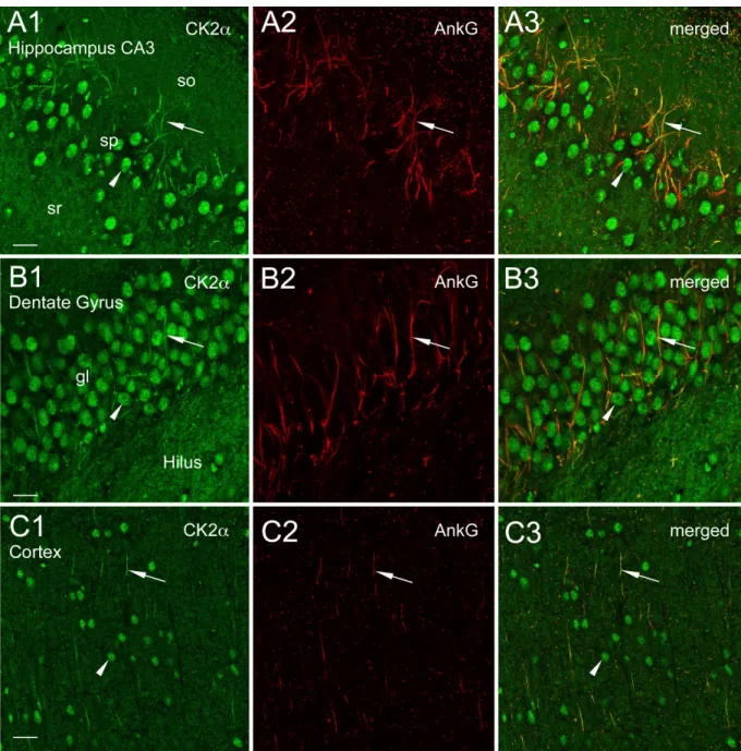
Figure S1. CK2a is concentrated at axonal initial segments in different rat brain regions. (A and B) Confocal views showing CK2a im-munoreactivity within initial segments (arrows) and nuclei (arrowhead) of neurons in the CA3 area of hippocampus (A1–A3), dentate gyrus (B1–B3), and cortex (layers 4–6, C1–C3). Note that diffuse and light CK2a labeling is visible throughout the neuropil. gl, granular layer; so, stratum; sp, stratum pyra-midale; sr, stratum radiatum. Bars: (A) 10 µm; (B) 26.8 µm; (C) 11.37 µm.
on January 6, 2009
jcb.rupress.org