HAL Id: inserm-00663858
https://www.hal.inserm.fr/inserm-00663858
Submitted on 27 Jan 2012
HAL is a multi-disciplinary open access
archive for the deposit and dissemination of
sci-entific research documents, whether they are
pub-lished or not. The documents may come from
teaching and research institutions in France or
abroad, or from public or private research centers.
L’archive ouverte pluridisciplinaire HAL, est
destinée au dépôt et à la diffusion de documents
scientifiques de niveau recherche, publiés ou non,
émanant des établissements d’enseignement et de
recherche français ou étrangers, des laboratoires
publics ou privés.
To cite this version:
Elise Hamard-Peron, Delphine Muriaux. Retroviral matrix and lipids, the intimate interaction..
Retrovirology, BioMed Central, 2011, 8 (1), pp.15. �10.1186/1742-4690-8-15�. �inserm-00663858�
R E V I E W
Open Access
Retroviral matrix and lipids, the intimate
interaction
Elise Hamard-Peron, Delphine Muriaux
*Abstract
Retroviruses are enveloped viruses that assemble on the inner leaflet of cellular membranes. Improving biophysical techniques has recently unveiled many molecular aspects of the interaction between the retroviral structural protein Gag and the cellular membrane lipids. This interaction is driven by the N-terminal matrix domain of the protein, which probably undergoes important structural modifications during this process, and could induce membrane lipid distribution changes as well. This review aims at describing the molecular events occurring during MA-membrane interaction, and pointing out their consequences in terms of viral assembly. The striking
conservation of the matrix membrane binding mode among retroviruses indicates that this particular step is most probably a relevant target for antiviral research.
Introduction
Retroviruses are enveloped single-stranded RNA (+) viruses; they include some human pathogens such as human immunodeficiency virus (HIV), and oncoviruses such as the murine leukemia virus (MLV). Regardless of their diversity and the high divergence in their sequences, they share functional and viral protein struc-ture similarities. Their genome contains the three retro-viral genes: gag, pol, and env, and regulatory proteins in the case of complex retroviruses. One of the important steps in the process of retoviral infection is the forma-tion of new infectious particles. It consists of the assem-bly of the viral core at the cellular membrane, budding, and maturation of the viral particles. In this review, we will focus especially on the events that occur at the molecular level during the interaction between Gag and membranes, more particularly between the Matrix domain of retroviral Gag proteins and the phospholi-pids, and we will place it in the context of the viral assembly process. Retroviral assembly relies on the viral Gag protein, and especially its ability to interact with the viral genomic RNA (gRNA) and cellular membranes. Gag is a polyprotein with three domains: the matrix domain, MA, that binds membranes, the capsid domain, CA, that contains Gag multimerization motifs and is responsible for the viral capsid formation (see [1] for
review), and the nucleocapsid domain, NC, that recruits the RNA genome and also promotes Gag multimeriza-tion [2,3]. The assembly process most probably initiates with the formation of a ribonucleoprotein complex com-posed of a few Gag molecules and the gRNA, which is going to interact with membranes [4,5]. Beta-retro-viruses and spumaBeta-retro-viruses are exceptions, that fully assemble in the cytosol before reaching membranes (see [6] for review on spumaviruses, and [7] for study on the role of MA in promoting cytosolic assembly of M-PMV). The formation of higher order Gag multimers leads to the formation of the viral particle at the plasma membrane, and subsequent budding and maturation, which consist of the proteolytic cleavage of Gag and structural rearrangement of the particle. The MA domain is not only carrying Gag trafficking and mem-brane binding determinants, but also dictating the speci-ficity of the bound lipid. Many data have been recently published partially unveiling the molecular mechanism of MA lipid binding, enhancing the understanding of the role played by MA during Gag membrane targeting and assembly. In the light of the literature and our experiences, this review aims at proposing biochemical models for MA-lipid interactions for different retro-viruses, and replacing the consequences of such interac-tions in the context of retroviral assembly. We will identify the elements conserved through retroviral evo-lution, and those that are specific to particular retroviral strains.
* Correspondence: [email protected]
Human Virology Department, Inserm U758, Ecole Normale Superieure de Lyon, 36 Allee d’Italie, IFR128, Universite de Lyon, Lyon, France
© 2011 Hamard-Peron and Muriaux; licensee BioMed Central Ltd. This is an Open Access article distributed under the terms of the Creative Commons Attribution License (http://creativecommons.org/licenses/by/2.0), which permits unrestricted use, distribution, and reproduction in any medium, provided the original work is properly cited.
Matrix proteins: a structural point of view
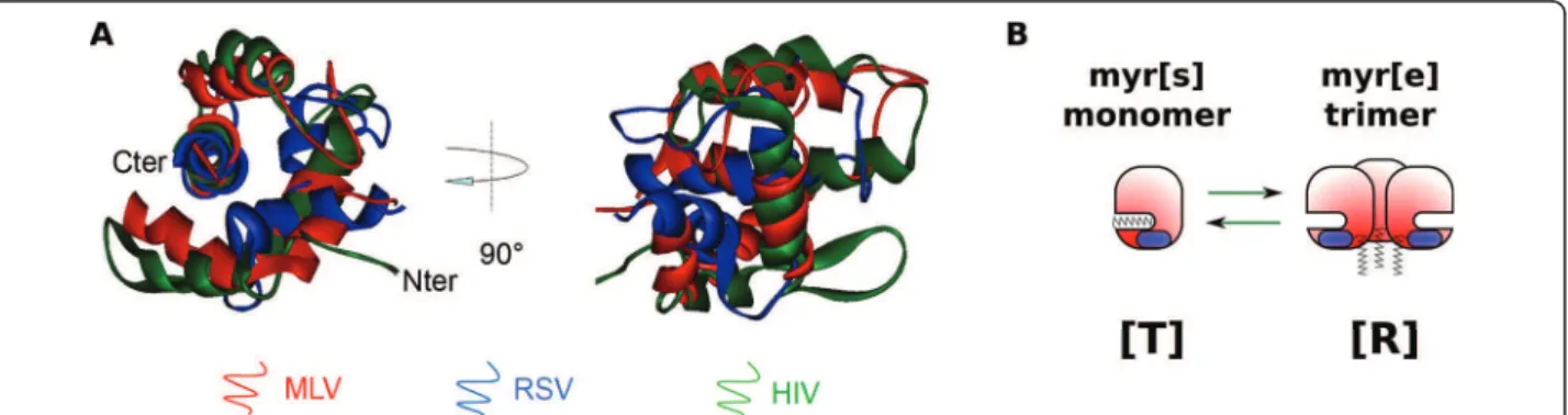
Despite low sequence similarity, MAs from different ret-roviruses share a conserved function in anchoring the viral Gag polyprotein to the plasma membrane. Indeed, most Gag chimeras with heterologous MA domains remain able to drive particle assembly [8-11]. One ele-ment allowing the interaction with the cellular mem-brane is N-terminal myristylation, a post-tranlational modification found in MAs from all retroviral families (myrMAs), including human immunodeficiency virus (HIV) [12], human T-lymphotropic virus (HTLV) [13], Mason-Pfizer monkey virus (M-PMV) [14] and exogen-ous murine leukemia virus (MLV) strains [15,16]. This myristate moiety is a common signal for membrane tar-geting of proteins, as it can insert into membrane bilayers. There are some exceptions, however, as Rous sarcoma virus (RSV), Visna virus, caprine arthritis-ence-phalitis virus (CAEV) and equine infectious anemia virus (EIAV) MAs are not myristylated [14]. Therefore, myristylation cannot be the only element involved in this targeting. Structural analysis of MA domains offers some clues for understanding its conserved biological role regarding membrane anchoring. Matrix structures from nine retroviruses have been resolved to date: HIV-1 [HIV-17-20] and 2 [2HIV-1], SIV [22], HTLV-2 [23], bovine leu-kemia virus (BLV) [24], M-PMV [25], RSV [26], EIAV [27], and MLV [28]. They are all made of a globular core composed of four a-helices, whose overall organi-zation is conserved among the retroviridae family [29,30] as shown by the superimposition Figure 1A. In the case of HIV-1, the unmyr-MA structure was resolved both by NMR [17,18] and crystallography [19], while the myr-MA structure was resolved by NMR only [20]. HIV-1 unmyr-MA (as well as SIV, but neither EAIV nor MLV MAs) crystallized as trimers, while it appeared mainly monomeric in classical NMR condi-tions. Overall structure was conserved between myr and
unmyr-MA, but some differences arose, notably in the putative trimerization region and in the first alpha helix. As suggested earlier by Zhou and Resh [31], Tang and colleagues [20] showed that there is an equilibrium between two conformations of HIV-1 myrMA in solu-tion. In the myr[s] conformation, the myristate moiety is sequestrated inside the core of the protein (see scheme in Figure 1B). This is the conformation adopted by the majority of myr-MA at a concentration of 150-200 μM. The other conformation, myr[e], promotes the exposure of the myristate and tends to assemble in trimers. This conformation is probably close to the conformation observed for unmyr-MA. The conversion from one state to the other is entropically regulated [20]. In particular, high concentration of MA (more than 400 μM) pro-motes trimerization and stabilizes the myr[e] conforma-tion. This will be extensively discussed in the next sections. Whether these myr[s] and myr[e] conforma-tions exist for other retroviral MAs has never been demonstrated formally. However, a NMR study carried out on EIAV-MA (which is not myristylated) evidenced amino acid shifts at high MA concentration, and corre-lated with an increase of the trimeric versus monomeric state [32]. Even if no major conformation change was noticed, this may correspond to an entropic switch between two slightly different conformations, similar to HIV. We, therefore, propose a new nomenclature for the MA conformations, that can also apply for unmyris-tylated MAs. By analogy with the enzymology, the mem-brane-binding prone conformation will be denoted hereafter as relaxed [R], while the other conformation will be denoted as tensed [T] (Figure 1B). Another important element of MA necessary for membrane binding is most probably the highly basic region (HBR). Indeed, an exposed patch of basic amino acids has been observed or predicted on all retroviral MAs [30]. A comparison between structurally superimposed
Figure 1 A structural overview of retroviral MAs. (A): Structural superimposition of MLV (1MN8), RSV (1A6S) and HIV (1TAM) MA proteins. Superimposition obtained using the combinatorial extension method (CE) and the image was generated with Viewer Pro software (Accelrys), thanks to E. Derivery. (B): Scheme of the [T] to [R] switch. [T] conformation sequesters the myristate of myristylated MAs, and remains monomeric, while [R] conformation associates in trimers and exposes the myristate (when present).
retroviral MAs shows that this domain “migrates” on the surface of the protein, but is always found in the proxi-mity of the N-terminus [30]. This supports the idea that the N-terminus and the polybasic region of MA coop-erate for efficient membrane binding, as HBR was hypothetized to promote interaction of MA with acidic phospholipid heads [30]. Moreover, other amino acids could be involved in Gag membrane anchoring, such as the N-terminal amino acids invovled in [T] to [R] con-version in HIV-MA [33,34].
Acidic lipid binding: the biochemical characterization
In cells, analysis confirmed that Gag membrane binding depends on this bipartite signal for most retroviruses. On one hand, the myristate moiety is, as expected, necessary to ensure membrane binding for all myristylated MAs, as shown for MLV [16,35], HIV [36], or M-PMV [37]. On the other hand, mutations in the HBR disrupted Gag membrane-binding and assembly of HIV [38-41], MLV [42,43], feline immunodeficiency virus (FIV) [44], RSV
[45], HTLV-1 [46] and M-PMV [47], suggest that MA may interact with acidic membrane lipids.
To precisely identify the lipids that interact with retro-viral Gag proteins, researchers focused on the lipids potentially present at the budding site. Phospholipids, including glycerophospholipids and sphingolipids, are the main components of cellular membranes, among which the most abundant are phosphatidylcholine (PC) and phosphatidylethanolamine (PE), both containing a neutral polar head. Some less abundant species, how-ever, like phosphatidyl serine (PS), phosphatidyl glycerol (PG) or phosphatidylinositol phosphates (PIPs), contain acidic polar heads (cf. Figure 2). Apart from phospholi-pids, cellular membranes also contain other liphospholi-pids, such as cholesterol, and an important proportion of trans-membrane proteins. The composition of a trans-membrane depends on its localization (internal/plasma membrane, inner/outer leaflets, etc.) and defines its functionality. Thus, retroviral assembly location restricts the panel of lipids potentially involved in the interaction with MA. Indeed, budding is mainly observed at the plasma
Figure 2 Some lipid components of the internal leaflet of cellular membranes. Main lipid components of the internal leaflet of cellular membranes are represented: phosphatidylcholine (PC), Phosphatidylethanolamine (PE), phosphatidylserine (PS), Phosphatidylinositol phosphates (here, PI(4,5)P2) and cholesterol. In membrane bilayers, the polar heads (top) face the cytosol, while the hydrophobic fatty acid chains (bottom) face the hydrophobic tails of the other leaflets’s lipids.
membrane for most retroviruses, including HIV [48], M-PMV, MLV [42,49], FIV, RSV, HTLV, but may also occur on internal membranes such as endosomes (see [50] and [51] for review). Moreover, the MA domain of Gag interacts with the inner leaflet of cellular mem-branes, whose main lipids are PC, PE, PS, PIP (here PI (4,5)P2), and cholesterol [52], thereby succeptible to
interact with MA (Figure 2).
Interaction between proteins and lipids can be studied in vitro using biomimetic membranes, and in particular large unilamellar vesicles (see [53] for review on using LUVs). The dissociation constant (Kd) can be measured, and corresponds to the lipid concentration at which half the protein is associated with the lipids: the lower the Kd, the higher the affinity. Most experiments were per-formed using recombinant MA proteins, because purifi-cation of the entire Gag protein is not easy. MA domain is separated from the rest of Gag by a flexible linker, thus isolated recombinant MA should recapitulate most functions of MA domain in Gag. It must be taken into account, however, that HIV-MA alone seems to have decreased affinity for membranes in comparison to the entire Gag [31]. Recombinant MA is also directly repre-sentative of the maturated MA domain function in mature particles and during early stage of viral infection. As expected, purified recombinant MAs from RSV [54] and HIV-1 [55,56] can bind containing an acidic phos-pholipid, the phosphatidylserine (PS), which is an abun-dant specy in the internal layer of cellular membranes. The order of magnitude of the Kd measurements made for recombinant RSV-MA and HIV-1 myristylated MA (myrMA) were of 10-3M, and about one order of
magni-tude lower upon forced dimerization of MA [54,55]. Nevertheless, the method used in these studies, i.e. LUV flotation, may underestimate the actual affinity, as the sucrose gradient may dilute the lipids. Indeed, we and others reported a value closer to 10-5M for
unmyristy-lated HIV MA (HIV unmyrMA) by sedimentation assay [42] or by intrinsic fluorescence measurement [56,57]. Ehrlich and colleagues [56] showed that HIV-1 MA is also able to bind in vitro to another basic phospholipid, the phosphatidylglycerol (PG). These later studies were contested, however, because the authors also observed a binding of the CA domain of Gag to PG and PS that other authors questioned [54]. Recently, Barrera and colleagues [58] confirmed that CA has acidic lipid bind-ing properties [58,59], rehabilitatbind-ing the previous find-ings. It was also reported that EIAV MA can interact with PS (Kd <10-6M at 0.1 M NaCl) and PC [60].
The binding of retroviral MAs to lipids was thus con-sidered to be purely electrostatic, as the interaction with PS was inhibited at high ionic strength. The Kd values found would fit well with the computational models considering electrostatic interaction between acidic
lipids and basic MAs [30]. These reported Kd values would be rather low, though, to fully explain the binding of Gag to the plasma membrane in cells, and multimeri-zation was invoked to explain MA binding to mem-branes [54,55].
Several retroviruses, however, show a dependency on a particular acidic phospholipid, the PI(4,5)P2, for efficient
particle production in cells. These include HIV [61,62], M-PMV [47] and MLV [42,62]. Phosphatidylinositol phosphates are a family of acidic glycerophospholipids, with a polar head made of an inositol ring that can be mono-, bi-or tri-phosphorylated (Figure 2 shows the example of PI(4,5)P2). The sub-cellular localization of
the different species is highly regulated by cellular kinases and phosphatases, such that they stand as major determinants of the identity of organelles’ membranes (see [63], [64] and [65] for review).
The interaction between MAs and PI(4,5)P2has been
observed in vitro by NMR (EIAV [32], HIV-1 [66] and HIV-2 [21]), using LUVs (HIV-1 [67-69] and MLV [42]), by mass spectrometric footprinting (HIV-1 [70]) and by surface plasmon resonance (SPR)(HIV-1, [71]). The Kd values measured by NMR were rather high for all tested lentiviruses (EIAV, HIV-1, and HIV-2), ranging from 125 to 185 μM, and cannot account for membrane binding in cells. It is noteworthy though that these interactions were observed with short chain PIPs (Di-C4-PI(4,5)P2). In
con-trast, SPR analysis was performed both with Di-C4-and Di-C8-PI(4,5)P2 (longer acyl chains), and Kd values
decreased significantly in the case of Di-C8-PI(4,5)P2,
suggesting that acyl chains are involved in the interaction between MA and PI(4,5)P2[71]. The Kd of this
interac-tion could not be calculated in the LUV systems, how-ever, neither for the recombinant HIV MA domain [42,55], nor for the recombinant RSV MA domain [54]. This suggests that unlike PS binding, the mechanism of PIP/HIV-MA interaction could be more complex than a simple electrostatic interaction. The region of HIV-MA involved in the interaction with PI(4,5)P2differs slightly
depending on the method used (NMR [66] or footprint-ing [70]), but mapped to the HBR in both cases. New NMR techniques, using reverse micelle encapsidation instead of soluble lipids could settle it, but only prelimin-ary results have been published to date [72].
We recently reported a definite different behavior in the case of MLV-MA [42]. UnmyrMLV-MA was able to bind PIPs-containing LUVs in a dose-dependant manner. An interaction is observed not only with PI(4,5)P2, but also
with all the PIPs species, with Kd values ranging from 20 to 50 μM. To the contrary, unmyrMLV-MA does not bind PS containing LUVs, even if the residues involved in the interaction with PIPs map to the HBR. However, adding PI(4,5)P2and PS together in the same LUV dramatically
for the other PIPs. Therefore, as for HIV, interaction with PIPs appears to result from a specific interaction, rather than a purely electrostatic mechanism [42].
Specificity and regulation of the interaction with acidic phospholipids
In the light of the data presented above, we can ques-tion the specificity and the biological relevance of the interaction of retroviral Gag with the different acidic phospholipid species, as MA can interact in vitro with different acidic phospholipids, with important differ-ences in Kd and interaction mode.
The lipidomics data emerging from the analysis of viral particles, however, seems to confirm the specificity for both PI(4,5)P2and PS, as they are highly enriched in
MLV particles [73]. This is consistent with the in vitro data obtained with MLV-MA, showing that there is in fact a cooperation between PI(4,5)P2 and PS which
allows strong MA anchoring to the membrane. Indeed, even if MLV-MA can bind any PIPs but not PS-contain-ing LUVs, the protein actually displayed a strong stereo-specificity for PI(4,5)P2, but exclusively when PS is
added to the same LUV (resulting in a fourfold decrease in Kd, [42]). Thus, MA probably interacts with both PI (4,5)P2 and PS, but we hypothesize that PS binding may
occur only after initial docking of MA on the PI(4,5)P2.
In HIV particles, PI(4,5)P2 is enriched, while PS is
pre-sent at high concentrations. Together with data emer-ging from MLV study, these results indicate that in vitro binding of HIV-MA to both PI(4,5)P2 and PS may be
biologically relevant. Other families of lipids may also regulate MA association with membranes. In particular, HIV myrMA show more affinity for cholesterol-contain-ing biomimetic membranes [57], and cholesterol enhances the binding specificity of HIV-MA to PI(4,5)P2
[67], in accordance with the finding that retroviruses can bud in cholesterol-enriched membrane domains such as lipid rafts [74-76].
Surprisingly enough, another element, the RNA, was recently found to be involved in the regulation and the specificity of HIV-MA membrane binding [69]. Indeed, HIV-MA has long been known to bind RNA efficiently in vitro [67,70,77-79], as does BLV-MA [80] and RSV-MA [81]. Moreover, HIV-RSV-MA specifically interacts with RNA, bearing a high degree of homology to a region within the Pol open reading frame of the HIV-1 gen-ome, suggesting that the RNA molecule interacting with MA in cells might be the viral gRNA [79]. Interestingly, the basic residues of HIV-1 MA involved in the interac-tion with RNA are also necessary for PI(4,5)P2 binding
[66,70,77,79]. Thus, RNA might be a competitive inhibi-tor of the interaction with PI(4,5)P2. As a matter of fact,
Chukkapalli and colleagues observed that RNAse treat-ment increased binding of Gag to both neutral and
acidic LUVs (PC, +/- PS, +/- PI(4,5)P2) [69]. The
hypothesis is that RNA would inhibit the entropic switch, stabilizing the [T] conformation (Figure 3Ab), thus preventing membrane-binding in general. On the other hand, Alfadhli and colleagues [67] simultaneously found that PI(4,5)P2 is the only lipid that can remove
nucleic acids bound to HIV-1 myrMA recombinant pro-tein. This favors the idea that RNA would ensure the specificity of the interaction of MA with the PI(4,5)P2,
which therefore appears as a relevant cellular partner of Gag during the assembly process, allowing MA to switch from a “transport” [T] conformation to a “mem-brane binding” [R] conformation. RNA-meditated regu-lation of HIV-MA binding to PI(4,5)P2 seems to be
supported by the data emerging from in cellulo studies. A functional link between the genomic RNA exporting pathway and the HIV-1 MA-driven assembly has been established recently, even if the precise mechanism has not been elucidated [82-85]. Whether gRNA plays a role in MA/lipid interaction for other retroviruses is not known as yet. EIAV or MLV does not seem to have the same dependency on gRNA export pathway for proper assembly [84,85] as compared to HIV-1. In contrast, RSV-MA is able to interact with both PS [54] and RNA [81]. The measured affinity for PI(4,5)P2 was found to
be low in the case of RSV MA alone [54], but given the results obtained with HIV-MA, further investigation could prove useful. Thus, from an evolutionary point of
Figure 3 A model for [T] to [R] equilibrium in different conditions. Some elements are susceptible to influence the MA [T] vs [R] equilibrium, in the context of MA alone (in the mature particle, during the early step of infection, or in vitro), or as a domain of the Gag polyprotein. The “initial” equilibrium (in solution, purified protein, concentration around 1 μM) between the [T] and [R] conformations of HIV (A) and MLV (B) MAs (a) or Gag (b) are depicted, the size of the protein representing the relative amount of each form. The factors susceptible to induce a majority of a given conformation are written in bold characters. Others, such as PI(4,5)P2 in the case of HIV-MA, are only able to (slightly) displace the equilibrium, even at a saturating concentration (Aa).
view, it would be interesting to determine if these regu-lation modes involving PI(4,5)P2 and RNA are conserved
among retroviruses, including those lacking MA myristylation.
In summary, we have proposed a model in which two different retroviral MAs use alternative mechanisms to bind membrane lipids, but end up with the same lipid specificity. MLV-MA is able to interact initially with PI (4,5)P2, and this interaction triggers a conformational
modification that allows PS binding. In contrast, HIV-MA would have initially low affinity for PI(4,5)P2,
espe-cially in the presence of gRNA [69]. However, PI(4,5)P2
seems to be the only compound able to compete with RNA for HIV-MA binding [67], and once RNA is removed, HIV-MA would be able to interact both with PI(4,5)P2 and PS, and this interaction may be stabilized
by other elements, as discussed in the next section. Therefore, in spite of different lipid binding modes, the specificity of binding could be highly conserved among retroviruses.
Let’s switch again! Stabilization of the [R] conformation
The interaction of retroviral MAs with PI(4,5)P2seems
to be a conserved, highly specific, and regulated feature among retroviruses. As previously mentioned, PI(4,5)P2
binding seems to be associated with conformational changes, as shown by NMR for HIV-1 MA [66] and EIAV-MA [86]. For HIV, it corresponds to the myr[s] and myr[e] conformations ([T] and [R] respectively) evi-denced by structural studies [20], and it is probably also the case for EIAV except that it is not myristylated. This supports a pre-existing hypothesis first proposed by Zhou et al [31]: the existence of a “myristyl switch”, that is actually an entropic equilibrium between the [T] conformation that sequesters the myristate inside the protein, and the [R] conformation that promotes trimer-istation and exposure of the myristate moiety allowing its insertion in the cellular membranes. A refinement of this model was proposed by Saad and colleagues, as the NMR data on HIV-MA suggested that the insertion of the myristate into the lipidic bilayer may be compen-sated by the extraction of the 2’ fatty acid chain of the PI(4,5)P2 out of the membrane, that would then be
sequestrated into the hydrophobic core of the MA domain (Figure 4Ad) [66]. Anraku and colleagues com-pared the affinity of HIV-1 MA and Gag for phophory-lated inositol ring alone and for medium length fatty acid chain lipids (Di-C8-PI(4,5)P2), in order to compare
the relative contribution of electrostatic interactions (with inositol phosphate ring) and hydrophobic interac-tions (with acyl chains) [71]. In accordance with the data from Saad et al. [66], acyl chains were found to have a major contribution in the interaction. This
model, however, is built on data obtained with short chain fatty acids, and needs further confirmation in lipid bilayer conditions.
As a model for HIV-MA/PI(4,5)P2interaction, we
pro-pose that the [T] conformation has a high affinity for RNA, and a low affinity for PI(4,5)P2. On the contrary,
the [R] conformation has a high affinity for PI(4,5)P2. PI
(4,5)P2 would compete with RNA for HIV-MA binding
as recently proposed [69,87] and its interaction with MA would in turn stabilize the [R] conformation as shown by Saad and colleagues [66] (Figure 3Aa). In this model, PI(4,5)P2 has two roles: in addition to being the
“substrate” (i.e. the bound molecule), it is also an effec-tor, stabilizing the binding prone conformation, [R] (Fig-ure 3Aa). In other words, PI(4,5)P2 is able to displace a
pre-exiting equilibrium toward the [R] conformation, as suggested by Tang et al [20]. Symmetrically, RNA would have an “allosteric inhibitor” effect in stabilizing the [T] conformation (Figure 3Aa). This property may prevent a specific binding to membranes lacking PI(4,5)P2. This
model could explain why many authors were unable to measure the affinity of HIV-1 MA for PI(4,5)P2 in the
LUV system [42,55]. At low HIV-MA concentrations (from 1μM to 20μM), the equilibrium would be only slightly displaced toward the [R] conformation, even at a saturating PI(4,5)P2 concentration (Figure 3Aa[42,54]).
The [T] conformation had very low affinity for the lipid; we and others concluded that the affinity of MA for PI (4,5)P2 was negligible in these conditions [42,55]. Many
other elements could also influence the [T] to [R] equili-brium in vivo, to allow specific interaction of Gag with membranes. As mentioned earlier, a high concentration of MA promotes trimerization, and at the same time stabilizes the [R] conformation [20] (cf. Figure 3Aa). In addition, multimerization of Gag seems to correlate with the appearance of the [R] state, as multimerizing regions in CA promote myristate exposure [20] and increase lipid binding of MA-CA constructs [55]in vitro. In cells, it has been shown that proteolytic cleavage of Gag induces partial dissociation of p17MA from the mem-brane, confirming that uncleaved Gag stabilizes the [R] conformation of MA [31,88,89]. Another parameter that seems to influence the [T] to [R] transition is pH, as shown recently by Fledderman et al. [90]. High pH sta-bilizes the [T] form, while acidification favors myristate exposure. In addition, the same laboratory also reported that Calmodulin (CalN), a Ca2+sensor protein determi-nant that interacts with HIV MA, promotes the myristyl switch [91].
The equilibrium constant between the [T] and [R] con-formations also seems to vary greatly from one MA to another. As a matter of fact, in NMR conditions (high MA concentration, around 0.5 mM), HIV-1 and HIV-2 MAs behave differently in the presence of PI(4,5)P2, the
[R] conformation remains undetectable for HIV-2 MA [21], unlike HIV-1 [66]. As far as other viruses are con-cerned, less data are available. It is possible that PI(4,5)P2
also stabilizes an [R] conformation of EIAV-MA as sug-gested by 2-D NMR data obtained by Chen et al [86], showing a slight amino acid shift upon PI(4,5)P2binding.
In contrast, MLV MA may display a more complex beha-vior. We were able to calculate two Kd values for MA/PI (4,5)P2interaction, either in the presence or absence of
PS. The [T] conformation might be able to bind PI(4,5)P2
with a Kd of 25 μM, while the [R] conformation might be stabilized by the presence of PS, allowing PI(4,5)P2 to
switch to the extended lipid conformation, with a result-ing Kd value approachresult-ing 5 μM (Figure 3Ba) [42]. Another hypothesis is that the majority of MA is already in the [R] conformation, and that PS modulates the affi-nity of the interaction with PI(4,5)P2.
The switch from the [T] to the [R] conformation may have further implications at the level of the entire Gag protein, thus influencing the assembly process. Indeed, Datta et al. recently proposed a model in which HIV-Gag would be in a bent conformation in solution, with MA and NC in close proximity [92,93] (Figure 3Ab). This model is supported by the fact that both NC and MA can bind IP6 (an inositol ring containing six phos-phorylations, thus somewhat homologous to PI(4,5)P2)
in vitro, and is consistent with hydrodynamic and small-angle neutron scattering data. This is also in agreement with the idea that RNA can bind both NC and MA [67,70,77-79]. This is not compatible, however, with the immature particle organization, in which Gag is in an extended rod-shaped conformation [94]. Consequently, the authors propose that viral assembly is coupled with major conformational modifications of Gag (Figure
Figure 4 Models for retroviral Gag membrane binding. Aa and Ba: formation of Gag dimers, association on gRNA. Ab: inhibition of HIV-MA membrane binding by gRNA. Ac: removal of gRNA resulting from competition between gRNA and PI(4,5)P2 for HIV-MA binding. Ad: Stabilization of the [R] conformation of MA by interaction with PI(4,5)P2, Gag trimerization, stabilization of membrane anchoring by PS, lateral targeting of Gag to assembly microdomains. Bb: Binding of MLV-MA to PI(4,5)P2. Bc: Secondary binding of MLV-MA to PS, stabilization the MA [R] conformation. Bd: lateral targeting of Gag to assembly microdomains.
3Ab). The same group showed that correct in vitro assembly of viral like particles necessitates both RNA and IP6 (that can be considered as an analog of PI(4,5) P2). It is still the case when the NC domain is replaced
by a multimerization domain such as a leucine zipper, suggesting that RNA not only plays a role in assembly via its interaction with the NC domain, but probably also at the level of the MA domain [95].
The ability of HIV-Gag to auto-assemble into viral-like particles in vitro seems to be linked with a switch from Gag dimers to Gag trimers that can be mediated by IP6 [93,95]. As it has been shown that PI(4,5)P2 promotes
HIV-MA trimeric association [87], the effect of IP6 addition could mimic the effect of PI(4,5)P2 binding in
cells, in stabilizing the [R] conformation and promoting the formation of MA trimers. This could further trigger Gag structural reorganization via dimer to trimer transi-tion (Figure 4Ad). A similar mechanism could drive the assembly of all retroviruses, as other retroviral MAs have multimerization properties upon PI(4,5)P2 binding.
For exemple, MLV-MA multimerizes in the presence of PI(4,5)P2under certain conditions (unpublished personal
data), and EIAV-MA forms trimers [32].
MLV-Gag, however, seems to differ in some points from lentiviral Gag proteins. Datta et al. showed that in vitro recombinant MLV-Gag is readily in a rod-shaped conformation in solution, with a much more rigid structure (Datta, Zuo, Campbell, Wang, Rein: Personnal communication) (Figure 3Bb). This property might argue for an absence of an RNA mediated main-tenance of the [T] conformation for MLV-MA. This correlates with the fact that the [R] conformation of MLV-MA appears more stable, as 100% of MLV-Gag is associated with membranes in cells [42], in contrast with HIV-Gag which is no more than 60% membrane bound [96]. However, we cannot exclude the possibility that RNA could regulate the interaction of MLV-MA with lipids.
The mechanisms of interaction between retroviral MAs and lipids are quite original, and whether some particula-rities of these binding modes can also apply to other viral or cellular proteins is not known. For instance, other ret-roviral proteins could interact with lipids using a similar mechanism. For example, Nef and Tat, two regulatory proteins of HIV, also bind membranes. In fact, Nef is a myristylated protein able to bind acidic phospholipids, but the curvature of the membrane induced upon Nef binding is not consistent with the extraction of a fatty acid out of the membrane [97] as in the model proposed for HIV-MA [66]. A myristyl switch mechanism is still possible, however, as the binding of Nef to biomimetic membranes is a biphasic process, with a first phase of electrostatic interaction with acidic phospholipids, and a second phase of structural modifications (in particular,
the formation of an amphiphatic helix) [97]. As for Tat, it was recently shown that it also interacts with PI(4,5)P2
before crossing the plasma membrane and being secreted into the extracellular environment [98-100].
Conclusion: Cellular consequence of Gag binding to PI(4,5)P2 and PS
Taking all the previously discussed data together allowed us to propose a model for the role played by MA during HIV and MLV assembly initiation, at the molecular level (Figure 4). In this model, Gag first poly-merizes on gRNA (Aa and Ba), but adopts a bent con-formation in the case of HIV (Aa), with both MA and NC interacting with gRNA, while MLV-Gag is readily in a rod-shaped conformation (Ba). For both viruses, the [T] conformation of MA is initially dominant, with myr-istate trapped in the protein core. When HIV-MA reaches PM (Ab), PI(4,5)P2 is able to compete with
gRNA for MA binding (Ac). Removal of gRNA and interaction with PI(4,5)P2stabilize the [R] conformation
of MA (exposed myristate), which in turn promotes the trimerization and the reorganization of Gag into its rod-shapped conformation (Ad). The presence of PS could stabilize the interaction between MA and PI(4,5)P2(Ad).
Gag would then be laterally targeted to membrane microdomains containing high levels of saturated lipids, such as lipid rafts (Ad). In the case of MLV, initial bind-ing to PI(4,5)P2(Bb) is followed by a secondary binding
to PS (Bc) that would further stabilize the [R] conforma-tion of MA, exposing the myristate. Like HIV, lateral targeting of Gag to rafts or other microdomains is likely to occur afterwards (Bd).
These mechanistic observations are useful to re-evalu-ate the data available regarding assembly and budding localization in cells. Analysis of the retroviral particle envelope content evidenced that budding membranes resemble the plasma membrane in terms of lipid compo-sition [73,75,101-105]. The ratio between lipids, however, differs from the average plasma membrane composition. In particular, viral particles of HIV and MLV are enriched not only in PI(4,5)P2and PS, but also in
choles-terol, ceramides, GM3 and sphingolipids [73]. This can reflect the fact that viral particles are produced in specific membrane microdomains. Moreover, HIV virions are also enriched in lipid raft markers such as GPI-anchored proteins [106], actin and actin-associated proteins, such as Ezrin-Radixin-Moesin proteins (ERMs) [107,108], and in tetraspanins [108-116]. ERM and tetraspanins are also found in particles of MLV [107,117,118]. In consequence, retroviral budding has been proposed to occur preferen-tially in two types of membrane microdomains associated with actin cytoskeleton: lipid rafts and tetraspanin enriched microdomains (TEMs). There is a spatial and functional distinction, however, between these two kind
of domains [119-121], even if they are adjacent and may interact [122-124].
Lipid rafts are membrane domains enriched in choles-terol and sphingolipids, but can also be enriched in PI (4,5)P2 and PS under specific conditions [125-128].
Rafts were initially identified as detergent-resistant membranes, and this property was widely utilized to characterize raft-associated lipids and proteins, includ-ing HIV-Gag [74,129-137], MLV-Gag [76,136] and HTLV-1-Gag [136,138]. The existence in living cell, the exact nature, and the actual size of lipid rafts has, how-ever, been intensely debated over the past decades. The current consensus is that lipid rafts are nanoscale con-centrations of specific lipids, notably cholesterol and sphingolipids, and proteins (reviewed in [128,139]). Their size is around 10 to 20 nm but they can coalesce and organize membrane bioactivity in many ways.
The association of HIV-Gag with lipid rafts depends on both membrane association signals of MA, the myr-istate and the HBR (reviewed in [140,141]). Lower order multimerization is also necessary because the association of CA mutants with lipid rafts is delayed [74], however, higher order association appears to be dispensable as demonstrated by NC mutants [142]. Lipid raft targeting is a slower process than membrane association, giving the idea that initial docking of Gag at the plasma mem-brane is followed by lateral transport to assembly micro-domains as proposed by Ono and Freed [74].
Saad and colleagues [66] proposed a very elegant model in agreement with a preferential budding of HIV in raft microdomains. Their NMR data suggests that the 2’-fatty acid of the PI(4,5)P2 is extracted from the
mem-brane bilayer upon MA binding, and sequestrated inside the protein, in the same hydrophobic pocket the myris-tate occupied. Unlike the 2’-chain, the 1’-chain is usually saturated, as is the myristate (cf. Figure 4). If this model proves to be correct, Gag would then be anchored to the membrane via two saturated chains (myristate and 1’-chain) and this could result in a lateral targeting of Gag to lipid rafts, where saturated lipids are enriched (Figure 4d, Bd).
The trapping of PI(4,5)P2 into lipid rafts by Gag may
have important consequences in terms of cellular responses. Indeed, in non-infected cells, it seems that the ratio of raft-associated PI(4,5)P2 versus raft-excluded
PI(4,5)P2 is finely regulated. Any modification of one
pool seems to have profound consequences, in particular on cytoskeleton remodelling, cell morphology and mod-ulation of signaling pathways, such as the PI3K-Akt pathway [143].
Whether Gag, and in particular the MA domain, is able to aggregate lipid raft microdomains (directly or indirectly) or bind to pre-formed platforms is not as yet known, even if recent findings argue for dynamic
aggregation of raft components by Gag [116]. Annexin 2 could potentially play a role, as this protein interacts with Gag [108,144] and is able to aggregate lipids, in particular cholesterol, PS, and PI(4,5)P2 [145,146]. Other
viral proteins may be involved too. It was recently shown that gPr80[gag], a long glycosylated form of MLV-Gag, increases the release of MLV and HIV particles via lipid rafts [76]. A similar role has been observed for HIV-Nef [147], which also increases the “raft-like” prop-erties of HIV particles [105] and modifies the choles-terol metabolism of producer cells [148]. However, it is not known how these two proteins act to relocate assembly in these microdomains.
On the other hand, several authors have reported that retroviral assembly occurs in association with tetraspa-nins [108-116,149-151]. Some tetraspatetraspa-nins can modulate viral infectivity and regulate cell to cell transmission [115], while the role of others, such as CD63, is currently debated [152]. The tetraspanins are a family of small transmembrane proteins that operate as major lateral organizers of membrane domains. They form tetraspa-nin-enriched microdomains (TEMs) or tetraspanin webs, in close relation with the cytoskeleton (reviewed in [153]). TEMs are enriched in cholesterol, GM1 and sphingolipids, but only a small fraction of the tetraspa-nins are found in the detergent resistant membrane (DRM) fractions, unlike raft proteins. Some tetraspanins, including CD9, CD63, CD81, and CD51 are associated with PI4K, a kinase that allows the synthesis of PI(4)P, the main precurssor of PI(4,5)P2. In particular, HIV-Gag
seems to associate specifically with CD63 and CD81 and less with CD82 [108,109,113-115] while HTLV-1 Gag associates preferentially with CD82 at the plasma mem-brane [149-151]. It is noteworthy that CD82 does not associate with PI4K and that this may be related to the unusual particle production mode of HTLV, with prefer-ential budding at the cell-to-cell contact areas and low production of cell-free virions. One unresolved question is whether there is a collaboration between rafts and TEMs during particle assembly or whether distinct bud-ding microdomains exist in the cell. In support of the first hypothesis, it was observed that some tetraspanins are able to address protein complexes toward lipid rafts, inducing the activation of specific signalization pathways. In particular, CD81 is necessary to partition the B cell receptor (BCR) and the CD19/CD21/CD81 complex into rafts [122,123], while CD82 links the actin cytoskeleton, T cell receptors and raft domains [124]. This suggests that tetraspanins may help to target Gag to lipid rafts, or, the other way around, that Gag could recruit tetraspanins and lipid raft components in order to activate particular signalization pathways necessary for sustaining HIV infection. This later model is supported by recent work by Krementsov et al. showing the strong trapping of
CD9 and the transient trapping of cholesterol, GM1 and CD55 into the HIV-1 assembly microdomains [116]. Interaction between TEMs and lipid rafts could result in the activation of TCR signalization pathway from which HIV could benefit. This pathway comprises, for example, the protein Lck, a Src-kinase participating in T-cell acti-vation [154], that interacts with HIV-Gag and increases particle production [155]. Moreover, the activation of TCR not only causes the accumulation of raft lipids in the membrane areas involved in the TCR signaling path-way but also recruits PS, which is probably necessary for Gag stabilization in PM microdomains during particle formation [127].
The enriched literature on retroviral assembly has allowed us to postulate a quite precise model of the molecular events that drive the anchoring of Gag to cel-lular membranes preceding particle formation, but these models remain to be tested experimentally. The high conservation of the overall process is striking, especially concerning the specificity of the interaction between Matrix domain of Gag and cellular lipids (PI(4,5)P2, PS, cholesterol), and suggests that targeting retroviral assembly by therapeutical approaches may be a good strategy to combat HIV infection.
Acknowledgements
We especially want to thank Dr Robin Buckland for his critical reading of the manuscript. This work was supported by INSERM and CNRS. EHP is a fellowship receiver of the French Government.
Authors’ contributions
EH wrote the manuscript and made the figures. DM contributed to the manuscript writing and editing. All authors read and approved the final manuscript.
Competing interests
The authors declare that they have no competing interests. Received: 29 October 2010 Accepted: 7 March 2011 Published: 7 March 2011
References
1. Adamson CS, Jones IM: The molecular basis of HIV capsid assembly-five years of progress. Rev Med Virol 2004, 14(2):107-21.
2. Darlix JL, Lapadat-Tapolsky M, de Rocquigny H, Roques BP: First glimpses at structure-function relationships of the nucleocapsid protein of retroviruses. J Mol Biol 1995, 254(4):523-37.
3. Rein A: Retroviral RNA packaging: a review. Arch Virol Suppl 1994, 9:513-22.
4. Jouvenet N, Simon SM, Bieniasz PD: Imaging the interaction of HIV-1 genomes and Gag during assembly of individual viral particles. Proc Natl Acad Sci USA 2009, 106(45):19114-9.
5. Ott DE, Coren LV, Shatzer T: The nucleocapsid region of human immunodeficiency virus type 1 Gag assists in the coordination of assembly and Gag processing: role for RNA-Gag binding in the early stages of assembly. J Virol 2009, 83(15):7718-27.
6. Delelis O, Lehmann-Che J, Saïb A: Foamy viruses-a world apart. Curr Opin Microbiol 2004, 7(4):400-6.
7. Choi G, Park S, Choi B, Hong S, Lee J, Hunter E, Rhee SS: Identification of a cytoplasmic targeting/retention signal in a retroviral Gag polyprotein. J Virol 1999, 73(7):5431-7.
8. Parent LJ, Wilson CB, Resh MD, Wills JW: Evidence for a second function of the MA sequence in the Rous sarcoma virus Gag protein. J Virol 1996, 70(2):1016-26[http://view.ncbi.nlm.nih.gov/pubmed/8551559].
9. Reed M, Mariani R, Sheppard L, Pekrun K, Landau NR, Soong NW: Chimeric human immunodeficiency virus type 1 containing murine leukemia virus matrix assembles in murine cells. J Virol 2002, 76:436-43.
10. Chen BK, Rousso I, Shim S, Kim PS: Efficient assembly of an HIV-1/MLV Gag-chimeric virus in murine cells. Proc Natl Acad Sci USA 2001, 98(26):15239-44.
11. Manrique ML, Gonzalez SA, Affranchino JL: Functional relationship between the matrix proteins of feline and simian immunodeficiency viruses. Virology 2004, 329:157-67.
12. Veronese FD, Copeland TD, Oroszlan S, Gallo RC, Sarngadharan MG: Biochemical and immunological analysis of human immunodeficiency virus gag gene products p17 and p24. J Virol 1988, 62(3):795-801. 13. Ootsuyama Y, Shimotohno K, Miwa M, Oroszlan S, Sugimura T:
Myristylation of gag protein in human T-cell leukemia virus type-I and type-II. Jpn J Cancer Res 1985, 76(12):1132-5.
14. Schultz AM, Oroszlan S: In vivo modification of retroviral gag gene-encoded polyproteins by myristic acid. J Virol 1983, 46(2):355-61. 15. Henderson LE, Krutzsch HC, Oroszlan S: Myristyl amino-terminal acylation
of murine retrovirus proteins: an unusual post-translational proteins modification. Proc Natl Acad Sci USA 1983, 80(2):339-43.
16. Rein A, McClure MR, Rice NR, Luftig RB, Schultz AM: Myristylation site in Pr65gag is essential for virus particle formation by Moloney murine leukemia virus. Proc Natl Acad Sci USA 1986, 83(19):7246-50. 17. Massiah MA, Starich MR, Paschall C, Summers MF, Christensen AM,
Sundquist WI: Three-dimensional structure of the human immunodeficiency virus type 1 matrix protein. J Mol Biol 1994, 244(2):198-223.
18. Matthews S, Barlow P, Boyd J, Barton G, Russell R, Mills H, Cunningham M, Meyers N, Burns N, Clark N, et al: Structural similarity between the p17 matrix protein of HIV-1 and interferon-gamma. Nature 1994, 370(6491):666-8.
19. Hill CP, Worthylake D, Bancroft DP, Christensen AM, Sundquist WI: Crystal structures of the trimeric human immunodeficiency virus type 1 matrix protein: implications for membrane association and assembly. Proc Natl Acad Sci USA 1996, 93(7):3099-104.
20. Tang C, Loeliger E, Luncsford P, Kinde I, Beckett D, Summers MF: Entropic switch regulates myristate exposure in the HIV-1 matrix protein. Proc Natl Acad Sci USA 2004, 101(2):517-22.
21. Saad JS, Ablan SD, Ghanam RH, Kim A, Andrews K, Nagashima K, Soheilian F, Freed EO, Summers MF: Structure of the myristylated human immunodeficiency virus type 2 matrix protein and the role of phosphatidylinositol-(4,5)-bisphosphate in membrane targeting. J Mol Biol 2008, 382(2):434-47.
22. Rao Z, Belyaev AS, Fry E, Roy P, Jones IM, Stuart DI: Crystal structure of SIV matrix antigen and implications for virus assembly. Nature 1995, 378(6558):743-7.
23. Christensen AM, Massiah MA, Turner BG, Sundquist WI, Summers MF: Three-dimensional structure of the HTLV-II matrix protein and comparative analysis of matrix proteins from the different classes of pathogenic human retroviruses. J Mol Biol 1996, 264(5):1117-31, [Plein de refs pour trucs de base: basic residues, myr, etc HTLV-II: 4 helices alpha, une “3-10” (helice courte) patch basique]..
24. Matthews S, Mikhailov M, Burny A, Roy P: The solution structure of the bovine leukaemia virus matrix protein and similarity with lentiviral matrix proteins. EMBO J 1996, 15(13):3267-74.
25. Conte MR, Klikova M, Hunter E, Ruml T, Matthews S: The three-dimensional solution structure of the matrix protein from the type D retrovirus, the Mason-Pizer monkey virus, and implications for the morphology of retroviral assembly. EMBO J 1997, 16(19):5819-26.
26. McDonnell JM, Fushman D, Cahill SM, Zhou W, Wolven A, Wilson CB, Nelle TD, Resh MD, Wills J, Cowburn D: Solution structure and dynamics of the bioactive retroviral M domain from Rous sarcoma virus. J Mol Biol 1998, 279(4):921-8[http://view.ncbi.nlm.nih.gov/pubmed/9642071]. 27. Hatanaka H, Iourin O, Rao Z, Fry E, Kingsman A, Stuart DI: Structure of
equine infectious anemia virus matrix protein. J Virol 2002, 76(4):1876-83. 28. Riffel N, Harlos K, Iourin O, Rao Z, Kingsman A, Stuart D, Fry E: Atomic
and its relationship to other retroviral matrix proteins. Structure 2002, 10(12):1627-36.
29. Conte MR, Matthews S: Retroviral matrix proteins: a structural perspective. Virology 1998, 246(2):191-8.
30. Murray PS, Li Z, Wang J, Tang CL, Honig B, Murray D: Retroviral matrix domains share electrostatic homology: models for membrane binding function throughout the viral life cycle. Structure 2005, 13(10):1521-31. 31. Zhou W, Resh MD: Differential membrane binding of the human
immunodeficiency virus type 1 matrix protein. J Virol 1996, 70(12):8540-8. 32. Chen K, Bachtiar I, Piszczek G, Bouamr F, Carter C, Tjandra N: Solution NMR characterizations of oligomerization and dynamics of equine infectious anemia virus matrix protein and its interaction with PIP2. Biochemistry 2008, 47(7):1928-37.
33. Paillart JC, Gottlinger HG: Opposing effects of human immunodeficiency virus type 1 matrix mutations support a myristyl switch model of gag membrane targeting. J Virol 1999, 73(4):2604-12.
34. Saad JS, Loeliger E, Luncsford P, Liriano M, Tai J, Kim A, Miller J, Joshi A, Freed EO, Summers MF: Point mutations in the HIV-1 matrix protein turn off the myristyl switch. J Mol Biol 2007, 366(2):574-85.
35. Hansen M, Jelinek L, Whiting S, Barklis E: Transport and assembly of gag proteins into Moloney murine leukemia virus. J Virol 1990, 64(11):5306-16. 36. Bryant M, Ratner L: Myristoylation-dependent replication and assembly of
human immunodeficiency virus 1. Proc Natl Acad Sci USA 1990, 87(2):523-7. 37. Rhee SS, Hunter E: Myristylation is required for intracellular transport
but not for assembly of D-type retrovirus capsids. J Virol 1987, 61(4):1045-53.
38. Yuan X, Yu X, Lee TH, Essex M: Mutations in the N-terminal region of human immunodeficiency virus type 1 matrix protein block intracellular transport of the Gag precursor. J Virol 1993, 67(11):6387-94.
39. Freed EO, Englund G, Martin MA: Role of the basic domain of human immunodeficiency virus type 1 matrix in macrophage infection. J Virol 1995, 69(6):3949-54.
40. Ono A, Orenstein JM, Freed EO: Role of the Gag matrix domain in targeting human immunodeficiency virus type 1 assembly. J Virol 2000, 74(6):2855-66.
41. Zhou W, Parent LJ, Wills JW, Resh MD: Identification of a membrane-binding domain within the amino-terminal region of human immunodeficiency virus type 1 Gag protein which interacts with acidic phospholipids. J Virol 1994, 68(4):2556-69.
42. Hamard-Peron E, Juillard F, Saad JS, Roy C, Roingeard P, Summers MF, Darlix JL, Picart C, Muriaux D: Targeting of murine leukemia virus gag to the plasma membrane is mediated by PI(4,5)P2/PS and a polybasic region in the matrix. J Virol 2010, 84:503-15.
43. Soneoka Y, Kingsman SM, Kingsman AJ: Mutagenesis analysis of the murine leukemia virus matrix protein: identification of regions important for membrane localization and intracellular transport. J Virol 1997, 71(7):5549-59.
44. Manrique ML, Celma CC, Gonzalez SA, Affranchino JL: Mutational analysis of the feline immunodeficiency virus matrix protein. Virus Res 2001, 76:103-13.
45. Callahan EM, Wills JW: Repositioning basic residues in the M domain of the Rous sarcoma virus gag protein. J Virol 2000, 74(23):11222-9. 46. Le Blanc I, Rosenberg AR, Dokhelar MC: Multiple functions for the basic
amino acids of the human T-cell leukemia virus type 1 matrix protein in viral transmission. J Virol 1999, 73(3):1860-7.
47. Stansell E, Apkarian R, Haubova S, Diehl WE, Tytler EM, Hunter E: Basic residues in the Mason-Pfizer monkey virus gag matrix domain regulate intracellular trafficking and capsid-membrane interactions. J Virol 2007, 81(17):8977-88.
48. Hoxie JA, Haggarty BS, Rackowski JL, Pillsbury N, Levy JA: Persistent noncytopathic infection of normal human T lymphocytes with AIDS-associated retrovirus. Science 1985, 229(4720):1400-2.
49. Suomalainen M, Hultenby K, Garoff H: Targeting of Moloney murine leukemia virus gag precursor to the site of virus budding. J Cell Biol 1996, 135(6 Pt 2):1841-52.
50. Benaroch P, Billard E, Gaudin R, Schindler M, Jouve M: HIV-1 assembly in macrophages. Retrovirology 2010, 7:29.
51. Corbin A, Grigorov B, Roingeard P, Darlix JL, Muriaux D: [Revisiting HIV-1 assembly]. Med Sci (Paris) 2008, 24:49-55.
52. van Meer G, Voelker DR, Feigenson GW: Membrane lipids: where they are and how they behave. Nat Rev Mol Cell Biol 2008, 9(2):112-24.
53. Cho W, Bittova L, Stahelin RV: Membrane binding assays for peripheral proteins. Anal Biochem 2001, 296(2):153-61.
54. Dalton AK, Murray PS, Murray D, Vogt VM: Biochemical characterization of rous sarcoma virus MA protein interaction with membranes. J Virol 2005, 79(10):6227-38.
55. Dalton AK, Ako-Adjei D, Murray PS, Murray D, Vogt VM: Electrostatic interactions drive membrane association of the human immunodeficiency virus type 1 Gag MA domain. J Virol 2007, 81(12):6434-45.
56. Ehrlich LS, Fong S, Scarlata S, Zybarth G, Carter C: Partitioning of HIV-1 Gag and Gag-related proteins to membranes. Biochemistry 1996, 35:3933-3943.
57. Provitera P, El-Maghrabi R, Scarlata S: The effect of HIV-1 Gag myristoylation on membrane binding. Biophys Chem 2006, 119:23-32. 58. Barrera FN, Hurtado-Gomez E, Lidon-Moya MC, Neira JL: Binding of the
C-terminal domain of the HIV-1 capsid protein to lipid membranes: a biophysical characterization. Biochem J 2006, 394(Pt 1):345-53.
59. Barrera FN, del Alamo M, Mateu MG, Neira JL: Envelope lipids regulate the in vitro assembly of the HIV-1 capsid. Biophys J 2008, 94(2):L8-10. 60. Provitera P, Bouamr F, Murray D, Carter C, Scarlata S: Binding of equine
infectious anemia virus matrix protein to membrane bilayers involves multiple interactions. J Mol Biol 2000, 296:887-898.
61. Ono A, Ablan SD, Lockett SJ, Nagashima K, Freed EO: Phosphatidylinositol (4,5) bisphosphate regulates HIV-1 Gag targeting to the plasma membrane. Proc Natl Acad Sci USA 2004, 101(41):14889-94.
62. Chan WT, Sherer NM, Uchil PD, Novak EK, Swank RT, Mothes W: Murine leukemia virus spreading in mice impaired in the biogenesis of secretory lysosomes and Ca2+-regulated exocytosis. PLoS ONE 2008, 3(7): e2713.
63. Di Paolo G, De Camilli P: Phosphoinositides in cell regulation and membrane dynamics. Nature 2006, 443(7112):651-7.
64. Krauss M, Haucke V: Phosphoinositides: regulators of membrane traffic and protein function. FEBS Lett 2007, 581(11):2105-11.
65. Krauss M, Haucke V: Phosphoinositide-metabolizing enzymes at the interface between membrane traffic and cell signalling. EMBO Rep 2007, 8(3):241-6.
66. Saad JS, Miller J, Tai J, Kim A, Ghanam RH, Summers MF: Structural basis for targeting HIV-1 Gag proteins to the plasma membrane for virus assembly. Proc Natl Acad Sci USA 2006, 103(30):11364-9.
67. Alfadhli A, Still A, Barklis E: Analysis of human immunodeficiency virus type 1 matrix binding to membranes and nucleic acids. J Virol 2009, 83(23):12196-203.
68. Chukkapalli V, Hogue IB, Boyko V, Hu WS, Ono A: Interaction between the human immunodeficiency virus type 1 Gag matrix domain and phosphatidylinositol-(4,5)-bisphosphate is essential for efficient gag membrane binding. J Virol 2008, 82(5):2405-17.
69. Chukkapalli V, Oh SJ, Ono A: Opposing mechanisms involving RNA and lipids regulate HIV-1 Gag membrane binding through the highly basic region of the matrix domain. Proc Natl Acad Sci USA 2010.
70. Shkriabai N, Datta SAK, Zhao Z, Hess S, Rein A, Kvaratskhelia M: Interactions of HIV-1 Gag with assembly cofactors. Biochemistry 2006, 45(13):4077-83.
71. Anraku K, Fukuda R, Takamune N, Misumi S, Okamoto Y, Otsuka M, Fujita M: Highly sensitive analysis of the interaction between HIV-1 Gag and phosphoinositide derivatives based on surface plasmon resonance. Biochemistry 2010, 49(25):5109-16.
72. Valentine KG, Peterson RW, Saad JS, Summers MF, Xu X, Ames JB, Wand AJ: Reverse Micelle Encapsulation of Membrane-Anchored Proteins for Solution NMR Studies. Structure 2010, 18:9-16.
73. Chan R, Uchil PD, Jin J, Shui G, Ott DE, Mothes W, Wenk MR: Retroviruses human immunodeficiency virus and murine leukemia virus are enriched in phosphoinositides. J Virol 2008, 82(22):11228-38[http://view.ncbi.nlm.nih. gov/pubmed/18799574].
74. Ono A, Freed EO: Plasma membrane rafts play a critical role in HIV-1 assembly and release. Proc Natl Acad Sci USA 2001, 98(24):13925-30. 75. Brugger B, Glass B, Haberkant P, Leibrecht I, Wieland FT, Krausslich HG: The
HIV lipidome: a raft with an unusual composition. Proc Natl Acad Sci USA 2006, 103(8):2641-6.
76. Nitta T, Kuznetsov Y, McPherson A, Fan H: Murine leukemia virus glycosylated Gag (gPr80gag) facilitates interferon-sensitive virus release through lipid rafts. Proc Natl Acad Sci USA 2010, 107(3):1190-5.
77. Cimarelli A, Luban J: Translation elongation factor 1-alpha interacts specifically with the human immunodeficiency virus type 1 Gag polyprotein. J Virol 1999, 73(7):5388-401, [Montre que MA interagit avec facteur+ARN]..
78. Lochrie MA, Waugh S, Pratt DGJ, Clever J, Parslow TG, Polisky B: In vitro selection of RNAs that bind to the human immunodeficiency virus type-1 gag polyprotein. Nucleic Acids Res type-1997, 25(type-14):2902-type-10.
79. Purohit P, Dupont S, Stevenson M, Green MR: Sequence-specific interaction between HIV-1 matrix protein and viral genomic RNA revealed by in vitro genetic selection. RNA 2001, 7(4):576-84. 80. Wang H, Norris KM, Mansky LM: Involvement of the matrix and
nucleocapsid domains of the bovine leukemia virus Gag polyprotein precursor in viral RNA packaging. J Virol 2003, 77(17):9431-8. 81. Parent LJ, Cairns TM, Albert JA, Wilson CB, Wills JW, Craven RC: RNA
dimerization defect in a Rous sarcoma virus matrix mutant. J Virol 2000, 74:164-72.
82. Sherer NM, Swanson CM, Papaioannou S, Malim MH: Matrix mediates the functional link between human immunodeficiency virus type 1 RNA nuclear export elements and the assembly competency of Gag in murine cells. J Virol 2009, 83(17):8525-35.
83. Hubner W, Chen BK: Inhibition of viral assembly in murine cells by HIV-1 matrix. Virology 2006, 352:27-38.
84. Jin J, Sturgeon T, Chen C, Watkins SC, Weisz OA, Montelaro RC: Distinct intracellular trafficking of equine infectious anemia virus and human immunodeficiency virus type 1 Gag during viral assembly and budding revealed by bimolecular fluorescence complementation assays. J Virol 2007, 81(20):11226-35.
85. Jin J, Sturgeon T, Weisz OA, Mothes W, Montelaro RC: HIV-1 matrix dependent membrane targeting is regulated by Gag mRNA tracking. PLoS One 2009, 4(8):e6551.
86. Chen C, Jin J, Rubin M, Huang L, Sturgeon T, Weixel KM, Stolz DB, Watkins SC, Bamburg JR, Weisz OA, Montelaro RC: Association of gag multimers with filamentous actin during equine infectious anemia virus assembly. Curr HIV Res 2007, 5(3):315-23.
87. Alfadhli A, Barklis RL, Barklis E: HIV-1 matrix organizes as a hexamer of trimers on membranes containing phosphatidylinositol-(4,5)-bisphosphate. Virology 2009, 387(2):466-72.
88. Hermida-Matsumoto L, Resh MD: Human immunodeficiency virus type 1 protease triggers a myristoyl switch that modulates membrane binding of Pr55(gag) and p17MA. J Virol 1999, 73(3):1902-8.
89. Resh MD: A myristoyl switch regulates membrane binding of HIV-1 Gag. Proc Natl Acad Sci USA 2004, 101(2):417-8.
90. Fledderman EL, Fujii K, Ghanam RH, Waki K, Prevelige PE, Freed EO, Saad JS: Myristate Exposure in the Human Immunodeficiency Virus Type 1 Matrix Protein Is Modulated by pH. Biochemistry 2010.
91. Ghanam RH, Fernandez TF, Fledderman EL, Saad JS: Binding of calmodulin to the HIV-1 matrix protein triggers myristate exposure. J Biol Chem 2010.
92. Datta SAK, Curtis JE, Ratcliff W, Clark PK, Crist RM, Lebowitz J, Krueger S, Rein A: Conformation of the HIV-1 Gag protein in solution. J Mol Biol 2007, 365(3):812-24.
93. Datta SAK, Zhao Z, Clark PK, Tarasov S, Alexandratos JN, Campbell SJ, Kvaratskhelia M, Lebowitz J, Rein A: Interactions between HIV-1 Gag molecules in solution: an inositol phosphate-mediated switch. J Mol Biol 2007, 365(3):799-811.
94. Ako-Adjei D, Johnson MC, Vogt VM: The retroviral capsid domain dictates virion size, morphology, and coassembly of gag into virus-like particles. J Virol 2005, 79(21):13463-72.
95. Crist RM, Datta SAK, Stephen AG, Soheilian F, Mirro J, Fisher RJ, Nagashima K, Rein A: Assembly properties of human immunodeficiency virus type 1 Gag-leucine zipper chimeras: implications for retrovirus assembly. J Virol 2009, 83(5):2216-25.
96. Ono A, Freed EO: Binding of human immunodeficiency virus type 1 Gag to membrane: role of the matrix amino terminus. J Virol 1999, 73(5):4136-44.
97. Gerlach H, Laumann V, Martens S, Becker CFW, Goody RS, Geyer M: HIV-1 Nef membrane association depends on charge, curvature, composition and sequence. Nat Chem Biol 2010, 6:46-53.
98. Yezid H, Konate K, Debaisieux S, Bonhoure A, Beaumelle B: Mechanism for HIV-1 Tat insertion into the endosome membrane. J Biol Chem 2009, 284(34):22736-46.
99. Rayne F, Debaisieux S, Bonhoure A, Beaumelle B: HIV-1 Tat is
unconventionally secreted through the plasma membrane. Cell Biol Int 2010, 34(4):409-13.
100. Rayne F, Debaisieux S, Yezid H, Lin YL, Mettling C, Konate K, Chazal N, Arold ST, Pugniere M, Sanchez F, Bonhoure A, Briant L, Loret E, Roy C, Beaumelle B: Phosphatidylinositol-(4,5)-bisphosphate enables efficient secretion of HIV-1 Tat by infected T-cells. EMBO J 2010, 29(8):1348-62. 101. Richieri SP, Bartholomew R, Aloia RC, Savary J, Gore R, Holt J, Ferre F, Musil R,
Tian HR, Trauger R, Lowry P, Jensen F, Carlo DJ, Maigetter RZ, Prior CP: Characterization of highly purified, inactivated HIV-1 particles isolated by anion exchange chromatography. Vaccine 1998, 16(2-3):119-29. 102. Aloia RC, Jensen FC, Curtain CC, Mobley PW, Gordon LM: Lipid
composition and fluidity of the human immunodeficiency virus. Proc Natl Acad Sci USA 1988, 85(3):900-4.
103. Aloia RC, Tian H, Jensen FC: Lipid composition and fluidity of the human immunodeficiency virus envelope and host cell plasma membranes. Proc Natl Acad Sci USA 1993, 90(11):5181-5.
104. Lorizate M, Brugger B, Akiyama H, Glass B, Muller B, Anderluh G, Wieland FT, Kra¨usslich HG: Probing HIV-1 membrane liquid order by Laurdan staining reveals producer cell-dependent differences. J Biol Chem 2009, 284(33):22238-47.
105. Brugger B, Krautkra¨mer E, Tibroni N, Munte CE, Rauch S, Leibrecht I, Glass B, Breuer S, Geyer M, Krausslich HG, Kalbitzer HR, Wieland FT, Fackler OT: Human immunodeficiency virus type 1 Nef protein modulates the lipid composition of virions and host cell membrane microdomains. Retrovirology 2007, 4:70.
106. Saifuddin M, Parker CJ, Peeples ME, Gorny MK, Zolla-Pazner S, Ghassemi M, Rooney IA, Atkinson JP, Spear GT: Role of virion-associated
glycosylphosphatidylinositol-linked proteins CD55 and CD59 in complement resistance of cell line-derived and primary isolates of HIV-1. J Exp Med 1995, 182(2):501-9.
107. Ott DE, Coren LV, Kane BP, Busch LK, Johnson DG, Sowder RCn, Chertova EN, Arthur LO, Henderson LE: Cytoskeletal proteins inside human immunodeficiency virus type 1 virions. J Virol 1996, 70(11):7734-43.
108. Chertova E, Chertov O, Coren LV, Roser JD, Trubey CM, Bess JWJ, Sowder RCn, Barsov E, Hood BL, Fisher RJ, Nagashima K, Conrads TP, Veenstra TD, Lifson JD, Ott DE: Proteomic and biochemical analysis of purified human immunodeficiency virus type 1 produced from infected monocyte-derived macrophages. J Virol 2006, 80(18):9039-52.
109. Orentas RJ, Hildreth JE: Association of host cell surface adhesion receptors and other membrane proteins with HIV and SIV. AIDS Res Hum Retroviruses 1993, 9(11):1157-65.
110. Jolly C, Sattentau QJ: Human immunodeficiency virus type 1 assembly, budding, and cell-cell spread in T cells take place in tetraspanin-enriched plasma membrane domains. J Virol 2007, 81(15):7873-84. 111. Nydegger S, Khurana S, Krementsov DN, Foti M, Thali M: Mapping of
tetraspanin-enriched microdomains that can function as gateways for HIV-1. J Cell Biol 2006, 173(5):795-807.
112. Sato K, Aoki J, Misawa N, Daikoku E, Sano K, Tanaka Y, Koyanagi Y: Modulation of human immunodeficiency virus type 1 infectivity through incorporation of tetraspanin proteins. J Virol 2008, 82(2):1021-33. 113. Grigorov B, Attuil-Audenis V, Perugi F, Nedelec M, Watson S, Pique C,
Darlix JL, Conjeaud H, Muriaux D: A role for CD81 on the late steps of HIV-1 replication in a chronically infected T cell line. Retrovirology 2009, 6:28.
114. Krementsov DN, Weng J, Lambele M, Roy NH, Thali M: Tetraspanins regulate cell-to-cell transmission of HIV-1. Retrovirology 2009, 6:64. 115. Thali M: The roles of tetraspanins in HIV-1 replication. Curr Top Microbiol
Immunol 2009, 339:85-102.
116. Krementsov DN, Rassam P, Margeat E, Roy NH, Schneider-Schaulies J, Milhiet PE, Thali M: HIV-1 assembly differentially alters dynamics and partitioning of tetraspanins and raft components. Traffic 2010. 117. Nermut MV, Wallengren K, Pager J: Localization of actin in Moloney
murine leukemia virus by immunoelectron microscopy. Virology 1999, 260:23-34.
118. Ott DE, Coren LV, Johnson DG, Kane BP, Sowder RCn, Kim YD, Fisher RJ, Zhou XZ, Lu KP, Henderson LE: Actin-binding cellular proteins inside human immunodeficiency virus type 1. Virology 2000, 266:42-51. 119. Espenel C, Margeat E, Dosset P, Arduise C, Le Grimellec C, Royer CA,
dynamics and partitioning reveals multiple modes of interaction in the tetraspanin web. J Cell Biol 2008, 182(4):765-76.
120. Wright MD, Moseley GW, van Spriel AB: Tetraspanin microdomains in immune cell signalling and malignant disease. Tissue Antigens 2004, 64(5):533-42.
121. Yang X, Kovalenko OV, Tang W, Claas C, Stipp CS, Hemler ME: Palmitoylation supports assembly and function of integrin-tetraspanin complexes. J Cell Biol 2004, 167(6):1231-40.
122. Cherukuri A, Shoham T, Sohn HW, Levy S, Brooks S, Carter R, Pierce SK: The tetraspanin CD81 is necessary for partitioning of coligated CD19/CD21-B cell antigen receptor complexes into signaling-active lipid rafts. J Immunol 2004, 172:370-80.
123. Cherukuri A, Carter RH, Brooks S, Bornmann W, Finn R, Dowd CS, Pierce SK: B cell signaling is regulated by induced palmitoylation of CD81. J Biol Chem 2004, 279(30):31973-82.
124. Delaguillaumie A, Harriague J, Kohanna S, Bismuth G, Rubinstein E, Seigneuret M, Conjeaud H: Tetraspanin CD82 controls the association of cholesterol-dependent microdomains with the actin cytoskeleton in T lymphocytes: relevance to co-stimulation. J Cell Sci 2004, 117(Pt 22):5269-82.
125. Munro S: Lipid rafts: elusive or illusive? Cell 2003, 115(4):377-88. 126. Simons K, Vaz WLC: Model systems, lipid rafts, and cell membranes. Annu
Rev Biophys Biomol Struct 2004, 33:269-95.
127. Zech T, Ejsing CS, Gaus K, de Wet B, Shevchenko A, Simons K, Harder T: Accumulation of raft lipids in T-cell plasma membrane domains engaged in TCR signalling. EMBO J 2009, 28(5):466-76.
128. Lingwood D, Simons K: Lipid rafts as a membrane-organizing principle. Science 2010, 327(5961):46-50.
129. Campbell S, Gaus K, Bittman R, Jessup W, Crowe S, Mak J: The raft-promoting property of virion-associated cholesterol, but not the presence of virion-associated Brij 98 rafts, is a determinant of human immunodeficiency virus type 1 infectivity. J Virol 2004, 78(19):10556-65. 130. Ding L, Derdowski A, Wang JJ, Spearman P: Independent segregation of human immunodeficiency virus type 1 Gag protein complexes and lipid rafts. J Virol 2003, 77(3):1916-26.
131. Halwani R, Khorchid A, Cen S, Kleiman L: Rapid localization of Gag/GagPol complexes to detergent-resistant membrane during the assembly of human immunodeficiency virus type 1. J Virol 2003, 77(7):3973-84. 132. Holm K, Weclewicz K, Hewson R, Suomalainen M: Human
immunodeficiency virus type 1 assembly and lipid rafts: Pr55(gag) associates with membrane domains that are largely resistant to Brij98 but sensitive to Triton X-100. J Virol 2003, 77(8):4805-17.
133. Lindwasser OW, Resh MD: Multimerization of human immunodeficiency virus type 1 Gag promotes its localization to barges, raft-like membrane microdomains. J Virol 2001, 75(17):7913-24.
134. Nguyen DH, Hildreth JE: Evidence for budding of human
immunodeficiency virus type 1 selectively from glycolipid-enriched membrane lipid rafts. J Virol 2000, 74(7):3264-72.
135. Ono A, Waheed AA, Freed EO: Depletion of cellular cholesterol inhibits membrane binding and higher-order multimerization of human immunodeficiency virus type 1 Gag. Virology 2007, 360:27-35. 136. Pickl WF, Pimentel-Mui¨nos FX, Seed B: Lipid rafts and pseudotyping. J
Virol 2001, 75(15):7175-83.
137. Bhattacharya J, Repik A, Clapham PR: Gag regulates association of human immunodeficiency virus type 1 envelope with detergent-resistant membranes. J Virol 2006, 80(11):5292-300.
138. Feng X, Heyden NV, Ratner L: Alpha interferon inhibits human T-cell leukemia virus type 1 assembly by preventing Gag interaction with rafts. J Virol 2003, 77(24):13389-95.
139. Lingwood D, Kaiser HJ, Levental I, Simons K: Lipid rafts as functional heterogeneity in cell membranes. Biochem Soc Trans 2009, 37(Pt 5):955-60.
140. Ono A: HIV-1 Assembly at the Plasma Membrane: Gag Trafficking and Localization. Future Virol 2009, 4(3):241-257.
141. Waheed AA, Freed EO: Lipids and membrane microdomains in HIV-1 replication. Virus Res 2009, 143(2):162-76.
142. Ono A, Waheed AA, Joshi A, Freed EO: Association of human immunodeficiency virus type 1 gag with membrane does not require highly basic sequences in the nucleocapsid: use of a novel Gag multimerization assay. J Virol 2005, 79(22):14131-40.
143. Johnson CM, Chichili GR, Rodgers W: Compartmentalization of phosphatidylinositol 4,5-bisphosphate signaling evidenced using targeted phosphatases. J Biol Chem 2008, 283(44):29920-8.
144. Harrist AV, Ryzhova EV, Harvey T, Gonzalez-Scarano F: Anx2 interacts with HIV-1 Gag at phosphatidylinositol (4,5) bisphosphate-containing lipid rafts and increases viral production in 293T cells. PLoS One 2009, 4(3): e5020.
145. Chasserot-Golaz S, Vitale N, Umbrecht-Jenck E, Knight D, Gerke V, Bader MF: Annexin 2 promotes the formation of lipid microdomains required for calcium-regulated exocytosis of dense-core vesicles. Mol Biol Cell 2005, 16(3):1108-19.
146. Menke M, Gerke V, Steinem C: Phosphatidylserine membrane domain clustering induced by annexin A2/S100A10 heterotetramer. Biochemistry 2005, 44(46):15296-303.
147. Zheng YH, Plemenitas A, Linnemann T, Fackler OT, Peterlin BM: Nef increases infectivity of HIV via lipid rafts. Curr Biol 2001, 11(11):875-9. 148. Zheng YH, Plemenitas A, Fielding CJ, Peterlin BM: Nef increases the
synthesis of and transports cholesterol to lipid rafts and HIV-1 progeny virions. Proc Natl Acad Sci USA 2003, 100(14):8460-5.
149. Pique C, Lagaudrielare-Gesbert C, Delamarre L, Rosenberg AR, Conjeaud H, Dokhelar MC: Interaction of CD82 tetraspanin proteins with HTLV-1 envelope glycoproteins inhibits cell-to-cell fusion and virus transmission. Virology 2000, 276(2):455-65.
150. Mazurov D, Heidecker G, Derse D: HTLV-1 Gag protein associates with CD82 tetraspanin microdomains at the plasma membrane. Virology 2006, 346:194-204.
151. Mazurov D, Heidecker G, Derse D: The inner loop of tetraspanins CD82 and CD81 mediates interactions with human T cell lymphotrophic virus type 1 Gag protein. J Biol Chem 2007, 282(6):3896-903.
152. Ruiz-Mateos E, Pelchen-Matthews A, Deneka M, Marsh M: CD63 is not required for production of infectious human immunodeficiency virus type 1 in human macrophages. J Virol 2008, 82(10):4751-61.
153. Hemler ME: Tetraspanin functions and associated microdomains. Nat Rev Mol Cell Biol 2005, 6(10):801-11.
154. Chichili GR, Westmuckett AD, Rodgers W: T cell signal regulation by the actin cytoskeleton. J Biol Chem 2010, 285(19):14737-46.
155. Strasner AB, Natarajan M, Doman T, Key D, August A, Henderson AJ: The Src kinase Lck facilitates assembly of HIV-1 at the plasma membrane. J Immunol 2008, 181(5):3706-13.
doi:10.1186/1742-4690-8-15
Cite this article as: Hamard-Peron and Muriaux: Retroviral matrix and lipids, the intimate interaction. Retrovirology 2011 8:15.
Submit your next manuscript to BioMed Central and take full advantage of:
• Convenient online submission
• Thorough peer review
• No space constraints or color figure charges
• Immediate publication on acceptance
• Inclusion in PubMed, CAS, Scopus and Google Scholar
• Research which is freely available for redistribution
Submit your manuscript at www.biomedcentral.com/submit

![Figure 3 A model for [T] to [R] equilibrium in different conditions. Some elements are susceptible to influence the MA [T]](https://thumb-eu.123doks.com/thumbv2/123doknet/14527253.532712/6.892.458.806.657.897/figure-model-equilibrium-different-conditions-elements-susceptible-influence.webp)

