Glycobiology vol. 5 no.l pp. 97-104, 1995
A 23 kDa membrane glycoprotein bearing
NeuNAcoc2-3Gaipi-3GalNAc O-linked carbohydrate chains acts as a receptor for
Streptococcus sanguis OMZ 9 on human buccal epithelial cells
Jean-Richard Neeser
3, Roland C.Grafstrom
1,
Arlette Woltz, Dominique Brassart, Vincent Fryder
and Bernhard Guggenheim
2NestM Research Centre, Nestec Limited, Vers-chez-les-Blanc, PO Box 44, CH-1000 Lausanne 26, Switzerland, 'Institute of Environmental Medicine, Division of Toxicology, Karolinska Institute, Box 210, S-17177 Stockholm, Sweden and 2Department of Oral Microbiology and General Immunology, Dental Institute, University of Zurich, CH-8038 Zurich, Switzerland T o whom correspondence should be addressed
Streptococcus sanguis colonizes several human oral surfaces,
including both hard and soft tissues. Large salivary
mucin-like glycoproteins bearing sialic acid residues are known to
bind various S.sanguis strains. However, the molecular basis
for the adhesion of S.sanguis to human buccal epithelial cells
(HBEC) has not been established. The present study shows
that S.sanguis OMZ 9 binds to exfoliated HBEC in a sialic
acid-sensitive manner. The desialylation of such cells
invari-ably abolishes adhesion of S.sanguis OMZ 9 to the cell surface.
A soluble glycopeptide bearing short sialylated O-linked
carbohydrate chains behaves as a potent inhibitor of the
attachment of S.sanguis OMZ 9 to exfoliated HBEC. The
resialylation of desialylated HBEC with CMP-sialic add and
Galpl,3GalNAc a2,3-sialyltransferase specific for O-glycans
restores the receptor function for Ssanguis OMZ 9, whereas
a similar cell resialylation with the Gal(3l,4GlcNAc
a2,6-sialyl-transferase specific for iV-glycans is without effect. Finally, the
same resialylation reaction carried out with
CMP-9-fluores-ceinyl-sialic acid as a substrate yields exfoliated HBEC
bear-ing fluorescence on a sbear-ingle 23 kDa protein, when usbear-ing the
o2^-sialyltransferase as the catalyst The latter finding
dem-onstrates that this 23 kDa cell surface glycoprotein bears
NeuNAcct2-3Galpl-3GalNAc O-linked sugar chains, a
car-bohydrate sequence which is recognized by S.sanguis OMZ
9 on exfoliated HBEC. In similar experiments carried out
with a buccal carcinoma cell line termed SqCC/Yl, S.sanguis
OMZ 9 did not attach in great numbers to such cultured
cells, and these cells were shown to not express membrane
glycoprotein bearing a2
r3-sialylated O-linked carbohydrate
chains.
Key words: bacterial adhesion/buccal
mucosa/receptor/sialyltrans-ferase/'Streptococcus sanguis
Introduction
Essential prerequisites for bacteria to become permanent
mem-bers of the oral microbia] ecology are sorption and adherence
to oral surfaces (Christensen et al, 1985). Oral bacteria exhibit
specific tropisms towards the various types of biological
sur-faces within the mouth (enamel, epithelium, bacteria themselves)
(Gibbons, 1989). In this regard, it has recently become apparent
that among the specific molecular recognition mechanisms
involved, many are based on lectin-carbohydrate interactions
(Mergenhagen et al, 1987). Compared with the multitude of
specificities already described for microbial lectins (Ofek and
Sharon, 1990), carbohydrate-binding proteins of oral bacteria
found so far express specificity for a limited number of
com-plex oligosaccharides. Thus, most oral Actinomyces can bind
lactose and P-galactosides (Mergenhagen etai, 1987), whereas
various strains of Streptococcus sanguis specifically bind sialic
acid-containing structures (Murray et al., 1982; Demuth et al.,
1990). Streptococcus sanguis colonizes several human oral
surfaces, including both hard and soft tissues (Gibbons, 1989).
The success of tooth colonization by S.sanguis is supposed to
depend upon an equilibrium between adherence of the
micro-organism to the saliva-coated tooth surface and
saliva-depend-ent bacterial aggregation, the latter process being understood as
a host defence mechanism for preventing mouth colonization.
Terminal sialic acid residues on salivary glycoproteins are known
to play a major role in modulating saliva-induced aggregations
of S.sanguis (McBride and Gisslow, 1977; Levinee/a/., 1978).
Moreover, a lectin on S.sanguis G9B cell surface was seen to
specifically interact with sialylated sugar chains of human
salivary glycoproteins (Murray et al., 1982; Bergey et al.,
1986). A large acidic mucin-like salivary glycoprotein has also
been shown to interact through a calcium-dependent binding
with a specific bacterial receptor on S.sanguis M5, using sialic
acid as the primary ligand (Demuth et al, 1990). Besides this
clear involvement of sialylated glycoproteins in such
saliva-induced bacterial aggregations, the role of sialylated
glyco-proteins as receptors for S.sanguis adhesion to saliva-coated
hydroxyapatite has not been clearly established (Liljemark
et al, 1989). Morris and McBride (1984) have proposed to
distinguish between a pH-sensitive and a sialidase-sensitive
binding site. On the other hand, Cowan et al. (1987a) have
allotted only a modest role to sialic acids in the initial sorbtion
of S.sanguis 10556 to saliva-coated hydroxyapatite, these
residues appearing responsible for the transition to
high-affinity binding sites however.
By contrast with the numerous studies devoted to sialylated
carbohydrates as ligands mediating saliva-dependent S.sanguis
aggregations, or addressing questions regarding their precise
role in S.sanguis adhesion to hard surfaces, studies focused on
sialylated cell surface glycoconjugates as possible receptors for
the adhesion of S.sanguis to human oral epithelium have not
been reported. Streptococcus sanguis OMZ 9 has been seen to
bind to many types of surfaces, including polystyrene,
saliva-coated hydroxyapatite, guinea pig erythrocytes and human
buccal epithelial cells (HBEC) (Neeser et al, 1988, 1994). The
aim of the present work was to determine the structure of the
receptor for S.sanguis OMZ 9 on HBEC. For this purpose,
measurement of bacterial adhesion to normal exfoliated HBEC
J.-R.Neeser et al
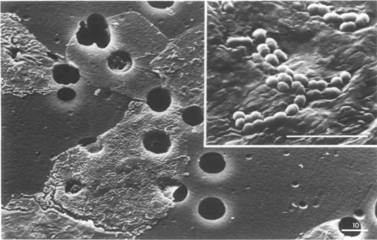
Fig. 1. Scanning electron micrographs of S.sanguis OMZ 9 binding to HBEC held by polycarbonate filters. Bars in Jim. Magnifications are 1200x (main panel)
and 7600x (inset panel).
was used as the main experimental assay. The effect of
sialidase treatment of the cells on S.sanguis OMZ 9 adhesion
was evaluated, as well as the effect of a competition with a
soluble glycopeptide bearing sialylated sugar chains of known
structures. Purified sialyltransferases were used to produce cell
surface sialyloligosaccharides of defined sequences (Rogers and
Paulson, 1983), and the same enzymes were also used in
conjunction with CMP-9-fluoresceinyl sialic acid to bring
fluorescence on the molecule(s) acting as receptor(s). For
comparative purposes, similar experiments were performed
with cultured HBEC obtained from a squamous cell carcinoma
termed SqCC/Yl (Reiss et al., 1985). The successful
restoration of a receptor function for S.sanguis OMZ 9 on
desialylated exfoliated HBEC by using a pure sialyltransferase
correlated with the fluorescent labelling of a single cell surface
glycoprotein. As a result, the precise carbohydrate sequence
and the cell surface glycoprotein bearing such sugar chains
were jointly identified, leading to the complete characterization
of the receptor for S.sanguis OMZ 9 on normal exfoliated
HBEC.
Results
Adhesion of S.sanguis OMZ 9 to exfoliated HBEC
An assay previously established for measuring the adhesion of
pathogenic yeast to HBEC (Brassart et al., 1991) was found to
be useful for the present purpose. Negligible background values
were obtained in the determination of microbial adhesion,
since the presence of bovine serum albumin (BSA) in the
incubation buffer totally prevented bacterial adhesion to the
polycarbonate filters. Preliminary experiments indicated that
incubation of 50 x 10
3HBEC with increasing concentrations of
a
l4C-labelled bacterial suspension led to an increase in the ratio
of bacteria attaching to the cells, until saturation was reached
(final bacterial OD > 1). Scanning electron micrographs
showed bacterial binding to HBEC held by the filter (Figure 1).
This result illustrates the usefulness of the assay conditions at
bacterial OD = 0.5 for determining specific bacterial binding
to exfoliated cells, whereas adhesion to the filter was
vir-tually absent (Figure 1). Based on the specific radioactivity
associated with the bacterial populations incubated in this assay,
an estimate of the extent of S.sanguis OMZ 9 attachment to
HBEC can be calculated to a mean value of 300 bacteria/buccal
cell.
To control both the degree of sialylation of exfoliated HBEC
and the effect of the sialidase treatment used for preparing
the desialylated cells, the amount of /V-acetylneuraminic acid
(NeuNAc) residues hydrolysed by this enzyme was
quan-titated. Exfoliated HBEC released -40 ng NeuNAc/10
4cells, a
value which is in good agreement with previously reported
determinations (Davis and Gibbons, 1990). The effect of HBEC
desialylation on the extent of bacterial adhesion was then
investigated. The adhesion of S.sanguis OMZ 9 to HBEC was
seen to be strictly dependent on sialic acid-bearing receptors,
since desialylation of HBEC prior to the adhesion experiment
invariably led to the total prevention of bacterial attachment
(As-Cells; Figure 2). Experiments were also carried out to
examine whether sialic acid or sialylated soluble complex
Adhesion receptor for S.sanguis on human buccal cells As-CGP CGP -As-Cells Control
A
/
1
)
-1
/
/
A 1.6+/-0.3 J 0.6 +/-0.3 / \ 4.0+/-0.8 J/ ]
5.5 +/- 1 J 0 1 2 3 4 5 6Adhesion (%)
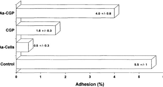
Fig. 2. Adhesion of 14C-labelled S.sanguis OMZ 9 to HBEC. The effects of either cell desialylation (As-Cells) or 10 mg/ml of CGP and As-CGP arc shown. They are expressed as the proportion of the total radioactivity in suspension remaining with the adhering microorganisms attached to HBEC after washing. The data represent mean values of three experiments with their SD.
a) NeuNAca(2-»~3)Galj3(l-^3)GalNAc b) Gal/S(l-^3)GalNAc 6 NeuNAc<*2 c) NeuNAca(2-^3)G«l/3(l-~3)GalNAc 6 NeuNAca2
Fig. 3. Structures of the three main carbohydrate chains of bovine CGP.
(NeuNAc = W-acetylneuraminic acid; Gal = galactose; GalNAc = N-acetylgalactosamine), From Van Halbeek et al. (1980).
carbohydrates inhibited the binding of S.sanguis OMZ 9 to
HBEC. At a concentration of 10 mg/ml, the
caseinoglyco-macropeptide (CGP)—a glycopeptide from bovine whey
which bears the short sialylated O-linked carbohydrate chains
shown in Figure 3—behaved as an excellent competitive
adhesion inhibitor. By contrast, its desialylated analogue
(As-CGP) was much less effective (Figure 2).
For comparative purposes, adhesion of S.sanguis OMZ 9 to
cultured monolayers of HBEC obtained from a squamous
car-cinoma was also assayed. It was found that S.sanguis OMZ 9
failed to attach in great numbers to these SqCC/Y 1 cell layers.
Indeed, the average score of microbial attachment to such
cultured cells ranged around 40 bacteria/buccal cell (results not
shown).
Host cell receptor specificity for S.sanguis OMZ 9
For defining the terminal oligosaccharide structure recognized
by the streptococcal adhesion factor on HBEC, commercially
available purified mammalian sialyltransferases (ST) were used.
For this purpose, aliquots of asialo-HBEC were reacted with
CMP-NeuNAc and one of the two following sialyltransferases:
the Gaipi,3GaTNAc a2,3-ST specific for sugar chains O-linked
to glycoproteins or located on glycolipids, and the Gaipi,4GlcNAc
a2,6-ST specific for the sequences found on oligosaccharides
N-linked to glycoproteins. Preliminary assays indicated that
exfoliated HBEC were very resistant to structural breakdown,
even after prolonged incubations in diluted solutions
contain-ing Triton X-100 and glycerol. Consequently, these
compon-ents were not removed from the commercial enzyme solutions.
The results of the resialylation experiments are shown in Figure
4. Clearly the reaction with the a2,3-ST yielded HBEC with
a restored receptor function for S.sanguis OMZ 9, whereas
the resialylation of asialo-cells with the a2,6-ST specific for
/V-glycans had no effect (Figure 4).
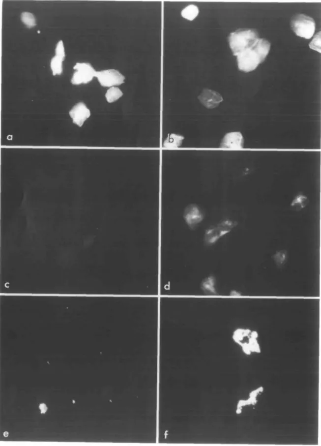
By using a fluorescent neuraminic acid derivative for the
resialylation reactions, we also investigated the feasibility of
visualizing such resialylated buccal cells bearing different
oligosaccharide sequences. With the commercial
CMP-9-fluor-esceinyl-sialic acid donor, we found that asialo-, exfoliated
HBEC were readily resialylated with both the a2,3-ST (Figure
5a) and the a2,6-ST (Figure 5b). This result indicates that such
desialylated cells expressed different sugar chains at their
sur-face, acting as acceptors either for one or for the other ST. By
contrast, the same experiment performed with cultured tumorous
buccal cells (SqCC/Yl) yielded fluorescent resialylated cells
only after reaction with the a2,6-ST specific for N-glycans
(Figure 5f), but not after reaction with the a2,3-ST enzyme
(Figure 5e). Finally, fluorescent cell samples freshly
resialyl-ated with the different sialyltransferases and stemming from
both exfoliated and cultured cells were dissolved in
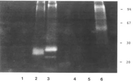
SDS and subjected to SDS-PAGE analysis. Interestingly,
from exfoliated HBEC, resialylated glycoproteins of low
mol. wt (20-30 kDa) were identified as bearing most of the
fluorescence attached to the cell surface after reaction with
either one or the other ST (Figure 6, lanes 2 and 3). In addition,
exfoliated HBEC sequentially desialylated and resialylated
with the oc2,3-ST yielded a single 23 kDa fluorescent
J.-R.Neeser et aL
ST2.6
ST2.3
As-Cells
Control
A
/
I
/
1
/
I
/
/
1.3 +/-1.4 +/ s O.I -0.2 ) / 4.6 + /-0.9 ) 3.6 +/- 0.4 1 0 0.5 1 1.6 2 2.5 3 3.5 4 4.5Adhesion (%)
Fig. 4. Adhesion of 14C-labelled S.sanguis OMZ 9 to native (Control), asialo (As-Cells) and resialylated (ST 2,3 and ST 2,6) HBEC. The data are expressed as in Figure 2.
glycoprotein (Figure 6, lane 2). By contrast, SDS-PAGE
analysis of fluorescence-bearing resialylated SqCC/Yl
cultured cells showed only fluorescent glycoproteins of higher
mol. wt (50-100 kDa), and only after reaction with the
a2,6-ST (Figure 6, lanes 5 and 6).
Discussion
The ability of several S.sanguis strains to specifically bind to
sialylated carbohydrate chains of salivary glycoproteins has
already been demonstrated and discussed (Me Bride and
Gisslow, 1977; Bergey et al, 1986; Demuth et ai, 1990).
However, it was never clearly established that the activity
of such a streptococcal lectin could also mediate bacterial
adhesion to HBEC. Streptococcus sanguis OMZ 9 has been
shown to bind to saliva-coated hydroxyapatite beads by a
mechanism sensitive to acidic polypeptides, including bovine
milk casein, CGP, its desialylated analogue (As-CGP) and
caseinophosphopeptides (Neeser et al, 1994). It was therefore
apparent that the adhesion of S.sanguis OMZ 9 to hard surfaces
involved mainly ionic interactions, as already observed for
other S.sanguis strains and other oral streptococci (Doyle et al.,
1982; Cowan et al, 1987b; Satou et al., 1988; Koga et al,
1990). By contrast, we show here that the adhesion of
S.sanguis OMZ 9 to HBEC is strictly dependent on the
presence of sialic acid residues on the cell surface, and that
such an adhesion is potently inhibited by CGP but not by
As-CGP. Thus, sialic acids seem to be the key configuration for the
specific attachment of S.sanguis OMZ 9 to the buccal mucosal
epithelium.
For analysing the sialyloligosaccharide receptor specificity
involved in the binding of S.sanguis OMZ 9 to HBEC, we used
the methodology previously developed by James Paulson and
co-workers in their studies of cell surface receptors for animal
viruses (Paulson, 1985). We took advantage of the
commer-cial availability of two purified sialyltransferases: the Gaipi,
3GalNAc a2,3-ST and the Galfi l,4GlcNAc a2,6-ST. It is clearly
established that both these enzymes exhibit a strict specificity
for their respective terminal oligosaccharide structures
recog-nized as acceptors. The majority of the studies involving such
enzymatic restorations of cell surface sialyloligosaccharides
with defined sequences have been performed from desialylated
red blood cells (Paulson, 1985). However, similar treatments of
tissue culture cells for investigating receptor determinants in
viral infections have also been reported (Markwell and
Paulson, 1980; Fried et al, 1981). In the present study, the
<x2,3-ST clearly restored the receptor function for S.sanguis
OMZ 9, previously destroyed by the sialidase treatment of
HBEC. The use of a fluorescent neuraminic acid derivative for
performing similar resialylation reactions from desialylated
HBEC finally led to the identification of the single 23 kDa
fluorescent glycoprotein acting as an acceptor for the Ct2,3-ST.
Thus, this HBEC membrane glycoprotein bears
NeuNAca2-3Gal(3l-3GalNAca O-linked sugar chains, the carbohydrate
sequence specifically restored by the a2,3-ST, and specifically
recognized by S.sanguis OMZ 9 on HBEC.
It should be noted that SqCC/Yl tumorous buccal epithelial
cells showed minimal microbial attachment, as compared to the
exfoliated cells from normal mucosa (HBEC). Tumorigenesis
is generally coupled to multiple genetic changes, resulting in
altered phenotypic properties of tumorous cells as compared to
normal cells. Moreover, aberrations in the cell surface
carbo-hydrate structures have now been established as a universal
characteristic of malignant transformation of cells, and cancer
has been referred to as a molecular disease of the cell membrane
glycoconjugates (Hakomori, 1989; Bhavanandan, 1991). In
this regard, glycosylation of annexins I and II by SqCC/Yl
cells has already been examined (Goulet et al, 1992). Also, the
SqCC/ Yl cell line has been seen to exhibit significant changes
in glycolipid composition, when differentiated in the absence
of serum (Tatsumura et al, 1988). Finally, the ability of these
cells to synthesize glycosaminoglycans differed markedly from
Adhesion receptor for Sjanguis on human buccal cells
Fig. 5. Fluorescence micrographs of exfoliated HBEC (a-d) and cultured SqCC/Yl cells (e, f). The desialylated cells were treated with the
CMP-9-fluoresceinyl-sialic acid donor, in conjunction with either a2,3-ST(a, e) or ct2,6-ST (b, f). Controls were obtained by incubating exfoliated HBEC without the fluorescent reagent (c) or without sialyltransferase (d).
J.-R.Neeser et al
- 67
- 30
- 20
1
2 3 4 5 6
Fig. 6. SDS-PAGE analysis of total cell extracts from exfoliated HBEC (lanes 1-3) and cultured SqCC/Yl cells (lanes 4-6). The desialylatcd cells were treated with the CMP-9-fluoresceinyl-sialic acid donor, in conjunction with either a2,3-ST (lanes 2 and 5) or ot2,6-ST (lanes 3 and 6). Controls were obtained by incubating the fluorescent reagent without sialyltransferase (lanes 1 and 4).
that of normal keratinocytes (Reiss et al, 1986). Thus, changes
in cell surface glycosylation may affect the cell-to-cell
interactions that participate in regulating growth and
differ-entiation of the epithelium, also the ability of such pheno
plastic and neoplastic cells to be identified in vivo by
immun-ologically based recognitions, and finally the expression of
particular determinants which can act as specific receptors for
bacteria.
Another explanation may be envisaged, to understand the
difference observed between exfoliated HBEC and cultured
tumorous SqCC/Yl cells, regarding the expression of receptors
for S.sanguis OMZ 9. The labellings yielded by the treatment
of both these cell types with the specific sialyltransferases and
a fluorescent neuraminic acid derivative revealed very different
membrane glycoprotein patterns. It has been reported that in
the oral cavity, certain salivary components may interact with
the epithelial cells to be finally cross-linked to the cell
cyto-skeleton by epithelial transglutaminases (Bradway et al,
1989). In such a case, the surface of HBEC obtained by
exfoli-ation would be very different from that of (normal and
tumor-ous) cultured cells. Interestingly, the determination of type I
transglutaminase in differentiating normal and neoplastic
(SqCC/Yl) human keratinocytes has recently retained attention
(Moore et al, 1993). Thus, further studies are required,
involv-ing the growth of various normal and tumorous cell types from
the oral mucosa, cultured in the presence and in the absence of
saliva, to understand the origin of the 23 kDa cell surface
receptor for S.sanguis OMZ 9.
Materials and methods
All reagents were of the highest quality available from Sigma Chemical Co. (St Louis, MO) or Fluka (Buchs, Switzerland), unless otherwise stated. Caseinoglycopeptide derivatives
Bovine CGP was isolated from a whey protein concentrate (Danmark protein, Videback, Denmark) by protein precipitation with a trichloroacetic acid solution (12% final, w/v). CGP desialylation was performed by mild acid hydrolysis (HjSCv, 25 mM, 2 h at 80°C). Both these CGP derivatives were purified on a G-50 Sephadex column (Pharmacia, Uppsala, Sweden), using a 0.1 M acetic acid buffer.
Bacterial growth conditions and radiolabelling
Streptococcus sanguis OMZ 9 was cultured in fluid universal medium (FUM), a previously defined medium (GmUr and Guggenheim, 1983). The bacteria were pre-cultured for 7 h at 37°C and then grown for 14 h before harvesting. The bacteria were metabolically labelled by the addition of [14C]acetic acid (94 mCi/mmol; 100 u.Ci/10 ml tube; CEA, Gif-sur-Yvette, France). Specific radioactivity (c.p.mVIO8 cells) was between 300 000 and 350 000.
Bacterial adhesion to HBEC and inhibition studies
HBEC were freshly collected by gently rubbing the buccal mucosa of several donors in the authors' laboratory. The cells were pooled, washed as previously described (Brassart et al., 1991) and finally suspended in a Hank's balanced salt solution containing 0.2% BSA, 0.02% each Ca2+, Mg2+ (Hank's/BSA), in a concentration of 105 cells/ml. After harvesting, l4C-labelled bacteria were washed three times and suspended (OD = 1.0, X = 650 nm) in Hank's/BSA solution, with or without an inhibitor to be tested (10 mg/ml). Then, 0.5 ml aliquots of the HBEC suspensions were mixed with 0.5 ml of the bacterial suspensions in plastic tubes which were rotated at 80 r.p.m. for 1 h at 25°C. These mixtures were finally filtered as previously described (Brassart et al., 1991). Bacterial adhesion was determined by radiometric counting. Desialylated HBEC were obtained by incubating 1.5 x 10⁵ washed cells (1 h at 37°C) in 0.2 ml of 150 mM NaCl containing 10 mM CaCl2 and 0.2 U of siahdase from Vibno cholerae (Behring, Marburg, FRG). Then the cells were collected, washed three times with Hank's/BSA and used in adhesion studies as described above. The sialic acid residues released from HBEC membranes by this enzymatic hydrolysis were quantitatively determined by colorimetry (Jourdian etal, 1971).
Scanning electron microscopy
For ultrastructural studies, bacteria attached to HBEC were fixed on the filter for 30 min at room temperature with 2.5% glutaraldehyde in 0.1 M phosphate buffer (pH 7.4). After washing with this buffer, cells were dehydrated in a graded series (50-100%) of ethanol. Wet cells were dried after substitution with liquid CO2 in a critical-point dryer (Polaron Equipment Ltd, Watford, UK) and coated with gold (SEM coating unit E5100; Polaron). Samples were viewed using a Philips 505 SEM microscope.
Culture conditions for SqCC/Yl cells and measurement of bacterial adhesion SqCC/Yl cells were kindly provided by Prof. A.C.Sartorelli, New Haven, CT. The cells were routinely grown in a serum-containing medium termed SqCC/Y 1 medium, as previously described (Sundq vist etal, 1991). Prior to an assay for bacterial adhesion, the cells were cultured in 6-well plates in SqCC/Yl medium until the cells covered the surface area. To avoid serum-dependent interactions in the assay, the cells were dien incubated for 48 h in a serum-free medium termed EMA, developed for both normal and tumorous
Adhesion receptor for S.sanguis on human buccal cells
buccal epithelial cell cultures (Sundqvist et al, 1991). Bacterial adhesion was measured by using metabolically l4C-labelled bacteria, as previously described (Neeser et al., 1989). The cell layers were then washed with Hank's/BSA before they were exposed to the bacterial suspensions (OD = 0.5 in 1 ml of Hank's/BSA) for 1 h at 37°C. Subsequently, unbound bacteria were removed by multiple washing, and the buccal cells were dissolved with the adhered or-ganisms in a NAOH solution (0.2 N). Bacterial adhesion was quantitated using liquid scintillation counting, as previously described (Neeser el al., 1989). Resialylation of desiatylated HBECfor adhesion studies
Freshly collected HBEC were pooled and pre-treated as described above. Exfo-liated cells (1.2 x 10⁶) were subjected to desialylation by incubation 0 h at 37°C) in 2 ml of 150 mM NaCl containing 10 mM CaCl2 and 2 U of sialidase from V.cholerae. These cells were then washed twice in a resialylation buffer [25 mM MOPS (pH 6.25) containing 75 mM NaCl, 100 mM glucose, 10 mg/ml BSA, and supplemented with 115 U/ml penicillin, 115 U/ml streptomycin, 1.3 u.g/ml amphotericin B and 150 Hg/ml gentamicin]. Such desialylated HBEC were divided into three samples. Resialylation reactions were performed by using either GalpM,3GalNAc ct2,3-ST from porcine liver or Gaipi,4GlcNAc a2,6-ST from rat liver, with CMP-NeuNAc in both cases; all these reagents being from Boehringer Mannheim, FRG. The procedure was adapted from that described by Rogers and Paulson (1983), except that the enzyme solutions were used as received from the supplier, without removing Triton X-100 and glycerol. Typically, 4 x 10⁵ desialylated HBEC were sus-pended in the resialylation buffer described above (40 u.1) and mixed with CMP-NeuNAc (500 ng in 10 ul of resialylation buffer). Then, either 5 mU of the a2,3-ST solution or 25 mU of the cx2,6-ST solution were added to the cell suspensions, which were incubated for 24 h at 37°C. A sample of 4 x 10³ native and intact exfoliated HBEC was incubated in 75 u.1 of resialylation buffer supplemented with Triton X-100 (0.22%) and glycerol (17%), to be used as a control. After completion of the enzymatic reactions, the cells were collected, washed three times with Hank's/ BSA and used in adhesion studies as above. Resialylation of desialylated HBEC and SqCC/Yl cells with fluoresceinyl-NeuNAc residues
Experiments were performed either with freshly collected exfoliated HBEC (1.6 x 10⁶ cells), or with SqCC/Yl cells (4 x 10*) obtained from cell layers cultured as described above, and further treated with a dispase D solution (Boehringer Mannheim, FRG) to gently detach the cells from the plastic. All cells were washed, desialylated, and again washed prior to rcsialylation, as already described. Samples of each cell type (exfoliated HBEC and SqCC/Yl) were subjected to the different rcsialylation reactions studied here (with the O2.3-ST and the tx2,6-ST, respectively), but in two steps: a first incubation period (4 h at 37°C) was carried out by using CMP-9-fluoresceinyl-NeuNAc (Boehringer Mannheim, FRG, 5 jig in 10 JJJ of resialylation buffer) as a glycosyl-donor nucleotide; after that time, a solution of CMP-NeuNAc (625 |ig in 15 u.1 of rcsialylation buffer) was added to these samples, which were further incu-bated for 20 h at 37°C. Samples of desialylated cells were used as controls after an incubation period (4 h at 37°C) with the fluorescent probe, but in the absence of any sialyltransferase. All cell samples were washed three times with a phosphate-buffered saline solution (pH 7.2) before being examined in a fluorescence microscope equipped with an epi-illuminator.
Gel electrophoresis
Fluorescent cell samples obtained as above were also suspended in 50 u.1 portions of a solution of Tnton X-100 (1%) and thoroughly shaken, prior to being mixed and heated with the incubation buffer for gel electrophoresis. SDS—PAGE (gradient from 10 to 20% acrylamide) was performed by the method of Laemmli (1970). The gel was finally examined under a UV lamp. Molecular weight markers were from Pharmacia (Uppsala, Sweden).
Acknowledgements
We thank ICSundqvist and A.Pfeifer for help and advice with culturing the buccal epithelial cells, and E.Kolodziejczyk for expert assistance in fluores-cence microscopy.
Abbreviations
As-CGP, asialo-caseinoglycomacropeptide; BSA, bovine serum albumin; CGP, caseinoglycomacropeptide; FUM, fluid universal medium; HBEC, human
buccal epithelial cell; MOPS, morpholinopropanesulphonic acid; NeuNAc, A'-acetylneuraminic acid; OD, optical density; ST, sialyltransferase.
References
Bergey.EJ., Levine,MJ., Reddy,M.S., Bradway.S.D. and Al-HashimiJ.. (1986) Use of the photoaffinity cross-linking agent /V-hydroxysuccinimidyl^ azidosalicylic acid to characterize salivary-glycoprotein-bacterial interactions. Biochem. J., 234, 43-48.
Bhavanandan.V.P. (1991) Cancer-associated mucins and mucin-type glyco-proteins. Clycobiology, 1, 493-503.
Bradway.S.D., Bergey.EJ., Jones.P.C. and Levine,MJ. (1989) Oral mucosal pellicle. Adsorption and transpeptidation of salivary components to buccal epithelial cells. Biochem. J., 261, 887-896.
Brassart.D., Woltz,A., Golliardjvl. and NeeserJ.-R. (1991) In vitro inhibition of adhesion of Candida albicans clinical isolates to human buccal epithelial cells by Fuc al—>2Galp"-bearing complex carbohydrates. Infect. Immun., 59,
1605-1613.
Christensen.G.D., Simpson.W.A. and Beachey^E.H. (1985) Adhesion of bacteria to animal tissues: complex mechanisms. In Savage.D.C. and Fletcherjvl. (eds), Bacterial Adhesion. Plenum Publishing Corp., New York, pp. 279-306.
Cowan.M.M., Grant Taylor.K. and Doyle,RJ. (1987a) Role of sialic acid in the kinetics of Streptococcus sanguis adhesion to artificial pellicles. Infect. Immun., 55, 1552-1557.
Cowan.M.M., Taylor,K.G. and Doyle.RJ. (1987b) Energetics of the initial phase of adhesion of Streptococcus sanguis to hydroxylapatite. J. BacterioL,
169, 2995-3000.
Davis.G. and Gibbons J U . (1990) Accessible sialic acid content of oral epithelial cells from healthy and gingivitis subjects. J. Ptriodont. Res., 25, 250-253. DemuthJD.R., Golub,E.E. and MalamudJD. (1990) Streptococcal-host
inter-actions. Structural and functional analysis of Streptococcus sanguis receptor for a human salivary glycoprotein. /. Biol. Chenu, 265, 7120-7126. Doyle.RJ., Nesbitt.W.E. and Taylor.K.G. (1982) On the mechanism of
adher-ence of Streptococcus sanguis to hydroxylapatite. FEMS Microbiol. Lett., 15, 1-5.
FriedJrl., CahanJL.D. and PaulsonJ.C. (1981) Polyoma virus recognizes specific sialyloligosaccharide receptors on host cells. Virology, 109, 188-192. GibbonsJU. (1989) Bacterial adhesion to oral tissues: a model for infectious
diseases. /. Dent. Res., 68, 750-760.
GmUr,R. and GuggenheimJ3. (1983) Antigenic heterogeneity of Bacteroides intermedius as recognized by monoclonal antibodies. Infect. Immun., 42, 459-470.
Goulet,F., Moore.K.G. and Sartorelli.A.C. (1992) Glycosylation of annexin I and annexin n. Biochem. Biophys. Res. Commun., 188, 554—558.
Hakomori.S. (1989) Aberrant glycosylation in tumors and tumor-associated carbohydrate antigens. Adv. Cancer Res., 52, 257-331.
Jourdian.G.W., Dean,L. and Roseman.S. (1971) The sialic acids. XI. A periodate-resorcinol method for the quantitative estimation of free sialic acids and their glycosides. J. Biol. Chem., 246, 430-435.
Koga,T, Okahashi.N., TakahashiJ., Kanamoto.T., Asakawa,H. and lwaki,M. (1990) Surface hydrophobicity, adherence, and aggregation of cell surface protein antigen mutants of Streptococcus mutans serotype c. Infect. Immun., 58, 289-296.
Laemmli.U.K. (1970) Cleavage of structural proteins during assembly of the head of bacteriophage T4. Nature, 227, 680-685.
Levine.M J., Herzberg.M.C, Levine,M.S., Ellison.S.A., Stinson.M.W., Li.H.C. and Van Dyke.T. (1978) Specificity of salivary-bacterial interactions: role of terminal sialic acid residues in the interaction of salivary glycoproteins with Streptococcus sanguis and Streptococcus mutans. Infect. Immun., 19,107-115. Liljemark.W.F., Bloomquist,C.G., Fenner.LJ., Antonelli.PJ. and Cecilia
Coulter.M. (1989) Effect of neuraminidase on the adherence to salivary pellicle of Streptococcus sanguis and Streptococcus mitis. Caries Res., 23, 141-145.
Markwell,M.A.K, and PaulsonJ.C. (1980) Sendai virus utilizes specific sialyl-oligosaccharides as host cell receptors determinants. Proc. Natl Acad. Sci. USA, 77, 5693-5697.
Me Bride.B.C. and Gisslow,M.T. (1977) Role of sialic acid in salivary-induced aggregation of Streptococcus sanguis. Infect. Immun,, 18, 35-40.
Mergenhagen.S.E-, Sandberg.A.L, Chassy.B.M., Brennan,MJ., Yeung.M.K., DonkerslootJ.A. and CisarJ.O. (1987) Molecular basis of bacterial adhesion in the oral cavity. Rev. Infect. Dis., 9, S467-474.
Moore,K.G., DonadioAC. and SartorelliAC. (1993) Determination of type I transglutaminase in differentiating normal and neoplastic human keratino-cytes by an in situ radioimmunoassay. Biochem. Biophys. Res. Commun.,
192, 381-385.
J.-R.Neeser el al
Morris.EJ. and McBride.B.C. (1984) Adherence of Streptococcus sanguis to saliva-coated hydroxyapatite: evidence for two binding sites. Infect. Immun.,
43, 656-663.
Murray.P.A., Levine.MJ., TabakJLA. and ReddyJvl.S. (1982) Specificity of salivary-bacterial interactions: II. Evidence for a lectin on Streptococcus sanguis with specificity for a NeuAca 2,3Galp'l,3GalNAc sequence. Biochem. Biophys Res. Commun., 106, 390-396.
NeeserJ.-R., Chambaz^., Del Vedovo.S., PrigentJvlJ. and Guggenheim,!). (1988) Specific and nonspecific inhibition of adhesion of oral Actinomyces and streptococci to erythrocytes and polystyrene by caseinoglycopeptide derivatives. Infect. Immun., 56, 3201-3208.
NeeserJ.-R., Chambaz.A., Golliard.M., Link-Amster.H., Fryder.V. and Kolodziejczyk.E. (1989) Adhesion of colonization factor antigen II-positive enterotoxigenic Eschcrichia coli strains to human enterocytelike differenti-ated HT-29 cells: a basis for host-pathogen interactions in the gut. Infect. Immun., 57, 3727-3734.
Neeser,J.-R., Golliard.M., Woltz.A., Rouvet.M., Dilimann,M.-L. and Guggenheim.B. (1994) In vitro modulation of oral bacteria] adhesion to saliva-coated hydroxyapatite beads by milk casein derivatives. Oral MicrobioL Immunol, 9, 193-201.
Ofek.I. and Sharon.N. (1990) Adhesins as lectins: specificity and role in infection. Curr. Top. Microbiol. Immunol, 151, 91-113.
PaulsonJ.C. (1985) Interactions of animal viruses with cell surface receptors. In Conn.RM. (ed.), The Receptors. Academic Press, UK, Vol. II, pp. 131-219.
Reiss.M., Pitman.S.W. and Sartorelh,A.C. (1985) Modulation of the terminal differentiation of human squamous carcinoma cells in vitro by ail-trans-retinoic acid. J. Natl Cancer Inst., 74, 1015-1023.
Reiss,M., Maniglia,C.A. and SartorelliAC. (1986) Modulation of cell shed-ding and glycosaminoglycan synthesis of human malignant keratinocytes by all-lrans-retinoic acid and hydrocortisone in vitro. /. Invest. Dermatol, 86, 683-688.
Rogers.G.N. and PaulsonJ.C. (1983) Receptor determinants of human and animal influenza virus isolates: differences in receptor specificity of the H3 hemagglutinin based on species of origin. Virology, 127, 361-373. Satoujvl., SatouJ., Shintam.H. and Okuda,K. (1988) Adherence of streptococci
to surface-modified glass. /. Gen. Microbiol, 134, 1299-1305.
Sundqvist.K., Kulkami.P, Hybbinette,S.S., Bertolero.E, Liu,Y. and Grafstrom, R.C. (1991) Serum-free growth and karyotype analyses of cultured normal and tumorous (SqCCVYl) human buccal epithelial cells. Cancer Commun., 3,331-340.
Tatsumura,T., Ariga,T., Yu.R.K. and Sartorelli^i.C. (1988) Changes in glyco-sphingolipids accompanying the differentiation of human squamous SqCC/ Yl cells. Cancer Res., 48, 2121-2124.
Van Halbeek.H., Dorland.L., VhegenthartJ.F.G., Hat,A.-M. and Jolles.P. (1980) A 360-MHz 'H-NMR study of three oligosaccharides isolated from cow K-casein. Biochim. Biophys. Acta, 623, 295-300.