HAL Id: hal-01793279
https://hal.archives-ouvertes.fr/hal-01793279
Submitted on 16 May 2018
HAL is a multi-disciplinary open access
archive for the deposit and dissemination of
sci-entific research documents, whether they are
pub-lished or not. The documents may come from
teaching and research institutions in France or
abroad, or from public or private research centers.
L’archive ouverte pluridisciplinaire HAL, est
destinée au dépôt et à la diffusion de documents
scientifiques de niveau recherche, publiés ou non,
émanant des établissements d’enseignement et de
recherche français ou étrangers, des laboratoires
publics ou privés.
Copyright
Bifunctional Enzyme from the Human Microbiome
Coupling Galactosidase and Kinase Activities
Laëtitia Bruel, Gerlind Sulzenbacher, Marine Tison, Ange Pujol, Cendrine
Nicoletti, Josette Perrier, Anne Galinier, David Ropartz, Michel Fons,
Frederique Pompeo, et al.
To cite this version:
Laëtitia Bruel, Gerlind Sulzenbacher, Marine Tison, Ange Pujol, Cendrine Nicoletti, et al..
α-Galactosidase/Sucrose Kinase (AgaSK), a Novel Bifunctional Enzyme from the Human Microbiome
Coupling Galactosidase and Kinase Activities. Journal of Biological Chemistry, American Society for
Biochemistry and Molecular Biology, 2011, 286 (47), pp.40814 - 40823. �10.1074/jbc.M111.286039�.
�hal-01793279�
␣-Galactosidase/Sucrose Kinase (AgaSK), a Novel
Bifunctional Enzyme from the Human Microbiome Coupling
Galactosidase and Kinase Activities
*
□SReceived for publication, August 1, 2011, and in revised form, September 3, 2011Published, JBC Papers in Press, September 20, 2011, DOI 10.1074/jbc.M111.286039 Laëtitia Bruel‡§, Gerlind Sulzenbacher¶, Marine Cervera Tison‡, Ange Pujol‡, Cendrine Nicoletti‡,
Josette Perrier‡, Anne Galinier§, David Ropartz储, Michel Fons‡, Frédérique Pompeo§, and Thierry Giardina‡1
From the‡Faculté des Sciences et Techniques Saint-Jérôme, Université Paul Cézanne, ISM2/BiosCiences UMR CNRS 6263, service 342, 13397 Marseille Cedex 20, the¶Architecture et Fonction des Macromolécules Biologiques UMR CNRS 6098, Université Aix-Marseille, Campus Luminy, Case 932, F-13288 Marseille Cedex 09, the§IMM/Laboratoire de Chimie Bactérienne, UPR CNRS 9043, 31 chemin Joseph Aiguier, 13402 Marseille Cedex 20, and the储Laboratoire de Spectrométrie de Masse, Plate-forme Biopolyme`res-Biologie Structurale, INRA UR1268 Biopolymères Interactions Assemblages, Rue de la Géraudière, B.P. 71627, F-44316 Nantes cedex 3, France
Background:Raffinose, an abundant carbohydrate in plants, is degraded into galactose and sucrose by intestinal microbial enzymes.
Results:AgaSK is a protein coupling galactosidase and sucrose kinase activity. The structure of the galactosidase domain sheds light onto substrate recognition.
Conclusion:AgaSK produces sucrose-6-phosphate directly from raffinose.
Significance: Production of sucrose-6-phosphate directly from raffinose points toward a novel glycolytic pathway in bacteria.
␣-Galactosides are non-digestible carbohydrates widely dis-tributed in plants. They are a potential source of energy in our daily food, and their assimilation by microbiota may play a role in obesity. In the intestinal tract, they are degraded by microbial glycosidases, which are often modular enzymes with catalytic domains linked to carbohydrate-binding modules. Here we intro-duce a bifunctional enzyme from the human intestinal bacterium
Ruminococcus gnavus E1,␣-galactosidase/sucrose kinase (AgaSK).
Sequence analysis showed that AgaSK is composed of two domains: one closely related to␣-galactosidases from glycoside hydrolase family GH36 and the other containing a nucleotide-binding motif. Its biochemical characterization showed that AgaSK is able to hydrolyze melibiose and raffinose to galactose and either glucose or sucrose, respectively, and to specifically phospho-rylate sucrose on the C6 position of glucose in the presence of ATP. The production of sucrose-6-P directly from raffinose points toward a glycolytic pathway in bacteria, not described so far. The crystal structures of the galactosidase domain in the apo form and in complex with the product shed light onto the reaction and sub-strate recognition mechanisms and highlight an oligomeric state
necessary for efficient substrate binding and suggesting a cross-talk between the galactose and kinase domains.
Recent studies have suggested that the human intestinal microbiome plays an important role in the weight differ-ences between human beings, namely that it could be signif-icant in obesity (1–3). Indeed, obesity can be linked to an alteration of the ratio of Bacteroidetes and Firmicutes in the microbiome (4). Together, these bacteria represent over 90% of the bacterial phylotypes in the intestinal tract (5). This advocates a change in the fermentation pattern and a more efficient energy extraction from foodstuff operated by Firmi-cutes (6, 7). In particular, non-digestible carbohydrates can be metabolized by bacteria residing in the intestinal tract, and the imbalance of calorie consumption and expenditure causing obesity can be attributed to variations of the intesti-nal microbial ecology (8).
Among non-digestible carbohydrates, essentially originat-ing from plant cell walls,␣-galactosides are among the most abundant oligosaccharides. For example, they represent from 39 to 53 g/kg in soybean meals (9). They consist of galactose units␣-(1,6) linked to a mannopyranose backbone (galactomannans) or␣-(1,6) linked to glucose (melibiose) or sucrose (raffinose) or raffinose (stachyose), with sucrose and raffinose being the most abundant soluble carbohydrates found in plant cell walls (9). However, there exists no ␣-(1,6)-galactosidase activity in the human intestine mucosa, and ␣-galactosides are exclusively fermented by microbial␣-(1,6)-galactosidases (EC 3.2.1.22). Studies of the bacterial metabolism of␣-galactosides (10, 11) have shown that melibiose and raffinose penetrate in the cell, where they
*This work was supported by the CNRS, the Agence Nationale de la Recherche (ANR) contract “P-loop proteins,” and the “Ministère de l’Enseignement Supérieur.”
The atomic coordinates and structure factors (codes 2YFN and 2YFO) have been deposited in the Protein Data Bank, Research Collaboratory for Structural Bioinformatics, Rutgers University, New Brunswick, NJ (http://www.rcsb.org/). The nucleotide sequences of the genes reported in this paper have been submit-ted to the EMBL Nucleotide Sequence Database (ENA) with accession numbers FQ790377–FQ790383.
□S The on-line version of this article (available at http://www.jbc.org) contains
supplemental Figs. 1– 6.
1To whom correspondence should be addressed. Tel.: 33-4-91-28-84-45; Fax: 33-4-91-28-84-40; E-mail: thierry.giardina@univ-cezanne.fr.
THE JOURNAL OF BIOLOGICAL CHEMISTRY VOL. 286, NO. 47, pp. 40814 –40823, November 25, 2011 © 2011 by The American Society for Biochemistry and Molecular Biology, Inc. Printed in the U.S.A.
at CNRS, on November 30, 2011
www.jbc.org
Downloaded from
http://www.jbc.org/content/suppl/2011/09/19/M111.286039.DC1.html
are hydrolyzed into galactose and glucose and into galactose and sucrose, respectively. Sucrose and galactose are subse-quently phosphorylated by a kinase and utilized in the gly-colytic pathways (12, 13).
According to the sequence-based classification of glycoside hydrolases (GHs)2 (Carbohydrate-Active Enzymes Database,
CAZy, classification) (14), ␣-galactosidases form eukaryotic organisms are mainly grouped into families GH27 and GH36, but those from microbial sources are also found in glycoside hydrolase families GH4, GH57, GH97, and GH110. Iterative database searches have shown that families GH27 and GH36, together with families GH31 and GH66, share a common evo-lutionary origin with family GH13 (15). For that matter, fami-lies GH27 and GH36, together with family GH31, form glyco-side hydrolase clan D (16). Crystal structures of the clinically important eukaryotic ␣-galactosidase and ␣-N-acetylgalac-tosaminidase have been reported by the Garman group almost a decade ago (17, 18), and a wealth of supplementary structural information for other family GH27 members has become avail-able later on. On the basis of the high level of sequence homol-ogy between members of family GH27 and GH36, which reflects intrinsically structural homology, Comfort et al. (19) were able to infer the catalytic residues of Thermotoga maritima
TmGalA, as well as to confirm their identity by elegant biochemi-cal studies. For family GH36, only the crystal structures of T.
mari-tima TmGalA (Protein Data Bank (PDB) 1zy9) and Lactobacillus
brevis LbR11 (PDB 3mi6) have been deposited within the Protein Data Bank by Structural Genomics Consortia, and the crystal structure of Lactobacillus acidophilus NCFM LaMel36A (PDB 2xn0) has been solved very recently (20).
Family GH36 members originate from bacteria, fungi, and plants and carry out hydrolysis with a net retention of the ano-meric configuration, which is why many enzymes from this family have been shown to possess transglycosylation activity (21–27). For their ability to hydrolyze␣-galactosides non-di-gestible by humans and to synthesize diverse oligosaccharides, GH36␣-galactosidases have potential applications for the pro-duction of prebiotics. These non-digestible oligosaccharides are beneficial for human health by their ability to stimulate selectively the growth of microbiota with advantageous physi-ological effects on the host (28 –30).
In this work, we have characterized from a functional point of view a bifunctional enzyme, hereafter called␣-galactosidase/ sucrose kinase (AgaSK), from Ruminococcus gnavus E1. R.
gna-vusis a strict anaerobic Gram-positive bacterium from the phy-lum Firmicutes. It belongs to the most common 57 species present in 90% of individuals (31). The enzyme AgaSK is com-posed of a GH36␣-galactosidase and a kinase domain and is able to release galactose from␣-galactosides and to phospho-rylate sucrose provided either by raffinose hydrolysis or by the environmental medium. The crystal structures of the galacto-sidase domain in the apo form and in complex with the product shed light onto the reaction and substrate recognition
mecha-nisms and highlight an oligomeric state necessary for efficient substrate binding and suggesting a cross-talk between the galactose and kinase domains.
EXPERIMENTAL PROCEDURES
Cecal Content Collection and Extraction of RNA—Animal
experiments were performed according to the guidelines of the French Ethics Committee. Fisher axenic rats came from the Animalerie Axe´nique de Micalis (ANAXEM) platform (INRA Jouy-en-Josas, France) and were reared in Trexler type isolators (La Calhène, Vélizy-Villacoublay, France). They were inocu-lated with⬃109cells of the R. gnavus E1 strain (0.5 ml of fresh
culture) by intragastric route. After 1 week, the animals were sacrificed, and the cecal contents were collected. Two hundred mg of fresh material were used for total RNA extraction accord-ing to the protocol described by Doré et al. (32) and cleaned up with the RNeasy mini kit (Qiagen). TURBOTM DNase
(Ambion威) was used for elimination of genomic DNA as rec-ommended by the supplier.
RT-PCR—cDNA synthesis was performed by reverse
tran-scription (RT) of 100 ng of RNA primed with 50 ng of random primers. The reaction was carried out at 50 °C for 1 h with the SuperScriptTMIII reverse transcriptase (Invitrogen), as
recom-mended by the supplier. The enzyme was inactivated by heating for 15 min at 70 °C, and 1l of the mixture was used directly as a template for PCR amplifications. PCR reactions were per-formed in a 50-l volume by using the Platinum威 Taq DNA polymerase (Invitrogen) under the following conditions: 94 °C for 2 min; 30 cycles at 94 °C for 30 s, 50 °C for 30 s, and 72 °C for 1 min; and a final elongation step at 72 °C for 10 min. As a control, additional reactions were performed by using as a tem-plate an RT mixture without enzyme, an RT mixture without RNA, and chromosomal DNA.
Bioinformatic Analysis—Manual validation of the automatic annotation of the R. gnavus E1 genome was performed using the MaGe (Magnifying Genomes) web-based interface from Genoscope. Putative transcriptional terminators were pre-dicted in silico using the RNAfold program. Functional predic-tions were based on sequence comparisons with the help of the sequence databases Profils pour l’Identification Automatique du Métabolisme (PRIAM), Clusters of Orthologous Groups of protein (COG) (www.ncbi.nlm.nih.gov/COG), and Swiss-Prot the domain/motif database InterProScan.
agaSK Amplification and Cloning into pOPIN E Expression
Vector—The agaSK gene was cloned from the genomic DNA of
R. gnavus E1 using DNA polymerase (PrimeSTAR HS DNA
polymerase, TAKARA BIO Inc.). PCR reaction primers
(for-ward, 5
⬘-AGGAGATATACCATGGCAATTATATACAAT-CCA-3⬘; reverse, 5⬘-GTGATGGTGATGTTTCTGTTCATA-AACACTTCC-3⬘) were used for 30 cycles of denaturation (1 min, 98 °C), annealing (1.5 min, 48 °C), and extension (3 min, 60 °C) in a Mastercycler威 gradient thermocycler (Eppendorf, Hamburg, Germany). The PCR products amplified with the In-Fusion primers were combined with the expression vector pOPIN E, kindly provided by Dr. Ray Owens (Oxford Univer-sity), following the manufacturer’s instructions (In-FusionTM,
Clontech). The recombinant vectors were checked by double-stranded DNA sequencing (Genome Express, Meylan, France).
2The abbreviations used are: GH, glycoside hydrolase; AgaSK, ␣-galactosid-ase/sucrose kinase; PNPG, p-nitrophenyl-␣-D-galactopyranoside; ABC, ATP-binding cassette transporter; Q-TOF, quadrupole time-of-flight; Gal-DNJ,D-galacto-1-deoxynojirimycin; PTS, phosphotransferase system.
at CNRS, on November 30, 2011
www.jbc.org
The In-Fusion system led to the production of AgaSK with a His6tag at the C terminus.
Expression and Purification of AgaSK—Recombinant AgaSK
was synthesized from Escherichia coli BL21 (DE3) grown in Luria broth, containing 50 mg䡠liter⫺1ampicillin, for 4 h. Bacte-rial lysis was carried out with a cell disruptor (Constant Systems Ltd.) in the binding buffer for affinity chromatography (50 mM sodium phosphate buffer, pH 8, 40 mMimidazole) at 1.37 kbar. After a centrifugation run at 33,000⫻ g, the soluble fraction was loaded onto a nickel-nitrilotriacetic acid column, and the recombinant protein was eluted with 400 mM imidazole in sodium phosphate buffer, pH 8. Fractions were assayed for ␣-galactosidase activity, pooled, and dialyzed against McIl-vaine’s buffer, pH 6. The protein was purified to 95% homoge-neity, with an overall yield of 20 mg per liter of culture.
␣-Galactosidase Activity Measurement and Analysis of
Mono- and Disaccharides—␣-Galactosidase activity was
assayed by monitoring the absorbance at 410 nm of p-nitrophe-nol released from 2 mMp-nitrophenyl-␣-D-galactopyranoside (PNPG) in McIlvaine’s buffer, pH 6, at 37 °C, with 9.8 nMof enzyme. One unit of␣-galactosidase activity was defined as the amount of enzyme required to liberate onemol of p-nitrophe-nol per minute at 37 °C. Hydrolysis products formed by recom-binant AgaSK with 2 mMmelibiose or raffinose in McIlvaine’s buffer, pH 6, at 37 °C for 30 min were detected by thin-layer chromatography (TLC, Silica gel 60, Merck) and separated by high performance anion exchange chromatography coupled with pulsed amperometric detection (Dionex, Sunnyvale, CA). TLC plates were developed with n-propyl alcohol/water (8:2; v/v), and the sugars were detected by heating at 140 °C for 5 min after spraying with 0.1% orcinol dissolved in 5% sulfuric acid (H2SO4).
The release of products from raffinose and melibiose by the action of AgaSK was analyzed by HPLC coupled with pulsed amperometric detection equipped with a Carbo-PacPA-100 analytical column (250⫻ 4 mm), a GP40 gradient pump, and an AS3500 auto-sampler. The protocol used is the same as described by Cervera Tison et al. (33), except that the A buffer was composed of 5 mMNaOAc and 80 mMNaOH. To evaluate the activity of the AgaSK on␣-galactosides (melibiose and raf-finose), the initial slopes of progress curves were used to deter-mine the catalytic efficiency (kcat/Km) of the reaction, following
the equation of Matsui et al. (34). All assays were carried out in triplicate.
Crystallization—Crystals of the glycosidase domain of
AgaSK (AgaSK-tru) were grown at room temperature by the vapor diffusion method in hanging drops consisting of equal volumes (1⫹ 1l) of full-length AgaSK solution at 7 mg/ml and crystallization solution composed of 14% (w/v) polyethylene glycol 8000 and 0.1MHepes, pH 7.5. Due to spontaneous in situ proteolysis, only crystals of the␣-galactosidase domain could be obtained. Crystals of the complex between AgaSK-tru and galactose were obtained by soaking crystals of AgaSK-tru obtained as described above in a stabilizing solution containing 18% (w/v) polyethylene glycol 8000, 0.1MHepes, pH 7.5, and 20 mMgalactose.
Data Collection and Structure Determination—Crystals
selected for data collection were rapidly transferred into the
relevant mother liquor supplemented with 30% glycerol and were flash-frozen in liquid nitrogen. X-ray data for AgaSK-tru, extending to 2.0 Å resolution, were measured at beam line Proxima1 (SOLEIL, Gif-sur-Yvette, France), and a data set at 1.45 Å resolution for AgaSK-tru and a data set at 1.35 Å reso-lution for AgaSK-tru in complex with galactose were subse-quently measured at beam lines ID14-1 and ID29, respectively (European Synchrotron Radiation Facility (ESRF), Grenoble, France). Oscillation images of native data sets were integrated with Mosflm (35), and data for the complex of AgaSK-tru with galactose were processed with XDS (36). All scaling was per-formed with SCALA (37).
The structure of AgaSK-tru was determined with PHASER (38) using the structure of the ␣-galactosidase LbR11 from
L. brevis(PDB 3mi6) as the search model. The resulting model was rebuilt in an automatic fashion with the program ARP/ wARP (39) and completed manually using the program COOT (40). The resulting model was refined with REFMAC (41). For the structure of the enzyme-galactose complex, the final native model was subjected to rigid body refinement and further refined with REFMAC (41).
The final protein structures encompass residues Ala-2 to Leu-720 and, in addition to galactose in the complex structure, also contain several glycerol and polyethylene glycol molecules and a number of sodium and phosphate ions, arising form the purification, crystallization, and cryo buffers. The root mean square deviation between the apo- and sugar-bound forms is 0.07 Å for 720 Calpha atoms. The stereochemistry of the model was analyzed with MolProbity (42), and only one residue, Ser-225, was found in the disallowed regions of the Ramachandran plot. This residue is part of a loop region of the -supersand-wich domain contacting the central (/␣)8barrel. The PISA
server (43) was used for quaternary structure analysis, and fig-ures were generated with PYMOL (44). Data collection and refinement statistics are reported in Table 1. The homology model of the kinase domain was derived with the Phyre server (45).
Fluorescence Measurements—All experiments were
per-formed at 25⫾ 0.1 °C using a SAFAS flx Xenius 5117 spectro-fluorometer. All spectra were corrected for buffer fluorescence. Fluorescence measurements were routinely carried out for 2 ml of AgaSK at 0.1Min 50 mMHepes/KOH buffer, pH 6, con-taining 1.0 mMMgCl2. Increasing concentrations of ATP (from
25 to 250M) were added, and the emission fluorescence was scanned in the range of 310 –380 nm, upon excitation at 282 nm. ATP binding was monitored by the variation of tryptophan intrinsic fluorescence of AgaSK produced by each addition of ATP. Corrections for the inner filter effect of the ligand were performed under the same conditions by using N-acetyl-tryp-tophanamide (Sigma). Peak integration was carried out at each ATP concentration, and the observed changes in fluorescence intensity were used for the calculation of affinity. Data collec-tion was performed in triplicate, and the curve fitting of the data was performed by using the GraFit 4.0 software, as described by Forouhar et al. (46).
Kinase Activity Measurements—Sugar phosphorylation
assays were carried out in a total volume of 10 l. 0.44 M AgaSK was incubated for 45 min at 37 °C with 2 mM of the
AgaSK, a Bifunctional Galactosidase/Sucrose Kinase
at CNRS, on November 30, 2011
www.jbc.org
tested sugar, 0.4 mM ATP, 1 mM MgCl2, and 0.05 l of
[␥-33P]ATP (0.5Ci) in 50 mMHepes/KOH, pH 6. The phos-phorylation reaction was stopped by incubating the samples for 5 min at 80 °C and then placing them on ice. 1.5l of the reac-tion mixtures were analyzed by TLC using silica gel plates (Merck). Plates were developed at room temperature in a satu-rated chamber containing n-propyl alcohol and water (8:2, v/v) as a solvent system. After development, the plates were dried and exposed overnight to autoradiography. The plates were then sprayed with 0.1% orcinol dissolved in 5% H2SO4 and
heated at 150 °C for 15 min for visualization of the sugar spots.
Matrix-assisted Laser Desorption Ionization Time-of-Flight
Mass Spectrometry—MALDI-TOF-MS experiments were
per-formed on an Autoflex III TOF/TOF mass spectrometer (Bruker Daltonics, Bremen, Germany), equipped with a Smart-beam laser (355 nm, 200 Hz) in positive and negative ionization mode with a reflector detection. The matrix was prepared by diluting 50 mg of 2,5-dihydroxybenzoic acid in 490l of a solu-tion of H2O/acetonitrile 1:1. 10l of N,N-dimethylaniline
(pur-chased from Fisher Scientific) were added to the mixture. Sam-ples were diluted 10-fold and mixed with matrix solution of
N,N-dimethylaniline/2,5-dihydroxybenzoic acid in a 1:1 ratio (v/v). 1 l of the mixture was deposited on a polished steel MALDI target plate. Mass data were recorded using FlexCon-trol and processed using FlexAnalysis (Bruker Daltonics). The
m/z range was 180 –1000.
Electrospray Ionization Quadrupole Time-of-Flight Mass
Spectrometry—ESI-Q-TOF-MS experiments were performed
on a Micromass Q-TOF Ultima Global mass spectrometer (Waters, Manchester, UK) in positive and negative ion mode. Samples were diluted 1000-fold in 1:1 MeOH/H2O and intro-duced at a flow rate of 2.5 l/min into the electrospray ion source (the ion spray voltage was maintained at 3.1 kV). For collision-induced dissociation experiments, argon was used as the collision gas. The collision energy was optimized for each sample. The fragments were annotated according to the nomenclature of Domon and Costello (47). Mass data were recorded using MassLynx 4 (Waters). The m/z range was 150 –1200.
RESULTS AND DISCUSSION
R. gnavus E1 Is Able to Metabolize Raffinose in the Intestinal Tract—R. gnavus E1 is a Gram-positive bacterium found in the human alimentary tract. To study its ability to hydrolyze non-digestible carbohydrates, and in particular ␣-linked galacto-sides, we first tested its ability to grow on melibiose and raffi-nose as a sole carbon and energy source. We observed that
R. gnavusE1 was able to grow on these two␣-galactosides, sug-gesting that the bacterium possesses not only a suitable sugar uptake system but also an intracellular␣-galactosidase and an efficient metabolic pathway for stepwise degradation of the released monosaccharides. By activity assays using extracts of TABLE 1
Data collection and refinement statistics
AgaSK-tru AgaSK-tru-galactose complex Data processing
Beamline ID14–1 ID29
Space group I222 I222
Cell dimensions, a, b, c (Å) 105.06, 111.65, 154.89 105.02, 111.60, 155.29
Resolution (Å)a 36–1.45 (1.53–1.45) 47–1.35 (1.42–1.35)
No. of observations 454,881 1,029,088
No. of unique observations 158,241 198,265
Rsym
b 0.077 (0.459) 0.093 (0.470)
Mean I/(I) 11.0 (2.2) 11.8 (3.0)
Redundancy 2.9 (2.6) 5.2 (5.1)
Completeness (%) 99.0 (80.2) 98.6 (98.1)
B-factor from Wilson plot (Å2) 10.50 7.99
Refinement Resolution (Å) 36–1.45 (1.488–1.45) 47–1.35 (1.385–1.35) Rwork c (%) 13.01 (24.0) 12.43 (19.40) Rfree d (%) 14.96 (25.8) 14.17 (20.09)
No. of reflections used in refinement 150,207 (10,384) 188,194 (13,810)
No. of free reflections 8033 (525) 10,028 (735)
r.m.s. deviationse Bond length (Å) 0.013 0.014 Bond angles (°) 1.45 1.01 Chiral volume (Å3 ) 0.091 0.093 Mean B-factors (Å2 ) Main/side chain 11.03/13.31 9.64/12.08 Galactose 8.29 Solvent/other ligands 22.85/21.25 25.02/20.60 r.m.s. deviations on B-factors (Å2 ) Main chain 0.534 0.589 Side chain 1.491 1.480
Ramachandran plot statisticsf
% of residues in favored regions 97.35 97.63
% of residues in allowed regions 99.86 99.86
% of outliers 0.14 0.14
a
Throughout the table, the values in parentheses are for the outermost resolution shell.
b
Rsym⫽ 兺hkl(兺i兩Ihkl-具Ihkl典兩)/兺hkl兩具Ihkl典兩. c
Rwork⫽ 兺hkl储Fo兩 ⫺ k兩Fc储/兺hkl兩Fo兩. d
Rfreeis calculated for randomly selected reflections excluded from refinement. e
Root mean square deviation from ideal geometry.
f
Ramachandran plot statistics have been calculated with the MolProbity server.
at CNRS, on November 30, 2011
www.jbc.org
R. gnavusE1 grown on several sugars, as well as Tryptone, we confirmed the presence of a constitutively expressed enzyme, capable of hydrolyzing PNPG.
To find the enzyme(s) and the corresponding gene(s) respon-sible for this carbon source utilization, an analysis of the R.
gna-vus E1 genome was undertaken. This analysis revealed an 8.5-kb cluster, composed of six genes, potentially involved in raffinose and sucrose metabolism (supplemental Fig. 1). The first ORF, agaR, could encode a transcriptional regulator of the AraC family that is widespread among bacteria and that regu-lates genes having diverse functions, ranging from carbon metabolism to stress response (48). The following five genes are transcribed in the opposite direction of the first ORF. The prod-ucts of agaE, agaF, and agaG could constitute part of an ABC importer specific for raffinose. Indeed, they present significant homologies with the MsmFII, MsmGII, and MsmEII proteins, the high affinity solute-binding protein, and the transmem-brane domains of an ABC transporter involved in raffinose uptake in L. acidophilus NCFM (10). However, the gene encod-ing for the correspondencod-ing nucleotide-bindencod-ing domain, nec-essary for an active ABC transporter (49), was not found in this cluster, but a gene with significant homology to the gene encoding this nucleotide-binding domain in L. acidophilus NCFM has been found elsewhere on the genome. The fourth gene encodes for a putative␣-galactosidase, hereafter called AgaSK, and the last gene encodes for a putative sucrose phosphorylase, an enzyme that converts sucrose to fructose and glucose-1-phosphate.
To determine whether AgaSK could be involved in the ␣-ga-lactosidase activity detected previously, we analyzed whether it is expressed in vivo. In particular, RT-PCR with mRNA from the cecal content obtained from germ-free mice associated with
R. gnavusE1 was thus performed. A high level of agaSK tran-script was detected, suggesting the presence of the enzyme in the intestinal tract (supplemental Fig. 2).
AgaSK Possesses␣-Galactosidase Activity—Sequence
analy-sis of AgaSK suggests that it does not contain a signal peptide and that it is composed of two domains (supplemental Fig. 3). The N-terminal domain encompasses residues 1–720, and a BLAST search (50) reveals homology with ␣-galactosidases belonging to family GH36 of the CAZy classification (14). Sur-prisingly, the C-terminal domain (residues 721–935) shares sequence homologies with several kinases acting on small mol-ecules, like phosphoribulokinases, uridine kinases, or adenylyl sulfate kinases, and contains a Walker A motif ((G/A)X4GK(T/
S)) (51), typical for nucleotide-binding proteins. The presence of these two domains indicates that AgaSK would not be a clas-sical␣-galactosidase but could be a bifunctional enzyme com-posed of two domains, each having their own enzymatic property.
To undertake a thorough biochemical characterization of AgaSK, recombinant AgaSK was heterologously expressed in
E. coliBL21 (DE3). After purification, the molecular mass of the protein as determined by SDS-PAGE was about 105 kDa, con-sistent with its theoretical mass of 105,510 Da, deduced from the peptide sequence. The molecular mass of native AgaSK, as estimated by size-exclusion chromatography, was about 490 kDa, indicating that AgaSK is a tetramer in solution.
Then, its ability to hydrolyze carbohydrates, and in particular ␣-1,6 glycosidic linkages, was tested toward different molecules (Table 2). AgaSK efficiently hydrolyzed the␣-galactosidic link-age in PNPG, melibiose, raffinose, and stachyose, but the activ-ity was low toward o-nitrophenyl--D-galactopyranoside,
p-nitrophenyl-␣-D-glucopyranoside, p-nitrophenyl-␣- D-xylo-pyranoside, locus bean, and guar gum. The release of products from raffinose and melibiose was analyzed by high performance anion exchange chromatography coupled with pulsed ampero-metric detection (supplemental Fig. 4). Galactose, sucrose, and glucose, but not fructose, were present in the hydrolysates of raffinose and melibiose. We then determined the enzymatic parameters of AgaSK, and we observed that the enzyme was active between pH 4.0 and 6.5 and exhibited highest activity on PNPG at pH 6.0 and 50 °C, with a specific activity of 150⫾ 17 units/mg. Using PNPG as substrate, the pH profile and the kinetic parameters, Km⫽ 1.37 ⫾ 0.20 mMand kcat/Km⫽ 1850 ⫾
24 s⫺1.mM⫺1, were similar to those of other GH36 ␣-galactosi-dases described previously (23–25, 27, 52). All these results indicate that AgaSK is an␣-galactosidase specific for ␣-1,6 linkages.
AgaSK Is a Tetrameric Protein—Full-length AgaSK was used
for crystallization trials, but due to spontaneous in situ prote-olysis, only crystals of a truncated form, AgaSK-tru, corre-sponding to the␣-galactosidase domain, could be obtained. The structure of AgaSK-tru was solved by x-ray crystallography to 1.45 Å resolution, and the structure of a complex with galac-tose was obtained at 1.35 Å resolution (Table 1). The three-dimensional structure of AgaSK-tru discloses three domains: an N-terminal-supersandwich domain (residues 2–327) fol-lowed by a canonical (/␣)8barrel (residues 328 – 626) and a
C-terminal -sheet domain (residues 627–720) (Fig. 1A). AgaSK-tru forms a tetramer in the crystal structure by applica-tion of two crystallographic 2-fold axes, which is consistent with results from size-exclusion chromatography (Fig. 1B). The association of AgaSK-tru into a tetramer results in a buried surface of 21,770 Å2as calculated by the PISA server (43),
rep-resenting 26% of the total tetramer surface area. A virtually identical tetrameric assembly can be observed in the crystal structures of L. brevis␣-galactosidase LbR11 and L. acidophilus NCFM LaMel36A, reflecting 35 and 46% sequence identity, respectively, with AgaSK, whereas T. maritima TmGalA (28% sequence identity with AgaSK) presents itself as a monomer. TABLE 2
Specific activity for hydrolysis of different natural or synthetic mole-cules by AgaSK
Parameters are calculated from the release of nitrophenolate from nitrophenylated substrates, and of galactose from natural substrates.
Substrate Specific activity
units䡠mg⫺1
PNPG 162⫾ 43
Melibiose 420⫾ 8
Raffinose 231⫾ 7
Stachyose 213⫾ 19
Locus bean gum 21⫾ 2
Guar gum 11⫾ 0.6
Xylose␣-D-PNP 0.96⫾ 0.6 Glucose␣-D-PNP 1.80⫾ 4.7 ONPGa
0.70⫾ 0.3 aONPG, o-nitrophenyl-alpha-D-galactopyranoside.
AgaSK, a Bifunctional Galactosidase/Sucrose Kinase
at CNRS, on November 30, 2011
www.jbc.org
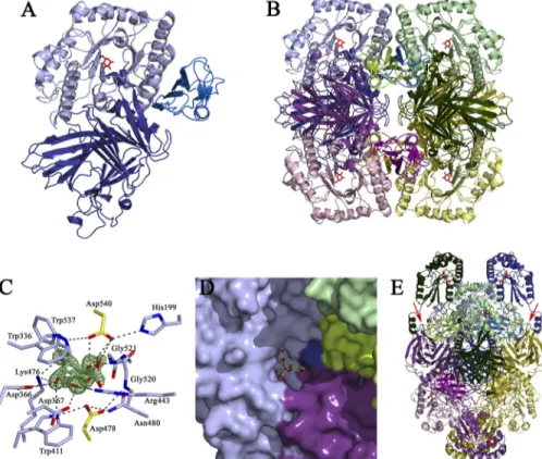
The Active Site Exemplifies Substrate Specificity—The struc-ture of AgaSK-tru in complex with the reaction product galac-tose allows us to identify unambiguously the catalytic center, which is located in a shallow pocket within the (/␣)8barrel
domain (Fig. 1C). The identity of the catalytic residues can be inferred from a superposition with the crystal structure of
T. maritima TmGalA, for which the catalytic nucleophile and general acid/base had been identified previously to be Asp-327 and Asp-387, respectively (19). In AgaSK-tru, Asp-478 is found at 3.0 Å from the anomeric center of the sugar and thus well positioned for nucleophilic attack. Conversely, the general acid/base Asp-540 is ideally poised to donate a hydrogen to the sugar O1 hydroxyl, found at a distance of 2.95 Å.
Asp-478 is held in place by hydrogen bonds with the side chains of Trp-411 and Asn-480 and with the main chain nitro-gen of Gly-520. The protonation state of Asp-540 is modulated by interactions with the side chains of His-199 and Trp-537. The galactose, found in the standard4C
1chair conformation, is
tightly enveloped by the enzyme and establishes an extensive network of hydrogen bonds with surrounding residues, most of them conserved in family GH36. The OH-2 hydroxyl interacts with the enzyme via the side chain of the acid/base Glu-540 and
FIGURE 1. Structure of the␣-galactosidase domain of AgaSK, tetrameric assembly, substrate-binding site, and model for full-length AgaSK. A, graphic presentation of the␣-galactosidase domain of AgaSK, with the N-terminal domain in deep blue, the catalytic (/␣)8barrel in light blue, and the C-terminal in marine blue. Galactose is shown as red sticks. B, tetrameric assembly of AgaSK-tru, with one monomer oriented and colored as in A and the other monomers shown in pink, green, and yellow hues, respectively. C, the␣-galactosidase active site. Residues interacting with the product are shown as sticks, with carbons color-coded as in B. Galactose is shown as sticks with green carbons, and the catalytic residues Asp-478 and Asp-540 are shown as
sticks with yellow carbons. Oxygen and nitrogen atoms are colored in red and blue, respectively. Fo⫺ Fcelectron density calculated prior to incorporation
of galactose into the model and contoured at 4 is shown in green. D, the association of three monomers within the tetrameric assembly creates a deep tunnel for substrate binding. The substrate-binding groove is shown in surface representation, with different monomers color-coded as in B. Galactose is shown as sticks with yellow carbons, and a model of raffinose, derived from a superposition of AgaSK-tru with S. cerevisiae␣-galactosidase 1, Mel1, complexed with raffinose (PDB 3lrm), is shown as sticks with green carbons. E, model for full-length AgaSK. The tetrameric assembly is color-coded as in
B. The modeled C-terminal domains are colored in the same hue as the N-terminal domains of each monomer. The polypeptide chain at the junction of
both domains has to bend backwards toward the (/␣)8barrel of an adjacent subunit to avoid steric clashes. This places each nucleotide-binding domain in proximity to an␣-galactosidase active site. Galactose and modeled ATP molecules are shown as red sticks. Two junctions between the galactose and the kinase domain are indicated with red arrows.
FIGURE 2. Characterization of sugar phosphorylation by AgaSK. A and B, phosphorylation of several sugars was tested in reaction mixtures containing AgaSK, ATP, MgCl2, [
␥-33P]ATP (0.5Ci), and different sugars. The radioactive spots were detected on the autoradiogram (A). The sugar spots were detected with 0.1% orcinol (B). Lanes 1, without AgaSK; lanes 2, with AgaSK;
lanes 3, with raffinose; lanes 4, with melibiose; lanes 5, with sucrose; lanes 6,
with glucose; lanes 7, with fructose; lanes 8, with galactose. C, phosphoryla-tion of raffinose and sucrose was tested in reacphosphoryla-tion mixtures containing AgaSK, ATP, and MgCl2. Lane 1, without AgaSK; lane 2, with AgaSK; lane 3, with raffinose; lane 4, with raffinose and 10MGal-DNJ; lane 5, with raffinose and 100MGal-DNJ; lane 6, with sucrose; lane 7, with sucrose and 10MGal-DNJ;
lane 8, with sucrose and 100MGal-DNJ. The sugar spots were detected with 0.1% orcinol.
at CNRS, on November 30, 2011
www.jbc.org
the main chain nitrogen of Gly-521. The OH-3 atom hydrogen bonds to Lys-476, which in turn stabilizes the axial OH-4, together with the side chains of Trp-411 and Asp-366. The OH-6 hydroxyl contacts the side chains of Asp-367 and Arg-443. Finally, the indole ring of Trp-336 provides a stacking plat-form for the sugar. Some family GH36 members exhibit ␣-N-acetylgalactosaminidase activity, but in AgaSK-tru, the tight interactions of the substrate with the enzyme do not allow for exocyclic substitutions on the galactose moiety.
The Tetrameric Assembly Provides a Platform for Efficient
Substrate Binding—Due to the tetrameric assembly of
AgaSK-tru, the shallow active site pocket extends toward a deep sub-strate-binding tunnel formed by loop regions of the central (/␣)8barrel and loop regions of the N- and C-terminal region
of different subunits (Fig. 1D). A superposition, based on the position of galactose, of the structure of the AgaSK-tru galac-tose complex, with the structure of a family GH27 active site
mutant of Saccharomyces cerevisiae␣-galactosidase 1, Mel1, in complex with raffinose (PDB 3lrm), allows us to define struc-tural elements possibly involved in aglycone binding. Phe-55, from the N-terminal region of a neighboring subunit, inserts tightly into a pocket situated below the active site pocket, thereby anchoring adjacent loop regions in close proximity to the putative⫹1 and ⫹2 binding sites (53). Residues likely to interact with the aglycone part of raffinose are His-199, Asp-376, and Arg-443, as well as Asp-52, Arg-66, and the backbone Gly-53-Gly-54 of a neighboring subunit. Most importantly, the shallowness of the substrate-binding tunnel does not allow accommodation of substrates with glycosidic linkages other than␣-1,6, confirming the strict specificity of AgaSK for ␣-1,6-linked galactose. Our observations let us conclude that the self-association of AgaSK into a tetramer is likely to be physiologi-cally relevant because different subunits join near the active site to provide a platform for efficient substrate binding.
FIGURE 3. Positive ESI-Q-TOF-MS mass spectra and chemical structure of phosphorylated sugar. All reaction products are deduced from the MS/MS data at m/z 365.107 (sucrose monosodiated form), 445.072 (phosphorylated sucrose monosodiated form), 467.060 (phosphorylated sucrose disodiated form), and 203.018 (galactose monosodiated form). The structural features of phosphorylated sucrose are deduced from an ion m/z 445.059 MS/MS spectrum. A, full scan MS of the sample from enzymatic reaction (solvent ions are indicated by black triangles). B, MS/MS of the sodiated ion at 445,1 with corresponding annotated fragments. In two areas of the MS/MS spectrum, the peak intensity was scaled up 24 and 36 times, respectively. C, chemical structure of the phosphorylated oligosaccharide annotated with corresponding fragments.
AgaSK, a Bifunctional Galactosidase/Sucrose Kinase
at CNRS, on November 30, 2011
www.jbc.org
AgaSK, an ␣-Galactosidase with a Kinase Activity—The
presence of the Walker A motif in the C-terminal domain of AgaSK (supplemental Fig. 3) suggested that it was also able to bind nucleotides. We took advantage of the presence of trypto-phans in the sequence of AgaSK to study nucleotide binding by intrinsic fluorescence measurements, and we showed that AgaSK binds ATP (Kdapp⬃61 ⫾ 13M) (supplemental Fig. 5). We then tested its potential ability to phosphorylate several sugars, especially sugars that are substrates of the ␣-galactosid-ase domain, in the presence of ATP and [␥-33P]ATP (Fig. 2A).
Raffinose and melibiose were tested (lanes 3 and 4), and a new radioactive spot was observed only when raffinose was used as substrate. Because raffinose is degraded by AgaSK into sucrose and galactose, we also tested sucrose, galactose, and the two monosaccharides composing the sucrose unit, glucose and fructose (lanes 5– 8). We observed the same radioactive spot in
lane 5 only, showing that sucrose was phosphorylated by
AgaSK. Furthermore, to test whether AgaSK was able to phos-phorylate both sugars, raffinose and sucrose, we added the ␣-galactosidase inhibitorD-galacto-1-deoxynojirimycin to the phosphorylation reaction mix (Fig. 2B, lanes 4, 5, 7, and 8). In the presence of the inhibitor, phosphorylated sucrose was not pro-duced anymore when raffinose was used as substrate (lanes 4 and
5 compared with lane 3), but phosphorylated sucrose was still observed when the substrate was sucrose (lanes 6 – 8). We there-fore conclude that in the presence of ATP, AgaSK specifically phosphorylates the sucrose unit liberated by the hydrolysis of raf-finose. This specific sucrose phosphorylation by AgaSK was con-firmed by MALDI-MS (Fig. 3). The control sample spectrum high-lighted raffinose and adducts of ATP (supplemental Fig. 6A), whereas the spectrum of the reaction products formed by AgaSK showed two products and adducts of ADP (supplemental Fig. 6B).
The structural features of phosphorylated sucrose were deduced from an MS/MS spectrum, and a set of specific related fragments was obtained, which allowed to locate the phosphate on position C6 of glucose (Fig. 3). Altogether these results show that AgaSK is also a sucrose kinase able to phosphorylate specifically sucrose on the C6 position of glucose.
Model of Full-length AgaSK—As all attempts to obtain crys-tals of full-length AgaSK failed, we used the Phyre server (45) to build a homology model for the kinase domain based on the crystal structure of putative fructose transport system kinase (YP_612366.1) from Silicibacter sp. TM1040 (PDB 3c8u). The model of the kinase domain could not be joined to the crystal structure of AgaSK-tru in an arbitrary fashion as the tetrameric assembly positions the C-terminus of the galactosidase domain from two adjacent subunits face to face (Fig. 1E). Due to these steric constraints, the kinase model had to be placed in a way such that the polypeptide chain at the galactosidase-kinase junction bends backwards toward the (/␣)8barrel, thereby
locating the active sites of the kinase domains near the galacto-sidase active sites of adjacent subunits within the tetramer (Fig. 1E). The proximity of the two active sites, as proposed in this model, might assure efficient cross-talk between the two activ-ities of AgaSK, but the full-length structure of this bifunctional enzyme will be necessary to corroborate our hypothesis.
Raffinose and Sucrose Uptake and Metabolism—To date, two
different pathways for sucrose utilization have been character-ized in bacteria (13) (Fig. 4). The first involves a phosphoenol-pyruvate-dependent phosphotransferase system (PTS), where sucrose is phosphorylated into sucrose-6-phosphate and simul-taneously transported into the cell. Sucrose-6-phosphate is then cleaved by a hydrolase into glucose-6-phosphate and fruc-tose. In the second pathway, a PTS-independent transport sys-FIGURE 4. Sucrose and raffinose pathways in R. gnavus E1 (adapted from Reid and Abratt (13)). Two pathways for sucrose transport are well characterized, the PTS-dependent sucrose system (left) and the non-PTS transport system (middle). The raffinose pathway (right) could be another possibility to provide sucrose to the bacterium.
at CNRS, on November 30, 2011
www.jbc.org
tem allows incorporation of sucrose under unmodified form into the cell, where it is hydrolyzed by sucrose phosphorylase (54, 55). Glucose-1-phosphate is subsequently transformed by a phosphoglucomutase into glucose-6-phosphate. In both mech-anisms, fructose is afterward phosphorylated by a fructokinase. Certain bacteria possess ABC transporters for the direct uptake of raffinose, which is subsequently cleaved by an ␣-ga-lactosidase into galactose and sucrose, and the sucrose pro-duced thereby is taken in charge by sucrose phosphorylase (54, 55). Genes coding for a putative ABC transporter for raffinose have been found in R. gnavus E1. This observation suggests that raffinose is transported within the cytoplasm of R. gnavus E1 by an ABC transporter and subsequently hydrolyzed by the ␣-ga-lactosidase activity of AgaSK into galactose and sucrose. The sucrose produced thereby is instantly taken in charge by the kinase activity of AgaSK to release sucrose-6-phosphate, the substrate of sucrose-6 phosphorylase (Fig. 4). The sucrose, which escapes the kinase activity of AgaSK, is probably cleaved by sucrose phosphorylase, encoded by the gene adjacent to the AgaSK gene. These findings place AgaSK at the crossroad of the two glycolytic pathways for sucrose utilization described so far.
Concluding Remarks—In conclusion, sucrose and raffinose
are the most abundant soluble carbohydrates found in plants (9), and they are probably part of the human carbohydrate energy intake. Although sucrose from diet is the substrate of the human intestinal sucrase-isomaltase, raffinose is degraded into galactose and sucrose only by intestinal microbial enzymes. Therefore, for the intestinal microbiota, a major external source of sucrose is probably raffinose, pointing out the impor-tance of its metabolism for bacteria. In the strict anaerobic Gram-positive bacterium R. gnavus, a single enzyme, AgaSK, is able to produce sucrose-6-phosphate directly from raffinose. The two distinct enzymatic activities are related to two domains. The N-terminal domain is responsible for the␣-galactosidase activity of AgaSK, whereas the C-terminal domain catalyzes the phospho-rylation of the sucrose produced thereby on the C6 position of glucose. We can speculate about the interest for R. gnavus E1 to possess a bifunctional protein having two enzymatic activities within the same scaffold. We can hypothesize that it could exem-plify a very efficient metabolic strategy developed by this human microbiota to metabolize raffinose, a potentially major source of carbohydrate available in the gut. Our studies contribute to a deeper understanding of the mechanisms by which Firmicutes use nutrients that are non-digestible by the host and optimize their metabolism for survival in such an extreme competitive environ-ment as the intestinal tract.
Acknowledgments—We are indebted to S. Rabot (INRA Jouy-en-Josas, France) for providing axenic rats of the animal facility ANAXEM platform and to C. Bridonneau and P. Guillaume for skillful technical assistance. Genome sequencing was carried out by Genoscope, AP05/06 Project 27. We thank also Dr. Ray Owens from the Depart-ment for Structural Biology at the University of Oxford for the pOPIN E vector. We thank the European Synchrotron Radiation Facility and the Synchrotron Soleil for provision of beam time and assistance with data collection.
REFERENCES
1. Ley, R. E., Turnbaugh, P. J., Klein, S., and Gordon, J. I. (2006) Nature 444, 1022–1023
2. Turnbaugh, P. J., Ley, R. E., Mahowald, M. A., Magrini, V., Mardis, E. R., and Gordon, J. I. (2006) Nature 444, 1027–1031
3. Greiner, T., and Bäckhed, F. (2011) Trends Endocrinol. Metab. 22, 117–123
4. Turnbaugh, P. J., Hamady, M., Yatsunenko, T., Cantarel, B. L., Duncan, A., Ley, R. E., Sogin, M. L., Jones, W. J., Roe, B. A., Affourtit, J. P., Egholm, M., Henrissat, B., Heath, A. C., Knight, R., and Gordon, J. I. (2009) Nature 457, 480 – 484
5. Eckburg, P. B., Bik, E. M., Bernstein, C. N., Purdom, E., Dethlefsen, L., Sargent, M., Gill, S. R., Nelson, K. E., and Relman, D. A. (2005) Science 308, 1635–1638
6. Armougom, F., Henry, M., Vialettes, B., Raccah, D., and Raoult, D. (2009)
PLoS One 4,e7125
7. Venema, K. (2010) Curr. Opin. Clin. Nutr. Metab. Care 13, 432– 438 8. Neyrinck, A. M., and Delzenne, N. M. (2010) Curr. Opin. Clin. Nutr.
Metab. Care 13,722–728
9. Trugo, L. C., Farah, A., and Cabral, L. (1995) Food Chem. 52, 385–387 10. Barrangou, R., Azcarate-Peril, M. A., Duong, T., Conners, S. B., Kelly,
R. M., and Klaenhammer, T. R. (2006) Proc. Natl. Acad. Sci. U.S.A. 103, 3816 –3821
11. Hugouvieux-Cotte-Pattat, N., and Charaoui-Boukerzaza, S. (2009) J.
Bac-teriol. 191,6960 – 6967
12. Caputto, R., Leloir, L. R., et al. (1949) J. Biol. Chem. 179, 497– 498 13. Reid, S. J., and Abratt, V. R. (2005) Appl. Microbiol. Biotechnol. 67,
312–321
14. Cantarel, B. L., Coutinho, P. M., Rancurel, C., Bernard, T., Lombard, V., and Henrissat, B. (2009) Nucleic Acids Res. 37, D233–D238
15. Rigden, D. J. (2002) FEBS Lett. 523, 17–22
16. Henrissat, B., and Bairoch, A. (1996) Biochem. J. 316, 695– 696 17. Garman, S. C., Hannick, L., Zhu, A., and Garboczi, D. N. (2002) Structure
10,425– 434
18. Garman, S. C., and Garboczi, D. N. (2004) J. Mol. Biol. 337, 319 –335 19. Comfort, D. A., Bobrov, K. S., Ivanen, D. R., Shabalin, K. A., Harris, J. M.,
Kulminskaya, A. A., Brumer, H., and Kelly, R. M. (2007) Biochemistry 46, 3319 –3330
20. Fredslund, F., Abou Hachem, M., Jonsgaard Larsen, R., Gerd Sørensen, P., Coutinho, P. M., Lo Leggio, L., and Svensson, B. (2011) J. Mol. Biol. 412, 466 – 480
21. Hashimoto, H., Katayama, C., Goto, M., Okinaga, T., and Kitahata, S. (1995) Biosci. Biotechnol. Biochem. 59, 619 – 623
22. Van Laere, K. M., Hartemink, R., Beldman, G., Pitson, S., Dijkema, C., Schols, H. A., and Voragen, A. G. (1999) Appl. Microbiol. Biotechnol. 52, 681– 688
23. Spangenberg, P., André, C., Dion, M., Rabiller, C., and Mattes, R. (2000)
Carbohydr. Res. 329,65–73
24. Tzortzis, G., Jay, A. J., Baillon, M. L., Gibson, G. R., and Rastall, R. A. (2003)
Appl. Microbiol. Biotechnol. 63,286 –292
25. Zhao, H., Lu, L., Xiao, M., Wang, Q., Lu, Y., Liu, C., Wang, P., Kumagai, H., and Yamamoto, K. (2008) FEMS Microbiol. Lett. 285, 278 –283 26. Goulas, T., Goulas, A., Tzortzis, G., and Gibson, G. R. (2009) Appl.
Micro-biol. Biotechnol. 82,471– 477
27. Nakai, H., Baumann, M. J., Petersen, B. O., Westphal, Y., Hachem, M. A., Dilokpimol, A., Duus, J. Ø., Schols, H. A., and Svensson, B. (2010) FEBS J. 277,3538 –3551
28. Blaut, M. (2002) Eur. J. Nutr. 41, Suppl. 1, I11–I16
29. Tzortzis, G., Goulas, A. K., Baillon, M. L., Gibson, G. R., and Rastall, R. A. (2004) Appl. Microbiol. Biotechnol. 64, 106 –111
30. Rastall, R. A., Gibson, G. R., Gill, H. S., Guarner, F., Klaenhammer, T. R., Pot, B., Reid, G., Rowland, I. R., and Sanders, M. E. (2005) FEMS Microbiol.
Ecol. 52,145–152
31. Qin, J., Li, R., Raes, J., Arumugam, M., Burgdorf, K. S., Manichanh, C., Nielsen, T., Pons, N., Levenez, F., Yamada, T., Mende, D. R., Li, J., Xu, J., Li, S., Li, D., Cao, J., Wang, B., Liang, H., Zheng, H., Xie, Y., Tap, J., Lepage, P., Bertalan, M., Batto, J. M., Hansen, T., Le Paslier, D., Linneberg, A., Nielsen,
AgaSK, a Bifunctional Galactosidase/Sucrose Kinase
at CNRS, on November 30, 2011
www.jbc.org
H. B., Pelletier, E., Renault, P., Sicheritz-Ponten, T., Turner, K., Zhu, H., Yu, C., Li, S., Jian, M., Zhou, Y., Li, Y., Zhang, X., Li, S., Qin, N., Yang, H., Wang, J., Brunak, S., Doré, J., Guarner, F., Kristiansen, K., Pedersen, O., Parkhill, J., Weissenbach, J., Bork, P., Ehrlich, S. D., and Wang, J. (2010)
Nature 464,59 – 65
32. Doré, J., Sghir, A., Hannequart-Gramet, G., Corthier, G., and Pochart, P. (1998) Syst. Appl. Microbiol. 21, 65–71
33. Cervera Tison, M., André-Leroux, G., Lafond, M., Georis, J., Juge, N., and Berrin, J. G. (2009) Biochim. Biophys. Acta 1794, 438 – 445
34. Matsui, I., Ishikawa, K., Matsui, E., Miyairi, S., Fukui, S., and Honda, K. (1991) J. Biochem. 109, 566 –569
35. Leslie, A. G. (1992) Joint CCP4/ESF-EACBM Newsletter, Vol. 26, pp. 27–33, Collaborative Computational Project No. 4, Daresbury Labora-tory, Daresbury, Warrington, UK
36. Kabsch, W. (2010) Acta Crystallogr. D Biol. Crystallogr. 66, 125–132 37. Evans, P. R. (2006) Acta Crystallogr. D Biol. Crystallogr. 62, 72– 82 38. McCoy, A. J. (2007) Acta Crystallogr. D Biol. Crystallogr. 63, 32– 41 39. Perrakis, A., Morris, R., and Lamzin, V. S. (1999) Nat. Struct. Biol. 6,
458 – 463
40. Emsley, P., Lohkamp, B., Scott, W. G., and Cowtan, K. (2010) Acta
Crys-tallogr. D Biol. CrysCrys-tallogr. 66,486 –501
41. Murshudov, G. N., Vagin, A. A., and Dodson, E. J. (1997) Acta Crystallogr.
D Biol. Crystallogr. 53,4449
42. Chen, V. B., Arendall, W. B., 3rd, Headd, J. J., Keedy, D. A., Immormino, R. M., Kapral, G. J., Murray, L. W., Richardson, J. S., and Richardson, D. C. (2010) Acta Crystallogr. D Biol. Crystallogr. 66, 12–21
43. Krissinel, E., and Henrick, K. (2007) J. Mol. Biol. 372, 774 –797
44. DeLano, W. L. (2010) The PyMOL Molecular Graphics System, version 1.3r1, Schrödinger, LLC, New York
45. Kelley, L. A., and Sternberg, M. J. (2009) Nat. Protoc. 4, 363–371 46. Forouhar, F., Abashidze, M., Xu, H., Grochowski, L. L., Seetharaman, J.,
Hussain, M., Kuzin, A., Chen, Y., Zhou, W., Xiao, R., Acton, T. B., Mon-telione, G. T., Galinier, A., White, R. H., and Tong, L. (2008) J. Biol. Chem. 283,11832–11840
47. Domon, B., and Costello, C. (1988) Glycoconj. J. 5, 397– 409
48. Aslanidis, C., Schmid, K., and Schmitt, R. (1989) J. Bacteriol. 171, 6753– 6763
49. Davidson, A. L., Dassa, E., Orelle, C., and Chen, J. (2008) Microbiol. Mol.
Biol. Rev. 72,317–364
50. Altschul, S. F., Madden, T. L., Schäffer, A. A., Zhang, J., Zhang, Z., Miller, W., and Lipman, D. J. (1997) Nucleic Acids Res. 25, 3389 –3402 51. Walker, J. E., Saraste, M., Runswick, M. J., and Gay, N. J. (1982) EMBO J. 1,
945–951
52. Ademark, P., Larsson, M., Tjerneld, F., and Stålbrand, H. (2001) Enzyme
Microb. Technol. 29,441– 448
53. Davies, G. J., Wilson, K. S., and Henrissat, B. (1997) Biochem. J. 321, 557–559
54. Silverstein, R., Voet, J., Reed, D., and Abeles, R. H. (1967) J. Biol. Chem. 242,1338 –1346
55. Kitaoka, M., and Hayashi, K. (2002) Trends Glycosci. Glycotechnol. 14, 35–50
at CNRS, on November 30, 2011
www.jbc.org