HAL Id: hal-03048110
https://hal.archives-ouvertes.fr/hal-03048110
Submitted on 9 Dec 2020
HAL is a multi-disciplinary open access
archive for the deposit and dissemination of
sci-entific research documents, whether they are
pub-lished or not. The documents may come from
teaching and research institutions in France or
abroad, or from public or private research centers.
L’archive ouverte pluridisciplinaire HAL, est
destinée au dépôt et à la diffusion de documents
scientifiques de niveau recherche, publiés ou non,
émanant des établissements d’enseignement et de
recherche français ou étrangers, des laboratoires
publics ou privés.
The Puzzling Conservation and Diversification of Lipid
Droplets from Bacteria to Eukaryotes
Josselin Lupette, Eric Marechal
To cite this version:
Josselin Lupette, Eric Marechal. The Puzzling Conservation and Diversification of Lipid Droplets
from Bacteria to Eukaryotes. Kloc M. Symbiosis: Cellular, Molecular, Medical and Evolutionary
Aspects. Results and Problems in Cell Differentiation, 69, Springer, pp.281-334, 2020,
978-3-030-51848-6. �10.1007/978-3-030-51849-3_11�. �hal-03048110�
2
The Puzzling Conservation
3
and Diversi
fication of Lipid Droplets from
4
Bacteria to Eukaryotes
5
Josselin Lupette and Eric Maréchal
6
Abstract Membrane compartments are amongst the most fascinating markers of
7
cell evolution from prokaryotes to eukaryotes, some being conserved and the others
8
having emerged via a series of primary and secondary endosymbiosis events.
9
Membrane compartments comprise the system limiting cells (one or two membranes
10
in bacteria, a unique plasma membrane in eukaryotes) and a variety of internal
11
vesicular, subspherical, tubular, or reticulated organelles. In eukaryotes, the internal
12
membranes comprise on the one hand the general endomembrane system, a dynamic
13
network including organelles like the endoplasmic reticulum, the Golgi apparatus,
14
the nuclear envelope, etc. and also the plasma membrane, which are linked via direct
15
lateral connectivity (e.g. between the endoplasmic reticulum and the nuclear outer
16
envelope membrane) or indirectly via vesicular trafficking. On the other hand,
semi-17
autonomous organelles, i.e. mitochondria and chloroplasts, are disconnected from
18
the endomembrane system and request vertical transmission following cell division.
19
Membranes are organized as lipid bilayers in which proteins are embedded. The
20
budding of some of these membranes, leading to the formation of the so-called lipid
21
droplets (LDs) loaded with hydrophobic molecules, most notably triacylglycerol, is
22
conserved in all clades. The evolution of eukaryotes is marked by the acquisition of
23
mitochondria and simple plastids from Gram-positive bacteria by primary
endosym-24
biosis events and the emergence of extremely complex plastids, collectively called
25
secondary plastids, bounded by three to four membranes, following multiple and
26
independent secondary endosymbiosis events. There is currently no consensus view
27
of the evolution of LDs in the Tree of Life. Some features are conserved; others show
Josselin Lupette and Eric Maréchal contributed equally with all other contributors.
J. Lupette (*)
MSU-DOE Plant Research Laboratory, Michigan State University, East Lansing, MI, USA AU1
e-mail:lupettej@msu.edu
E. Maréchal (*)
Laboratoire de Physiologie Cellulaire et Végétale, CNRS, CEA, INRA, Université Grenoble Alpes, Institut de Recherche Interdisciplinaire de Grenoble, Grenoble, France
e-mail:eric.marechal@cea.fr
© Springer Nature Switzerland AG 2020
M. Kloc (ed.), Symbiosis: Cellular, Molecular, Medical and Evolutionary Aspects, Results and Problems in Cell Differentiation 69,
28 a striking level of diversification. Here, we summarize the current knowledge on the
29 architecture, dynamics, and multitude of functions of the lipid droplets in
prokary-30 otes and in eukaryotes deriving from primary and secondary endosymbiosis events.
31 Keywords Lipid droplets · Evolution · Architecture · Biogenesis · Catabolism
32
11.1
Introduction
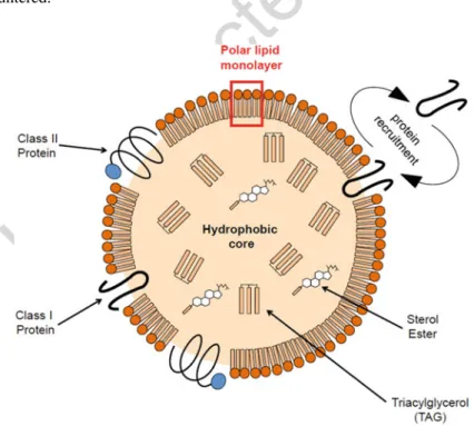
AU233 Lipid droplets (LDs) are conserved structures in prokaryotic and eukaryotic cells
34 (Walther et al. 2017; Zhang and Liu2017). Their architecture consists of a core,
35 loaded with hydrophobic carbon-rich molecules (polyhydroxyalkanoates or PHAs,
36 triacylglycerol or TAG, steryl esters, isoprenoids such as squalene, etc.) bounded by
37 a monolayer of polar glycerolipids, generally phospholipids that can be associated
38 with sterols. Proteins are transiently or permanently associated with its surface
39 (Walther et al.2017) (Fig. 11.1). Two main classes have been identified amongst
40 LD closely associated proteins, based on their structure (Kory et al.2016). Class I
41 proteins have a hydrophobic“hairpin” pattern (Bersuker and Olzmann2017) and
42 class II proteins have at least one amphipathic helix (Bersuker and Olzmann2017)
43 (Fig.11.1). Nevertheless, proteins that do not belong to these two classes are also
44 encountered.
45
LDs derive from the budding of a cell membrane. Membrane compartments are
46
amongst the most fascinating markers of cell evolution from prokaryotes to
eukary-47
otes, some being conserved and the others having emerged via a series of primary
48
and secondary endosymbiosis events. Membrane compartments comprise the system
49
limiting cells (one or two membranes in bacteria, a unique plasma membrane in
50
eukaryotes) and a variety of internal vesicular, subspherical, tubular, or reticulated
51
organelles. In eukaryotes, the internal membranes comprise on the one hand
52
the general endomembrane system, a dynamic network including organelles like
53
the endoplasmic reticulum (ER), the Golgi apparatus, nuclear envelope, etc. and also
54
the plasma membrane, which are linked via direct lateral connectivity (e.g. between
55
the endoplasmic reticulum and the nuclear outer envelope membrane) or indirectly
56
via vesicular trafficking. On the other hand, semi-autonomous organelles,
57
i.e. mitochondria and chloroplasts, are disconnected from the endomembrane system
58
and request vertical transmission following cell division. Membranes are organized
59
as lipid bilayers in which proteins are embedded. The evolution of eukaryotes is
60
marked by the acquisition of mitochondria and simple plastids from Gram-negative
61
bacteria by primary endosymbiosis events (Maréchal2018), and the emergence of
62
extremely complex plastids, collectively called secondary plastids, bounded by three
63
to four membranes, following multiple and independent secondary endosymbiosis
64
events (Fussy2018). There is currently no consensus view on the evolution of cell AU3
65
membranes and that of LDs.
66
Due to their hydrophobic core loaded with carbon-rich molecules, LDs have long
67
been considered as simple carbon and energy storage organelles. Based on the
68
analyses of LD proteomes in various cell models, it is now considered that LDs
69
have other functions that depend on their protein composition (Walther et al.2017;
70
Den Brok et al.2018; Henne et al.2018).
71
Research on LDs has increased strikingly in recent decades motivated by the
72
multitude of applications ranging from nutrition, health to green chemistry and
73
bioenergy. In 2020, the keyword“lipid droplet” returns as many as 12,500 hits in
74
the Pubmed bibliographic database. Concerning human obesity-related diseases
75
(Faucher and Poitou 2016; Madrigal-Matute and Cuervo 2016), protein actors
76
(CGI-58, SEIPIN, ATGL, LAL) at the surface of LDs have been extensively studied
77
in mammalian models (summarized in Table11.1). CGI-58-ABHD5 (Comparative
78
Gene Identification 58—α/β hydrolase domain-containing 5; 349 amino acids—
79
45 kDa) is particularly studied because its mutation is responsible for the
80
Chanarin-Dorfman syndrome, an autosomal recessive disease (Missaglia et al.
81
2014). In mammals, the CGI-58 protein is located on cytosolic LDs interacting
82
with PLIN1 (Subramanian et al.2004). A second important player in the
mecha-83
nisms of LD biogenesis in mammals is the SEIPIN protein. A mutation in the human
84
Seipin gene leads to severe forms of generalized Berardinelli-Seip congenital
85
lipodystrophy (Magré et al. 2001). The deletion of ATGL (Adipose triglyceride
86
lipase) in mice reduces the mechanism of lipolysis and promotes the accumulation of
87
lipids in oxidative tissues of the body, leading to the death of mice in 3 months
88
(Zimmermann et al.2004). The last example is the Wolman disease, which is an
89
90 LIPA gene (Wolman et al. 1961). The LIPA mutation leads to the synthesis
91 deficiency of lysosomal acid lipase (LAL) (Onal et al.2017). This disease causes
92 accumulation of cholesterol esters and TAG in leukocyte lysosomes,fibroblasts, and
93 hepatocytes generally leading to the death of the child by liver failure (Pericleous
94 et al.2017). It is a very rare disease with only 14 cases detected so far, half of which
95 are from the consanguineous union (Ikari et al.2018).
96 TAG-rich LDs produced by oleaginous organisms, mainly plants and algae, but
97 sometimes also fungi or animals, are also key to numerous biotechnological
appli-98 cations. Molecules of TAG are composed of a glycerol-3-phosphate backbone on
99 which three fatty acids are esterified (Lupette and Maréchal2018). Fatty acids (FAs)
100 are carboxylic acids. Their carbon chain length and number of unsaturations
101 (or double bonds, C¼C) allow assessing whether they can be used for different
102 applications. Oleaginous crops are an essential resource for human nutrition.
103 Microalgae, whose interest in the scientific community is currently exponential,
t1:1 Table 11.1 Human diseases related to LD formation AU4
Diseases Anatomical pathology Pathophysiology References
t1:2
Atherosclerosis Accumulation of
atheroma-tous plaques (cholesterol) in the arteries
ACAT1; ABCA1; ADRP
Paul et al. (2008) t1:3
Obesity Accumulation of fat
reserves
Multifactorial (genetic, envi-ronmental, psychological)
Faucher and Poitou (2016) AU5
t1:4
Fatty liver Accumulation of TAGs in
the cytoplasm of hepatocytes Alcohol, hepati-tis B and C Madrigal-Matute and Cuervo (2016) AU6 t1:5 Chanarin-Dorfman syndrome Accumulation of lipid droplets in lymphocytes and many tissues Mutation of CGI-58/ABHD5 Dorfman et al. (1974), Chanarin et al. (1975), Lefèvre et al. (2001), Samuelov et al. (2011), Missaglia et al. (2014),
Jordans (1953), Gupta and Kaur (2005), Waheed et al. (2016) t1:6 Myopathy Mutation of PNPLA2 Zimmermann et al. (2004) t1:7 Congenital generalized Lipodystrophy (CGL)
Dystrophy of adipose tissue Mutation of
AGPAT2, BSCL2, CAV1 or PTRF
Magré et al. (2001),
Agarwal et al. (2002), Kim et al. (2008), Hayashi et al. (2009), Rajab et al. (2010), Quinn and Purcell (2017) t1:8
Lysosomal acid lipase
deficiency
Lysosomal acid lipase de
fi-ciency causing an accumu-lation of TAGs and cholesterol esters in leuko-cytes, hepatoleuko-cytes, and fibroblasts
Mutation of LIPA
Wolman et al. (1961), Onal
et al. (2017), Pericleous
et al. (2017), Ikari et al.
(2018)
104
can also produce TAGs. Microalgae enriched in FAs with short or medium carbon
105
chains without unsaturation are an interesting feedstock for green chemistry or the
106
development of biofuels (Lupette and Maréchal2018). Microalgae containing high
107
levels of very long-chain fatty acids (carbon number greater than 20) with multiple
108
unsaturations (1–6), called VLC-PUFAs, with unsaturation at the ω-3 position
109
(i.e. eicosapentaenoic acid or EPA, 20:5, and docosahexaenoic acid or DHA,
110
22:6), are promising for human health applications (Lupette and Maréchal2018).
111
There is currently no consensus view of the evolution of LDs in the Tree of Life.
112
Some features are conserved; others show a striking level of diversification. Here, we
113
summarize the current knowledge on the architecture, dynamics, and multitude of
114
functions of the lipid droplets in prokaryotes and eukaryotes deriving from primary
115
and secondary endosymbiosis events.
116
11.2
Studying Lipid Droplets
117
11.2.1
Imaging Lipid Droplets
118
Microscopic observation by confocal or epifluorescence imaging is the main method
119
of detection of LDs in a cell or an organelle. The most commonly usedfluorophores
120
are Nile Red (Greenspan et al.1985) and BODIPY 505/515 (Rumin et al.2015) or
121
BODIPY 493/503 (Gocze and Freeman 1994). More recently, new fluorophores
122
have been developed (Yang et al.2012; Gidda et al.2016). These molecules are
123
compatible with the parallel measurement of the fluorescence of GFP (Green
124
Fluorescent Protein), RFP (Red Fluorescent Protein), and of chlorophyll (Kuntam
125
et al.2015). Other compounds including AC-202 were recently used in two model
126
species of microalgae Chlamydomonas reinhardtii and Phaeodactylum tricornutum
127
(Harchouni et al.2018). Thesefluorophores make it possible to determine the size
128
and number of LDs in a semi-quantitative manner, as well as their cellular
129
localization.
130
11.2.2
Purifying Lipid Droplets
131
The general strategy for studying LD architecture is similar regardless of the
132
organism studied. A culture of cells in a medium promoting the development of
133
LDs is used (e.g. a nutrient deficiency). The LD purification starts by a gentle cell
134
disruption step (French press, cell disruptor, etc.) in a suitable buffer releasing
135
droplets as well as other cellular components. It is then necessary to perform a
136
density gradient purification (Brasaemle and Wolins2016). Due to their low density,
137
LDs rise to the surface of the gradient during ultracentrifugation (Brasaemle and
138
Wolins2016). LDs are harvested and washed to limit the presence of contaminants.
139
140 components of the hydrophobic core and on the composition of the monolayer of
141 polar lipids and proteins on the surface of the droplet (Walther et al. 2017).
142 Proteomic analyses allow the identification of proteins but also the characterization
143 of some post-translational modifications (phosphorylation, nitrosylation,
144 ubiquitinylation, sumoylation, N and O-glycosylation, farnesylation). The proteome
145 must then be validated by biochemical studies (western blot), the imaging of fusion
146 proteins (with afluorescent marker to verify the location on the surface of LDs), and
147 functional genetic studies (the study of mutants with altered expression such as
148 knockout, silencing, and overexpression of genes coding for droplet proteins allows
149 their functional characterization).
150
11.2.3
Biophysical Properties of Lipid Droplets
151 Biophysical studies of LDs have proven to be critical to advance our understanding
152 of LD biogenesis. After the removal of proteins and polar lipids, it is possible to
153 consider LDs as the product of an emulsion of oil in water (Thiam et al.2013b). The
154 cytosol of the cell represents the aqueous phase and LDs, the dispersed oily phase.
155 The interface between oil and water generates a surface tension due to the lack of
156 cohesive integrations between the two phases. These emulsions are metastable in the
157 absence of external disturbance. The presence of surfactants makes it possible to
158 reduce the surface tension, thus increasing the (meta)stability of the emulsion and the
159 cohesion energy cost (Georgieva et al.2009). The polar glycerolipids at the
periph-160 ery of the droplet then act as surfactants. Mastering this system in vitro is probably
161 one of the most important challenges to understand how such anisotropic
hydropho-162 bic cores can be maintained in a cell, where all other components are highly
self-163 assembled and organized (membranes can be considered as two-dimensionalfluid
164 crystals, DNA and polymers have three-dimensional architectures, polypeptides
165 form protein structures with rigorous three-dimensional folds). It is also possible
166 to use biophysical methods such as Pulsed Field Gradient-Nuclear Magnetic
Reso-167 nance (PFG-NMR) to determine the mobility of TAGs inside the volume set by LDs
168 with or without LD-to-LD connections (Gromova et al. 2015). Since the overall
169 structure of LDs appears conserved in the Tree of Life, whereas components may
170 differ, a key to LD conservation may rely on biophysical properties, which now need
171 to be evaluated in different systems.
172
11.3
Lipid Droplets in the Tree of Life
173
11.3.1
Lipid Droplets in Prokaryotic Cells
174 The vast majority of bacterial species have the capacity to accumulate lipid
176
made according to accumulated hydrophobic molecules, with either“lipid droplets”
177
(containing fluid acyl esters, triacylglycerols, or TAGs) or “granules” (containing
178
semi-solid lipopolymers called polyhydroxyalkanoates or PHAs). Here, we describe
179
the formation of LD structures in bacteria, as a possible basis for their evolution in
180
eukaryotes following primary endosymbiosis events. Since primary endosymbiosis
181
events are believed to be facilitated by the presence of pathogenic partners (Maréchal
182
2018), we also describe how some pathogenic bacteria (and viruses) are known to
183
interact with host cell LDs.
184
11.3.1.1 Polyhydroxyalkanoate Granules
185
Polyhydroxyalkanoates (PHAs) are polyesters produced by fermentation of lipids or
186
carbohydrates. They are linear polyesters consisting of hydroxy acid monomers
187
(HA) linked together by an ester bond (Możejko-Ciesielska and Kiewisz 2016).
188
PHAs include poly(3-hydroxybutyrate) (PHB) and polyhydroxyvalerate (PHV)
189
(Murphy2012). PHAs are also classified into two groups according to the number
190
of carbons per monomer: short-chain (3–5 carbons) PHAs and medium-chain PHAs
191
(6–14 carbons). PHAs are synthesized when the C/N ratio is altered (a nitrogen
192
deficiency coupled with an excess of carbon), stopping growth and division,
193
resulting in an entry into the quiescent phase. Special attention has been given to
194
these polyesters for several decades because they are biodegradable (Pötter and
195
Steinbüchel2006). PHAs are used in a wide range of applications such as resorbable
196
materials for medical purposes (implants, biodegradable sutures, stents, etc.),
mate-197
rials (paper coating, shape memory gel, etc.), fuel additives, and as metabolic
198
regulators (Możejko-Ciesielska and Kiewisz2016).
199
Ralstonia eutropha H16 (new name: Cupriavidus necator) is the study model for
200
PHB granules (Reinecke and Steinbüchel2009). It is a Gram-negative bacterium
201
that can accumulate 10–20 granules per cell, measuring 500 nm in diameter and
202
representing 90% of the dry weight (Anderson and Dawes1990). A recent study has
203
shown that R.eutropha H16 PHB granules do not have a monolayer of polar lipids
204
but only superficial proteins essential for their synthesis and degradation (Bresan
205
et al.2016). The main proteins detected on their surface are PHB synthase (PhaC),
206
phasin (PhaP), PhaR (PhaR), and PHB depolymerase (PhaZ). The R. eutropha H16
207
PhaC gene has been cloned by three independent laboratories. The localization of the
208
PHB synthase on the surface of the granules was confirmed by immunocytochemical
209
staining with colloidal gold (Gerngross et al.1993). The study of PHA granules is
210
still too scarce to assess whether they are linked to TAG containing LDs and whether
211
they may also be present in some eukaryotic clades.
212
11.3.1.2 TAG and Wax Ester Droplets
213
Bacteria are also able to accumulate TAG in LDs. Bacterial species producing LDs
214
215 Rhodococcus, Micromonospora, Dietzia, and Gordonia as well as several
Strepto-216 mycetes (Alvarez and Steinbüchel2002; Wältermann and Steinbüchel2005;
Mur-217 phy 2012). γ-Proteobacteria (Marinobacter, Alcanivorax, etc.) are
hydrocarbon-218 based bacteria capable of accumulating LDs (TAGs and wax esters) when entering
219 dormancy (Kalscheuer et al.2007). Theseγ-proteobacteria are often found in the
220 oceans associated with microalgae in a system called the phycosphere (Lupette et al.
221 2016). These bacteria are also able to use petroleum hydrocarbons as a source of
222 carbon raising possible applications in the degradation of hydrocarbons during oil
223 spills (Murphy2012).
224 Rhodococcus are oleaginous bacteria containing large amounts of TAGs (Alvarez
225 2016). Proteomic studies of LDs of several Rhodococcus species have been
226 performed: Rhodococcus opacus and Rhodococcus ruber (Kalscheuer et al.2001),
227 Rhodococcus jostii RHA1 (Ding et al. 2012b), and Rhodococcus opacus PD630
228 (Kalscheuer et al. 2001; Chen et al. 2014). 228 proteins have been identified in
229 R. jostii RHA1 including two putative structural proteins representing 15% of LD
230 proteins: Microorganism Lipid Droplet Small (MLDS) and Phage shock protein A
231 (Psp A). Ribosomes and translational regulators have also been isolated in the
232 proteome of the LD of R. jostii RHA1 (Ding et al.2012b). By a functional genetic
233 study, deletion of the MLDS causes the formation of larger LDs (Ding et al.2012b).
234 A recent study showed that LDs of R. jostii RHA1 are bound to genomic DNA via
235 MLDS protein, which increases the survival rate of bacterial cells during nutritional
236 deficiency or genotoxic stress (Zhang et al.2017).
237 11.3.1.3 Pathogenic Bacteria
238 Pathogenic bacteria can divert lipids and even‘hijack’ LDs from an infected host.
239 Well-known examples include bacteria of the genera Mycobacterium and
Chla-240 mydia. Mycobacteria are bacilli with pathogenic potency: Mycobacterium
tubercu-241 losis (Menon et al. 2019), M. bovis, and M. avium are causative agents of
242 tuberculosis, M. leprae is the agent of leprosy, and M. ulcerans is responsible for
243 Buruli ulcer. Mycobacteria are able to disrupt human lipid homeostasis during
244 infection following the formation of foamy macrophages containing LDs in the
245 cytoplasm (Kim et al.2010; Caire-Brändli et al.2014). LDs produced in infected
246 cells serve as a platform for the production of signaling molecules (prostaglandins
247 and leukotrienes eicosanoids) regulating the immune response and inflammation
248 (Melo and Weller2016). LDs also serve to concentrate and deliver iron via lipophilic
249 siderophores (mycobactins) secreted by the bacteria (Luo et al. 2005). The LD
250 proteome of Bacillus Calmette and Guerin (BCG: a non-replicating hypoxic
atten-251 uated strain of M. bovis) used in the tuberculosis vaccine allowed the identification
252 of five proteins: two triacylglycerol synthases, Tgs1 (BCG3153c) and Tgs2
253 (BCG3794c), as well as three other proteins, BCG1169c, BCG1489c, and
254 BCG1721 (Low et al.2010). BCG1169c is a specific protein of the Mycobacterium
256
the Kennedy pathway. A deletion of BCG1489C causes a decrease in the amount
257
of TAG.
258
Chlamydia trachomatis is an obligate intracellular bacterium responsible for
259
sexually transmitted infections as well as eye diseases. This bacterium has a biphasic
260
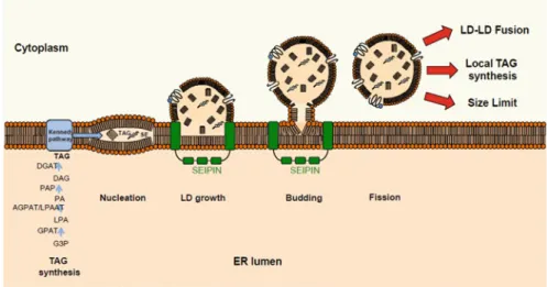
life cycle: the elementary body representing the infectious form and the
261
non-infectious reticulate body. C. trachomatis is able to translocate an LD from
262
the cytoplasm of the host to the parasitophorous vacuole lumen containing the
263
bacterium via an endocytosis process. Internalization occurs after LDs are coated
264
with a family of Lda proteins (Kumar et al.2006). The proteome of LDs of HeLa
265
human cervical adenocarcinoma epithelial cells infected with C. trachomatis
266
LGV-L2 434/Bu showed that they are enriched in PLIN2, PLIN3, ACSL-3, and
267
ACSL-4 proteins (Saka et al.2015).
268
11.3.2
Hijacking of Lipid Droplets by Viruses
269
LDs can serve as a source of energy for the dispersion of viruses such as hepatitis C
270
virus (HCV), dengue virus (DENV), poliovirus (PV), or rotavirus (RV). This section
271
presents several examples of‘hijacking’ of lipid metabolism by viruses.
272
HCV is the most widely used model for studying LD diversion (Roingeard and
273
Melo 2017). The virion initially circulates in a form of lipoviroparticles (rich in
274
cholesterol esters and apolipoproteins apoB and apoE) in the blood of infected
275
patients (Boyer et al.2014). Its entry into the cell is dependent on LDL receptors
276
(Low Density Lipoproteins). The replication of the viral RNA then induces
exten-277
sive alterations of the membrane and the formation of vesicular structures that
278
exhibit features similar to lipid rafts (Aizaki et al. 2004). The assembly of HCV
279
requires the localization of some of the HCV proteins in LDs of the host, and the
280
release of virions is strongly associated with the secretion pathway of very
281
low-density lipoproteins (VLDL). Proteins that are important to complete the viral
282
cycle, i.e. the nucleocapsid of HCV and the nonstructural protein NS5A co-localize
283
with the LD in HCV-infected cells and interact with LD proteins, such as DGAT1
284
(Camus et al.2013).
285
Other examples of viruses depending on host cell LDs to complete their cycle
286
include DENV in mosquitoes (Mayer et al. 2017), PV causing poliomyelitis
287
(Nchoutmboube et al. 2013), Flock House Virus (Castorena et al.2010), Brome
288
mosaic virus (BMV) (Zhang et al.2016), or RV causing gastroenteritis in infants and
289
young children (Gaunt et al.2013).
290
11.3.3
Lipid Droplets in Eukaryotic Cells
291
Eukaryotes including unicellular and multicellular organisms are characterized by
292
293 trans-Golgi network, peroxisomes, lysosomes, vacuoles, and numerous cytosolic
294 vesicles for the endomembrane system, mitochondria, and chloroplast for
295 semi-autonomous organelles). Primary endosymbiosis events are at the origin of
296 mitochondria and primary plastids. In a very simplistic scheme, mitochondria are
297 considered to derive from Gram-negative alpha-proteobacteria and primary plastids,
298 from Gram-negative cyanobacteria. Based on molecular evidence, other partners
299 have been involved, including pathogenic bacteria (Maréchal2018). In contrast to
300 prokaryotes, the synthesis of LDs from the ER seems to occur nearly in all
eukary-301 otes studied to date.
302 In this section, we first consider non-photosynthetic eukaryotes, containing
303 mitochondria as unique semi-autonomous organelles, then photosynthetic
eukary-304 otes containing primary plastids, andfinally those containing secondary plastids.
305 11.3.3.1 Opisthokonta: Non-photosynthetic Eukaryotes
306 Metazoa
307 Chordata
308 LDs of mammals are composed of a hydrophobic core consisting of TAGs and
309 cholesteryl esters, generally considered as a form of storage. The monolayer of polar
310 lipids is mainly composed of PC, PE, and PI (Bartz et al.2007). In mammals, LD
311 surface proteins are grouped into a family formerly called PAT (for Perilipin—
312 Adipophilin—Tail-interacting protein of 47 kDa) (Bickel et al.2009) but whose
313 nomenclature has evolved since 2010 under the name of Perilipin (PLIN) (Kimmel
314 et al.2010). PLIN1 has four splice variants PLIN1a, PLIN1b, PLIN1c, and PLIN1d
315 (Kimmel et al.2010). PLINs contain a hydrophobic PAT domain of 100 amino acids
316 defining an N-terminal region. The study of a mutant PLIN2-N1 (deficient for the
317 PAT domain) reveals that this domain is not involved in lipid binding, but in the
318 stabilization of lipid droplets, and lipid accumulation and degradation of PLIN2 by
319 the proteasome (Najt et al.2014). The PAT domain is followed by an 11-mer helical
320 pattern of variable size, which might interact with phospholipids (Bussell and Eliezer
321 2003). Some PLIN proteins can be post-translationally modified by
phosphoryla-322 tions, via PKA, during lipolysis (PLIN3 and PLIN4 do not have a phosphorylation
323 site (Kimmel and Sztalryd 2016)). Recently, the sixth clade of Perilipin, called
324 PLIN6, has been discovered, specific to teleosts (Granneman et al.2017).
Interest-325 ingly, PLIN6 is not expressed in tissues associated with lipid metabolism but in the
326 xanthophores of teleost skin. Biochemical analyses have shown that PLIN6 is
327 associated with the surface of droplets enriched in carotenoids and regulates the
328 pigment synthesis pathways (Granneman et al.2017).
329 The expression of PLINs (with the exception of PLIN3) is regulated by a family
330 of transcription factors called PPAR (Peroxisome Proliferator Activated Receptor).
331 These transcription factors are activated by the binding of lipid ligands (Poulsen
333
Since the early 2000s, at least 25 proteomic studies of the LD of mammalian cells
334
or tissues have been published (Table11.2). In all these studies, proteomics reveals
335
the presence of at least one of the classes of PLIN on the surface of the mammalian
336
LD. It can also be seen that the distribution of the PLINs varies according to
337
mammalian cells and tissues. PLIN1 and PLIN4 are present on the surface of LDs
338
of adipocytes (Ding et al.2012a) and adipose tissue (Yu et al. 2015). PLIN2 and
339
PLIN3 are ubiquitous in non-adipose tissues. PLIN2 is strongly expressed on the
340
surface of LDs of the liver and hepatocytes. PLIN5 is present in the oxidative tissues
341
i.e. the heart, brown adipose tissue, and skeletal muscles (Kimmel and Sztalryd
342 2014).
343
Functional studies of PLIN have been performed upstream and downstream of
344
proteomic studies. KO mice for PLIN1 have a phenotype of reduction of fat mass, an
345
increase of the lipolytic activity, but also a glucose intolerance and peripheral insulin
346
resistance (Tansey et al.2001). Stimulation of lipolytic activity by tumor necrosis
347
factor (TNF-α) showed, in a first study, a decrease in the expression of PLIN1
348
variants. However, the overexpression of PLIN1a and PLIN1b blocks the ability of
349
TNF-α to increase lipolysis in 3T3-L1 cells (Souza et al.1998). KO mice for PLIN2
350
present unchanged adipose differentiation, a 60% decrease in hepatic TAGs, but a
351
level of VLDL identical to control mice suggesting the retention of TAGs in the
352
microsomes (Chang et al.2006). Overexpression of PLIN2 fused to GFP causes an
353
increase in the number and size of LDs in hepatocytes (Imamura et al.2002). Afirst
354
antisense study of hepatic PLIN2 causes a decrease in hepatic steatosis,
355
hypertriglyceridemia, and insulin resistance in obese mice without altering the
356
level of expression of PLIN3 and PLIN4 (Imai et al. 2007). A second antisense
357
study of PLIN2 shows the same type of result with a decrease in DAG and TAG in
358
the liver as well as an improvement in insulin production (Varela et al. 2008).
359
Finally, deletion of the PLIN2 exon 5 (Plin2Δ5) in mice causes resistance to obesity
360
induced by a diet rich in fats indicating the role of PLIN2 in obesity and hepatic
361
steatosis (Mcmanaman et al.2013). The deletion of PLIN3 in mice induces cold
362
tolerance (Lee et al.2018), probably by regulating beige adipocyte formation and
363
thermogenic activities. The deletion of PLIN4 leads to decreased expression of
364
PLIN5 reducing lipid accumulation in the cardiac muscle (Chen et al.2013). KO
365
of PLIN6 is responsible for stopping the concentration of carotenoids in the droplets
366
(Granneman et al.2017).
367
Arthropoda
368
Research on LDs of Arthropoda was mainly carried out in the fruitfly Drosophila
369
melanogaster. LD research began with two RNAi screens (Beller et al.2008; Guo
370
et al. 2008) showing that approximately 370 genes, or 1.5% of the expressed
371
genome, were involved in LD physiology. Afirst proteomic study was conducted
372
on the abdominal fat body, the fat tissue of thefly, which allowed the identification
373
of 248 proteins (Beller et al. 2006). A second study was conducted on whole
374
embryos (Cermelli et al.2006). The proteomes of the LD of D. melanogaster have
375
t2 :1 Table 11.2 Analyses of LDs in representative prokaryotic and eukaryotic study models AU7 Phylogeny Species LD Proteome/ LD protein characterized LD lipidome Other informations References t2 :2 Prokaryota Proteobacteria β-Proteobacteria Cupriavidus necador PhaC/PhaP/ PhaR/PhaZ PHB PHB granules Bresan et al. ( 2016 ), Peoples et al. (1989), Schubert et al. (1988), Slater et al. (1988), Gerngross et al. ( 1993 ), Dennis et al. (1998), Tariq et al. (2015), Han et al. (2007) t2 :3 γ-Proteobacteria Marinobacter TAGs Kalscheuer et al. ( 2007 ) t2 :4 Alcanivorax t2 :5 Actinobacteria Mycobacteriaceae Mycobacterium tuberculosis PLIN2, PLIN3, ARL8B TAGs Pathogenic bacteria Menon et al. ( 2019 ), Kim et al. ( 2010 ), Caire-Brändli et al. ( 2014 ), Melo and Weller ( 2016 ) AU8 , Luo et al. ( 2005 ), Low et al. ( 2010 ) t2 :6 Mycobacterium bovis / BCG Tgs1, Tgs2, BCG1169c, BCG1489c, BCG1721 t2 :7 Mycobacterium avium t2 :8 Nocardiaceae Rhodococcus jostii RHA1 MLDS/Psp A TAGs Ding et al. ( 2012a , b ) AU9 t2 :9 Rhodococcus opacus PD630 LPD06283 Kalscheuer et al. ( 2001 ), Chen et al. ( 2014 ) t2 :10 Rhodococcus ruber Ribosomal protein L7 Kalscheuer et al. ( 2001 ) t2 :11 Chlamydiae Chlamydia trachomatis PLIN2, PLIN2,
ACSL-3, ACSL-4, CGI-58
TAGs Pathogenic bacteria Saka et al. ( 2015 ) t2 :12
Eukaryota Unikonta Opisthokontha Metazoa Cnidaria Euphyllia glabrescens SLDP TAGs, sterol, WE, PE, PC, Lyso-PC Peng et al. ( 2011 ) t2 :13 Chordata Cricetulus griseus
PLINs, CGI-58, ATGL,
HSL, CNX TAGs/SE Analysis in mul-tiple cell and tis-sue types Bartz et al. ( 2007 ), Bickel et al. ( 2009 ), Kimmel et al. ( 2010 ), Greenberg et al. (1991), Orlicky et al. (2008), Najt et al. ( 2014 ), Bussell and Eliezer ( 2003 ), Hickenbottom et al. (2004), Kimmel and Sztalryd ( 2016 ) AU10 , Granneman et al. ( 2017 ), Mandard et al. (2004), Poulsen et al. ( 2012 ), Targett-Adams et al. (2005), Dalen et al. (2007), Edvardsson et al. (2006), Chawla et al. (2003), Schmuth et al. (2004), Tobin et al. (2006), Bindesbøll et al. (2013), Stenson et al. (2011), Langhi et al. (2014), Liu et al. (2004) t2 :14 (continued )
t2 :16 Table 11.2 (continued) Phylogeny Species LD Proteome/ LD protein characterized LD lipidome Other informations References t2 :17 Mus musculus Wu et al. (2000), Brasaemle et al. (2004), Cho et al. (2007), Kanshin et al. (2009), Blouin et al. (2010), Zhang et al. (2011), Ding et al. ( 2012a , b ), Crunk et al. (2013), Goo et al. (2014), Yu et al. ( 2015 ), Yamagushi et al. (2015), Wang et al. (2015), Kramer et al. (2018) t2 :15 Rattus norvegicus Turró et al. (2006), Larsson et al. (2012), Eichmann et al. (2015), Khor et al. (2014) t2 :16 Homo sapiens Fujimoto et al. (2004), Sato et al. (2006), Bouchoux et al. (2011), Moessinger et al. (2011), Beilstein et al. (2013), Dahlhoff et al.
(2015), Pataki et al. (2018) t2 :17 Danio renio Granneman et al. ( 2017 ) t2 :18 Bos taurus Orban et al. (2011), Talbott et al. (2017) t2 :19 Arthropoda Drosophila melanogaster PLIN1, PLIN2, Lsdh1, H2Av, CGI-58 TAG/CE Whole embryos and abdominal fat body Beller et al. ( 2006 ), Cermelli et al. ( 2006 ) t2 :20 Manduca sexta LSD-1, LSD-2, ATGL TAG/DAG Soulages et al. ( 2012 ) t2 :21 Anopheles aquasalis LSD-2, apoli-poprotein-o Dias-Lopes et al. ( 2016 ) t2 :22 Nematoda Caenorhabditis elegans PLIN1, DHS-3, MDT-28 TAG/DAG/ MAG Zhang et al. ( 2012 ), Na et al. ( 2015 ), Vrablik et al. ( 2015 ) t2 :23 Fungi Saccharomyces cerevisiae Yes
TAGs/SE/ squalene/ste- rol/PL
Czabany et al. ( 2008 ), Leber et al. ( 1994 ), Tauchi-sato et al. ( 2002 ), Grillitsch et al. ( 2011 ), Athenstaedt et al. ( 1999 ) t2 :24 Pichia pastoris TAGs/PL/ DMPE Ivashov et al. ( 2013 ) t2 :25 Yarrowia lipolytica TAGs/SE Athenstaedt et al. ( 2006 ) t2 :26 Mortierella alpina Neutral and Polar lipids Yu et al. ( 2017 ) t2 :27 Trichosporon fermentans Shen et al. ( 2016 ) t2 :28 (continued )
t2 :30 Table 11.2 (continued) Phylogeny Species LD Proteome/ LD protein characterized LD lipidome Other informations References t2 :31 Schizosaccharomyces pombe TAG/SE Venkata and Shigeto (2012) t2 :29 Rhodosporidium toruloides Zhu et al. ( 2015 ) t2 :30 Amoebozoa Dyctiostelium discoideum plnA TAGs/FFA Du et al. ( 2013 ) t2 :31 Bikonta Excavata Euglenozoa Trypanozoma cruzi LDs induction D ’Avila et al. ( 2011 ) t2 :32 Leishmania amazonensis Lecoeur et al. (2013) t2 :33 Leishmania major Rabhi et al. (2012, 2016) t2 :34 Viridiplantae Streptophyta Arabidopsis thaliana Seeds — SH oleosin, SL oleosin, U oleosin, pol-len — U
oleosin, Caleosin, Stereoleosin
TAGs OLDs (seeds, pollen, leaves) Jolivet et al. ( 2004 ), Vermachova et al. ( 2011 ) t2 :35 Brassica napus Katavic et al. ( 2006 ), Jolivet et al. ( 2009 ) t2 :36 Sesamum indicum Lin et al. ( 2005 ) t2 :37 Jatropha curcas Popluechai et al. ( 2011 ), Liu et al. ( 2015 ) t2 :38 Madia sativa Acevedo et al. ( 2012 ) t2 :39 Camelina sativa Jolivet et al. ( 2013 ) t2 :40 Gevuina avellana Acevedo et al. ( 2012 ) t2 :41 Zea mays Tnani et al. ( 2011 ) t2 :42
Arachis hypogea Jolivet et al. ( 2013 ) t2 :43 Nicotiana tabacum Kretzschmar et al. ( 2018 ) t2 :44 Brassica tapetum T-oleosin TAGs Tapetosome Huang ( 2018 ) t2 :45 Persea americana
LDAP1, LDAP2, M-oleosin
TAGs OLDs — mesocarp Horn et al. ( 2013 ) t2 :46 Allium cepa U-oleosin/ caleosin TAGs OLDs — LDs cluster Huang ( 2018 ) t2 :47 Vanilla planifolia U-oleosin t2 :48 Aloe vera U-oleosin t2 :49 Hevea brasiliensis SRPP/REF cis -1,4-polyisopropene Rubber parti-cles — NOLDs Berthelot et al. ( 2014 ), Oh et al. (1999), Sando et al. ( 2009 ) t2 :50 Capsicum annum PAP/Fibrillin Yes Plastoglobules — NOLDs Van Wijk and Kessler ( 2017 ) t2 :51 Chlorophyta Chlamydomonas reinhardtii MLDP TAGs Moellering and Benning ( 2010 ), Nguyen et al. ( 2011 ), James et al. ( 2011 ) t2 :52 Dunaliella bardawil TAGs Davidi et al. ( 2012 ) t2 :53 Dunaliella salina t2 :54 Dunaliella parva t2 :55 Scenedesmus quadricauda TAGs Javee et al. ( 2016 ) t2 :56 Haematococcus pluvialis HOGP TAGs/DGTS/ PC/SQDG/ DGDG Pigments in LDs Peled et al. ( 2011 ) t2 :57 Chlorella sp. Caleosin TAGs/DAG Lin et al. ( 2012 ) t2 :58 Lobosphaera incisa TAGs (continued )
t2 :60 Table 11.2 (continued) Phylogeny Species LD Proteome/ LD protein characterized LD lipidome Other informations References t2 :61
LiMLDP, LiLBP62, LiLBP36
Siegler et al. ( 2017 ), Bigogno et al. (2002) t2 :59 Chromista Hacrobia Haptista Haptophyta Tisochrysis lutea Yes Alkenone Alkenone body Marlowe et al. ( 1984a ), Marlowe et al. ( 1984b ), Song et al. ( 2013 ), Shi et al. ( 2015 ), Shi ( 2019 ) t2 :60 SAR super-group/ Harosa Alvaria Heterokonta Nannochloropsis oceanica LDSP TAGs Vieler et al. ( 2012 ) t2 :61 Fistulifera solaris DOAP1 TAGs Nojima et al. ( 2013 ) t2 :62 Phaeodactylum tricornutum StLDP TAGs/DGTA/ SQDG/PC Yoneda et al. ( 2016 ), Lupette et al. ( 2019 ) t2 :63 Auranthiochytrium limacinum TLDP1 TAGs Watanabe et al. ( 2017 ) t2 :64 Alveolata Apicomplexa Toxoplasma gondii LDs induction Hu et al. ( 2017 ) t2 :65 Dinophyta Symbiodinium sp. SLDP TAGs/CE Pasaribu et al. ( 2014b ), Jiang et al. ( 2014 ) t2 :66 Rhizaria Cercozoa Plasmodiophora brassicae Yes TAGs Bi et al. (2016) t2 :67 Retaria Ammonia tepida LDs detection LeKieffre et al. (2017), Le Cadre et al. (2006) t2 :68 Ammonia beccarii Le Cadre et al. (2006) t2 :69
376
PLIN family (DmPLIN1 and DmPLIN2) in the fat body abdominal, as well as the
377
CGI-58 protein in the whole embryo proteome. As a result, Drosophila has been
378
considered an interesting model for studying the role of LDs in the context of human
379
pathologies. The conservation of LD-associated proteins is not complete, since
380
PLIN3, PLIN4, and PLIN5 appear to be restricted to vertebrates. DmPLIN1 is
381
present only on the surface of LDs and is involved in promotion/prevention
mech-382
anisms for lipolysis (Bi et al.2012). DmPLIN1 contains four helices in the central
383
region of the protein, capable of binding lipid compounds (Arrese et al.2008; Lin
384
et al.2014). Mutantflies deficient of DmPLIN1 have larger LDs. Single and giant
385
LDs within the fat body of these mutants have also been found to confer an obesity
386
phenotype (Beller et al. 2010). DmPLIN2 is present in the cytoplasm and on the
387
surface of LDs (Beller et al.2010). DmPLIN2 only plays a role in the prevention of
388
lipolysis (Bi et al.2012). DmPLIN2 mutants have smaller LDs (Li et al.2012). The
389
double mutant fly DmPLIN1/DmPLIN2 presents a marked reduction of LD size;
390
however, LDs are still present in these mutants suggesting that there is an additional
391
mechanism regulating lipid storage and lipolysis. DmHSL
(Hormone-Sensitive-392
Lipase) is a lipase participating in lipolysis and interacting with DmPLIN1
393
(Bi et al.2012). A complementary analysis of the CG2254 protein identified in the
394
proteome of LDs from Drosophila abdominal cells (Beller et al.2006) showed that it
395
was LD subset dehydrogenase 1 (Lsdsh1) (Thul et al.2017).
396
Interestingly, the study of Drophila LDs has highlighted an unsuspected role in
397
the homeostasis of histones within the cell, a function that may be more frequent in
398
eukaryotes than initially thought. Histones werefirst identified in the LD proteome
399
of Drosophila embryos (Cermelli et al.2006). These histones are not detected in the
400
fat body abdominal proteome (Beller et al.2006). These results were confirmed by a
401
secondary study of the Jabba protein that co-immuno-precipitates with histones
402
(Li et al.2012). The presence of histones H2A, H2B, and H4 was also observed in
403
the tobacco sphinx Manduca sexta (Soulages et al.2012). Finally, it has recently
404
been shown that histone H2Av was dynamically associated with D. melanogaster
405
LD during cleavage and syncytial blastoderm stages (Johnson et al.2018).
406
A recent study also investigated the protein ABHD4/ABHD5 (CGI-58) in
Dro-407
sophila (Hehlert et al.2019). The mutation of the pummelig (puml) gene encoding
408
CGI-58 causes abnormal accumulation of TAG in mutantflies as well as a change in
409
the FA profile of TAGs in Malpighian tubules (kidneys). In contrast to mammals, the
410
Drosophila puml does not stimulate ATGL lipase activity (brummer) in vitro
411
(Hehlert et al.2019).
412
Proteomic studies of LDs have also been performed in other arthropod models
413
such as the tobacco sphinx Manduca sexta (Soulages et al. 2012) or Anopheles
414
aquasalis (Dias-Lopes et al.2016), an important vector of Plasmodium virax, the
415
main human malarial parasite in the Americas.
416
Nematoda
417
The nematode Caenorhabditis elegans is a worm constituting a popular study model
418
419 nematode has LDs measuring 1–1.5 μm in diameter in its intestine and in the
420 hypodermis. Three proteomic studies of C. elegans LDs have been performed
421 (Zhang et al.2012; Na et al.2015; Vrablik et al.2015). Thefirst shotgun proteomic
422 analysis allowed the identification of 306 proteins of which 193 were known to be
423 associated with mammalian LDs (Zhang et al.2012). Thisfirst study identified the
424 DHS-3 protein on the surface of the LD via a GFP fusion. A second proteome of the
425 LD of C.elegans allowed the identification of 154 proteins of which 113 are common
426 with the first proteome (Na et al.2015). DHS-3 and MDT-28 are the two major
427 proteins in C. elegans LD. The deletion of the dhs-3 gene causes a decrease in the
428 size of LDs as well as the amount of their TAG. The mdt-28 mutant causes the
429 formation of LD aggregations (Na et al.2015). A third proteome compared the LD
430 protein composition of a C. elegans wild type and high daf-2 (e1370) fat mutant
431 (Vrablik et al. 2015). Using a GFP construct, the ACS-4 protein, an acyl-CoA
432 synthase, was localized at the surface of the C. elegans LD. It has long been thought
433 that PLINs were lost in C. elegans. However, three isoforms of mammalian PLINs
434 have been identified: PLIN-1a, PLIN-1b, and PLIN-1c. These isoforms have an
435 N-terminal PAT domain, an amphiphilic region with imperfect helices, and four
436 C-terminal helices (Chughtai et al.2015). The C. elegans genome seems to also code
437 for several sequences of the LD protein actors ABHD4 (CeLid-1) and
ABHD5/CGI-438 58 (CeAbhd5.2) (Lee et al.2014; Xie and Roy2015).
439 Fungi
440 Saccharomyces cerevisiae is a widely used model for studying lipid biology because
441 the synthetic pathways in the ER are similar to those of plants and animals (Koch
442 et al.2014). LDs measure about 400 nm in this organism. The hydrophobic core of
443 LDs of S. cerevisiae is composed of TAGs grouped in the center and surrounded by
444 the steryl ester molecules (Leber et al.1994; Czabany et al.2008). There is also a
445 minor proportion of squalene and sterols. The monolayer of phospholipids consists
446 of PC, PI, PE, PA, and PS (Tauchi-Sato et al.2002; Grillitsch et al. 2011). The
447 proteome of the LD highlighted proteins that contribute to the synthesis of the
448 hydrophobic core, such as sterol-Δ24-methyltransferase, squalene epoxidase, and
449 lanosterol synthetase (Leber et al.1994; Athenstaedt et al.1999) (Table11.2). In
450 Pichia pastoris, the polar lipid monolayer is mainly composed of PC and PE but
451 there is also a lower proportion of PI, PS, PA, cardiolipin, lysophospholipids, and
452 1,2-dimyristoyl-sn-glycero-3-phosphoethanolamine (DMPE) (Ivashov et al.2013)
453 (Table11.2). Studies have also been conducted in other models such as Yarrowia
454 lipolytica (Athenstaedt et al.2006), Mortierella alpina (Yu et al.2017),
Cryptococ-455 cus albidus (Shi et al. 2013), Trichosporon fermentans (Shen et al. 2016),
456 Schizosaccharomyces pombe (Noothalapati Venkata and Shigeto 2012), or
457 Rhodosporidium toruloides (Zhu et al.2015). Identified proteins comprise a majority
458 of orthologues of S. cerevisiae and one can deduce from this analysis the enzymatic
460
also proteins involved in the metabolism of fatty acids and degradation of non-polar
461
lipids (Table11.2).
462
11.3.3.2 Amoebozoa
463
Only one study of LD was recorded for Amoebozoa in the model species
464
Dyctyostelium discoideum. D. discoideum is an amoeba living on dead leaves in
465
forests, phagocyting bacteria, or yeasts (Malchow et al.1967). LDs of D. discoideum
466
are composed of TAG, free fatty acid, and more than 10% of an unknown lipid
467
(Table 11.2). Proteomic analysis of the LD of D. discoideum reveals 72 proteins
468
including one perilipin (plnA) (Du et al.2013). The expression of plnA in CHO cells
469
allowed their localization to the surface of LDs (Miura et al.2002). Fifteen lipid
470
metabolism enzymes, 31 small GTPases belonging to the Rab family, eleven
471
endoplasmic reticulum component proteins, and six cytoskeletal associated proteins
472
were also identified.
473
In non-photosynthetic eukaryotes, LD studies support the general conservation of
474
the architecture including some classes of perilipins, but with a striking diversi
fica-475
tion of proteins associated with the LDs likely associated with specialized functions.
476
A role in histone homeostasis may be an important innovation in eukaryotes. The
477
biogenesis process from the ER seems also conserved, with SEIPIN-associated
478
machineries. Enzymes involved in the biosynthesis of TAGs, also lipases and
pro-479
teins involved in lipolysis, such as CGI-58, seem to be the markers of LD evolution.
480
However, the molecular function of CGI-58 seems to differ in the various clades of
481
non-photosynthetic eukaryotes studied so far, a functional‘flexibility’ which is also
482
observed in photosynthetic eukaryotes (see below).
483
11.3.3.3 Photosynthetic Eukaryotes Originating from Primary
484
Endosymbiosis
485
The acquisition of the primary chloroplast occurred when an unknown eukaryotic
486
organism integrated a Gram-negative cyanobacterium (Petroutsos et al. 2014;
487
Maréchal 2018). This event led to the emergence of a photosynthetic organelle
488
with two membranes (inner and outer membranes of the envelope) called the plastid.
489
Based on the machinery of photosynthetic pigments (Archibald and Keeling2002;
490
Petroutsos et al.2014; Maréchal2018), three lineages appeared. The green lineage of
491
primary endosymbionts corresponds to Viridiplantae. This lineage includes
492
Chlorophyta (“green algae”) and Streptophyta (commonly called “plants”). The
493
photosynthetic machinery is composed of chlorophyll a and b. The red lineage of
494
primary endosymbionts consists of Rhodophyta or “red algae”. These organisms
495
have chlorophylls a and c associated with phycobilin. The‘blue’ lineage of primary
496
endosymbionts corresponds to Glaucocystophytes (Cyanophora paradoxa), having
497
a chloroplast with a residual cell wall rich in peptidoglycans. Chlorophyll a is
498
499 long been considered a unique event during evolution. The study of Paulinella
500 chromatophora (a photosynthetic amoeba) has shown that a second, more recent,
501 primary endosymbiosis event (60–100 million years ago) occurred between
502 cyanobacteria and an amoeba (Maréchal 2018). This endosymbiosis led to the
503 formation of an organelle also limited by two membranes, called the chromatophore.
504 In contrast to non-photosynthetic eukaryotes, in which FA biosynthesis occurs in
505 the cytosol, FAs are synthesized in the stroma of the chloroplast and then exported to
506 the cytosol. On the one hand, chloroplasts contain LD, called plastoglobules. On the
507 other hand, the plastid appears to play a role in the production of TAG and
508 biogenesis of cytosolic LDs in some of the lineages of photosynthetic eukaryotes.
509 Stronger cooperation of the ER and plastid in LD formation may therefore be an
510 important innovation in these primary endosymbionts. This may also be related to
511 the loss of perilipins and the emergence of specific LD-associated proteins. Our
512 understanding of LD evolution in primary endosymbionts is mainly based on
513 analyses performed in Chlorophyta and land plants (Embryophyta, mostly in
514 Angiosperms).
515 Chlorophyta
516 Chlamydomonas reinhardtii is a green alga that accumulates oils in the form of LDs
517 following environmental stresses such as a nitrogen deficiency or an increase in
518 salinity. A proteomic study of LD performed in C. reinhardtii highlighted a Major
519 Lipid Droplet Protein (MLDP) of 27 kDa (Moellering and Benning2010; James
520 et al.2011; Nguyen et al. 2011). The phenotype of a mldp mutant suggests that
521 MLDP is involved in the regulation of LD size (Moellering and Benning2010). A
522 33 kDa homolog of MLDP was also found in Haematococcus pluvialis (Peled et al.
523 2011) as well as in three Dunaliella species (Davidi et al.2012). MLDP was also
524 detected in Scenedesmus quadricauda during salt stress or nitrogen deficiency
525 (Javee et al. 2016). MLDP orthologues are also present in several species of
526 Chlorophyta: Volvox carteri, Haematococcus pluvialis, Dunaliella salina,
527 Coccomyxa sp., Chlorella variabilis, Polytomella parva, Prototheca wickerhamii,
528 and Micromonas pusilla CCMP1545 (Goold et al.2015). A 28 kDa caleosin protein
529 was shown to be the major protein in the LD of Chlorella sp. (Lin et al.2012). The
530 size of LDs of Chlorella can reach 3μm (Lin et al.2012). The caleosin localization
531 was specifically determined on the surface of the LD by immunostaining with gold
532 beads (Pasaribu et al.2014a). In these studies, caleosinsfirst characterized in plants
533 (see below) seem to be conserved LD-associated proteins.
534 Plantae
535 Plants accumulate LDs in both vegetative and reproductive tissues (Chapman et al.
536 2012). The involvement of LDs in the physiology and development of plants are
538
groups: (1) oleosins; (2) caleosins, steroleosins, and dioxygenases; and (3) the
pro-539
teins associated with the LD.
540
Oleosins were thefirst proteins characterized on the surface of LDs of Zea mays
541
seeds (Qu et al.1986; Vance and Huang 1987). By their small molecular weight
542
(15–26 kDa), oleosins are very abundant proteins on the surface of LDs of plants.
543
Structurally, the oleosins are divided into three portions: (1) a short and amphiphilic
544
N-terminal peptide, (2) a C-terminal amphiphilic peptide of varying length, and (3) a
545
hydrophobic pin of nonpolar amino acids penetrating the monolayer of
phospho-546
lipids on the surface of the LD (Huang2018). The N- and C-terminal peptides form
547
receptor binding lipases and other proteins involved in TAG degradation (Huang and
548
Huang2015). The 72 amino acid pin is a specificity of oleosins (Kory et al.2016),
549
thus differing from major LD proteins of mammals (PLINs 1-6) or bacteria (Phasin).
550
The pin is also divided into three portions, consisting of two 30-amino acid arms
551
connecting a loop consisting of three prolines (P) and one serine (S) forming a
552
structure called the“Proline Knot” inserted into the hydrophobic core [19, 229]. The AU11
553
secondary structure of the loop has not been defined yet. Seventeen genes code for
554
the oleosins in Arabidopsis:five in the seed, three jointly in the seeds and pollen
555
grains, and nine in thefloral cells of the tapetum [230].
556
In a bioinformatic study, oleosins could be classified into six major lineages
557
(Huang and Huang 2015): the primitive lineage evolving from green algae to
558
Filicophyta (ferns), the universal lineage (U oleosin) for which genes are present,
559
and Bryophyta (mosses) to higher plants. The universal U line then evolved to
560
specialize in particular structures such as seed-specific oleosins with the Low and
561
High Molecular Weight Seed Oleosin (SH) lines in Angiosperms. Oleosins also
562
specialized in Brassicaceae with the tapetum T line and the M line for oleosins in the
563
Lauraceae mesocarp (avocado) (Kilaru et al.2015).
564
Caleosins, stereoleosins, and dioxygenases are grouped into a single cluster
565
because they have a common enzymatic function in the stress response (Huang
566
2018). Caleosins are enzymes that have been found in microsomes (Frandsen et al.
567
1996). Caleosins, like oleosins, have a hydrophobic as well as a“proline knot” motif
568
(Huang2018). A recent study showed that two of the hairpin prolines (P116 and
569
P125) were not essential for LD binding (Müller et al.2016). Caleosins have an
570
N-terminal EF hand-type calcium binding motif (Chen et al.1999), a peroxygenase
571
activity (Hanano et al. 2006), and several phosphorylation sites. The genome of
572
Arabidopsis thaliana codes for eight caleosins expressed in different structures
573
(Shimada and Hara-Nishimura2015).
574
Steroleosins (sterol dehydrogenases) have only two structural domains: a
hydro-575
phobic N-terminal region and a C-terminal region having a sequence close to the
576
mammalian hydroxysteroid dehydrogenase (HSD) domain. Steroleosins also have a
577
semi-conserved hydrophobic pin similar to that of oleosin, but of a size similar to
578
that of caleosin (Huang2018). They are also class I proteins such as oleosins and
579
caleosins (Kory et al.2016). In contrast to oleosins and caleosins, steroleosins do not
580
have a“proline knot” motif but a “proline knob” (Chapman et al.2012). Steroleosins
581
are particularly studied because they are capable of converting sterols into
582
583 Dioxygenases (α-DOX) have also recently been found to be associated with LDs
584 in leaves of senescent Arabidopsis thaliana cells (Shimada et al.2014). A. thaliana
585 has two homologs of these dioxygenases (Atα-DOX1 and Atα-DOX2). Atα-DOX1
586 is localized on LDs of leaves and Atα-DOX2 is located in the ER (Shimada et al.
587 2014). These enzymes produce an oxylipin (2-HOT) fromα-linolenic acid (18:3).
588 These molecules participate in defense mechanisms in response to biotic and abiotic
589 stresses (Shimada and Hara-Nishimura2015).
590 Not all LD proteins listed above belong to the same LDs. Indeed, in plants,
591 cytosolic LDs are divided into two groups: oleosin-based lipid droplets (OLDs) and
592 non-oleosin-based lipid droplets (NOLDs) (Laibach et al.2015).
593 Seeds are the most studied structure for the understanding of OLDs because they
594 are able to accumulate TAGs in the form of LDs reserve to support germination after
595 the end of the dormancy phase (Huang1996). In special cases such as jojoba seed
596 (Simmondsia chinensis), LDs can contain cerides (Yermanos1975). These LDs are
597 small (between 0.5 and 1.5μm) conferring a large surface area per unit of TAGs,
598 facilitating the binding of lipases during germination (Huang and Huang 2015).
599 Numerous proteomic studies have been performed in plant models: Brassica napus
600 (Katavic et al.2006; Jolivet et al.2009), Arabidopsis thaliana (Jolivet et al.2004;
601 Vermachova et al. 2011), Sesamum indicum (Lin et al. 2005), Jatropha curcas
602 (Popluechai et al. 2011; Liu et al. 2015), Madia sativa (Acevedo et al. 2012),
603 Gevuina avellana (Acevedo et al.2012), Zea mays (Tnani et al. 2011), Camelina
604 sativa (Jolivet et al. 2013), or Arachis hypogaea (Jolivet et al. 2013). These
605 proteomic analyses show that LDs are covered with oleosins with a minor presence
606 (less than 5%) of caleosin and steroleosin (Chapman et al. 2012; Murphy2012).
607 Oleosins are involved in regulating the size and stability of LDs of seeds (Chapman
608 et al. 2012). LDs have also been characterized in the tapetum cells of the anther
609 (Hsieh and Huang2004) and in pollen grains and pollen tubes (Kretzschmar et al.
610 2018). A focused study on PUX10 (Plant UBX Domain-containing Protein 10)
611 whose localization was confirmed by fusion with enhanced GFP (eGFP) on the
612 surface of LDs during embryonic development, seed germination, and pollen tubes,
613 showed that PUX10 recruits by its UBX domain an AAA-type ATPase Cell Cycle
614 48 (CDC48) that facilitates the transfer of polyubiquitinated protein to the 26S
615 proteasome (Kretzschmar et al.2018).
616 LDs are also present in Arabidopsis thaliana leaves. The number of LDs is very
617 low in healthy leaves. LDs accumulate more in the leaves in the senescence phase
618 (Shimada et al. 2015) with a variable size of 1–18 μm (Lersten et al. 2006). In
619 particular, the expression of A. thaliana caleosin-3 as well as Atα-DOX1 increases
620 during senescence (Shimada et al. 2014). Proteomic analysis of the LD of aging
621 leaves of A. thaliana was performed: 28 proteins including 9 enzymes involved in
622 the secondary defense metabolism of the plant were identified (Brocard et al.2017).
623 The analysis also revealed the presence of the Small Rubber Particle 1 (AtSRP1)
624 protein. Functional analysis of AtSRP1 reveals that this protein modulates the
625 expression of caleosin-3 in aging leaves. In addition, overexpression of AtSRP1
626 induces an increase in 18:3 enriched TAG accumulations from galactolipid recycling
628
LDs of the fruit mesocarp can reach sizes of 10–20 μm, placing them at the top of
629
the ranking of the largest observable LDs in eukaryotic cells (Horn et al. 2013).
630
Proteomic analysis of the mesocarp of the avocado (Persea americana) allowed the
631
identification of two LDAP1 and LDAP2 associated proteins (Lipid
Droplet-632
Associated Proteins 1 and 2), which also showed homologies of sequences with
633
Small Rubber Particle Proteins (SRPP) (Horn et al.2013). Type M oleosins specific
634
to the Lauraceae family have been described in avocado (Huang2018).
635
NOLDs include two special cases: rubber particles and plastoglobules. More than
636
20,000 species of higher plants can accumulate rubber particles within their
vegeta-637
tive organs (Hagel et al.2008). Hevea brasiliensis is the main source of latex used by
638
humans. Latex is a colloidal white suspension composed of rubbery and non-rubbery
639
particles, organelles, proteins, lipids, carbohydrates, and minerals. These particles
640
have a hydrophobic core consisting of cis-1,4-polyisopropene surrounded by a
641
monolayer of phospholipids in which proteins are bound (Berthelot et al. 2014).
642
The proteome of the rubber particles revealed two major proteins: the SRPP and the
643
Rubber Elongation Factor (Sando et al.2009).
644
Finally, plastoglobules are special LDs synthesized inside the chloroplast
645
(Bréhélin et al.2007). Plastoglobules are continuous with the outer monolayer of
646
thylakoids in higher plants, which is supposed to facilitate the exchange of
metab-647
olites (Van Wijk and Kessler 2017). Plastoglobules have been less studied than
648
cytosolic LDs. Analyses support that they have a hydrophobic core containing three
649
classes of molecules: (1) neutral lipids (TAGs, phytol esters, and free fatty acids),
650
(2) tocopherols and quinones (α-tocopherol, plastoquinol-9, plastochromanol-8, and
651
Vitamin K1), and (3) linear carotenoids (lycopene), cyclic carotenoids (lutein and
652
xanthophylls), and carotenoid esters (Van Wijk and Kessler2017). These molecules
653
are surrounded by a monolayer of amphiphilic lipids (monogalactosyldiacylglycerol,
654
MGDG; digalactosyldiacylglycerol, DGDG; sulfoquinovosyldiacylglycerol,
655
SQDG) in which proteins are embedded. Proteomic analyses of several study models
656
(the chromoplast of red pepper Capsicum annuum and the green microalgae
657
Dunaliella bardawil and Chlamydomonas reinhardtii) have allowed the identi
fica-658
tion of about 30 proteins (Kreimer2009; Davidi et al. 2015). The protein mainly
659
represented on the surface of plastoglobules is Plastid-lipid Associated Protein,
660
Fibrillin (PAP/Fibrillin) (Youssef et al.2010). This 30-kDa protein does not have
661
transmembrane segments.
662
11.3.3.4 Photosynthetic Eukaryotes and Non-photosynthetic Relatives
663
Originating from a Secondary Endosymbiosis
664
Secondary endosymbioses are events that have occurred several times during the
665
evolution of eukaryotes (Petroutsos et al.2014). Two main types of lineages have
666
emerged as a result of these evolutionary events: (1) green lines resulting from the
667
integration of a green alga within an unknown heterotrophic eukaryotic organism,
668
669 lines resulting from the integration of a red alga inside a eukaryotic organism
670 forming the polyphyletic group of Chromalveolata.
671 Organisms from the Green Lineage: Euglenozoa
672 At least two independent events of secondary endosymbiosis between a green alga
673 and an unknown heterotrophic eukaryotic organism led to the appearance of
674 Euglenozoa and Chlororarachniophytes (Petroutsos et al.2014; Füssy and Oborník
675 2018). Chlororarachniophytes (Bigelowiella natans) contain a four-membrane
chlo-676 roplast and a residual nucleus, called the nucleomorph, located between the two most
677 internal and external membranes of the chloroplast (Petroutsos et al.2014). To our
678 knowledge, no data are available on LD formation in Chlororarachniophytes.
679 Euglenozoa comprises photosynthetic species with a chloroplast bounded by three
680 membranes but also parasitic species devoid of any chloroplast (Petroutsos et al.
681 2014). Several studies report that parasitic organisms such as Trypanosoma cruzi,
682 the agent of Chagas disease (D’avila et al. 2011), are capable of inducing the
683 formation of large LDs in macrophages. In photosynthetic organisms, Euglena
684 gracilis is a microalga living in freshwater, interesting for the research of alternatives
685 to petroleum resources because it accumulates wax esters inside LDs in nitrogen
686 starvation. These wax esters come from the conversion of a crystallineβ-1,3-glucan,
687 paramylon. Wax esters can be used as fuel for aviation but also as biofuel after
688 refining (Guo et al. 2017). To our knowledge, there is no detailed study of the
689 structure of the LD of E. gracilis.
690 Organisms from the Red Lineage: Chromista/Chromalveolates
691 Apicomplexa (Containing a Non-photosynthetic Plastid)
692 Apicomplexa is a phylum grouping unicellular parasitic organisms responsible for
693 many diseases in metazoans such as malaria or toxoplasmosis. As noted previously
694 with pathogenic bacteria, parasitic Euglenozoa or HCV parasites, these organisms
695 are also able to divert the lipid metabolism of the host by inducing the formation of
696 LDs or by modifying their architecture. One of the most commonly studied
697 Apicomplexa models is Toxoplasma gondii, the toxoplasmosis agent. T. gondii
698 replicates in mammalian cells in a parasitophorous vacuole. Toxoplasma induces
699 an increase in the catalytic activity of the host DGAT, which leads to diversion of
700 lipid metabolism toward TAGs and guarantees the import of FAs (Hu et al.2017).
701 Dinophyta
702 Dinophyta or Dinoflagellates are photosynthetic protists, but also mixotrophic and
703 heterotrophic, with twoflagella allowing them to move (Sardet 2013). There are
![Radiosynthesis and Radiotracer Properties of a 7-(2-[18F]Fluoroethoxy)-6-methoxypyrrolidinylquinazoline for Imaging of Phosphodiesterase 10A with PET](data:image/gif;base64,R0lGODlhAQABAIAAAP///wAAACH5BAEAAAAALAAAAAABAAEAAAICRAEAOw==)