HAL Id: inserm-00612922
https://www.hal.inserm.fr/inserm-00612922
Submitted on 1 Aug 2011
HAL is a multi-disciplinary open access
archive for the deposit and dissemination of
sci-entific research documents, whether they are
pub-lished or not. The documents may come from
teaching and research institutions in France or
abroad, or from public or private research centers.
L’archive ouverte pluridisciplinaire HAL, est
destinée au dépôt et à la diffusion de documents
scientifiques de niveau recherche, publiés ou non,
émanant des établissements d’enseignement et de
recherche français ou étrangers, des laboratoires
publics ou privés.
Dynamics of NO interacting with soluble guanylate
cyclase from 1 ps to 0.1 s and induced structural
transitions
Byung-Kuk Yoo, Isabelle Lamarre, Jean-Louis Martin, Fabrice Rappaport,
Michel Negrerie
To cite this version:
Byung-Kuk Yoo, Isabelle Lamarre, Jean-Louis Martin, Fabrice Rappaport, Michel Negrerie. Dynamics
of NO interacting with soluble guanylate cyclase from 1 ps to 0.1 s and induced structural transitions.
BMC Pharmacology, BioMed Central, 2011, 11 (Suppl 1), pp.P77. �inserm-00612922�
P O S T E R P R E S E N T A T I O N
Open Access
Dynamics of NO interacting with soluble
guanylate cyclase from 1 ps to 0.1 s and induced
structural transitions
Byung-Kuk Yoo
1, Isabelle Lamarre
1, Jean-Louis Martin
1, Fabrice Rappaport
2, Michel Negrerie
1*From 5th International Conference on cGMP: Generators, Effectors and Therapeutic Implications
Halle, Germany. 24-26 June 2011
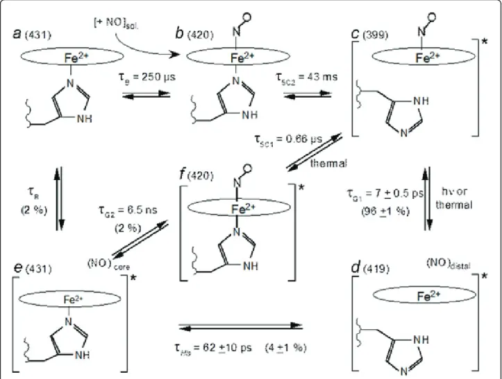
Results
We investigated the interaction between purified soluble
guanylate cyclase (sGC) from beef lung and NO by
time-resolved spectroscopy in a time-range which
encom-passes eleven orders of magnitude, from 1 ps [1] to 0.1 s
[2]. After its dissociation from the heme, NO either
recombines geminately to the 4-coordinate heme within
τ
G1= 7 ps [1] (96 ± 1 % of the population) or exits the
heme pocket (4 ± 1 %), allowing the proximal histidine to
rebind within 62 ± 10 ps. Then, NO is distributed in two
approximately equal populations (~2 %). One geminately
rebinds to the 5-coordinate heme (
τ
G2= 6.5 ns) while the
other migrates into the solution. NO can rebind from the
solution (bimolecular rebinding,
τ
B= 0.25 ms with [NO]
= 20 µM), forming a 6-coordinate heme with a rate
con-stant of 2 x 10
8M
-1s
-1(in vitro purified protein) very
close to that measured in platelets (3 × 10
8M
-1s
-1) [3].
The cleavage of Fe-His bond and subsequent
forma-tion of 5-coordinate NO-heme occurs with different
time constants for NO which geminately rebinds (τ
5C1=
0.66 µs) and for NO which binds from the solution
(τ
5C2= 43 ms). Thus, because the same structural event
occurs with rates separated by more than 4 orders of
magnitude, we must infer that sGC is not in the same
structural state in both cases, with a different strain
exerted on the Fe-His bond. This allosteric transition
between both states occurs in the time range 0.66 µs <
τ
R< 250 µs in sGC after His rebinding and NO release.
Conclusion
Since the discovery that NO binds to the proximal heme
side in cytochrome c
’ [4] (AXCP), several models of
sGC activation were proposed which include the binding
of NO to the proximal heme side despite the lack of
observation for such an activation step in sGC. After the
fast histidine rebinding in the picosecond range, we have
observed only four phases in the nano to millisecond
time range (assigned as indicated in Table 1). Thus,
* Correspondence: michel.negrerie@polytechnique.fr
1
Laboratoire d’Optique et Biosciences, INSERM, Ecole Polytechnique, Palaiseau, France
Full list of author information is available at the end of the article
Table 1 Rates of the transitions observed in kinetics.
Transition Time constants Transition rates
Bimolecular NO binding to 5c-His τB= 0.25 ms; [NO]= 20 µM kB= 2 x 10 8
M-1s-1 Conversion 6c-NO® 5c-NO τ5C2= 43 ms k5C2= 23 s
-1
Geminate NO rebinding to 5c-His τG2= 6.5 ns kG2= 0.15 x 10 9
s-1 Conversion 6c*-NO® 5c*-NO τ5C1= 0.66 µs k5C1= 1.5 x 10⁶s-1
His rebinding to 4c-heme τHis= 62 ps kHis= 1.4 x 10¹⁰s-1
Geminate NO rebinding to 4c-heme τG1= 7 ps kG1= 0.13 x 10¹²s-1
Structural relaxation sGC*® sGC 0.66 µs <τR< 250 µs 4 x 10³s-1<kR<1.5 x 10⁶s-1
Yoo et al. BMC Pharmacology 2011, 11(Suppl 1):P77 http://www.biomedcentral.com/1471-2210/11/S1/P77
© 2011 Yoo et al; licensee BioMed Central Ltd. This is an open access article distributed under the terms of the Creative Commons Attribution License (http://creativecommons.org/licenses/by/2.0), which permits unrestricted use, distribution, and reproduction in any medium, provided the original work is properly cited.
analysis of the entire NO dynamics from 1 ps to 0.1 s
did not detect NO binding to the proximal side of sGC
heme despite the fact that NO shows the same geminate
rebinding to the 4-coordinate heme in sGC and AXCP
[5]. Our data can be described with a one-site model,
without phases assigned to dinitrosyl formation or to
NO proximal binding.
Author details
1
Laboratoire d’Optique et Biosciences, INSERM, Ecole Polytechnique, Palaiseau, France.2Institut de Biologie Physico-Chimie, CNRS, Paris, France.
Published: 1 August 2011
References
1. Negrerie M, Bouzhir L, Martin JL, Liebl U: Control of nitric oxide dynamics by guanylate in its activated state. J Biol Chem 2001, 276:46815-46821.
2. Beal B, Rappaport F, Joliot P: A new high-sensitivity 10-ns time-resolution spectrophotometric technique adapted to in vivo analysis of the photosynthetic apparatus. Re Sc Instrum 1999, 70:202-207.
3. Roy B, Garthwaite J: Nitric oxide activation of guanylyl cyclase in cells revisited. Pro. Natl Acad Sci USA 2006, 103:12185-12190.
4. Lawson DM, Stevenson CEM, Andrew CR, Eady R: Unprecedented proximal binding of nitric oxide to heme: implications for guanylate cyclase. EMBO J 2000, 19:5661-5671.
5. Kruglik SG, Lambry JC, Cianetti S, Martin JL, Eady RR, Andrew CR, egrerie M: Molecular basis for nitric oxide dynamics and affinity with Alcaligenes xylosoxidans cytochrome c’. J Biol Chem 2007, 282:5053-5062.
doi:10.1186/1471-2210-11-S1-P77
Cite this article as: Yoo et al.: Dynamics of NO interacting with soluble guanylate cyclase from 1 ps to 0.1 s and induced structural transitions. BMC Pharmacology 2011 11(Suppl 1):P77.
Figure 1 Model describing the species and transitions involved in the time-resolved experiments. a: 5-c-His sGC in the resting state. b: 6-C sGC. c: 5-c-NO sGC, in the activate state. d: 4-c sGC after NO dissociation. e: 5-c-His sGC still in the activated conformation, immediately after His105 rebinding. f: 6-c sGC in the activated state, immediately after NO geminate rebinding. The value adjacent to the letter label is the wavelength of the species. The star at the right bracket in c, d, e denotes the activated state of sGC. The starting species of the kinetic measurements is c. The time constants indicated are those measured and the corresponding rates are given in Table 1. In species d, NO is located within the heme pocket whereas in species e, NO is located in another docking site in the protein.
Yoo et al. BMC Pharmacology 2011, 11(Suppl 1):P77 http://www.biomedcentral.com/1471-2210/11/S1/P77