Dysphagia in Elderly Women: Consider Tetanus
A. Rauch, S. Droz, S. Zimmerli, S. L. Leib
Abstract
Background: Dysphagia is seldom caused by tetanus;
however, it is a common symptom of tetanus. Treating
patients with tetanus is a rare event in industrialized
countries and awareness is needed to recognize early signs
of this serious disease. In Switzerland, the most recently
reported tetanus cases occurred in elderly women with
insufficient seroprotection.
Patients: We report on three elderly women presenting with
dysphagia as an initial symptom of tetanus.
Results: Generalized tetanus was diagnosed in two
patients upon admission, the third presented with cephalic
tetanus with secondary generalization. All three patients
had undetectable levels of tetanus antibodies and had no
documented prior tetanus immunizations. Cultures of wound
swabs grew Clostridium tetani in all cases. Electromyography
was highly suggestive for tetanus in two patients. Treatment
involved mechanical ventilation, intravenous benzodiazepine
and metronidazole therapy, and active and passive tetanus
immunization. The disease had a favorable outcome in two
cases and was fatal in one.
Conclusion: Tetanus remains a threat in patients with
insuf-ficient seroprotection and efforts are needed to improve
tetanus immunization in these individuals. Tetanus should
be considered in the differential diagnosis of dysphagia.
Infection 2006; 34: 35–38 DOI 10.1007/s15010-006-4151-7
Introduction
Tetanus has become a rare disease in industrialized
countries. In Switzerland, the annual incidence of tetanus
decreased from 4.3 to 0.4 cases/million population between
1979 and 2004 [1]. Zuber et al. [2] estimated that only about
10% of tetanus cases are reported to the Swiss Federal Office
of Health, but tetanus would be a rare event even if 100%
of cases were reported. Hullstrung et al. [1] found antibodies
to tetanus in 36 (92%) of 39 elderly patients in a Swiss
geriatric clinic, and interestingly, the median concentration
of anti-tetanus antibodies in males was more than twice as
high as in females. This gender-specific difference in
anti-tetanus serum titer is likely the result of repeated booster
immunizations of adult male Swiss citizens during their
annual weeks of compulsory military service. The fact that
over 75% of tetanus cases observed in Switzerland over the
last 5 years occurred in women older than 60 years further
supports the concept that gender-specific differences in
anti-tetanus serum titer leads to an increased incidence of
tetanus in this risk group. Surveillance data for 1998–2000
in the United States showed that adults older than 60 years
were at highest risk for tetanus (annual incidence of 0.35
cases/million population) and tetanus-related death
(case-fatality ratio of 40%) [3]. Dysphagia is a common symptom
of tetanus [4]. However, because dysphagia is seldom caused
by tetanus, the correct diagnosis and adequate therapy are
likely to be delayed. The purpose of this report is to outline
the presentation and clinical course of three patients with
dysphagia as a common symptom of tetanus.
Patients
Case 1
A 75-year-old woman was admitted to the emergency department with dysphagia and slight neck stiffness. Ten days before admission, the patient was in her usual good health when she fell with her left knee into a rusty iron rod which penetrated the skin and underlying structures to a depth of approximately 1 cm. The wound became red, hot and purulent 3 days after this incident. One day before admission she reported dysphagia and had increasing difficulties opening her mouth. The general practitioner performed wound debridement, active immunization for tetanus, and organized immediate hospitalization. Medical history was significant for bilateral knee prosthesis 15 years ago. The patient lived in a rural area in Switzerland and had not traveled recently. She had no known prior tetanus immunizations.
Physical examination revealed a slightly increased tone in the masseter muscles and a tender 3 × 4 cm wound with purulent discharge on the left knee (Figure 1). The patient was alert
S. Droz, S. Zimmerli, S.L. Leib (corresponding author)
Institute for Infectious Diseases, University of Bern, Friedbühlstrasse 51, P.O. Box 61, 3010 Bern, Switzerland; Phone: (+41/31) 632-4949, Fax: -3550, e-mail: [email protected]
A. Rauch, S. Zimmerli, S.L. Leib
Clinic and Policlinic for Infectious Diseases, University Hospital, Inselspital, Bern, Switzerland
Received: December 20, 2004
.
Revision accepted: June 6, 2005Infection
Brief Report
A. Rauch et al. Dysphagia and Tetanus
36
Infection 34 · 2006 · No. 1 © URBAN & VOGELand coherent, and all other physical and neurological findings were normal. Laboratory studies showed no abnormalities. Tetanus antibodies were not detectable by the enzyme-linked immunosorbent assay (ELISA). Electromyography of the masseter muscles showed spontaneous activity of motor units which could not be suppressed voluntarily and an absence of the silent period. Laboratory exams and clinical evaluation revealed no signs of strychnine intoxication, of hypocalcemia or of a dystonic reaction. The diagnosis of tetanus was made and therapy started including human tetanus immunoglobulin (4,000 IU i.m.), metronidazole (500 mg i.v. qid) and lorazepam (i.v. in 2-mg increments). Swabs taken from the bottom of the wound during debridment were cultured aerobically and anaerobically. All aerobic cultures revealed Staphylococcus aureus. After 48 h of incubation, the anaerobic cultures revealed two different morphologies. The two gram-positive rods were identified as Clostridium tetani and Clostridium perfringens. C. tetani isolates are routinely identified to the species level by volatile fatty acid analysis, and by sequencing of 1,507-bp of the 16S rRNA gene.
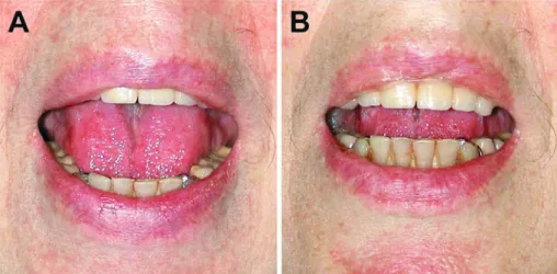
Within 1 day, the patient developed rapidly progressive trismus (Figure 2), painful muscle spasms, respiratory insufficiency, and autonomic dysfunction necessitating intubation under sedation and relaxation. She needed to be mechanically ventilated for 4 weeks. Repeated wound debridement resulted in complete healing of the soft-tissue infection. The patient was released from the intensive care setting after 6 weeks. She recovered almost completely; the only sequelae were slight dysphagia and an impaired gait.
Case 2
An 86-year-old woman sustained a head injury while gardening. Wound débridement was performed immediately. Although her tetanus immunization history was unknown, she received at that time neither tetanus toxoid nor tetanus immunoglobulin. One week later, the patient was hospitalized with progressive dysphagia and trismus and received active tetanus immunization, human tetanus immunoglobulin (500 IU i.m.) and metronidazole (500 mg i.v. tid) upon admission. Within 2 days of hospital admission, she developed respiratory failure and required mechanical ventilation. Clinical findings and electromyography were highly suggestive of generalized tetanus. The swabs taken from the facial wound were cultured aerobically and anaerobically. The aerobic cultures showed no growth after 48 h of incubation but anaerobic cultures revealed three different morphologies. The
three gram-positive rods were identified as C. tetani, C. sporogenes and C. hastidiforme. Tetanus antibodies were not detectable by ELISA. The patient developed severe autonomic dysfunction and progressive opisthotonus. Both spasms and autonomic dysfunction progressed rapidly despite adequate intensive care management. The patient died 6 days after admission.
Case 3
An 85-year-old woman presented to the emergency department with an (initially unrecognized) orbital floor fracture and a facial wound from a wooden stick. Wound débridement and active tetanus immunization were performed immediately, but no tetanus immunoglobulins were given at that time. She had no known prior tetanus immunizations. The patient was readmitted to the hospital 2 weeks later with an infraorbital wound infection and diplopia due to an orbital floor fracture, which required surgical reconstruction. Five days later, she developed dysphagia, facial paresis, trismus and subsequently progressive respiratory insufficiency. The aerobic cultures taken from the orbita abscess during operative revision revealed Serratia liquefaciens and mixed gram-positive bacteria which were not further investigated. No anaerobic bacteria were observed on the anaerobe solid media. Owing to the high clinical suspicion of tetanus, the thioglycolate medium was subcultured and grew multiple anaerobes. The gram-positive rod was identified as C. tetani. The other anaerobes were not further specified. Tetanus antibodies were not detectable by ELISA. Electromyographic findings were not conclusive. A diagnosis of cephalic tetanus with secondary generalization was considered and therapy was complemented with human tetanus immunoglobulin (1,000 IU i.m.), metronidazole (500 mg i.v. tid) and intravenous midazolam (in 2.5-mg increments). Initially, the muscle spasms involving head, neck, and the arms intensified despite sedative and anticonvulsant therapy. After 4 weeks, the spasms improved and the patient was extubated. She recovered well and was discharged 2 months after admission to a rehabilitation center.
Discussion
The three cases described above illustrate important
epidemiological, clinical, diagnostic and therapeutic aspects
of tetanus in industrialized countries. We discuss here these
issues with respect to our cases.
Figure 2. Case 1: Evolution of symptomatic trismus. The patient was instructed to maximally open
her mouth at admission (A) and on the following day (B).
Figure 1. Case 1: Macroscopic aspect
of the wound over the left knee at admission. The surrounding skin was erythematous and hot. Culture of the purulent wound secretion revealed growth of Clostridium tetani and Clostridium perfringens.
A. Rauch et al. Dysphagia and Tetanus
Infection 34 · 2006 · No. 1 © URBAN & VOGEL
37
All three cases presented in this report were elderly
women, had no documented prior tetanus immunizations
and had undetectable levels of tetanus antibodies. This
finding reflects the particular risk for tetanus in elderly
women outlined in the introduction and underscores the
importance of providing adequate tetanus immunization
to the whole population.
Tetanus diagnosis is primarily based on clinical
symptoms [5, 6]. Dysphagia as an initial symptom of
tetanus has been described in case reports, primarily from
otolaryngologic departments [7–13]. In a 1957 review of
91 patients with tetanus, dysphagia was noted in 10% at
admission to the hospital and developed subsequently in
50% of patients [4]. On admission to an Australian hospital,
trismus was present in 93% and dysphagia in 46% of
106 patients with tetanus [14]. In an otolaryngologic center
in Turkey, 92% of 37 patients with tetanus had trismus and
78% dysphagia as initial symptoms of the disease [15]. In
a Belgian hospital, all 27 patients with tetanus developed
dysphagia and trismus during hospitalization [16].
Dysphagia was the first symptom reported by our patients;
a detailed clinical history, however, revealed additional
symptoms of tetanus in all three cases which underscores the
importance of clinical awareness for tetanus in this setting.
Wound cultures grew C. tetani in all cases presented in this
report. However, the diagnostic value of positive cultures is
limited: A positive culture may be present without disease
and does not indicate whether the organism contains the
toxin-producing plasmid. Electromyography has shown to
be helpful in the diagnosis of tetanus, but as illustrated in
Case 3, inconclusive results do not rule out tetanus [17].
The patient presented in Case 3 suffered most probably
from cephalic tetanus with secondary generalization. This
special form of initially localized disease affects primarily
the cranial nerves and commonly follows an injury to the
head. It is characterized by lower motoneuron lesion,
frequently producing facial paresis [18]. Paralysis is
maximal close to the site of injury, while spasm is evident
at more distant sites [19]. Like in other forms of localized
tetanus, patients with cephalic tetanus often proceed to
develop generalized tetanus [18, 19].
Standard treatment of tetanus includes benzodiazepines,
opiates and vasoactive drugs to help reduce muscle spasms,
pain, and autonomic dysfunction [20, 21]. Benzodiazepines
remain the mainstay of sedative and anticonvulsant
treatment [22]. Some reports suggest an additional benefit
of intrathecal baclofen [23]. Passive immunization is given
to neutralize circulating toxin. Data on the effect of different
doses of tetanus immune globulin suggest that 500 U may
be as effective as higher doses of 3,000–10,000 U [24]. Prior
tetanus does not provide immunity, possibly due to the
minimal amounts of toxin needed to cause disease. Thus,
passive immunization and active immunization with tetanus
toxoid should commence early in the management of the
patient [20, 21]. The choice of antibiotic therapy remains
controversial. A study comparing oral metronidazole to
intramuscular penicillin showed improved survival in the
metronidazole group, possibly due to the GABA antagonist
effects of penicillin [25, 26].
The introduction of intensive care treatment has
substantially reduced mortality, but mortality rate is still
high, particularly in elderly persons [27]. The patient
with fatal outcome reported in Case 2 presented several
features which have been associated with an unfavorable
outcome: a short incubation period, an internal or facial
site of infection and lack of immune protection [28].
Tetanus remains a threat in persons with insufficient
seroprotection. Treating patients with tetanus is a rare
event in industrialized countries and awareness is needed
to recognize early signs of this rare but serious disease.
Tetanus should be considered in the differential diagnosis
of dysphagia.
Acknowledgments
Funding was received from the Swiss National Science Founda-tion, grant No. 632-66057.01.
References
1. Hullstrung HD, Mausezahl D, Feuz M, Herzog C, Conzelmann M, Zimmerli W: Tetanus immunisation in geriatric patients with accidental wounds: how much is needed? Swiss Med Wkly 2003; 133: 227–232.
2. Zuber PL, Schierz A, Arestegui G, Steffen R: Tetanus in Switzerland 1980–1989. Eur J Epidemiol 1993; 9: 617–624. 3. Pascual FB, McGinley EL, Zanardi LR, Cortese MM, Murphy TV:
Tetanus surveillance – United States, 1998–2000. MMWR Surveill Summ 2003; 52: 1–8.
4. Christensen NA, Thurber DL: Clinical experience with tetanus: 91 cases. Mayo Clin Proc 1957; 32: 146–158.
5. Farrar JJ, Yen LM, Cook T, Fairweather N, Binh N, Parry J, Parry CM: Tetanus. J Neurol Neurosurg Psychiatry 2000; 69: 292–301. 6. Henderson SO, Mody T, Groth DE, Moore JJ, Newton E: The
presentation of tetanus in an emergency department. J Emerg Med 1998; 16: 705–708.
7. Stephenson JW, Blacklay B: A case of tetanus presenting as dysphagia. J Laryngol Otol 1955; 69: 496–498.
8. Weider DJ, Tingwald FR: Dysphagia as initial and prime symptom of tetanus. Report of a case. Arch Otolaryngol 1970; 91: 479–481.
9. Wang L, Karmody CS: Dysphagia as the presenting symptom of tetanus. Arch Otolaryngol 1985; 111: 342–343.
10. Kasanzew M, Browne B, Dawes P: Tetanus presenting as dysphagia. J Laryngol Otol 1989; 103: 229–230.
11. Scholz DG, Olson JM, Thurber DL, Larson DE: Tetanus: an
uncommon cause of dysphagia. Mayo Clin Proc 1989; 64: 335–338. 12. Ross J, Murrant NJ: Dysphagia as a major symptom of tetanus.
J Laryngol Otol 1992; 106: 923–924.
13. Mastaglia FL, Thickbroom GW, Day T, Bond R: Craniocervical tetanus presenting with dysphagia: diagnostic value of electrophysiological studies. J Neurol 2001;248:903–904 14. Newton-John HF: Tetanus in Victoria, 1957–1980. Review of 106
patients managed in one hospital. Med J Aust 1984; 140: 194–200. 15. Aydin K, Caylan R, Bektas D, Koksal I: Otolaryngologic aspects of
A. Rauch et al. Dysphagia and Tetanus
38
Infection 34 · 2006 · No. 1 © URBAN & VOGEL16. Peetermans WE, Schepens D: Tetanus – still a topic of present interest: a report of 27 cases from a Belgian referral hospital. J Intern Med 1996; 239: 249–252.
17. Steinegger T, Wiederkehr M, Ludin HP, Roth F: Electromyography as a diagnostic aid in tetanus. Schweiz Med Wochenschr 1996; 126: 379–385.
18. Jagoda A, Riggio S, Burguieres T: Cephalic tetanus: a case report and review of the literature. Am J Emerg Med 1988; 6: 128–130. 19. Dastur FD, Shahani MT, Dastoor DH, Kohiyar FN, Bharucha
EP, Mondkar VP, Kashyap GH, Nair KG: Cephalic tetanus: demonstration of a dual lesion. J Neurol Neurosurg Psychiatry 1977; 40: 782–786.
20. Hsu SS, Groleau G: Tetanus in the emergency department: a current review. J Emerg Med 2001; 20: 357–365.
21. Cook TM, Protheroe RT, Handel JM: Tetanus: a review of the literature. Br J Anaesth 2001; 87: 477–487.
22. Okoromah CN, Lesi FE: Diazepam for treating tetanus. Cochrane Database Syst Rev 2004:CD003954.
23. Santos ML, Mota-Miranda A, Alves-Pereira A, Gomes A, Correia J, Marcal N: Intrathecal baclofen for the treatment of tetanus. Clin Infect Dis 2004; 38: 321–328.
24. Blake PA, Feldman RA, Buchanan TM, Brooks GF, Bennett JV: Serologic therapy of tetanus in the United States, 1965–1971. JAMA 1976; 235: 42–44.
25. Ahmadsyah I, Salim A: Treatment of tetanus: an open study to compare the efficacy of procaine penicillin and metronidazole. Br Med J (Clin Res Ed) 1985; 291: 648–650.
26. Weiss DS, Hablitz JJ: Interaction of penicillin and pentobarbital with inhibitory synaptic mechanisms in neocortex. Cell Mol Neurobiol 1984; 4: 301–317.
27. Anonymous: Leads from the MMWR. Tetanus – United States, 1982–1984. JAMA 1985; 254: 2873, 2877–2878.