In Vitro Inhibition of Coagulase-Negative
Staphylococci by Vancomycin/Aminoglycoside-
Loaded Cement Spacers
J.C. Streuli, G.U. Exner, C.L. Reize, C. Merkofer, C.P. Scott, R. Zbinden
Abstract
Background: Successful treatment of allograft infections by
the temporary implantation of an antibiotic-loaded
polymeth-ylmethacrylate cement spacer depends on the diffusion of
antibiotics out of the cement and inhibition of bacterial
growth in the surrounding tissue. We investigated with an in
vitro model how long antibiotics are released by the cement
and if gentamicin-resistant coagulase-negative staphylococci
(CNS) are inhibited by vancomycin mixed with the
gentamicin-loaded cement.
Materials and Methods: Four formulations of
antibiotic-loaded cement disks, i.e. gentamicin, tobramycin,
vancomycin and tobramycin combined with vancomycin,
respectively, were used to test the inhibition of eight isolates
of Staphylococcus epidermidis and two reference strains of
Staphylococcus aureus by an agar diffusion test on
Mueller-Hinton (MH) agar similar to the routine laboratory
disk diffusion method. Moreover, cement spacer cylinders
loaded with gentamicin alone or combined with vancomycin
were submerged in MH agar for weeks and the capacity to
inhibit five different isolates of S. epidermidis was measured.
Results: The size of the inhibition zones around the
antibiotic-loaded cement disks correlated with the minimal
inhibitory concentration (MIC) of the antibiotics against the
tested strains. All five strains of S. epidermidis were inhibited
by vancomycin-loaded cement spacers for at least 30 days.
However, two gentamicin-resistant S. epidermidis strains with
MICs of 4 mg/l and 16 mg/l could not be inhibited longer
than 3 days by the gentamicin-loaded cement spacer.
Conclusion: The in vitro data suggest that antibiotic-loaded
cement spacers inhibit susceptible bacteria for 4–6 weeks. The
addition of vancomycin to commercial aminoglycoside-loaded
cements might be helpful in allograft infections in tumor
patients to inhibit a broad range of bacteria including
genta-micin-resistant CNS very commonly found in such infections.
Infection 2006; 34: 81–86 DOI 10.1007/s15010-006-5039-2
Introduction
Buchholz and Engelbrecht [1] introduced the concept of
antibiotic-loaded polymethylmetacrylate (PMMA) bone
cement over 30 years ago. After successful prophylactic
use of gentamicloaded cement with a reduced rate of
in-fected endoprosthesis, they treated deep infections of total
hip replacements by revision arthroplasty that comprised,
in one stage, excision of soft tissue, removal of implant and
cement and replacement with an appropriate implant using
antibiotic-loaded cement [2]. Staged revision of infected
prosthesis by using temporary antibiotic-loaded cement
spacers may be preferred [3]. Systemic antibiotic treatment
of infected bone reconstructions in tumor patients may be
compromised by devascularization. Bacteria colonize the
surface of inert biomaterial and adjacent, damaged tissue
cells [4]. Therefore, antibiotic-impregnated cement spacers
should inhibit bacteria locally before they form a biofilm.
However, it is not well defined how long an
antibiotic-loaded cement spacer should be left in place before the
definitive reconstruction can take place.
Aminoglycosides are widely used because of their
broad-spectrum activity and thermal stability [5, 6]. The
high prevalence of highly resistant coagulase-negative
staphylococci (CNS) in infected reconstructions may justify
the use of vancomycin in bone cement. The present study
investigates the in vitro inhibition of clinical isolates of
Staphylococcus epidermidis by bone cement loaded with
single antibiotics, i.e. gentamicin, tobramycin and
vancomy-cin, and loaded with vancomycin combined with
tobramy-cin, respectively. In addition, we developed a simple method
to measure the time of the inhibition of bacteria which
com-bines the standardized method of disk diffusion and some of
the in vivo conditions of cement spacers. Our in vitro model
should demonstrate in an easier way that the antibiotic-
loaded bone cement can elute antibiotic over weeks.
C. Merkofer, R. Zbinden (corresponding author)
Institute of Medical Microbiology, University of Zurich, Gloriastr. 32, 8006 Zurich, Switzerland; Phone: (+41/44) 634-2608, Fax: -4906, e-mail: rzbinden@immv.unizh.ch
J.C. Streuli, G.U. Exner
Balgrist, Dept. of Orthopaedics, University of Zurich, Zurich, Switzerland C.L. Reize
Kantonsspital Olten, Olten, Switzerland C.P. Scott
Advanced Technology Group, Stryker Orthopaedics, Mahwah, NJ, USA
Materials and Methods
Eight isolates of S. epidermidis, isolated from patients with ortho-pedic infections and two reference strains of Staphylococcus aureus (ATCC 29213, susceptible to methicillin; ATCC 43300, resistant to methicillin) were included in our in vitro inhibition assay by anti-biotic-loaded cement disks. Five of those isolates were used to in-vestigate their inhibition over weeks by cylindrical antibiotic-loaded cement spacers.
The minimal inhibitory concentrations (MIC) of gentamicin, tobramycin and vancomycin for those bacteria were determined by Etest strips containing the corresponding antibiotic calibrated with MIC values covering a scale of 15 twofold dilutions (AB Biodisk, Solna, Sweden). In brief, staphylococcal colonies of an overnight culture on sheep blood agar were suspended in saline and adjusted to match a 0.5 McFarland turbidity standard, i.e. containing ap-proximately 1 to 2 × 10⁸ CFU/ml. A sterile cotton swab was dipped into the suspension and Mueller-Hinton (MH) agar plates were inoculated by streaking the swab over the entire agar surface before one Etest strip per plate was placed onto each agar. They were incu-bated for 24 h at 35 °C without CO2 and MICs were read where the growth inhibition ellipse intersected the scale. Synergy testing was also performed by Etest as above according to the instructions of the manufacturer. However, a first Etest strip with gentamicin was placed onto the inoculated MH agar for only 1 h and removed again before a second Etest strip with vancomycin was put in exactly the same place as the first; this experiment was also performed in paral-lel for each strain by placing first the Etest strip with vancomycin for 1 h followed by the Etest strip with gentamicin overnight. Plates were incubated as above but the read MIC is the MIC of the com-bination of both, gentamicin and vancomycin.
For the in vitro inhibition assay by antibiotic-loaded cement disks, four antibiotic-loaded cement formulations were used: (1) Palacos commercially prepared with 500 mg gen-tamicin per 40.8 g cement powder (Biomet-Merck, Warsaw, Indiana, USA) and (2) Simplex commercially prepared with 1 g tobramycin (Stryker Howmedica Osteonics, Limerick, Ireland); two other formulations were prepared by hand-mixing 3.0 g of vancomycin hydrochloride (Vancocin, Lilly, Indianapolis, In-diana, USA) with a tongue depressor into standard 40 g doses (3) of the standard Simplex P without antibiotic (Stryker How-medica Osteonics, Limerick, Ireland) and (4) of the Simplex bone cement with tobramycin (Stryker Howmedica Osteonics, Limerick, Ireland). Single doses of each bone cement formula-tion were vacuum mixed according to manufacturer’s instrucformula-tions using a commercial cement mixer and delivery system (How-medica Inc., Limerick, Ireland). Immediately after mixing, the cement was delivered into aluminum molds with cavities to produce uniform disk-shaped specimens with a diameter of 14 mm and thickness of 2 mm. Cured samples were inspected and any disks possessing gross voids or surface defects were discarded. The antibiotic-impregnated cement disks were used on the very same day of their production.
Suspensions of the eight isolates of CNS as well as of the two reference strains of S. aureus were prepared and streaked onto MH agar plates as above. After a few minutes the disks of antibi-otic-loaded cement were placed into the middle of the inoculated
MH agar plates and pressed down to ensure complete contact with the agar surface. Each strain was tested in duplicate with each cement disk formulation. Plates were incubated for 24 h at 35 °C without CO2. After 24 h the inhibition zones were measured.
Five of the eight isolates of S. epidermidis were included in the inhibition experiment with cylindrical antibiotic-loaded ce-ment spacers. Two antibiotic-loaded cece-ment formulations were used to prepare five of each cylindrical cement spacers: (1) Pala-cos R-40 (40.8 g powder) commercially prepared with 500 mg gentamicin (Biomet-Merck) and (2) a formulation prepared by hand-mixing 3 g of vancomycin hydrochloride into the standard 40 g doses of Palacos bone cement with 500 mg gentamicin (Biomet-Merck). Briefly, the Palacos R-40 with gentamicin was mixed with the entire contents of two ampoules of 18.8 g liquid component twice as much as normal in a bowl for 50 s with a sterile plastic spatula until a homogeneous fluid cement mixture was formed. The mixture was filled into a perfusor syringe OPS® 50 ml (Braun, Melsungen, Germany) and packed by the gum stamp of the syringe. The syringes were placed with the outflow part upside until the hardened spacers could be taken out of the syringes after 10 min; they were stored at room temperature for 24 h. Further five cylindrical cement spacers were produced in the same way but 3 g vancomycin was added to the solution into the mixing bowl. The cylindrical spacers had approximately the diameter of the spacers used in the tumor patients with allograft infections.
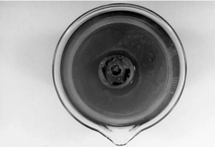
The cylindrical spacers were placed in the middle of cylindri-cal glasses (Duran®, Schott, Mainz, Germany) containing 440 ml of liquid (50 °C) sterile MH agar so that 2–3 cm of the spacer were still above the level of the agar surface. After the agar became solid at room temperature, suspensions of staphylococci were prepared as above and streaked with a swab tightly over the entire agar surface around the cylindrical cement spacer. The cylindri-cal glasses with the spacer in the agar were incubated at 37 °C without CO2 and diameters of inhibition were measured after 24 h and incubation continued for some days (Figure 1). The cylin-drical spacers that showed an inhibition of the staphylococci were then transferred to a new cylindrical glass with liquid (50 °C) sterile MH agar but the amount of agar was diminished by
Figure 1. Cement spacer in a cylindrical glass with a zone of
20 ml (Figure 2a, b) so that the contact of the agar with the spacer cement not yet submerged in the former experiment can be ex-cluded; otherwise, the contact of agar with the cement not sub-merged in the earlier experiments would allow diffusion of antibi-otics of the upper part of the spacer. Suspensions from overnight grown colonies from the same bacteria were streaked onto the agar and the inhibition zone was measured again after 24 h. At the beginning the intervals were short to detect an early loss of release of antibiotics by bone cement; the volume of the agar had to be reduced for each new transfer by 20 ml to be sure that the contact zone of the agar was not at a level of the cement spacer not yet submerged before; therefore, the interval was prolonged so that we could perform the experiment over 2 months. The
experi-ment was stopped earlier for those spacers that did not show any more inhibition zones or which were contaminated by molds.
Statistics
The MICs of the combination of gentamicin
plus vancomycin or vancomycin plus
gentami-cin (Table 1) were compared with the MICs of
the single compound with the lower MIC by the
Wilcoxon signed-rank test to detect synergy, i.e.
lower MICs of the combination than the MICs
of the single compounds. The mean values of the
duplicate zones of inhibition of the cement disks
with vancomycin combined with tobramycin
(Table 2) were compared with the mean values
of the single compound with the bigger zone
by the Wilcoxon signed-rank test to detect
synergy, i.e. greater inhibition zones of the disks
with combined antibiotics.
Results
The MICs of the eight strains of S.
epidermi-dis and of the two reference strains of S.
au-reus are shown in table 1. The MICs of the
syn-ergy testing by Etest were not different from
the MICs of the single component which was
most active against the corresponding strain
(p = 0.38 for the gentamicin plus vancomycin
combination, p = 0.69 for the vancomycin plus
gentamicin combination). Inhibition zones of
the antibiotic-loaded cement disks were
mea-sured after an overnight incubation (Table 2).
The duplicate values did not differ more than
2 mm with the following exception: the
in-hibition zones of the two cement disks with
vancomycin combined with tobramycin were
30 mm and 37 mm for strain V10 21204. The
diameters of the zones around the cement disks
loaded with vancomycin and tobramycin were
not different from those of the most active
sin-gle antibiotic (p = 0.96). Furthermore, the zones
were greater for strains with low MICs than for strains with
higher MICs and strains with MICs greater than 12 mg/l
did not show any inhibition zone with the corresponding
cement disks.
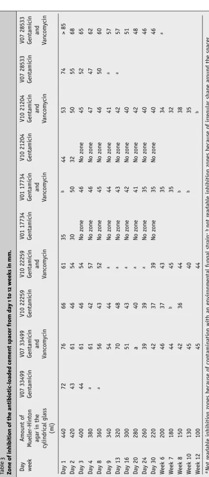
The zones of inhibition of the ten cement spacers are
shown in table 3. Only one of the spacers loaded with
gen-tamicin alone (V10 22259) showed a zone of inhibition
un-til the 8th week. The two spacers incubated with strains
with elevated MICs against gentamicin (V01 17734 and
V10 21204, see Table 2) stopped showing a zone of
inhibi-tion after 2 and 3 days. The corresponding spacers loaded
with gentamicin and vancomycin inhibited the same strains
of S. epidermidis over 7 weeks. The zones of inhibition of
Figure 2. a) Cement spacer in the cylindrical glass at the beginning of the
experiments; b) Cement spacer in the cylindrical glass 5 weeks later.
Table 1
Minimal inhibitory concentrations (MIC) of all isolates determined by Etest.
Organism MIC in mg/l Tobramycin Vanco-mycin Gentamicin plus vancomycin Vancomycin plus gentamicin Genta-micin Staphylococcus aureus ATCC 43300 > 256 1.5 0.75 0.38 32 ATCC 29213 1.0 1.0 1.0 1.0 0.38 Staphylococcus epidermidis V07 33499 0.38 2 0.19 0.125 0.125 V10 33001 2 2 0.25 0.125 0.125 V10 22259 8–12 1.0 0.38 0.19 0.25 V01 17734 > 256 1.5 2 2 16 V07 21204 > 256 2 1.5 1.5 4 V01 15748 > 256 2 2 2 4 V10 28533 > 256 2 0.25 0.19 0.19 V09 15128 > 256 2 2 2 > 256
the other two gentamicin-loaded spacers were only
mea-surable for 3 and 8 days, respectively, because they were
contaminated by environmental strains.
Three of five spacers loaded with gentamicin and
van-comycin (V07 33499, V10 22259, V10 21204) still showed a
zone of inhibition at the end of the study after more than
15 diffusion experiments; spacer V10 22259 showed a
tran-sient contamination by fungus. Two spacers (V01 17734,
V07 28533) produced after 7 and 4 weeks, respectively,
irregular zones and were not transferred for further
experi-ments.
Discussion
It is known that many commonly used antibiotics are
released from bone cement in such a way that the local
antibiotic levels vastly exceed the MICs needed for
treating most susceptible pathogens, and that the
lev-els are much higher than those achieved with
par-enteral therapy [7]. When used in PMMA, the
antibiotic agent must be heat stable and effective against
the common pathogens and exhibit good elution
char-acteristics [8]. Previous studies measured in vitro the
released antibiotics in fluids surrounding antibiotic-loaded
cement [9–11] or in vivo in wound drainage, urine and
serum [12–14].
The time period of antibiotic release out of the bone
cement reached days to several months in earlier
stud-ies and the diffusion is influenced by the cement-,
antibi-otic- and environment-dependent factors [15]. The
elu-tion characteristics of different bone cements depend on
handling, physical states and physical characteristics, i.e.
speed of creep and relaxation [16]. Some authors reported
that Palacos releases more gentamicin and vancomycin [12,
14, 17–20] and has more rapid elution characteristics than
Simplex-P, which is probably due to its greater porosity
than other cements [7, 21]. In contrast, Simplex-P bone
cement has superior handling characteristics [7]. Other
studies claimed that the elution capacity of CMV (DePuy
International, Blackpool, UK) may be greater than that of
Palacos R [20, 22].
Our freshly prepared antibiotic-loaded cement disks
released tobramycin, gentamicin and vancomycin into the
agar, as shown in previous studies [8, 19, 23]. The
con-centration of antibiotics is highest near the disk and the
bacterium is inhibited if the MIC is lower than this
con-centration. The staphylococci were only inhibited by the
aminoglycoside-loaded cement disks if the
correspond-ing MIC in the Etest was lower than 16. The
concentra-tion of the antibiotic at the edge of the inhibiconcentra-tion zone
corresponds to the MIC of the bacterium and the
inhi-bition zones correlated – with one exception – with the
MIC of the corresponding staphylococci, i.e. the lower
the MIC the bigger was the inhibition zone. The addition
of vancomycin to tobramycin resulted in an inhibition of
all staphylococci including those six strains against which
MICs of tobramycin were > 256. However, the inhibition
zones of tobramycin combined with vancomycin were
not significantly greater for the other staphylococci than
those of the more potent antibiotic alone. This was also
observed by Etest where the combination of gentamicin
with vancomycin revealed MIC similar to the MIC of the
most active antibiotic. We could not observe a synergistic
inhibition of tobramycin or gentamicin with vancomycin
in vitro. However, we did not measure the bactericidal
effect of the combination of vancomycin and gentamicin.
A bactericidal synergy of two substances that interact
with different targets of the bacteria might be helpful to
eliminate the bacteria from the bone similar to
synergis-tic action of vancomycin or penicillin with gentamicin in
enterococcal endocarditis [24]. Furthermore,
aminoglyco-sides alone might promote the formation of small colony
variants of staphylococci [25, 26]. Small colony variants
of staphylococci have been recovered from patients with
unusually persistent infections which are chronically
ex-posed to aminoglycosides. The addition of vancomycin
might prevent the formation of small colony variants but
this has to be studied further. The reason for the addition
of vancomycin is, however, to inhibit those staphylococci
that are not inhibited by the eluted amounts of
aminogly-cosides out of the cement.
Our experiments with cement spacers show that
enough antibiotic was eluted into the agar to inhibit
bac-teria. However, the two strains of S. epidermidis with MIC
4 and 16 against gentamicin were not inhibited after 3 days.
The spacers with vancomycin released antibiotics until the
end of the experiment over 8 weeks. This is in contrast
with previous reports that vancomycin is not well eluted
from antibiotic-impregnated bone cement in total hip
ar-throplasty [13].
Table 2
Zones of inhibition of the antibiotic-loaded cement disks with the agar diffusion method
Organism Zone of inhibition of duplicate
cement disks (first, second disk) in mm
Tobramycin Vanco-mycin Vancomycin plus tobramycin Genta-micin Staphylococcus aureus ATCC 43300 (MRSA) No zone 27, 28 26, 27 No zone ATCC 29213 30, 30 28, 28 32, 32 31, 31 Staphylococcus epidermidis V07 33499 35, 36 28, 28 38, 39 38, 37 V12 33001 26, 26 30, 29 29, 28 36, 37 V10 22259 23, 24 28, 30 29, 28 34, 33 V01 17734 No zone 31, 31 30, 28 No zone V10 21204 No zone 30, 31 30, 37 24, 23 V01 15748 No zone 34, 33 34, 32 26, 26 V07 28533 No zone 31, 32 31, 32 40, 40 V09 15128 No zone 28, 28 26, 28 No zone
However, the addition of
vancomy-cin might be a microbial ecological risk
[27]. Gradual development of vancomycin
resistance could occur as a result of wide
use of vancomycin with prolonged release
in subtherapeutic doses or the
employ-ment of large amounts. Therefore,
vanco-mycin-loaded cement is suggested only for
revisions of infected arthroplastic and for
high-risk procedures. Failure of infection
control in massive tumor reconstruction may
result in the loss of the extremity. Since most
CNS of orthopedic infections are resistant to
gentamicin, vancomycin might inhibit those
bacteria. Aminoglycosides are particularly
effective against gram-negative bacteria,
and cannot be replaced totally by
vanco-mycin.
We conclude that these in vitro
experi-ments support reasonable use of
vancomy-cin loaded spacers in tumour patients with
infected reconstructions by CNS. Although
we observed antibiotic elution over 8 weeks
we suggest that the spacer should be changed
every 4–6 weeks since the declining
antibi-otic concentration may no longer inhibit the
CNS, the most prevalent infectious
patho-gens in infected allografts. These data have
to be confirmed by in vivo experiments; we
recently started to investigate explanted
cement spacer from patients to detect their
inhibition of CNS.
References
1. Buchholz HW, Engelbrecht H: Über die Depot-wirkung einiger Antibiotica (sic!) bei Vermisch-ung mit dem Kunstharz Palacos. Chirurg 1970; 40: 511–515.
2. Buchholz HW, Elson RA, Engelbrecht E, Loden-kämpfer H, Röttger J, Siegel A: Management of deep infection of total hip replacement. J Bone Joint Surg Br 1981; 63: 342–353.
3. Donati D, Biscaglia R: The use of antibiotic-im-pregnated cement in infected reconstructions after resection for bone tumours. J Bone Joint Surg Br 1998; 80: 1045–1050.
4. Gristina AG: Biomaterial-centered infection: microbial adhesion versus tissue integration. Science 1987; 237: 1588–1595.
5. Bunetel L, Segui A, Cormier M, Langlais F: Com-parative study of gentamicin release from nor-mal and low viscosity acrylic bone cement. Clin Pharmacokinet 1990; 19: 333–340.
6. Seyral P, Zannier A, Argenson JN, Raoult D: The release in vitro of vancomycin and tobramycin from acrylic bone cement. J Antimicrob Che-mother 1994; 33: 337–339.
Table 3 Zone of inhibition of the antibiotic-loaded cement
spac er fr om da y 1 to 12 w eek s in mm. Day week Am oun t o f M ueller-Hin ton agar in th e cylin d ri cal glass (ml) V07 33499 Gen tami cin V07 33499 Gen tami cin an d Va n com ycin V10 22259 Gen tami cin V10 22259 Gen tami cin an d Va n com ycin V01 17734 Gen tami cin V01 17734 Gen tami cin an d Va n com ycin V10 21204 Gen tami cin V10 21204 Gen tami cin an d Va n com ycin V07 28533 Gen tami cin V07 28533 Gen tami cin an d Va n com ycin Day 1 440 72 76 66 61 35 b 44 53 74 > 85 Day 2 420 43 61 46 54 30 50 32 50 55 68 Day 3 400 44 61 46 54 N o zon e 4 6 N o zon e 4 5 5 2 6 5 Day 4 380 a 61 42 57 N o zon e 4 6 N o zon e 4 7 4 7 6 2 Day 8 360 a 56 43 52 N o zon e 4 5 N o zon e 4 6 5 0 6 0 Day 9 340 54 44 a N o zon e 4 4 N o zon e 4 1 a 57 Day 13 320 70 48 a N o zon e 4 3 N o zon e 4 2 a 57 Day 16 300 51 43 a N o zon e 4 2 N o zon e 4 0 5 1 Day 20 280 a 40 a N o zon e 4 1 N o zon e 4 2 4 8 Day 24 260 39 39 a N o zon e 3 5 N o zon e 4 0 4 6 Day 30 220 42 37 39 N o zon e 3 5 N o zon e 4 0 4 6 W eek 6 200 46 37 43 35 34 a W eek 7 180 44 b 45 35 32 W eek 8 150 42 36 44 b 38 W eek 10 130 45 40 b 35 W eek 12 100 45 44 b aN ot r ead able inhibiti on zon es because o f con tamin ati on with an envir onm en tal fun gal str ain; b n ot r ead able inhibiti on zon es because o f irr egular shape ar oun d th e spacer
7. Duncan CP, Masri BA: The role of antibiotic-loaded cement in the treatment of an infection after a hip replacement. J Bone Joint Surg Am 1994; 76: 1742–1751.
8. Scott CP, Higham PA, Dumbelton JH: Effectiveness of bone ce-ment containing tobramycin. An in vitro susceptibility study of 99 organisms found in infected joint arthroplasty. J Bone Joint Surg Br 1999; 81: 440–443.
9. Perry AC, Rouse MS, Khaliq Y, Piper KE, Hanssen AD, Osmon DR, Steckelberg JM, Patel R: Antimicrobial release kinetics from polymethylmethacrylate in novel continuous flow chamber. Clin Orthop 2002; 403: 49–53.
10. Lawson KJ, Marks KE, Brems J, Rehm S: Vancomycin vs tobra-mycin elution from polymethylmethacrylate: an in vitro study. Orthopedics 1990; 13: 521–524.
11. Nelson CL, Griffin FM, Harrison BH, Cooper RE: In vitro elution characteristics of commercially and noncommercially prepared antibiotic PMMA beads. Clin Orthop 1992; 284: 303–309. 12. Wahlig H, Dingeldein E: Antibiotics and bone cements.
Experi-mental and clinical long-term observations. Acta Orthop Scand 1980; 51: 49–56.
13. Brien WW, Salvati EA, Klein R, Brause B, Stern S: Antibiotic im-pregnated bone cement in total hip arthroplasty. Clin Orthop 1993; 296: 242–248.
14. Chapman MW, Hadley WK: The effect of polymethylmethacry-late and antibiotic combination on bacterial viability. An in vitro and preliminary in vivo study. J Bone Joint Surg Am 1976; 58: 76–81.
15. Bertazzoni Minelli E, Caveiari C, Benini A: Release of antibiotics from polymethylmethacrylate cement. J Chemother 2002; 14: 492–500.
16. Holm NJ: The relaxation of some acrylic bone cements. Acta Orthop Scand 1980; 51: 727–731.
17. Elson RA, Jephcott AE, McGechie DB, Verettas D: Antibiotic-loaded acrylic cement. J Bone Joint Surg Br 1977; 59: 200–205.
18. Greene N, Holtom PD, Warren CA, Ressler RL, Shepherd L, McPherson EJ, Patzakis MJ: In vitro elution of tobramycin and vancomycin polymethylmethacrylate beads and spacers from Simplex and Palacos. Am J Orthop 1998; 27: 201–205.
19. Kuechle DK, Landon GC, Musher DM, Noble PC: Elution of vanco-mycin, daptomycin and amikacin from acrylic bone cement. Clin Orthop 1991; 264: 302–308.
20. Cerretani D, Giorgi G, Fornara P, Bocchi L, Neri L, Ceffa R, Ghisel-lini F, Ritter MA: The in vitro elution characteristics of vancomy-cin combined with imipenem-cilastatin in acrylic bone-cements. J Arthroplasty 2002; 17: 619–626.
21. Taggart T, Kerry RM, Norman P, Stockley I: The use of vancomy-cin-impregnated cement beads in the management of infection of prosthetic joints. J Bone Joint Surg Br 2002; 84: 70–72. 22. Bayston R, Milner RD: The sustained release of antimicrobial
drugs from bone cement. An appraisal of laboratory investi-gations and their significance. J Bone Joint Surg Br 1982; 64: 460–464.
23. Penner MJ, Masri BA, Duncan CP: Elution characteristics of vancomycin and tobramycin combined in acrylic bone-cement. J Arthroplasty 1996; 11: 939–944.
24. Pankey GA, Sabath LD: Clinical relevance of bacteriostatic versus bactericidal mechanisms of action in the treatment of Gram-positive bacterial infections. Clin Inf Dis 2004; 38: 864–870. 25. Chuard C, Vaudaux PE, Proctor RA, Lew DP: Decreased
suscep-tibility to antibiotic killing of a stable small colony variant of
Staphylococcus aureus in fluid phase and on fibronectin-coated
surfaces. J Antimicrob Chemother 1997; 39: 603–608. 26. Proctor RA, Peters G: Small colony variants in staphylococcal
infections: diagnostic and therapeutic implications. Clin Inf Dis 1998; 27: 419–423.
27. Chohfi M, Langlais F, Fourastier J, Minet J, Thomazeau H, Cormier M: Pharmacokinetics, uses, and limitations of vancomycin-loaded bone cement. Int Orthop 1998; 22: 171–177.