HAL Id: hal-02905828
https://hal.archives-ouvertes.fr/hal-02905828
Submitted on 29 Sep 2020
HAL is a multi-disciplinary open access
archive for the deposit and dissemination of
sci-entific research documents, whether they are
pub-lished or not. The documents may come from
teaching and research institutions in France or
abroad, or from public or private research centers.
L’archive ouverte pluridisciplinaire HAL, est
destinée au dépôt et à la diffusion de documents
scientifiques de niveau recherche, publiés ou non,
émanant des établissements d’enseignement et de
recherche français ou étrangers, des laboratoires
publics ou privés.
Clinical impact of MUC1 and MUC4 expression in
Barrett-associated oesophageal adenocarcinoma
G. Piessen, A. Wacrenier, N. Briez, Jean-Pierre Triboulet, Isabelle van
Seuningen, Christophe Mariette
To cite this version:
G. Piessen, A. Wacrenier, N. Briez, Jean-Pierre Triboulet, Isabelle van Seuningen, et al.. Clinical
impact of MUC1 and MUC4 expression in Barrett-associated oesophageal adenocarcinoma. Journal
of Clinical Pathology, BMJ Publishing Group, 2009, 62, pp.1144-1146. �10.1136/jcp.2008.060780�.
�hal-02905828�
Queries for Author
Journal: Journal of Clinical Pathology Paper: cp60780
Title: Clinical impact of MUC1 and MUC4 expression in Barrett-associated oesophageal adenocarcinoma The proof of your manuscript appears on the following page(s).
Please note that this is a galley proof and the layout of the article may change before publication. Please read the manuscript carefully, checking for accuracy, verifying the reference order and double-checking figures and tables. When reviewing your page proof please keep in mind that a professional copyeditor edited your manuscript to comply with the style requirements of the journal. This is not an opportunity to alter, amend or revise your paper; it is intended to be for correction purposes only.
During the preparation of your manuscript for publication, the questions listed below have arisen (the query number can also be found in the gutter close to the text it refers to). Please attend to these matters and return the answers to these questions when you return your corrections.
Please note, we will not be able to proceed with your article and publish it in print if these queries
have not been addressed.
Query Reference
Query
1
Please check that the addresses are correct
2
Barrett’s has been changed to Barrett in accordance with journal style. Please
confirm that this is acceptable.
3
Please check the changes to the last sentence of Conclusions in the Summary
If you are happy with the proof as it stands, please email to confirm this. Changes that do not require a copy of the proof can be sent by email (please be as specific as possible).
Email: annshakespeare@ntlworld.com
If you have any changes that cannot be described easily in an email, please mark them clearly on the proof and email a scan of the changes by replying to the eProof email or by fax:+44 (0)844 443 1192.
Clinical impact of MUC1 and MUC4 expression in
Barrett-associated oesophageal adenocarcinoma
G Piessen,
1,2A Wacrenier,
3N Briez,
1,2J-P Triboulet,
1I Van Seuningen,
1C Mariette
1,21Inserm, U837, Centre de
Recherche Jean-Pierre Aubert, Place de Verdun, Lille Cedex, France;2Department of
Digestive and Oncological Surgery, University Hospital Claude Huriez, Lille, France;
3Department of Pathology
-Centre Hospitalier Re´gional et Universitaire de Lille, Lille Cedex, France
;
Correspondence to:
Guillaume Piessen, Department of Digestive and Oncological Surgery, University Hospital Claude Huriez, CHRU, Place de Verdun, 59037 Lille Cedex, France; g-piessen@chru-lille.fr
Accepted 2 March 2009
ABSTRACT
Aims: To study the expression of MUC1 and MUC4 mucins in Barrett-associated oesophageal adenocarci-noma and coexisting lesions of the carcinogenic sequence (normal mucosa, metaplasia, dysplasia) if present, and to investigate their
<
prognostic significance.Methods: The expression profiles of MUC1 and MUC4 were investigated by immunohistochemistry in tissue samples obtained from consecutive patients with primary surgically resected lower third oesophageal adenocarci-noma (OA) between 1997 and 2002. Histopathological parameters, recurrence and long-term survival were correlated with the number of cells stained.
Results: All 52 patients exhibited OA, with 25 patients (48.1%) having associated Barrett oesophagus lesions (metaplasia or/and dysplasia). MUC1 and MUC4 were expressed in 52 and 41 of the 52 patients with adenocarcinoma (100% and 78%), respectively. All samples expressed MUC1 strongly. The prevalence of MUC4 staining was significantly decreased in metaplasia compared with normal mucosa (53% versus 92%, p,0.001). No correlation was found between the level of MUC1 or MUC4 expression in OA and histopathological variables, recurrence or survival.
Conclusions: MUC1 and MUC4 are strongly expressed in OA. The results do not support a role for membrane-bound mucin as either a phenotypic or a prognostic marker for
=
the development of Barrett OA.Despite recent advances in multimodal therapy, the prognosis for invasive oesophageal adenocarci-noma (OA) developed in Barrett oesophagus (BO) remains poor, reflecting the early dissemination of this disease.1Early detection of malignant
progres-sion is the key factor for improving the outcome of OA. The use of molecular markers, in addition to endoscopic and histological evaluation, could sig-nificantly enhance the detection of OA.
Among the possible molecular markers, mucins appear to be good candidates for evaluation. Mucins are large glycoproteins that are either secreted or membrane bound. Membrane-bound mucins, including MUC1 and MUC4, are usually at the apical surface of polarised epithelia. Under normal conditions they provide a steric barrier and constitute a second line of defence.2
Membrane-bound mucins are thought to play important roles in tumour cell biology, cell proliferation, tumour progression and metastasis.2 They were recently
emphasised as potent actors in the carcinogenetic process of OA.3 4
In normal oesophagus, MUC1 and MUC4 are the main mucin genes expressed in the stratified squamous epithelium. In high-grade dysplasia and OA, expression of genes encoding MUC1 and
MUC4 is sustained in a variable proportion of patients, suggesting a potential role for MUC1 and MUC4 as phenotypic markers.5–7
The prognostic role of MUC1 and MUC4 expression has been demonstrated in other tumour locations such as breast, stomach, liver and pancreas, but it has not been evaluated in OA.3 8 9
The aim of this work was (i) to study the expression of MUC1 and MUC4 mucins in Barrett-associated OA and in coexisting lesions of the carcinogenic sequence, and (ii) to evaluate their potential value as phenotypic and prognostic markers.
MATERIALS AND METHODS
Histological analysis and immunohistochemistry
The study subjects consisted of 52 consecutive patients who underwent curative oesophagectomy without neoadjuvant treatment for lower third OA between 1997 and 2002. A consent form was obtained from each patient, and permission for removal of surgical samples was obtained from the institutional review board. For each patient, morphological examination of the surgical speci-men allowed us to obtain samples of tumour, normal control mucosa, and, if present, metaplasia and/or dysplasia associated with Barrett mucosa. Samples were processed for paraffin embedding. Tissue sections (4 mm) were stained with H&E to confirm histological diagnosis of adenocarcinoma and to identify the different steps of pathological sequence (intestinal metaplasia, low-grade and high-grade dysplasia) in Barrett mucosa. The diagnosis was assessed by two independent pathologists.
Immunohistochemistry was performed as pre-viously described, with the monoclonal antibodies anti-MUC1 (LICR-LON-M8, 1:50 dilution) and anti-MUC4 (clone 8G7, 1:20 000 dilution).3 4 A
positive control for MUC1 and MUC4 immunos-taining was included in each set of experiments on human lung sections. A negative control was run by omitting the primary antibody. The percentages of positively stained cells were classified as follows: none: 0, 1–10%, 11–50% and .50%. Patients were divided into two groups based on a cut-off value of 50% or 10% for statistical analysis.
Statistical analysis
The survival status of patients was ascertained in January 2008 and median follow-up was 50 months (range 12–228 months). Ordinal data and survival curves were compared with the x2test
and the log rank test, respectively. SPSS version 15.0 software (SPSS, Chicago, Illinois, USA) was used for statistical analysis.
RESULTS
MUC1 and MUC4 expression in OA and coexisting lesions
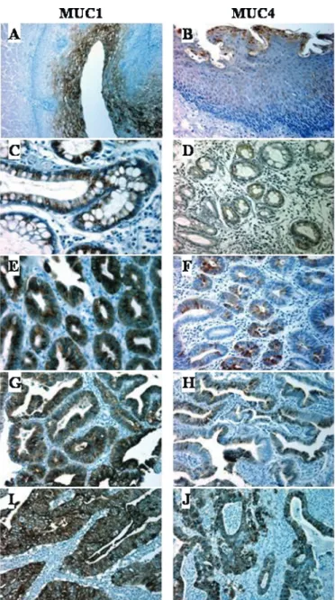
Figure 1 shows representative examples of the expression profiles of MUC1 and MUC4 apomucins in OA and coexisting lesions during the progression of OA. In normal mucosa, MUC1 and MUC4 were expressed in the cytoplasm of superficial epithelial cells (fig 1A, B). In metaplasia, MUC1 and MUC4 expression was in the cytoplasm of non-goblet columnar cells and goblet cells (fig 1C, D). In dysplasia (fig 1E–H) and adenocarcinoma (fig 1I, J), MUC1 and MUC4 expression was diffuse and heterogeneous in both the cytoplasm and on the apical membrane of cells (fig 1E–J).
The mucin staining patterns are detailed in table 1. Among 52 patients with OA, 25 patients (48.1%) had various BO lesions, including intestinal metaplasia (36.5%), low-grade dysplasia
(25.0%) and high-grade dysplasia (28.8%). Strong positive staining of MUC1 was seen in all but three specimens. Positive staining of MUC4 was seen in 92% of the normal mucosa specimens and it decreased to 53% (p,0.001) in metaplasia. No significant differences were found between metaplasia and low-grade dysplasia (53% versus 62%, p = 0.99), low- and high-grade dysplasias (62% versus 60%, p = 0.95), or high-grade dysplasia and OA (60% versus 78%, p = 0.99). The distribution of the number of stained cells did not differ significantly between the different steps of the carcinogenetic process (not shown).
Histopathological characteristics of resected OA and long term follow-up
The histopathological characteristics of resected OA and long-term follow-up results are summarised in table 2. The postoperative mortality rate was 4% (n = 2). Recurrence was only considered in long term survival patients who underwent microscopic and macroscopic complete (R0) resection (n = 41). At the time of writing, 28 (60%) patients had experienced recurrence, with median time to recurrence of 11 months (range 6–80 months). The median (SD) survival of patients was 36 (4.2) months.
Association between MUC1 and MUC4 expression in OA and clinicopathological features
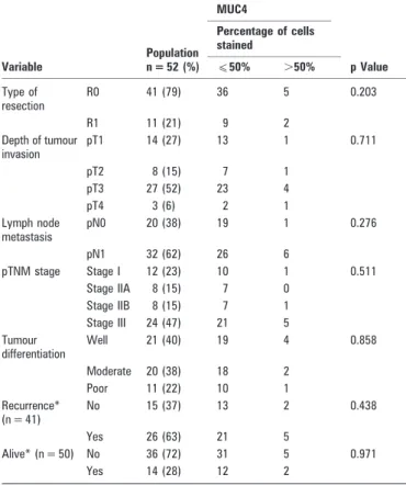
All but two cases expressed MUC1 strongly, so it was not descriptive of clinicopathological parameters. We evaluated the association between MUC4 expression and clinicopathological features using a cut-off value of 50% positively stained neoplastic cells (table 2). There was no significant correlation between MUC4 staining and the type of resection, depth of tumour invasion, lymph node metastasis, pTNM (tumour, node, metastases) stage, tumour differentiation, recurrence or survival. Statistical analyses were also performed by either (i) using a cut-off value of 10% or (ii) calculating the ratio between the number of cells stained in
Figure 1 Expression of MUC1 and MUC4 apomucins during the progression of Barrett-associated oesophageal adenocarcinoma. Immunohistochemical studies of normal oesophageal mucosa (A, B), Barrett metaplasia (C, D), low-grade dysplasia (E, F), high-grade dysplasia (G, H) and oesophageal adenocarcinoma (I, J) were carried out with anti-MUC1 (A, C, E, G, I) and anti-MUC4 (B, D, F, H, J) specific monoclonal antibodies (magnification 6200).
Table 1 Expression of MUC1 and MUC4 apomucins by immunohistochemistry according to histopathological analysis
Histological diagnosis No. of patients analysed Stained cells (%) MUC1 (% of specimen) MUC4 (% of specimen) Normal oesophageal mucosa 52 0 0 8 1–10 0 20 11–50 0 47 .50 100 25 Metaplasia 19 0 0 47 1–10 0 42 11–50 4 11 .50 96 0 Low-grade dysplasia 13 0 0 38 1–10 0 31 11–50 0 23 .50 100 8 High-grade dysplasia 15 0 0 40 1–10 0 13 11–50 0 34 .50 100 13 Adenocarcinoma 52 0 0 22 1–10 0 37 11–50 4 28 .50 96 13
adenocarcinoma and normal mucosa. These analyses also did not yield a statistical correlation between MUC4 expression and any clinicopathological feature (not shown).
DISCUSSION
Several smaller sample studies have reported downregulation of MUC1 and MUC4 between normal mucosa and metaplasia and upregulation in the progression of OA at the mRNA level and the protein level.3 5–7 The authors have consequently proposed
that a high level of MUC1 protein or MUC4 mRNA expression could serve as a reliable tumour marker in this process.5 7 In
accordance with other groups, and despite the fact that we deliberately analysed entire surgical specimens of resected OA, our results do not support any role for MUC1 or MUC4 protein expression as a diagnostic tool to facilitate early detection of high-grade dysplasia or OA.10 11An entire surgical specimen may
provide better information than random sample biopsies by allowing the analysis of both carcinomatous and coexisting lesions (metaplasia, dysplasia). The wide variability of results between studies raises the question of antibody specificity, especially when recognising glycan motifs that may be brought by surface glycoproteins and other mucins. In our study, we used an antibody that recognises a peptide motif specific to MUC4 and which does not react with other mucins.3
Two groups previously reported high expression of MUC1 in advanced OA.5 12In accordance with Flucke et al, our results do
not support any correlation between MUC1 or MUC4 with clinicopathological features in OA.10One could argue that there
is a lack of statistical power in our study. However, due to the absence of any statistical trend, we deduce no major clinical role
for MUC1 or MUC4 as prognostic markers in OA and, therefore the study will not be extended. The raised expression of MUC1 and MUC4 in adenocarcinomas is usually associated with more invasiveness and worse prognosis.6 9However, improved
survi-val and decreased recurrence have also been reported in breast and stomach cancers for MUC1, and in upper aerodigestive tract tumours for MUC4, suggesting a potential duality of function for membrane-bound mucins.3 6 8 13
The present study confirmed that most OAs express MUC1 and MUC4, but it did not support (i) any role as diagnostic tools to facilitate early detection of OA in BO, and (ii) any correlation between MUC1 or MUC4 expression and prognosis.
Acknowledgements: The authors wish to thank Dr D Swallow (MRC, London, England) for the kind gift of MUC1 antibody.
Funding: This work was funded by a grant from la Ligue Nationale contre le Cancer (comite´ du Pas-de-Calais) (IVS).
Competing interests: None.
Ethics approval: Ethics approval was obtained.
REFERENCES
1. Mariette C, Piessen G, Triboulet JP. Therapeutic strategies in oesophageal carcinoma: role of surgery and other modalities. Lancet Oncol 2007;8:545–53. 2. Hollingsworth MA, Swanson BJ. Mucins in cancer: protection and control of the
cell surface. Nat Rev Cancer 2004;4:45–60.
3. Piessen G, Jonckheere N, Vincent A, et al. Regulation of the human mucin MUC4 by taurodeoxycholic and taurochenodeoxycholic bile acids in oesophageal cancer cells is mediated by hepatocyte nuclear factor 1alpha. Biochem J 2007;402:81–91. 4. Mariette C, Piessen G, Leteurtre E, et al. Activation of MUC1 mucin expression by
bile acids in human esophageal adenocarcinomatous cells and tissues is mediated by the phosphatidylinositol 3-kinase signaling pathway. Surgery 2008;143:58–71. 5. Chinyama CN, Marshall RE, Owen WJ, et al. Expression of MUC1 and MUC2 mucin
gene products in Barrett’s metaplasia, dysplasia and adenocarcinoma: an immunopathological study with clinical correlation. Histopathology 1999;35:517–24. 6. Piessen G, Mariette C, Van Seuningen I. Mucin expression and regulation in oesophageal
cancer: New molecular targets in cancer progression from Barrett’s oesophagus to adenocarcinoma? In: Van Seuningen I, ed. The epithelial mucins: structure/function. Roles in cancer and inflammatory diseases. Kerala: Research Signpost, 2008:169–81. 7. Bax DA, Einerhand AWC, van Dekken H, et al. MUC4 is increased in high grade
intraepithelial neoplasia in Barrett’s oesophagus and is associated with a proapoptotic Bax to Bcl-2 ratio. J Clin Pathol 2004;57:1267–72.
8. van der Vegt B, de Roos MA, Peterse JL, et al. The expression pattern of MUC1 (EMA) is related to tumour characteristics and clinical outcome of invasive ductal breast carcinoma. Histopathology 2007;51:322–35.
9. Saitou M, Goto M, Horinouchi M, et al. MUC4 expression is a novel prognostic factor in patients with invasive ductal carcinoma of the pancreas. J Clin Pathol 2005;58:845–52. 10. Flucke U, Steinborn E, Dries V, et al. Immunoreactivity of cytokeratins (CK7, CK20) and mucin peptide core antigens (MUC1, MUC2, MUC5AC) in adenocarcinomas, normal and metaplastic tissues of the distal oesophagus, oesophago-gastric junction and proximal stomach. Histopathology 2003;43:127–34.
11. Glickman JN, Blount PL, Sanchez CA, et al. Mucin core polypeptide expression in the progression of neoplasia in Barrett’s esophagus. Hum Pathol 2006;37:1304–15. 12. Gulmann C, Counihan I, Grace A, et al. Cytokeratin 7/20 and mucin expression
patterns in oesophageal, cardia and distal gastric adenocarcinomas. Histopathology 2003;43:453–61.
13. Wang RQ, Fang DC. Alterations of MUC1 and MUC3 expression in gastric carcinoma: relevance to patient clinicopathological features. J Clin Pathol 2003;56:378–84.
Table 2 Histopathological characteristics, long-term follow-up results, and their correlation with MUC4 apomucin expression by
immunohistochemistry in 52 patients with oesophageal adenocarcinoma
Variable Population n = 52 (%) MUC4 p Value Percentage of cells stained (50% .50% Type of resection R0 41 (79) 36 5 0.203 R1 11 (21) 9 2 Depth of tumour invasion pT1 14 (27) 13 1 0.711 pT2 8 (15) 7 1 pT3 27 (52) 23 4 pT4 3 (6) 2 1 Lymph node metastasis pN0 20 (38) 19 1 0.276 pN1 32 (62) 26 6 pTNM stage Stage I 12 (23) 10 1 0.511 Stage IIA 8 (15) 7 0 Stage IIB 8 (15) 7 1 Stage III 24 (47) 21 5 Tumour differentiation Well 21 (40) 19 4 0.858 Moderate 20 (38) 18 2 Poor 11 (22) 10 1 Recurrence* (n = 41) No 15 (37) 13 2 0.438 Yes 26 (63) 21 5 Alive* (n = 50) No 36 (72) 31 5 0.971 Yes 14 (28) 12 2
*At the last follow-up.
R0, microscopically and macroscopically complete resection; R1, microscopically incomplete resection; TNM, tumour, node, metastases.
Take-home messages
c MUC1 and MUC4 membrane-bound mucins are frequently and strongly expressed in Barrett-associated oesophageal adenocarcinoma (OA) and coexisting precancerous lesions, suggesting their potential role in tumour progression. c The present study does not support any role for MUC1 or
MUC4 as diagnostic tools to facilitate early detection of OA. c MUC1 and MUC4 immunohistochemical expression was not associated with prognosis in this large cohort of 52 patients with resected OA.