HAL Id: hal-01957839
https://hal.sorbonne-universite.fr/hal-01957839
Submitted on 17 Dec 2018
HAL is a multi-disciplinary open access
archive for the deposit and dissemination of
sci-entific research documents, whether they are
pub-lished or not. The documents may come from
teaching and research institutions in France or
abroad, or from public or private research centers.
L’archive ouverte pluridisciplinaire HAL, est
destinée au dépôt et à la diffusion de documents
scientifiques de niveau recherche, publiés ou non,
émanant des établissements d’enseignement et de
recherche français ou étrangers, des laboratoires
publics ou privés.
Lipids and synaptic functions
Fanny Mochel
To cite this version:
Fanny Mochel. Lipids and synaptic functions. Journal of Inherited Metabolic Disease, Springer Verlag,
In press, �10.1007/s10545-018-0204-1�. �hal-01957839�
Lipids and synaptic functions
Fanny Mochel1,2,3
Abstract
Synaptic functions have long been thought to be driven by proteins, especially the SNARE complex, contrasting with a relatively passive role for lipids constituting cell membranes. It is now clear that not only lipids, i.e. glycerophospholipids, sphingolipids and sterols, play a determinant role in the dynamics of synaptic membranes but they also actively contribute to the endocytosis and exocytosis of synaptic vesicles in conjunction with synaptic proteins. On the other hand, a growing number of inborn errors of metabolism affecting the nervous system have been related to defects in the synthesis and remodelling of fatty acids, phospholipids and sphingolipids. Alterations of the metabolism of these lipids would be expected to affect the dynamics of synaptic membranes and synaptic vesicles. Still, only few examples are currently documented. It remains to be determined to which extent the pathophysiology of disorders of complex lipids biosynthesis and remodelling share common pathogenic mechanisms with the more traditional synaptopathies.
Role of lipids in synaptic functions
Lipids can be subdivided into simple lipids such as fatty acids (FA), sterols and acylglycerols, and complex lipids such as glycerophospholipids, ether phospholipids and sphingolipids (Fahy et al.2005). The fatty acyl group from FA and conjugates is made of a repeating series of methy-lene bridges rendering them hydrophobic. The fatty acyl structure is the major lipid backbone of complex lipids. Glycerolipids result from the link between FA and a glycerol backbone through ester bounds and encompass all glycerol-containing lipids. They include glycerophospholipids (phos-phatidyl-choline, -ethanolamine, -serine and -inositol) and glycerolipids (mono, di and triacylglycerol). Phospholipids are made of a glycerol molecule, two FA and a phosphate
group that is modified by an alcohol. The phosphate group is hydrophilic due to its negatively charged polar head, while the FA chains are hydrophobic due uncharged, non-polar tails (Fahy et al. 2005). Triacylglycerols differ from glycerophospholipids as they carry a third ester bound acyl chain instead of the phosphate group, making them completely hydrophobic (Fahy et al. 2005). Sphingolipids share a common structural feature, a sphingoid base back-bone that is synthesised de novo from serine and a long chain fatty acyl-CoA. The sphingoid bases are acylated with a FA in order to form ceramide, which can carry a hydro-philic head group, e.g. phosphorylcholine in the case of sphingomyelin or carbohydrate residues in the case of glycosphingolipids (Fahy et al. 2005). Most of these major lipid classes, e.g. phospholipids, sphingolipids and sterols, contain a wide range of metabolic and molecular species that differ in polar head and hydrophobic tail groups, phos-phorylation states and regulation by kinases and phospha-tases. Therefore, the signalling and regulatory potential of lipids is wide. Synaptic membranes are especially abundant in glycerophospholipids, sphingolipids and cholesterol.
The key role of lipids in synaptic functions is mediated by their implication in the dynamics of synaptic membranes and synaptic vesicles (Rohrbough and Broadie2005; Piomelli et al.2007; Lauwers et al.2016). Lipid composition and topol-ogy is indeed an important factor in the spatial regulation of membrane shape, curvature and fluidity during vesicle fusion (exocytosis) and fission (endocytosis). Let us first consider the
Communicated by: Sander M Houten * Fanny Mochel
fanny.mochel@upmc.fr
1 Sorbonne Université, UPMC-Paris 6, UMR S 1127 and Inserm U 1127, and CNRS UMR 7225, and ICM, F-75013, Paris, France 2
Sorbonne Université, GRC no. 13, Neurométabolisme, Paris, France 3
Department of Genetics and Reference Centre for Adult Neurometabolic Diseases, AP-HP, La Pitié-Salpêtriere University Hospital, Paris, France
role of lipids in the dynamics of synaptic membranes. Due to their physic properties, lipids directly control membrane bio-physical parameters such as curvature, fluidity and thickness. Indeed, the geometry of membrane lipids depends on the area occupied by the head group versus the hydrophobic part of the molecule. Cone-shaped lipids (ceramide, phosphatidic acid, diacylglycerol, phosphatidylethanolamine) have a small head group and promote negative membrane curvature. Conversely, lipids like phosphatidylcholine have a nearly cy-lindrical geometry, while lysophosphatidylcholine has an inverted cone shape. Likewise, presynaptic lipid bilayers un-dergo extensive and concerted shape changes as they transit through the different lipid shapes (cone, cylinder, inverted cone). These changes are driven by phospholipases, e.g. deacylation of phosphatidylcholine (cylinder) into lysophosphatidylcholine (inverted cone) by phospholipase A2 (Lauwers et al.2016). Hence, remodelling of membrane phospholipids by phospholipases is crucial for synaptic func-tion and maintenance as it directly affects membrane curvature but also permits the release of lipid signalling molecules like arachidonic acid by phospholipase A2 or diacylglycerol by phospholipase C.
Let us now consider the role of lipids in endocytosis and exocytosis of synaptic vesicles. Presynaptic endocytosis and synaptic vesicle release are linked in time and space. Several proteins involved in the priming and docking of synaptic ves-icle bind lipids that are negatively charged, e.g. phos-phatidylinositol phosphates, in order to cluster in the active zone where exocytosis takes place (Lauwers et al. 2016). Many of these proteins recognise only one given phos-phatidylinositol phosphate species (Hammond and Balla
2015). Therefore, the phosphatidylinositol phosphates-composition of a membrane determines which proteins are recruited, a process that has been well characterised in the endosomal system (Di Paolo and De Camilli 2006). Phosphatidylinositol phosphates are also inter-converted by specific kinases and phosphatases. These enzymes are deter-minant to generate rapid changes in the membrane concentra-tion of phosphatidylinositol phosphates so that the endocytic machinery can be sequentially recruited for the formation of synaptic vesicles (Ueda2014). Hence, phosphatidylinositol phosphates and their metabolising enzymes help coordinate endocytosis, via the sequential recruitment and subsequent d i s a s s e m b l y o f m e m b r a n e - r e m o d e l l i n g p r o t e i n s . Furthermore, the exocytosis of synaptic vesicles requires membrane fusion between the synaptic vesicle and plasma membrane. This process is mediated by the assembly of SNARE complexes (especially synaptobrevin, syntaxin and SNAP-25) and is assisted by cone-shaped, also called fusogenic, lipids and membrane-remodelling proteins. Likewise, phosphatidylinositol phosphates, but also phosphatidylserine (Williams et al.2009), promote the bind-ing of synaptobrevin and syntaxin by shieldbind-ing positive
charges on either protein as they approach each other during synaptic vesicle docking. Besides, polyunsaturated FA (PUFA) can induce a conformational change on syntaxin favouring its interaction with SNAP-25 (Darios et al.2007) while sphingosine mediates the relief of the cytoplasmic part of synaptobrevin, which is necessary for its interaction with the syntaxin/SNAP-25 heterodimer (Darios et al. 2009). Altogether, by regulating interactions between charged do-mains of the SNARE machinery, glycerophospholipids, sphingolipids and PUFA play an active role in exocytosis. Other lipids such as diacylglycerol and phosphatidic acid have a direct role in the regulation of synaptic vesicle exo/ endocytosis (Tu-Sekine et al.2015). Thus, diacylglycerol acts on priming of syntaxin for SNARE-mediated vesicle fusion (Wierda et al.2007).
It is noteworthy that cholesterol impacts both the dynamics of synaptic membranes and synaptic vesicles. Together with sphingolipids, cholesterol is indeed essential for raft integrity and function. Still, only cholesterol has the unique ability to engage in rapid trans-bilayer flip-flop, allowing for stress re-laxation during membrane bending and thereafter membrane remodelling (Bruckner et al.2009). On the other hand, cho-lesterol is enriched in secretory and synaptic vesicles and is important for neurosecretory and synaptic vesicle cycling and release (Rohrbough and Broadie2005). Indeed, syntaxins are concentrated in cholesterol-dependent clusters at which secre-tory vesicles preferentially dock and fuse (Lang et al.2001). Cholesterol also binds the synaptic vesicle protein synaptophysin in order to modulate its interaction with synaptobrevin (Mitter et al. 2003). The cholesterol–
synaptophysin–synaptobrevin interaction is thought to recruit and segregate essential synaptic vesicle-specific proteins from the plasma membrane (Thiele et al.2000).
Neurometabolic diseases caused by defects
in the synthesis or remodelling of complex
lipids
Defects in lipid catabolic pathways have been involved for decades in numerous inborn errors of metabolism (IEM), es-pecially lysosomal storage diseases and peroxisome biogene-sis disorders. These catabolic defects lead to the accumulation of lipid substrates that have become useful diagnostic biolog-ical markers such as lysosphingolipids (Pettazzoni et al.2017) and C26:0- lysophosphatidylcholine (Klouwer et al.2017). Conversely, IEM related to defects in the biosynthesis and remodelling of glycerophospholipids, sphingolipids and fatty acids have only been delineated recently (Lamari et al.2013) but rapidly expanded to over 100 diseases (Lamari et al.2015) thanks to the increased access to next-generation sequencing tools. Diagnostic biological markers are still missing for this large group of IEM of diseases as decrease in product is
usually more difficult to detect than substrate accumulation, and even more so with lipid species that are very redundant such as glycerophospholipids. Still, the measurement of sev-eral hundreds of lipid species using lipidomics may allow a better delineation of some of these metabolic defects (Seyer et al.2016). The clinical presentation of IEM involved in the synthesis and remodelling of complex lipids has been reviewed in details (Garcia-Cazorla et al.2015). All organs and systems may be affected—the central and peripheral ner-vous system, eye, muscle, heart, skin, bone, liver and immune system. We will focus here on IEM primarily affecting the nervous system and presenting with a combination of intellec-tual disability, epilepsy, movement disorder, sensorineural dysfunction, peripheral neuropathy and/or myopathy.
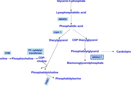
A first category of IEM relates to defects in the synthesis of phospholipids (Fig.1). ABHD5 is α/β-hydrolase domain-containing 5 that acts as a lysophosphatidic acid acyltransfer-ase and therefore plays a role in the synthesis of phosphatidic acid (Fig.1). Its defects cause Chanarin-Dorfman syndrome, a neutral lipid storage disease characterised by intellectual dis-ability, cerebellar ataxia and myopathy associated with cata-ract, hearing loss, erythrodermic ichthyosis and hepatomegaly with liver steatosis (Lefèvre et al.2001). Phosphatidic acid can be converted into diacylglycerol by a phosphatidic acid phos-phatase called Lipin 1 (Fig.1). Defect in Lipin-1 is the most common cause of rhabdomyolysis after FA oxidation disor-ders, and episodes are usually induced by fever, anaesthesia or fasting (Zeharia et al.2008). Choline kinase b (CHB) cataly-ses the first step in the de novo biosynthesis of phosphatidyl-choline (Fig.1). CHB deficiency causes intellectual disability, epilepsy, early onset muscular dystrophy and dilated cardio-myopathy (Mitsuhashi et al.2011). PCYT1A encodes the α-isoform of phosphate cytidylyltransferase 1 choline, which is responsible for converting phosphocholine into cytidine di-phosphate-choline, a key intermediate step in the phosphati-dylcholine biosynthesis pathway (Fig.1). Defect in PCYT1A c a u s e s a c o n e - r o d d y s t r o p h y a s s o c i a t e d w i t h a spondylometaphyseal dysplasia (Hoover-Fong et al. 2014). Phosphatidylserine synthase 1 (PSS1) synthesises phosphatidylserine through the exchange of L-serine with the choline moiety of phosphatidylcholine (Fig.1). PSS1 de-ficiency is responsible for the autosomal dominant Lenz-Majewski syndrome that is characterised by intellectual dis-ability,cutis laxa and a sclerosing bone dysplasia with hyper-ostotic dwarfism (Sousa et al.2014).
The second category of IEM relates to the remodelling of phospholipids (Fig.2). SERAC1 is involved in remod-elling 34:1 to phosphatidylglycerol-3 6 : 1 s p e c i e s , t h e l a t t e r b e i n g t h e p r e c u r s o r f o r bismonoacylglycerophosphate (BMP) and cardiolipin spe-cies (Fig. 1). Defect in SERAC1 causes MEGDEL (3-MEthylGlutaconic aciduria with deafness, encephalopathy and Leigh-like) syndrome, characterised by intellectual
disability, deafness, dystonia and spasticity (Wortmann et al. 2012). An alternative pathway for diacylglycerol and FA synthesis is their release from membrane phospholipids by specific phospholipases and lysophospholipases (Fig.
2). PLA2G6 is a phospholipase A2 that removes unsatu-rated FA chains and arachidonic acid, which is the starting molecule of many complex FA like prostaglandins, leuko-trienes (Fig. 2). PLA2G6 mutations are associated with three phenotypes: INAD (infantile neuroaxonal dystro-phy) that causes psychomotor regression, spasticity, pe-ripheral neuropathy and optic atrophy; NBIA (neurode-generation with brain iron accumulation) that causes cer-ebellar ataxia, dystonia and psychiatric symptoms; and a dystonia-parkinsonism syndrome (Paisan-Ruiz et al.2009; Illingworth et al., 2014). DDHD1 and DDHD2 are phos-pholipase A1 that remove saturated FA chains (Fig. 2). DDHD2 is also a triglyceride lipase. Mutations in DDHD1 and DDHD2 are associated with complex forms of spastic paraplegia—i.e. pigmentary retinopathy and in-tellectual disability, respectively (Tesson et al. 2012; Schuurs-Hoeijmakers et al. 2012). NTE (neuropathy tar-get esterase) is a patatin-like phospholipase that has a l y s o p h o s p h o l i p a s e a c t i v i t y , r e l e a s i n g glycerophospholipids (Fig. 2). NTE mutations have been associated with a large spectrum of neurological disor-ders—spastic paraplegia 39, Gordon Holmes syndrome, Boucher-Neuhauser syndrome, Olivier McFarlane syn-drome and Laurence-Moon synsyn-drome—that have in com-mon spastic paraplegia, cerebellar ataxia, chorioretinal dystrophy and hypogonadism (Synofzik et al. 2014). CYP2U1 is a cytochrome P450 that catalyses the hydrox-ylation of arachidonic acid and similar long-chain FA, leading to the formation of hydroxyeicosatetraenoic acids (Fig. 2). CYP2U1 mutations cause spastic paraplegia, which can be associated with intellectual disability, dys-tonia, peripheral neuropathy and pigmentary maculopathy (Tesson et al. 2012). Phospholipase C removes phosphate groups and leads to the release of diacylglycerol and its further conversion by diacylglycerol lipase into 2-arachidonoyl glycerol, which can then be hydrolysed into arachidonic acid byα/β-hydrolase-12, ABHD12 (Fig. 2). ABHD12 mutations are associated with PHARC syn-drome, characterised by peripheral neuropathy, hearing loss, cerebellar ataxia, pigmentary retinopathy and cata-ract (Fiskerstrand et al. 2010) or an isolated pigmentary retinopathy (Nishiguchi et al. 2014).
The third category of IEM relates to the synthesis of sphingolipids (Fig. 3). Serine palmitoyltransferase catalyses the condensation of palmitoyl-CoA and L-serine to generate ketosphinganine, which is then reduced to sphinganine. This is the first and rate limiting step in the de novo synthesis of sphingolipids.SPTLC1 and SPTLC2 mutations cause an au-tosomal dominant sensory and autonomic neuropathy due to
the abnormal production of toxic ceramides (Rotthier et al.
2010). Fatty acid 2 hydroxylase (FA2H) introduces a hydroxyl group at the C2-position of FA. The corresponding hydroxyl-ated fatty acyl-CoA is incorporhydroxyl-ated by ceramide synthases (CERS) into sphingolipids (Fig.3). FA2H mutations have been associated with a spectrum of spastic paraplegia (SPG35), intellectual disability, epilepsy, dystonia, white mat-ter disease and NBIA (Kruer et al.2010).CERS2 mutations were found in the Amish population causing progressive myo-clonic epilepsy (Mosbech et al.2014). GBA2 encodes the glucocerebrosidase that is mainly expressed in the endoplas-m i c r e t i c u l u endoplas-m a n d c a n p r o d u c e c e r a endoplas-m i d e f r o endoplas-m glucosylceramide (Fig.3).GBA2 mutations are responsible for a spastic ataxia often associated with intellectual disability, axonal neuropathy, cataract and/or hearing loss (Martin et al.
2013; Hammer et al.2013). GM3 synthase catalyses the for-mation of GM3 gangliosides using lactosylceramide as the substrate. Deficiency in GM3 synthase causes an early onset e p i l e p s y a s s o c i a t e d w i t h i n t e l l e c t u a l d i s a b i l i t y,
choreoathetosis, optic atrophy and hyperpigmented lesions (Simpson et al.2004).B4GALNT1 encodes β-1,4-N-acetyl-galactosaminyltransferase 1, that synthesises GM2 and GD2 gangliosides. B4GALNT1 mutations lead to intellectual dis-ability, cerebellar ataxia, dystonia, muscle wasting and axonal neuropathy (Boukhris et al.2013).
Abnormal lipid metabolism and synaptic
dysfunction?
How to reconcile this expanding list of IEM affecting the biosynthesis and remodelling of glycerophospholipids, sphingolipids and fatty acids with the key role of lipids in synaptic function? One can indeed hypothesise that the altered production of glycerophospholipids (e.g. phosphatidylcho-line, phosphatidylserine, phosphatidylinositol phosphates) or their precursors (e.g. phosphatidic acid, diacylglycerol), PUFA (e.g. arachidonic acid) or sphingolipids (e.g.
CDP Diacylglycerol Diacylglycerol Lipin 1 ABHD5 Glycerol-3-phosphate Lysophosphatidic acid Phosphatidic acid Phosphatidylcholine Choline Phosphocholine CDP-choline CHB PC cytidylyl-transferase Phosphatidylserine Phosphatidylglycerol Bismonoglycerolphosphate Cardiolipin SERAC1 Fig. 1 Phospholipids
biosynthetic pathways—selected steps (modified from Lamari et al.
2015). ABHD5α/β-hydrolase domain-containing 5, CHB cho-line kinase b, PCYT1Aα-isoform of phosphate cytidylyltransferase 1 choline, PSS1
phosphatidylserine synthase 1. Dashed boxes represent remodel-ling steps COOH Arachidonic acid PLA2G6 Prostaglandins Leukotrienes DDHD1,2 + Phosphoglycerol NTE + COOH OH COOH OH 20-HETE 19-HETE CYP2U1 Diacylglycerol 2-Arachidonoylglycerol ABHD12 PLC
Fig. 2 Phospholipids remodelling pathways—selected steps (modified from Lamari et al.
2015). PLA2G6 phospholipase A2 group VI, NTE neuropathy target esterase, ABHD12 α/β-hydrolase-12, HETE hydroxyeicosatetraenoic acids, PLC phospholipase C. Dashed boxes represent remodelling steps
sphingosine, ceramide) would affect membrane curvature and the dynamics of synaptic membranes, as well as the formation of synaptic vesicles and their release. Likewise, loss of func-tion of PLA2G6, which catalyses the deacylafunc-tion of phospha-tidylcholine with release of unsaturated FA chain and regu-lates membrane remodelling, has been associated with presyn-aptic membrane defects (Sumi-Akamaru et al. 2015). Furthermore, loss of function of phosphatidylinositol phos-phates metabolising enzymes have been associated with sev-eral neurodegenerative disorders. Mutations inPIP5K1C, which encodes an enzyme that phophorylates phosp h a t i d y l i n o s i t o l 4 phosp h o s phosp h a t e t o g e n e r a t e phosp h o s -phatidylinositol-4,5-bisphosphate, and mutations inERBB3, which encodes an activator of the phosphatidylinositol-3-ki-nase, cause lethal congenital contracture syndromes with at-rophy of the anterior horn of the spinal cord (Narkis et al.
2007a; Narkis et al. 2007b). Synaptojanin, encoded by SYNJ1, is a phosphatidylinositol phosphate phosphatase. SYNJ1 mutations in the SAC1 domain that dephosphorylates phosphatidylinositol 3-phosphate and phosphatidylinositol 4-phosphate, found on synaptic vesicle membranes, causes early onset Parkinson’s disease and epilepsy (Krebs et al.2013).
Altogether, both pre- and postsynaptic effects of altered lipid metabolism ought to have an impact on the development of neurological disorders. Similarly to IEM affecting lipid metabolism, diseases caused by defects in proteins involved in synaptic functions, traditionally called synaptopathies,
encompass epilepsy, intellectual disability, movement disor-ders, cerebellar ataxia, spastic paraplegia, peripheral neuropa-thy and myopaneuropa-thy (Cortès-Saladelafont et al. 2016). Therefore, it remains to be determined to which extent the pathophysiology of IEM affecting the biosynthesis and re-modelling of glycerophospholipids, sphingolipids and fatty acids is related to synaptic dysfunction and share common pathogenic mechanisms with these synaptopathies.
Acknowledgements The author is very grateful to Pr Jean-Marie Saudubray and Dr. Foudil Lamari for their pioneering and invaluable contribution to the delineation of the novel class of inborn errors of me-tabolism represented by disorders of complex lipids biosynthesis and remodelling.
Compliance with ethical standards
Details of ethics approval No ethics approval was required. Patient consent statement Patient consent was not required. Conflict of interest Fanny Mochel declares that she has no conflict of interest.
References
Boukhris A, Schule R, Loureiro JL et al (2013) Alteration of ganglioside biosynthesis responsible for complex hereditary spastic paraplegia. Am J Hum Genet 93:118–123
Bruckner RJ, Mansy SS, Ricardo A et al (2009) Flip-flop-induced relax-ation of bending energy: implicrelax-ations for membrane remodeling. Biophys J 97:3113–3122
Cortès-Saladelafont E, Tristán-Noguero A, Artuch R et al (2016) Diseases of the synaptic vesicle: a potential new group of neurometabolic disorders affecting neurotransmission. Semin Pediatr Neurol 23:306–320
Darios F, Connell E, Davletov B (2007) Phospholipases and fatty acid signalling in exocytosis. J Physiol 585:699–704
Darios F, Wasser C, Shakirzyanova A et al (2009) Sphingosine facilitates SNARE complex assembly and activates synaptic vesicle exocyto-sis. Neuron 62:683–694
Di Paolo G, De Camilli P (2006) Phosphoinositides in cell regulation and membrane dynamics. Nature 443:651–657
Fahy E, Subramaniam S, Brown HA et al (2005) A comprehensive clas-sification of lipids. J Lipid Res 46:839–861
Fiskerstrand T, H'mida-Ben Brahim D, Johansson S et al (2010) Mutations in ABHD12 cause the neurodegenerative disease PHARC: an inborn error of endocannabinoid metabolism. Am J Hum Genet 87:410–417
Garcia-Cazorla À, Mochel F, Lamari F et al (2015) The clinical spectrum of inherited diseases involved in the synthesis and remodeling of complex lipids. A tentative overview. J Inherit Metab Dis 38:19–40 Hammer MB, Eleuch-Fayache G, Schottlaender LV et al (2013) Mutations in GBA2 cause autosomal-recessive cerebellar ataxia with spasticity. Am J Hum Genet 92:245–251
Hammond GR, Balla T (2015) Polyphosphoinositide binding domains: key to inositol lipid biology. Biochim Biophys Acta 1851:746–758 Hoover-Fong J, Sobreira N, Jurgens J et al (2014) Mutations in PCYT1A, encoding a key regulator of phosphatidylcholine metabolism, cause
+ Palmitoyl-CoA L-Serine 3-Ketosphinganine Dihydrosphingosine NADP+ NADPH Fatty acid 2-OH-AcylCoA CoA-SH Dihydroceramide 2-OH fatty acid
Ceramide Glucosylceramide FAD+ FADH Lactosylceramide GM3 SPTLC1,2 FA2H GM3 synthase CERS GBA2 GA2 GM2 GD3 GD2 B4GALNT1 B4GALNT1
Fig. 3 Sphingolipids biosynthetic pathways—selected steps. SPTLC 1/2 serine palmitoyltransferase1/2, F FA2H fatty acid 2 hydroxylase, CERS ceramide synthase, GBA2 glucocerebrosidase type 2, B4GALNT1 β-1,4-N-acetyl-galactosaminyltransferase 1
spondylometaphyseal dysplasia with cone-rod dystrophy. Am J Hum Genet 94:105–112
Illingworth MA, Meyer E, Chong WK et al (2014) PLA2G6-associated neurodegeneration (PLAN): further expansion of the clinical, radio-logical and mutation spectrum associated with infantile and atypical childhood-onset disease. Mol Genet Metab 112:183–189
Klouwer FCC, Ferdinandusse S, van Lenthe H et al (2017) Evaluation of C26:0-lysophosphatidylcholine and C26:0-carnitine as diagnostic markers for Zellweger spectrum disorders. J Inherit Metab Dis 40: 875–881
Krebs CE, Karkheiran S, Powell JC et al (2013) The Sac1 domain of SYNJ1 identified mutated in a family with early-onset progressive Parkinsonism with generalized seizures. Hum Mutat 34:1200–1207 Kruer MC, Paisán-Ruiz C, Boddaert N et al (2010) Defective FA2H leads to a novel form of neurodegeneration with brain iron accumulation (NBIA). Ann Neurol 68:611–618
Lamari F, Mochel F, Sedel F et al (2013) Disorders of phospholipids, sphingolipids and fatty acids biosynthesis: toward a new category of inherited metabolic diseases. J Inherit Metab Dis 36:411–425 Lamari F, Mochel F, Saudubray JM (2015) An overview of inborn errors
of complex lipid biosynthesis and remodelling. J Inherit Metab Dis 38:3–18
Lang T, Bruns D, Wenzel D et al (2001) SNAREs are concentrated in cholesterol-dependent clusters that define docking and fusion sites for exocytosis. EMBO J 20:2202–2213
Lauwers E, Goodchild R, Verstreken P (2016) Membrane lipids in pre-synaptic function and disease. Neuron 90:11–25
Lefèvre C, Jobard F, Caux F et al (2001) Mutations in CGI-58, the gene encoding a new protein of the esterase/lipase/thioesterase subfamily, in Chanarin-Dorfman syndrome. Am J Hum Genet 69:1002–1012 Martin E, Schüle R, Smets K et al (2013) Loss of function of
glucocerebrosidase GBA2 is responsible for motor neuron defects in hereditary spastic paraplegia. Am J Hum Genet 92:238–244 Mitsuhashi S, Ohkuma A, Talim B et al (2011) A congenital muscular
dystrophy with mitochondrial structural abnormalities caused by defective de novo phosphatidylcholine biosynthesis. Am J Hum Genet 88:845–851
Mitter D, Reisinger C, Hinz B et al (2003) The synaptophysin/ synaptobrevin interaction critically depends on the cholesterol con-tent. J Neurochem 84:35–42
Mosbech MB, Olsen AS, Neess D et al (2014) Reduced ceramide syn-thase 2 activity causes progressive myoclonic epilepsy. Ann Clin Transl Neurol. 1: 88–98
Narkis G, Ofir R, Landau D et al (2007a) Lethal contractural syndrome type 3 (LCCS3) is caused by a mutation in PIP5K1C, which encodes PIPKI gamma of the phophatidylinsitol pathway. Am J Hum Genet 81:530–539
Narkis G, Ofir R, Manor E et al (2007b) Lethal congenital contractural syndrome type 2 (LCCS2) is caused by a mutation in ERBB3 (Her3), a modulator of the phosphatidylinositol-3-kinase/Akt path-way. Am J Hum Genet 81:589–595
Nishiguchi KM, Avila-Fernandez A, van Huet RA et al (2014) Exome sequencing extends the phenotypic spectrum for ABHD12 muta-tions: from syndromic to nonsyndromic retinal degeneration. Ophthalmology 121:1620–1627
Paisan-Ruiz C, Bhatia KP, Li A et al (2009) Characterization of PLA2G6 as a locus for dystonia-parkinsonism. Ann Neurol 65:19–23
Pettazzoni M, Froissart R, Pagan C et al (2017) LC-MS/MS multiplex analysis of lysosphingolipids in plasma and amniotic fluid: a novel tool for the screening of sphingolipidoses and Niemann-Pick type C disease. PLoS One 12:e0181700
Piomelli D, Astarita G, Rapaka R (2007) A neuroscientist’s guide to lipidomics. Nat Rev Neurosci 8:743–754
Rohrbough J, Broadie K (2005) Lipid regulation of the synaptic vesicle cycle. Nat Rev Neurosci 6:139–150
Rotthier A, Auer-Grumbach M, Janssens K et al (2010) Mutations in the SPTLC2 subunit of serine palmitoyltransferase cause hereditary sen-sory and autonomic neuropathy type I. Am J Hum Genet 87:513– 522
Schuurs-Hoeijmakers JH, Geraghty MT, Kamsteeg EJ et al (2012) Mutations in DDHD2, encoding an intracellular phospholipase A(1), cause a recessive form of complex hereditary spastic paraple-gia. Am J Hum Genet 91:1073–1081
Seyer A, Boudah S, Broudin S et al (2016) Annotation of the human cerebrospinal fluid lipidome using high resolution mass spectrome-try and a dedicated data processing workflow. Metabolomics 12:91– 104
Simpson MA, Cross H, Proukakis C et al (2004) Infantile-onset symp-tomatic epilepsy syndrome caused by a homozygous loss-of-function mutation of GM3 synthase. Nat Genet 36:1225–1229 Sousa SB, Jenkins D, Chanudet E et al (2014) Gain-of-function mutations
in the phosphatidylserine synthase 1 (PTDSS1) gene cause Lenz-Majewski syndrome. Nat Genet 46:70–76
Sumi-Akamaru H, Beck G, Kato S et al (2015) Neuroaxonal dystrophy in PLA2G6 knockout mice. Neuropathology 35:289–302
Synofzik M, Gonzalez MA, Lourenco CM et al (2014) PNPLA6 muta-tions cause Boucher-Neuhauser and Gordon Holmes syndromes as part of a broad neurodegenerative spectrum. Brain 137:69–77 Tesson C, Nawara M, Salih MA et al (2012) Alteration of
fatty-acid-metabolizing enzymes affects mitochondrial form and function in hereditary spastic paraplegia. Am J Hum Genet 91:1051–1064 Thiele C, Hannah MJ, Fahrenholz F et al (2000) Cholesterol binds to
synaptophysin and is required for biogenesis of synaptic vesicles. Nat Cell Biol 2:42–49
Tu-Sekine B, Goldschmidt H, Raben DM (2015) Diacylglycerol, phos-phatidic acid, and their metabolic enzymes in synaptic vesicle recycling. Adv Biol Regul 57:147–152
Ueda Y (2014) The role of phosphoinositides in synapse function. Mol Neurobiol 50:821–838
Wierda KD, Toonen RF, de Wit H et al (2007) Interdependence of PKC-dependent and PKC-inPKC-dependent pathways for presynaptic plastic-ity. Neuron 54:275–290
Williams D, Vicôgne J, Zaitseva I et al (2009) Evidence that electrostatic interactions between vesicle-associated membrane protein 2 and acidic phospholipids may modulate the fusion of transport vesicles with the plasma membrane. Mol Biol Cell 20:4910–4919 Wortmann SB, Vaz FM, Gardeitchik T et al (2012) Mutations in the
phospholipid remodeling gene SERAC1 impair mitochondrial func-tion and intracellular cholesterol trafficking and cause dystonia and deafness. Nat Genet 44:797–802
Zeharia A, Shaag A, Houtkooper RH et al (2008) Mutations in LPIN1 cause recurrent acute myoglobinuria in childhood. Am J Hum Genet 83:489–494