HAL Id: inserm-00146212
https://www.hal.inserm.fr/inserm-00146212
Submitted on 15 May 2007HAL is a multi-disciplinary open access
archive for the deposit and dissemination of sci-entific research documents, whether they are pub-lished or not. The documents may come from teaching and research institutions in France or abroad, or from public or private research centers.
L’archive ouverte pluridisciplinaire HAL, est destinée au dépôt et à la diffusion de documents scientifiques de niveau recherche, publiés ou non, émanant des établissements d’enseignement et de recherche français ou étrangers, des laboratoires publics ou privés.
Dystrophin Dp71f associates with the beta1-integrin
adhesion complex to modulate PC12 cell adhesion.
Joel Cerna, Doris Cerecedo, Arturo Ortega, Francisco García-Sierra, Federico
Centeno, Efrain Garrido, Bulmaro Cisneros, Dominique Mornet
To cite this version:
Joel Cerna, Doris Cerecedo, Arturo Ortega, Francisco García-Sierra, Federico Centeno, et al.. Dys-trophin Dp71f associates with the beta1-integrin adhesion complex to modulate PC12 cell adhesion.. Journal of Molecular Biology, Elsevier, 2006, 362 (5), pp.954-65. �10.1016/j.jmb.2006.07.075�. �inserm-00146212�
Dystrophin Dp71f Associates with the β1-Integrin Adhesion Complex to Modulate PC12 Cell Adhesion
Joel Cerna1, Doris Cerecedo2, Arturo Ortega1, Francisco García-Sierra3 Federico Centeno1, Efrain Garrido1, Dominique Mornet4 and Bulmaro Cisneros1∗
1
Departmento de Genética y Biología Molecular Centro de Investigación y de Estudios Avanzados del I.P.N. Av. Instituto Politécnico Nacional 2508, Col. San PedroZacatenco, México, D. F. 07360 México
2
Laboratorio de Hematología Escuela Superior de Medicina y Homeopatía, I.P.N. Wilfrido Massieu Helguera 239 Frac. La Escalera Ticomán. México, D. F. 07320, México
3
Departmento de Biología Celular, Centro de Investigación y de Estudios Avanzados del I.P.N., Av. Instituto Politécnico Nacional 2508 Col. San Pedro Zacatenco, México, D. F. 07360, México
4
Université de Montpellier1 UFR de Médecine Laboratoire de Physiologie des Interactions, EA 701 Institut de Biologie Boulevard Henry IV, 34062. Montpellier, France
*Corresponding author
Abstract:
Dystrophin Dp71 is the main product of the Duchenne muscular dystrophy gene in the brain; however, its function is unknown. To study the role of Dp71 in neuronal cells, we previously generated by antisense treatment PC12 neuronal cell clones with decreased Dp71 expression (antisense-Dp71 cells). PC12 cells express two different splicing isoforms of Dp71, a cytoplasmic variant called Dp71f and a nuclear isoform called Dp71d. We previously reported that antisense-Dp71 cells display deficient adhesion to substrate and reduced immunostaining of β1-integrin in the cell area contacting the substrate. In this study, we isolated additional antisenseDp71 clones to analyze in detail the potential involvement of Dp71f isoform with the β1-integrin adhesion system of PC12 cells. Immunofluorescence analyses as well as immunoprecipitation assays demonstrated that the PC12 cell β1-integrin adhesion complex is composed of β1-integrin, talin, paxillin, α-actinin, FAK and actin. In addition, our results showed that Dp71f associates with most of the β1-integrin complex components (β1-integrin, FAK, α-actinin, talin and actin). In the antisense-Dp71 cells, the deficiency of Dp71 provokes a significant reduction of the β1-integrin adhesion complex and, consequently, the deficient adhesion of these cells to laminin. In vitro binding experiments confirmed the interaction of Dp71f with FAK and β1-integrin. Our data indicate that Dp71f is a structural component of the β1integrin adhesion complex of PC12 cells that modulates PC12 cell adhesion by conferring proper complex assemblyand/ormaintenance.
Keywords: dystrophin Dp71; cell adhesion; PC12 cells; β1-integrin; FAK
E-mailaddressofthecorrespondingauthor: [email protected]
Abbreviations used: DMD, Duchenne muscular dystrophy; the DAPC,
dystrophin-associated protein complex; GST, glutathione-S-transferase
HAL author manuscript inserm-00146212, version 1
HAL author manuscript
Introduction
Duchenne muscular dystrophy (DMD) is an inherited disorder characterized by progressive muscle degeneration due to the absence of dystrophin.1 Dystrophin is a 427 kDa protein consisting of four major domains; an N-terminal actin-binding domain, a central spectrin-like rod domain, a cysteine-rich domain and a unique C-terminal domain.2 The cysteine-rich and C-terminal domains of dystrophin associate with a group of transmembrane glycoproteins and cytoplasmic proteins known collectively as the dystrophin-associated protein complex (DAPC).3,4 It is thought that dystrophin and DAPC help to maintain the sarcolemmal stability and participate in the signaling transduction occurring from the extracellular environment to the intracellular compartments5,6In addition to dystrophin, the DMD gene generates several C-terminal dystrophin isoforms by the alternative usage of different internal promoters; these dystrophin isoforms differ among them in structure and expression patterns, and are named according to their molecular mass as Dp260, Dp140, Dp116, and Dp71.1,7–9 Dp71 is the major dystrophin isoform in a wide variety of tissues and organs, including the brain.10,11 Dp71 contains a distinctive N terminus of seven residues fused to the cysteine-rich and C-terminal domains of dystrophin; consequently, Dp71 is also able to associate with DAPC.12 Dp71 mRNA is transcribed from a housekeeping promoter located between exons 62 and 63 of the DMD gene,13 and suffers alternative splicing of exons 71–74 and 78.14,15 Although the function of Dp71 is not known, the following experimental results suggest that this protein may play an important role in the nervous system: (a) Dp71 expression occurs in parallel with brain development;16 (b) Dp71 is required for the anchorage and/or organization of DAPC in the brain;17 and (c) C-terminal mutations in dystrophin, which would adversely affect Dp71 expression, are associated with mental retardation.18
To define the function of Dp71 in neuronal cells, we have established the PC12 cell line as an in vitro cell model.19 These cells express at least two different splicing isoforms of Dp71; both of them spliced out for exon 71 and with exon 78 either present (Dp71d variant) or spliced out (Dp71f variant).20 In PC12 cells, Dp71 protein isoforms display different subcellular distributions: Dp71f is a cytoplasmic protein that localizes to the cell body and neurites, while Dp71d accumulates mainly in nuclei.20Previously, by using an antisense strategy we generated PC12-derived clones with depleted levels of Dp71 protein (antisense-Dp71 clones), which exhibit impaired neurite outgrowth in response to NGF.21 In our recent preliminary study,22 we reported that the antisense-Dp71 cells display deficient adhesion to substrate. In addition, we observed that immunostaining of β1-integrin, which overlaps with that of Dp71f in the PC12 wild-type cells, decreases in the antisense-Dp71 cells, specifically at the basal area contacting the substrate. Integrins are heterodimeric cell adhesion molecules composed of α and β subunits that mediate interaction between the extracellular environment and the cytoplasm.23 In the PC12 cell line, the integrin heterodimer α1 β1 mediates adhesion to laminin;24 however, the integrinbased adhesion complex of these cells is poorly characterized.25 Our previous findings suggest that deficient levels of Dp71f may disturb in some way the β1-integrin adhesion system of PC12 cells.
In this study, we define the β1-integrin-based adhesion complex of PC12 cells, which is
composed of β1-integrin talin, paxillin, α-actin, FAK and actin. In addition, we present evidence that Dp71f associates with most of the β1-integrin adhesion complex components in PC12 cells (β1-integrin, talin, αactinin, FAK and actin), and that its deficient expression, displayed by the antisense-Dp71 cells, provokes a decrease of the β1-integrin complex with no reduction of the total protein levels of the adhesion-associated proteins and ultimately an adhesion failure. These data indicate that Dp71f is a new component of the β1 integrin adhesion complex of PC12 cells that modulates cell adhesion by providing proper complex assembly and/or stability.
Results
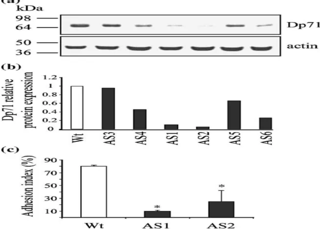
Antisense-Dp71 cells display decreased expression of Dp71 and deficient adhesion to laminin
We have shown that PC12 cell line-derived clones with depleted levels of Dp71 protein (antisense-Dp71 cells) elicit deficient cell adhesion and reduced immunostaining of β1-integrin in the basal area contacting the substrate,22 which suggests that Dp71 deficiency may disturb the β1-integrin-mediated cell adhesion of PC12 cells. To study the role of Dp71 in PC12 cell adhesion, we decided to analyze the potential interactions of Dp71f (the cytoplasmic isoform of Dp71, see Introduction) with the adhesion-associated proteins. First, we performed a second round of stable transfection to obtain additional antisenseDp71 PC12 clones. We isolated six different antisense-Dp71 clones that were further screened for antisense-Dp71 expression. Then, total protein extracts from PC12 cells and the different antisense-Dp71 clones were analyzed by Western blotting with the anti-Dp71 antibody 5F3 (see Table 1). Blots were stripped and reprobed with an anti-actin antibody (loading control). The expected immunoreactive bands for Dp71f (71 kDa) and actin (41 kDa) were revealed in the wild-type and antisense-Dp71 cells (Figure 1(a)); however, the Dp71 band intensity decreased in all the antisense-Dp71 clones (AS1 to AS6). Quantitative analysis demonstrated that clones AS1 and AS2 displayed the maximal reductions in Dp71 expression, 95% and 90%, respectively (Figure 1(b)), compared with wild-type cells. Therefore, these two antisense-Dp71 clones were selected for further studies. Next, the capability of AS1 and AS2 clones to adhere to laminin was evaluated. Compared with control cells, the AS1 and AS2 clones elicited deficient adhesion activity with decrements of 78% and 69%, respectively (Figure 1(c)). Since AS1 and AS2 clones exhibited similar phenotypic responses under the different analyses performed in this study, we decided to show hereafter representative results that are valid for both clones, unless indicated otherwise.
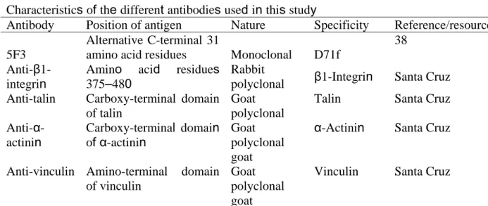
Table 1. Antibodies used in this study
Characteristics of the different antibodies used in this study
Antibody Position of antigen Nature Specificity Reference/resource
5F3
Alternative C-terminal 31
amino acid residues Monoclonal D71f
38
Anti-β1-integrin
Amino acid residues 375–480
Rabbit
polyclonal β1-Integrin Santa Cruz Anti-talin Carboxy-terminal domain
of talin
Goat polyclonal
Talin Santa Cruz
Anti-α-actinin Carboxy-terminal domain of α-actinin Goat polyclonal goat
α-Actinin Santa Cruz
Anti-vinculin Amino-terminal domain of vinculin
Goat polyclonal goat
Vinculin Santa Cruz
Anti-paxillin Central portion of paxillin Monoclonal Paxilin Signal transduction Anti-FAK Anti-actin Carboxy-terminal domain of FAK Amino-terminal domain of actin Rabbit polyclonal Monoclonal
FAK β-Actin Santa Cruz 39
Anti-GST GST Monoclonal GST Zymed
Anti-β-dystroglycan
C-terminal seven amino acid residues Monoclonal β-Dystroglycan Novocastra Anti-α-syntrophin
Amino acid residues 191–206
Rabbit polyclonal
α-Syntrophin Mornet D
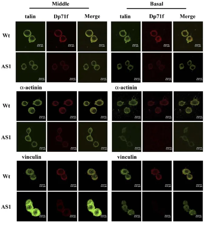
Subcellular distribution of Dp71f and the adhesion-associated proteins in the wild-type and antisense-Dp71 PC12 cells
The subcellular distribution of Dp71f was compared with that of the adhesion proteins (talin, α-actinin and vinculin) in the wild-type and antisenseDp71 cells. Wild-type (Wt) and antisense-Dp71 cells (AS1), plated on laminin-coated dishes, were double-stained with the anti-antisense-Dp71f antibody 5F3 and either anti-talin, anti-α-actinin or anti-vinculin antibody. Figure 2 shows the confocal microscopy analysis of the staining patterns of these proteins. In the wild-type cells, talin, α-actinin (green channel) as well as Dp71f (red channel) showed a diffuse cytoplasmic labeling at the middle area of cells, which became punctate at the basal area of cells. Staining of Dp71f overlapped that of talin and α-actinin at both the middle and basal portions of these cells (yellow channel). In the antisense-dp71 cells, the immunostaining of talin and α-actinin decreased at the basal area of cells contacting the substrate, compared with wild-type cells. The decrease of α-actinin was higher than that of talin. The drastic decrease of Dp71f immunolabeling in the antisense-Dp71 cells precluded the analysis of the overlapping of this protein with talin and α-actinin. On the other hand, the immunostaining of vinculin in the wild-type cells displayed a cytoplasmic grainy pattern that became more prominent at the basal area. The staining of Dp71 overlapped that of vinculin in both the middle and basal portions of these cells. The immunolabeling of vinculin was much more intense in the middle area of the antisense-Dp71cells, compared with the same cell portion of wild-type cells. However, such labeling decreased markedly and became diffuse in the basal area of the antisense-Dp71 cells. Overall, the immunofluorescence results indicate that Dp71f distribution overlaps partially with that of talin, vinculin and α-actinin, and that deficiency of Dp71 provokes a certain decrease in the immunolabeling of these proteins at the basal area of the antisense-Dp71 cells.
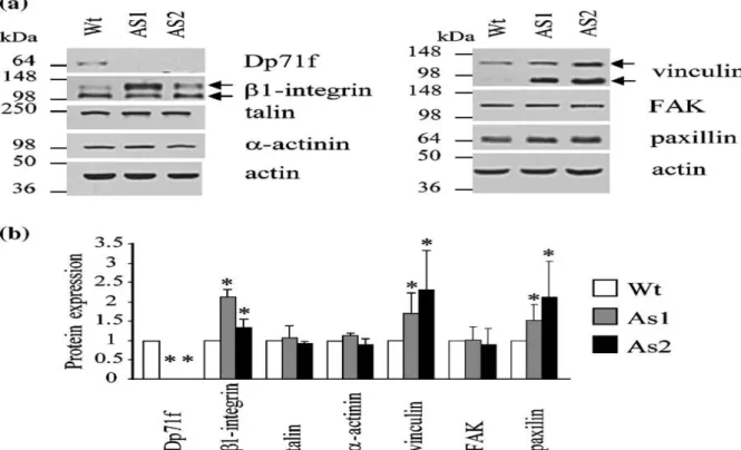
Expression of the adhesion-associated proteins in the wild-type and antisense-Dp71 PC12 cells
To explore the possibility that the adhesion failure displayed by the antisense-Dp71 cells is due to the deficient expression of the adhesion-associated proteins, total extracts from wild-type and antisense-Dp71 cells (AS1 and AS2), grown for 15 h on laminin-coated dishes, were analyzed by Western blotting using antibodies raised against Dp71f (5F3), β1-integrin, α-actinin, FAK, paxillin, talin or vinculin (Table 1). To normalize immunoblotting data, blots were stripped and reprobed with an anti-actin antibody. The expected immunoreactive bands for β1-integrin (135 kDa and 116 kDa protein bands) talin (250 kDa) paxillin (68 kDa), vinculin (130 kDa and 85 kDa), α-actinin (105 kDa), FAK (125 kDa) and actin (41 kDa) were observed in the lysates obtained from wild-type and antisense-Dp71 cells (Figure 3(a)). The 85 kDa immunoreactive band of vinculin, observed in the antisense-Dp71 cells, might correspond to its calpain-cleaved fragment.38 On the other hand, the immunoreactive band of Dp71f (71 kDa) was present in the wild-type cells but undetectable in the two antisense-Dp71 clones. Quantification of the immunoblots demonstrated that protein levels of talin, α-actinin, and FAK were similar between wild-type and antisense-Dp71 cells, whereas those of β1-integrin, vinculin and paxillin were increased in the antisense-Dp71 clones (Figure 3(b)). Similar results were obtained when the
expression of the adhesion-associated proteins was analyzed in non-adhering wild-type and antisense-Dp71 cells (data not shown). The increased expression of some adhesion-associated proteins in the antisense-Dp71 cells might be the response of a compensatory mechanism. We have reported that utrophin expression increases in response to the adhesion of PC12 cells to laminin.26 Therefore, given the structural similarity between Dp71f and utrophin, it is tempting to speculate that utrophin might mediate such compensation in the antisense-Dp71 cells. In summary, immunoblotting data indicated that the impaired adhesion activity of the antisense-Dp71 cells is not caused by deficient expression of the adhesion-associated proteins.
Figure 1. Decreased expression of Dp71f is related to deficient adhesion to laminin in the PC12 cells. (a) Immunodetection of Dp71 was carried out with the antiDp71f antibody 5F3. Membranes were stripped and reprobed with monoclonal antibody anti-actin. Position of the protein markers is shown on the left, and migration of Dp71f and actin is indicated on the right. (b) Dp71 protein levels were estimated by densitometric analysis of immunoblot autoradiograms and normalized against levels of actin . Data represent the average value of two independent experiments. AS1 and AS2, antisense-Dp71 clones. (c) The adhesion index was obtained as described in Materials and Methods. Values are the mean ± SD of three independent experiments. Asterisks (*) denote significant differences (p < 0.05). Wt, wild-type cells; AS1AS6, antisense-Dp71 clones.
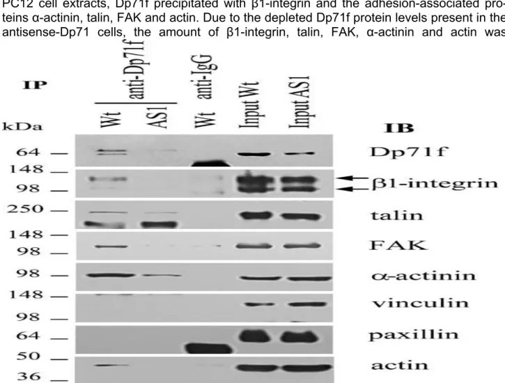
Dp71f protein associates with β1-integrin and the adhesion-associated proteins
Since no deficiency was observed in the expression of β1-integrin or the adhesion-associated protein in the antisense-Dp71 cells, we hypothesized that Dp71f may interact with components of the β1-integrin adhesion complex of PC12 cells in such a way that decreased expression of Dp71f, display by the antisense-Dp71 cells, may affect the stability of the adhesion complex components and ultimately impair the adhesion activity. To test our hypothesis, lysates obtained from wild-type and antisense-Dp71 cells were immunoprecipitated with the anti-Dp71f antibody 5F3, and the immunoprecipitated material was subjected to immunoblotting with antibodies raised against Dp71f, β1-integrin, talin, FAK, αactinin, vinculin, paxillin or actin (Figure 4). In the wild-type cells, the
immunoprecipitate of 5F3 contained the immunoreactive bands corresponding to Dp71f, β1-integrin, talin, FAK, α-actinin and actin. Since the antisense-Dp71 cells have deficient expression of Dp7f, the antibody 5F3 failed to immunoprecipitate this protein at detectable levels. In consequence, the amount of β1-integrin, talin, FAK, α-actinin and actin recovered in the immunoprecipitate of Dp71f, was greatly reduced, compared with wild-type cells (Figure 4). The immunoreactive bands corresponding to vinculin and paxillin were not recovered in the Dp71f immunoprecipitates of the wild-type or antisense-Dp71 cells; thus, it seems that Dp71f does not interact with these two proteins. None of the analyzed proteins was precipitated by irrelevant IgG antibodies (Figure 4), which confirmed the specificity of the immunoprecipitation assays.
Dp71f deficiency provokes a reduction of the β1-integrin adhesion complex
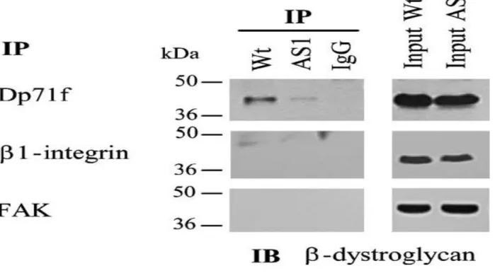
To confirm the interaction of Dp71f with β1integrin and the adhesion-associated proteins, lysates obtained from wild-type or antisense-Dp71 cells were immunoprecipitated with the anti-β1integrin antibody, and the precipitated proteins were analyzed by immunoblotting using antibo-dies directed against β1-integrin, Dp71f (5F3), talin, FAK, α-actinin, vinculin paxillin and actin. Figure 5 shows that talin, FAK, α-actinin, paxillin, actin and Dp71f, but not vinculin, copurified with β1integrin in the wild-type cells, In the antisenseDp71 cells, the immunoprecipitates of β1-integrin contained a marked decrease in the amounts of the adhesion proteins talin (90%), FAK (22%), α-actinin (99%) and actin (50%), while the protein levels of β1-integrin and paxillin were unaltered, compared with wild-type cells (Figure 5(a)). Statistical analysis of these results confirmed their significance (data not shown). None of the analyzed proteins was precipitated by irrelevant IgG. Immunoprecipitation with the anti-β1 integrin antibody did not distinguish the β1-integrin engaged in the adhesion complex from that unbounded. Therefore, to ascertain specifically whether the protein levels of β1-integrin engaged in the adhesion protein complex are altered in the antisense-Dp71 cells, lysates obtained from wild-type and antisenseDp71 cells were immunoprecipitated with an anti-FAK antibody, and the precipitated material was analyzed by immunoblotting with the anti-β1 integrin antibody and, after blot stripping, with the anti-FAK antibody. Figure 5(b) and 5(c) show that the amount of FAK-associated β1 integrin was reduced significantly in the antisense-Dp71 clones AS1 (40% decrease) and AS2 (45% decrease), compared with wild-type cells. Altogether, the immunoprecipitation data demonstrated that the PC12 cell β1-integrin adhesion complex is composed of β1-integrin, talin, α-actinin, actin, FAK paxilin and Dp71f, but not vinculin and, more interestingly, that Dp71 deficiency provokes a marked reduction of the adhesion complex, probably by disturbing its assembly and/or stability. β-Dystroglycan associates with Dp71f but not cells were immunoprecipitated with anti-Dp71f with the β1-integrin adhesion complex (5F3), anti-β1-integrin or anti-FAK antibody, and the immunoprecipitates were subsequently anaDp71 is a common component of the dystrophin-lyzed by SDS-PAGE and immunoblotting using an associated protein complex (DAPC) in non-muscle anti-β-dystroglycan antibody (see Table 1). The cells. Therefore, we decided to determine whether immunoreactive band of β-dystroglycan (43 kDa), the DAPC component β-dystroglycan interacts revealed in the input lanes, was recovered in the with Dp71f in the PC12 cells and, by this means, immunoprecipitates of Dp71f,but its intensity was with the β1-integrin adhesion complex. Cell extracts clearly lower in the antisense-Dp71 cells than in the obtained from the wild-type and antisense-Dp71 wild-type cells; in contrast, the immunoprecipitates of β1-integrin and FAK did not contain β-dystroglycan (Figure 6). These results indicate that β1dystroglycan binds to Dp71f out of the context of the β1-integrin adhesion complex.
Figure 2. Subcellular distribution of Dp71f and the adhesion-associated proteins in the wild-type and antisense-Dp71 PC12 cells. Cells, plated for 15 h on laminin-coated glass coverslips, were double stained with the dystrophin antibody 5F3 (red channel) and vinculin, anti-talin or anti-actinin antibody (green channel). Images were analyzed by confocal laser microscopy. Focus was adjusted to the middle or basal portion of cells, as indicated for the two panels of images. Merge staining of Dp71f with talin, α-actinin or vinculin is shown in yellow (right-hand side of each panel). Wt, wild-type cells; AS1, antisense-Dp71 clone. The white scale bar represents 8 μm.
Figure 3. Expression of the adhesion-associated proteins in the wild-type and antisense-Dp71 PC12 cells. (a) Total protein extracts from the wild-type and antisense-Dp71 cells, plated on laminin-coated dishes for 15 h, were resolved by SDS-PAGE and analyzed by immunoblotting with antibodies directed specifically against Dp71f, β1-integrin, talin, αactinin, vinculin, FAK or paxillin. Blots were stripped and reprobed with an anti-actin antibody to normalize protein measurements. Positions of molecular mass markers are shown on the left, migration of Dp71f and the adhesion proteins analyzed is shown on the right. (b) Data collected from densitometric analysis of immunoblot autoradiograms represent the mean ± SD deviation of three independent experiments, each one performed in duplicate. Asterisks (*) denote significance differences (p <0.05) from wild-type cell cultures. Wt, wild-type cells; AS1 and AS2, antisense-Dp71 clones.
In vitro binding of Dp71f to components of the β1-integrin adhesion complex
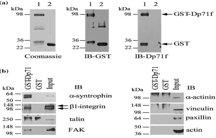
To corroborate the interaction of Dp71f with components of the β1-integrin adhesion protein complex by glutathione-S-transferase (GST) pull-down assays, a pGex-4T1-derived vector expressing the GST-Dp71f protein fusion was generated (see Materials and Methods). The GST and GST-Dp71f proteins fusions were isolated by incubation of JM109 bacterial lysates with glutathione-Sepharose beads, and visualized by staining the gel with Coomassie brilliant blue (Figure 7(a)). Next, GST and GST-Dp71f proteins were characterized by immunoblotting. As expected, the protein band corresponding to GST-Dp71f (97 kDa) was revealed using both the anti-GST and anti-Dp71 (5F3) antibodies, while the GST protein band (28 kDa) was observed only with the anti-GST antibody (Figure 7(a)). To perform pulldown assays, GST or GST-Dp71 fusion protein, previously immobilized on glutathione-Sepharose beads, was incubated with PC12 cell lysates, as described in Materials and Methods, and the endogenous proteins bound to GST were eluted and analyzed by SDS-PAGE and immunoblotting with antibodies to α-syntrophin, β1-integrin, talin, FAK, α-actinin, vinculin, paxillin and actin. Evaluation of the known interac-tion of Dp71 with the dystrophin-associated protein α-syntrophin served as positive control. As shown in Figure 7(b), α-syntrophin, β1-integrin and FAK protein bands were recovered bound to the GSTDp71f fusion protein, but not to GST alone. In contrast, no binding to GST-Dp71f was observed for talin, α-actinin, vinculin, paxillin or actin.
Discussion
We have shown that PC12-derived clones with deficient Dp71 expression (antisense-Dp71 cells) display altered adhesion activity.22 In addition, we observed that immunolabeling of β1-integrin decreased markedly in the cell area contacting the substrate of the antisense-dp71 cells. These results suggested the involvement of Dp71 in the β1-integrin adhesion system of PC12 cells. In the present study, we provide evidence indicating that the Dp71f isoform interacts with the β1-integrin adhesion complex in PC12 cells grown on laminin, and that such binding is required for the proper assembly and function of this protein complex. (1) Immunofluorescence studies revealed that Dp71f distribution overlapped with that of talin, α-actinin and vinculin in the PC12 cells, whereas in the antisense-Dp71 cells the immunostaining of these adhesion-associated proteins decreased markedly in the cell area contacting the substrate (Figure 2). (2) In the wild-type PC12 cell extracts, Dp71f precipitated with β1-integrin and the adhesion-associated pro-teins α-actinin, talin, FAK and actin. Due to the depleted Dp71f protein levels present in the antisense-Dp71 cells, the amount of β1-integrin, talin, FAK, α-actinin and actin was
Figure 4. Dp71f interacts in vivo with β1 integrin and the adhesion-associated proteins in the PC12 cells. Wild-type and antisense-Dp71 cells were cultured on laminincoated plates for 15 h before lysis in RIPA buffer (see Materials and Methods). Lysates were subjected to immunoprecipitation with the anti-Dp71f antibody (5F3) and the washed immunoprecipitates were analyzed by immunoblotting using antibodies to Dp71f, β1 integrin, talin, FAK, α-actinin, vinculin, paxillin, and actin. Irrelevant IgG was used as negative control. Input lanes, 5% of the crude extract used for immunoprecipitation was analyzed in parallel by immunoblotting. Migration of proteins standards is shown on the left whereas position of Dp71f and the adhesion-associated proteins is shown on the right. IP, immunoprecipitation; IB, immunoblotting; rabbit IgG, irrelevant antibody. Wt, wild-type cells; AS1, antisense-Dp71 cells.
reduced drastically in the Dp71f immunoprecipitates of these cells, which confirmed the specificity of the interactions of Dp71f with the β1-integrin adhesion complex components (Figure 4). (3) Complementary assays demonstrated that β1 integrin precipitated with Dp71f and the adhesion-associated proteins α-actinin, talin, paxillin, FAK and actin in PC12 cell extracts, whereas in the antisense-Dp71 cell extracts, the immunoprecipitated levels of talin α-actinin, actin and vinculin were clearly diminished (Figure 5(a)). Likewise, immunoprecipitates of FAK contained significantly less β1 integrin in the antisense-dp71 cells than in the wild-type cells (Figure 5(b)). Since total protein levels of the adhesion components (β1-integrin, talin, vinculin, paxillin and FAK) were not decreased in the antisense-Dp71 cells (Figure 3), it seems that Dp71f deficiency affects specifically the cell fraction of these proteins that is engaged in the β1-integrin adhesion complex. (4) Finally, in vitro pulldown assays using recombinant Dp71f and PC12 endo-genous adhesion complex confirmed the interaction of Dp71f with β1-integrin and FAK (Figure 7(b)).
Integrins are heterodimeric transmembrane molecules composed of α and β subunits that mediate cellular binding to components of the extracellular matrix.23 Binding of the cytoplasmic domain of β1integrin to focal adhesion proteins, such as talin, αactinin, paxillin and FAK, leads to recruitment of further proteins to form a complex hierarchical structure that directs cytoskeletal assembly and signal transduction cascades.23 Therefore, it is of interest that components of DAPC have been found to be related to the integrin adhesion system.27–29 The existence of a functional communication between DAPC and the integrin adhesion complex was shown in the rat L6 skeletal muscle cell line.28
PC12 cell attachment to laminin is mediated by adhesion sites, termed point contacts, that contain the integrin heterodimer α1β1.30 The composition of PC12 cell point contacts has been poorly characterized. Using PC12 cell extracts, Loster et al.(2001) demonstrated by in vitro binding assays that paxillin, FAK and α-actinin, but not talin or vinculin, bound to the cytoplasmic domain of β1-integrin.25 Here, by using an in vivo biochemical strategy, we report for the first time that the β1-integrin adhesion complex of PC12 cells is additionally composed of talin, α-actinin, paxillin, FAK and actin, but not vinculin. Furthermore, our finding of the interaction of Dp71f with the β1-integrin adhesion complex components shows that this short dystrophin is a novel adhesion component. Binding of Dp71f to the adhesion complex seems to have functional significance, because deficiency of Dp71f, displayed by the antisense-Dp71 cells, provokes a concomitant decrease in the levels of the β1-integrin adhesion complex ,and ultimately impairs the adhesion to laminin (Figure 1). We propose that Dp71f is required for the appropriate assembly of the adhesion complex or that, once the complex assembly takes place, Dp71f is required for its stability. As evidence of the role of Dp71 in cell adhesion, it has been found that Dp71 associated with the DAPC participates during the actin-cytoskeleton remodeling of platelets, promoting shape change, adhesion and aggregation.31 Furthermore, adhesion to collagen in response to thrombin was decreased significantly in platelets isolated from mdx3cv mice, a dystrophin-deficient animal model.32 and a C-terminal domain), which contain several Finally, it has been reported that Dp71f is localized protein–protein interaction modules (two EF-hand with vinculin and α-actinin at focal contacts and regions, an incomplete WW motif, a ZZ motif and lamellipodia of the astrocytoma cell line U373 MG.33 two coiled-coil regions) that potentially could
Figure 5. Dp71f deficiency causes a marked reduction of the β1 integrin adhesion complex in the PC12 cells. (a) Lysates from wild-type and Dp71-antisense cells were immunoprecipitated with an anti-β1-integrin antibody and the immunoprecipitates were subsequently analyzed by immunoblotting using antibodies directed against β1 integrin, talin, FAK, α-actinin, vinculin, paxillin, and actin. (b) Wild-type and antisense-Dp71 cell extracts were subjected to immunoprecipitation using an anti-FAK antibody, and the precipitated material was analyzed by immunoblotting with the anti-β1 integrin antibody and after stripping with the anti-FAK antibody (loading control). Input lanes, 5% of the crude extract used for immunoprecipitation was analyzed in parallel by immunoblotting. Irrelevant IgG was used as negative control. Migration of protein standards is shown on the left, migration of Dp71f and the adhesion proteins is shown on the right. (c) Data collected from densitometric analysis of immunoblot autoradiograms represent the mean ± SD deviation of three independent experiments, each performed in duplicate. Asterisks (*) denote significant differences (p < 0.05) from wild-type cell cultures. IP, immunoprecipitation; IB, immunoblotting; mouse IgG, irrelevant antibody. Wt, wild-type cells; AS1 and AS2, antisense-Dp71 clones.
At the present stage of our research, it is difficult to mediate its association with the adhesion complex establish whether Dp71f interacts alone with the components.34,35 It has been established by yeast integrin adhesion complex, or whether such binding two-hybrid and ligand-blotting techniques that αtakes place in the context of Dp71f assembled into actinin interacts directly with the last 200 amino acid DAPC. PC12 cells express utrophin,33 and some residues of the dystrophin C-terminal region, which DAPC components, such as β-dystroglycan and is present in Dp71.36 Such results are consistent with dystrobrevin;20,26 however, the presence of a func-our showing the co-immunoprecipitation of Dp7f tional DAPC remains to be shown in these cells. and α-actinin using antibodies to Dp71f and β1Furthermore, although β-dystroglycan was found to integrin. Therefore, it is plausible to propose that the be associated with Dp71f, we failed to recover this interaction of these proteins constitutes the commuprotein in the immunoprecipitates of β1-integrin nication link between Dp71f and the β1-integrin and FAK. Therefore, the Dp71f cellular fraction adhesion system. However, we were unable to associated with the adhesion
complex appears to be detect the interaction of Dp71f and α-actinin by in different from that interacting with β-dystroglycan vitro pulldown assays. The reason for this apparent or the whole DAPC. inconsistency is unclear, although obvious differ
Dp71f consists of three protein domains (an actin-ences in the experimental approaches could be binding N-terminal domain, a cysteine-rich region invoked. For instance, it could be that α-actinin interacts with Dp17f only when this dystrophin short product is assembled into the
Figure 6. β-Dystroglycan interacts with Dp71f but not with β1 integrin or FAK. Lysates from wild-type and antisense-Dp71 cell cultures, grown on laminin-coated plates, were immunoprecipitated with anti-Dp71f (5F3), anti-β1 integrin or anti-FAK antibodies and the immunoprecipitates were subsequently analyzed by immunoblotting with an anti-β-dystroglycan antibody. Irrelevant IgG was used as negative control. Input lanes, 5% of the crude extract used for immunoprecipitation was analyzed in parallel by immunoblotting. Migration of protein stan-dards is shown on the left. IP, immunoprecipitation; IB, immunoblotting; Wt, wild-type cells; AS1, antisense-Dp71 cells.
adhesion complex. On the other hand, pulldown assays confirm the interaction of Dp71 with β1-integrin and FAK. Since WW motifs are able to bind proline-rich sequences,34 it could be speculated that interaction between FAK and Dp71f occurs through the binding of the FAK proline-rich domain with the incomplete WW domain of Dp71f.23 Based on the in vivo and in vitro protein–protein interaction data, a schematic representation of Dp71f binding to the β1-integrin adhesion complex is shown in Figure 8. Further work will be required to define precisely the protein–protein interaction(s) linking Dp71f with the β1-integrin adhesion system of PC12 cells.
The β1-integrin-meditated cell adhesion modulates the neuronal migration and synaptogenesis during development of the central nervous system.37 Therefore, the involvement of Dp71f in PC12 cell adhesion constitutes an important advance towards understanding its role in neuronal development. In conclusion, we demonstrated that Dp71f interacts with components of the β1-integrin adhesion complex of PC12 cells, and that such interaction modulates the adhesion to laminin by providing proper complex assembly and/or stability.
Materials and Methods Cell culture
PC12 cells were grown in RPMI-1640 medium supplemented with 10% (v/v) horse serum, 5% (v/v) fetal bovine serum, 100 U/ml of penicillin and 0.1 mg/ml of streptomycin, and maintained at 37 °C in a humidified incubator with a 5% (v/v) CO 2 atmosphere. PC12-derived antisense-Dp71 clones were generated as described,21 and maintained with 500 μg/ml of neomycin analog G418 (Invitrogen, Carlsbab, CA). When indicated, cells were seeded onto plates coated with 20 μg/ml of laminin.
Cell adhesion assay
Cells were seeded onto laminin-coated six-well plates at a cell density of 2 × 10⁶. After incubation for 15 h, wells were washed with phosphate-buffered saline (PBS) by shaking gently for 30 s at 15 Hz, and the supernatant with detached cells was removed and counted by flow cytometry with an argon laser at 488 nm (Fac'sCalibur, Beckton Dickinson, Franklin Lakes, NJ). The adhesion index was calculated as follows: 100%-(number of detached cells x 100 / total number of seeded cells).
Immunoblotting assays
Cells, seeded onto laminin-coated plates, were washed with PBS and resuspended in 50 mM Tris–HCl (pH 8) containing complete Protease Inhibitor Cocktail (Roche, Mannheim, Germany). Homogenates were sonicated and protein concentrations were determined by the Bradford method (Bio-Rad, Hercules, CA). A portion (50 μg) of protein extract was mixed with an equal volume of 2 × sample buffer (50 mM Tris–HCl (pH 6.8), 15% (w/v) SDS, 5% (v/v), β-mercaptoethanol, 20% (v/v) glycerol, 0.1% (w/v) bromophenol blue) and boiled for 3 min. Proteins were resolved by SDS-PAGE (10% (w/v) polyacrylamide gel) and electrotransferred onto nitrocellulose membrane. Membranes were blocked for 1 h in TBS-T (100 mM Tris–HCl (pH 8.0), 150 mM NaCl, 0.5% (v/v) Tween-20) with 6% (w/v) low-fat dried milk and then incubated overnight at 4 °C with the respective primary antibody (Table 1). Following three washes in TBS-T, membranes were incubated with the appropriate horseradish peroxidase-conjugated secondary antibody (Amersham Biosciences, Bucks, England) and developed using the ECL Western blotting analysis system (Amersham Biosciences, Bucks, England). Quantitative analysis of immunoblots was performed with a Kodak digital Science v. 2.0.2 program (Kodak, New Haven, CT, USA).
Immunofluorescence and confocal microscopy analysis
Cells, plated onto laminin-coated 18 mm × 18 mm coverslips at a cell density of 2 × 10⁴, were washed with cytoskeleton buffer (10 mM Mes (pH 6.1), 150 mM NaCl, 5 mM EGTA, 5 mM MgCl2, 5 mM glucose), fixed for 5 min at room temperature in PBS containing 4% (v/v) paraformaldehyde and permeabilized for 5 min with PBS containing 0.5% (w/v) Triton X-100. Coverslips were washed three times with PBS, incubated for 1 h at room temperature with the blocking solution (Block ACE, Dainippon Pharmaceuticals) containing 3% (w/v) bovine serum albumin (BSA) and washed again three times with PBS containing 0.1% (v/v) Tween 20. Cells preparations were double-stained by overnight incubation at 4 °C with the following primary antibodies: an anti-Dp71f antibody as a positive control), β1 integrin, talin, FAK, α-actinin, vinculin, paxillin and actin. Input lane represents 5% of the PC12 cell
Figure 7. In vitro interaction of Dp71f with β1 integrin and FAK. GST and GST-Dp71f proteins, isolated by incubation of JM109 bacterial lysates with glutathione-Sepharose beads, were analyzed by SDS-PAGE followed by staining with Coomassie brilliant blue, and immunoblotting using anti-Dp71f and anti-GST antibodies (a). Lanes: 1, cell extracts expressing GST alone; 2, cell extracts expressing GST-Dp71f. Protein standards are indicated on the left. Arrows on the right denote the positions of GST and GST-Dp71f proteins. (b) To perform affinity pulldown assays, GST or GSTDp71f fusion protein was immobilized on glutathione-Sepharose beads and incubated with freshly prepared PC12 cell extracts. Beads were recovered by centrifugation, washed and heated in the presence of SDS-PAGE sample buffer to elute GST and GST-Dp71f proteins and associated proteins. Eluted protein samples were analyzed by immunoblotting using antibodies to αsyntrophin (to detect α-syntrophin
extract used for pulldown assay, subjected to immunoblotting. Protein standards are indicated on the left. Arrows on the right denote the position of the proteins analyzed. IB, immunoblotting. (5F3) and either anti-talin, anti α-actinin or anti-vinculin washing with PBS, cell preparations were mounted with antibody. The day after, cells were washed three times VectaShield (Vector Laboratories Inc., Burlingame, CA) with PBS and incubated for 1 h at 4 °C with PBS containing and viewed with a confocal laser scanning microscope both a rhodamine isothiocyanate (TRITC)-conjugated(TCP-SP2, Leica, Heidelberg Germany) using 63× and secondary anti-mouse antibody and an fluorescein iso-100× oil-immersion plan apochromat objectives (NA 1.32 thiocyanate (FITC)-conjugated secondary anti-goat anti-and NA 1.4, respectively). Six to eight consecutive single bodies (Zymed Laboratories Inc. San Francisco, CA). After sections were obtained simultaneously for one or two channels through the Z-axis of the sample. The resulting stack of images were projected and analyzed onto the two-dimensional plane using a pseudocolor display, green (FITC), and red (TRITC), for double-labeling experiments. Fluorochromes were excited at a wavelength of 488 nm (for FITC) for single labeling, and at 560 nm (for TRITC) in double-labeling experiments. Single optical sections of the stacks were selected to analyze the colocalization patterns between two markers. For comparison, all the confocal images were collected under the same scanning settings.
Immunoprecipitation assays
Cells were plated onto laminin-coated plates for 15 h before being washed in PBS and resuspended in 300 μlof RIPA buffer (10 mM Tris–HCl (pH 7.4), 1 mM EGTA, 158 mM NaCl, 10 mM Na3MoO4, 25 mM NaF, 1 mM phenylmethanesulphonyl fluoride (PMSF), 1 mM EDTA, 2mMNa3VO4). After determination of protein concentration by the Bradford method, cell lysates were added to an equal volume of ice-cold RIPA buffer containing 1% (w/v) Triton X-100 and 0.1% (w/v) SDS, clarified by centrifugation at 18,000g for 5 min and then the recovered supernatant was added to an equal volume of RIPA buffer. Next, 1.5 mg of cell lysate clarified by incubation with 10 μl of protein G-agarose (Invitrogen, Carlsbad, CA), was incubated overnight at 4 °C with the appropriate primary antibody, previously incubated for 2 h with 10 μlof protein G-agarose. When required, protein G-agarose was blocked with 2% (w/v) BSA for 2 h at 4 °C, before incubation with primary antibodies. Parallel incubations with irrelevant rabbit or mouse IgG were performed as a control for nonspecific interactions. The immune complexes were recovered by centrifuging for 4 min at 18,000g and washing three times with RIPA buffer. Precipitated proteins were eluted by boiling in sample buffer for 10 min, separated by SDS-PAGE (10% (w/v) polyacrylamide gel), and analyzed by SDS-SDS-PAGE immunoblotting.
GST binding assays
The human Dp71f cDNA was removed from pGFPDp71 vector21 by EcoRI digestion and cloned in-frame to the 3′-end of GST in the bacterial expression vector pGex4T1 (Amersham Biosciences Co., Piscataway, NJ) to generate the GST-Dp71f gene fusion. To express and characterize GST and GST-Dp71f fusion proteins, 300 ml of transformed strain JM109 bacterial cells, induced with 0.1 mM IPTG for 2 h, were centrifuged at 3800g for 10 min, resuspended in 2 ml of NETN buffer (20 mM Tris–HCl (pH 7.5),100 mM NaCl, 1 mM EDTA, 0.5% (v/v) NP40, 1 mM PMSF, 0.5% (w/v) low-fat dried milk, Complete Protease Inhibitor Cocktail) and sonicated on ice (4 × 30 s, Branson sonifier). After that,
300 μl of packed glutathione-Sepharose 4B beads (Amersham Biosciences Co., Piscat-away, NJ) were added to bacterial lysate and the mixture was incubated overnight at 4 °C while rotating. Beads, recovered by centrifugation at 1000g for 5 min, were washed five times with 1 ml of ice-cold NETN buffer and resuspended in 300 μl of NETN buffer. GST and GSTDp71f proteins, bound to glutathione-Sepharose, were eluted by adding an equal volume of 2× sample buffer and heating at 95 °C, and subsequently analyzed by staining with Coomassie blue brilliant and immunoblotting. To perform pulldown assays, a similar amount of GST or GST-Dp71f fusion protein, immobilized on 20 μlof glutathione-Sepahrose beads, was incubated for 2 h at 4 °C on a rotator with 1 mg of PC12 cell extract, prepared in RIPA buffer containing 1% (v/v) Triton X-100 and 0.1% (w/v) SDS. Beads were recovered by centrifugation at 6000 rpm for 5 min and washed five times with 1 ml of ice-cold NETN buffer. Finally, PC12 cell endogenous proteins, bound to glutathione-Sepharose, were eluted by adding an equal volume of 2× sample buffer and heating at 95 °C, and subsequently analyzed by SDS-PAGE and immunoblotting.
Statistical analysis
Statistical analyses were performed using the Mann-Whitney U-test and a significant difference was defined at p < 0.05.
Acknowledgements
We are grateful to Dr Manuel Hernández for supplying anti-actin monoclonal antibody. We thank Victor Tapia Ramírez for technical assistance. This work was supported by CONACyT-México grant 43285-M to B.C.
References
1 Ahn, A. H. & Kunkel, L. M. (1993). The structural and functional diversity of dystrophin. Nature Genet. 3, 283–291.
2 Koenig, M., Monaco, A. P. & Kunkel, L. M. (1988). The complete sequence of dystrophin predicts a rod-shaped cytoskeletal protein. Cell, 53, 219–226.
3 Blake, D. J., Weir, A., Newey, S.E. & Davies, K. E. (2002). Function and genetics of dystrophin and dystrophin-related proteins in muscle. Physiol. Rev. 82, 291–329.
4 Ervasti, J. M. & Campbell, K. P.(1991). Membrane organization of the dystrophin-glycoprotein complex. Cell, 66, 1121–1131.
5 Lapidos, K. A., Kakkar, R. & McNally, E. M. (2004). The dystrophin glycoprotein complex: signaling strength and integrity for the sarcolemma. Circulat. Res. 94, 1023–1031. 6 Petrof, B. J., Shrager, J. B., Stedman, H. H., Kelly, A. M. & Sweeney, H. L. (1993). Dystrophin protects the sarcolemma from stresses developed during muscle contraction. Proc. Natl Acad. Sci. USA, 90, 3710–3714.
7 Byers, T. J., Lidov, H. G. & Kunkel, L. M. (1993). An alternative dystrophin transcript specific to peripheral nerve. Nature Genet. 4,77–81.
8 D'Souza, V. N., Nguyen, T. M., Morris, G. E., Karges, W., Pillers, D. A. & Ray, P. N. (1995). A novel dystrophin isoform is required for normal retinal electrophysiology. Hum. Mol. Genet. 4, 837–842.
9 Lidov, H. G., Selig, S. & Kunkel, L. M. (1995). Dp140: a novel 140 kDa CNS transcript from the dystrophin locus. Hum. Mol. Genet. 4, 329–335.
10 Bar, S., Barnea, E., Levy, Z., Neuman, S., Yaffe, D. & Nudel, U. (1990). A novel product of the Duchenne muscular dystrophy gene which greatly differs from the known isoforms in its structure and tissue distribution. Biochem. J. 272, 557–560.
11 Hugnot, J. P., Gilgenkrantz, H., Vincent, N., Chafey, P., Morris, G. E., Monaco, A. P. et al. (1992). Distal transcript of the dystrophin gene initiated from an alternative first exon and encoding a 75-kDa protein widely distributed in nonmuscle tissues. Proc. Natl Acad. Sci. USA, 89, 7506–7510.
12 Dalloz, C., Claudepierre, T., Rodius, F., Mornet, D., Sahel, J. & Rendon, A. (2001). Differential distribution of the members of the dystrophin glycoprotein complex in mouse retina: effect of the mdx (3Cv) mutation. Mol. Cell Neurosci. 17, 908–920.
13 de Leon, M. B., Montanez, C., Gomez, P., Morales-Lazaro, S. L., Tapia-Ramirez, V., Valadez-Graham, V. et al. (2005). Dystrophin Dp71 expression is down-regulated during myogenesis: role of Sp1 and Sp3 on the Dp71 promoter activity. J. Biol. Chem. 280, 5290–5299. 14 Austin, R. C.,Howard, P. L., D'Souza,V. N., Klamut H. J. & Ray, P. N. (1995). Cloning and characterization of alternatively spliced isoforms of Dp71.Hum.Mol.Genet.4,1475–1483.
15 Ceccarini, M., Rizzo, G., Rosa, G., Chelucci, C., Macioce, P. & Petrucci, T. C. (1997). A splice variant of Dp71 lacking the syntrophin binding site is expressed in early stages of human neural development. Brain Res. Dev. Brain Res. 103,77–82.
16 Jung, D.,Filliol,D.,Metz-Boutigue,M.H.&Rendon A.(1993).Characterization and subcellular localization of the dystrophin-protein 71 (Dp71) from brain. Neuromuscul. Disord. 3, 515–518 17 Greenberg, D. S., Schatz, Y., Levy, Z., Pizzo, P., Yaffe, D.& Nudel, U. (1996). Reduced levels of dystrophin associated proteins in the brains of mice deficient for Dp71. Hum.Mol.Gene.t,5,1299–1303
18 Moizard, M. P., Toutain, A., Fournier, D., Berret, F., Raynaud, M., Billard, C. et al. (2000). Severe cognitive impairment in DMD: obvious clinical indication for Dp71 isoform point mutation screening. Eur. J. Hum. Genet. 8, 552–556.
19 Cisneros,B., Rendon, A., Genty, V., Aranda, G., Marquez, F., Mornet, D. & Montanez, C. (1996). Expression of dystrophin Dp71 during PC12 cell differentiation. Neurosci. Letters, 213, 107–110.
20 Marquez, F.G., Cisneros, B., Garcia, F., Ceja, V., Velazquez, F., Depardon, F. et al. (2003). Differential expression and subcellular distribution of dystrophin Dp71 isoforms during differentiation process. Neuroscience, 118, 957–966.
21 Acosta, R., Montanez, C., Fuentes-Mera, L., Gonzalez, E., Gomez, P., Quintero-Mora, L. et al. (2004). Dystrophin Dp71 is required for neurite outgrowth in PC12 cells. Expt. Cell Res. 296, 265–275.
22 Enriquez-Aragon, J. A., Cerna-Cortes, J., Bermudez de Leon, M., Garcia-Sierra, F., Gonzalez, E., Mornet, D. & Cisneros, B. (2005). Dystrophin Dp71 in PC12 cell adhesion. NeuroReport, 16, 235–238.
23 Lee,J. W. & Juliano, R. (2004). Mitogenic signal transduction by integrin-and growth factor receptor-mediated pathways. Mol. Cell, 17, 188–202.
24 Tomaselli, K. J., Damsky,C. H. & Reichardt, L. F. (1987). Interactions of a neuronal cell line (PC12) with laminin, collagen IV, and fibronectin: identification of integrin-related glycoproteins involved in attachment and process outgrowth. J. Cell Biol. 105, 2347–2358. 25 Loster, K., Vossmeyer, D., Hofmann, W., Reutter, W. & Danker, K. (2001). alpha1 Integrin cytoplasmic domain is involved in focal adhesion formation via association with intracellular proteins. Biochem. J. 356, 233–240.
26 Rosas-Vargas, H., Montanez, C., Rendon, A., Mornet, D., Garcia, F., Ceja, V. & Cisneros, B. (2000). Expression and localization of utrophin in differentiating PC12 cells. NeuroReport, 11, 2253–2257.
27 Driss, A., Charrier, L., Yan, Y., Nduati, V., Sitaraman, S. & Merlin, D. (2006). Dystroglycan receptor is involved in integrin activation in intestinal epithelia. Am. J. Physiol. Gastrointest. Liver Physiol. 290, G1228–G1242.
28 Yoshida, T., Pan, Y., Hanada, H., Iwata, Y. & Shigekawa, M. (1998). Bidirectional signaling between sarcoglycans and the integrin adhesion system in cultured L6 myocytes. J. Biol. Chem. 273, 1583–1590.
29 Matsumura, K., Chiba, A., Yamada, H., Fukuta-Ohi, H., Fujita, S., Endo, T. et al. (1997). A role of dystroglycan in schwannoma cell adhesion to laminin. J. Biol. Chem. 272, 13904–
13910.
30 Arregui, C. O., Carbonetto, S. & McKerracher,L. (1994). Characterization of neural cell adhesion sites: point contacts are the sites of interaction between integrins and the cytoskeleton in PC12 cells. J. Neurosci. 14, 6967–6977.
31 Cerecedo, D., Martinez-Rojas, D., Chavez, O., Martinez-Perez, F., Garcia-Sierra, F., Rendon, A. et al. (2005). Platelet adhesion: structural and functional diversity of short dystrophin and utrophins in the formation of dystrophin-associated-protein complexes related to actin dynamics. Thromb. Haemost. 94, 1203–1212.
32 Austin, R. C., Fox, J. E., Werstuck, G. H., Stafford, A. R., Bulman, D. E., Dally, G. Y. et al. (2002). Identification of Dp71 isoforms in the platelet membrane cytoskeleton. Potential role in thrombin-mediated platelet adhesion. J. Biol. Chem. 277, 47106–47113.
33 Garcia-Tovar, C. G., Luna, J., Mena, R., Soto-Zarate, C. I., Cortes, R., Perez, A. et al. (2002). Dystrophin isoform Dp7l is present in lamellipodia and focal complexes in human astrocytoma cells U-373 MG. Acta Histochem. 104, 245–254.
34 Ilsley, J. L., Sudol, M. & Winder, S. J. (2002). The WW domain: linking cell signalling to the membrane cytoskeleton. Cell Signal, 14, 183–189.
35 Ponting, C. P., Blake, D. J., Davies, K. E., Kendrick-Jones, J. & Winder, S. J. (1996). ZZ and TAZ: new putative zinc fingers in dystrophin and other proteins. Trends Biochem. Sci. 21,11–13.
36 Hance, J. E., Fu, S. Y., Watkins, S. C., Beggs, A. H. & Michalak, M. (1999). alpha-actinin-2 is a new component of the dystrophin-glycoprotein complex. Arch. Biochem. Biophys. 365, 216–222.
37 Graus-Porta, D., Blaess, S., Senften, M., Littlewood-Evans, A., Damsky, C., Huang, Z. et al. (2001). Beta1class integrins regulate the development of laminae and folia in the cerebral and cerebellar cortex. Neuron, 31, 367–379.
38 Fabbrizio, E., Nudel, U., Hugon, G., Robert, A., Pons, F. & Mornet, D. (1994). Characterization and localization of a 77 kDa protein related to the dystrophin gene family. Biochem. J. 299, 359–365.
39 Garcia-Tovar,C. G., Perez, A., Luna, J., Mena, R., Osorio, B., Aleman, V. et al. (2001). Biochemical and histochemical analysis of 71 kDa dystrophin isoform (Dp71f) in rat brain. Acta Histochem. 103, 209–224.