HAL Id: inserm-00842318
https://www.hal.inserm.fr/inserm-00842318
Submitted on 8 Jul 2013
HAL is a multi-disciplinary open access
archive for the deposit and dissemination of
sci-entific research documents, whether they are
pub-lished or not. The documents may come from
teaching and research institutions in France or
abroad, or from public or private research centers.
L’archive ouverte pluridisciplinaire HAL, est
destinée au dépôt et à la diffusion de documents
scientifiques de niveau recherche, publiés ou non,
émanant des établissements d’enseignement et de
recherche français ou étrangers, des laboratoires
publics ou privés.
Fast functional imaging of multiple brain regions in
intact zebrafish larvae using Selective Plane Illumination
Microscopy
Raphaël Candelier, Thomas Panier, Sebastián Romano, Raphaël Olive,
Thomas Pietri, Germán Sumbre, Georges Debrégeas
To cite this version:
Raphaël Candelier, Thomas Panier, Sebastián Romano, Raphaël Olive, Thomas Pietri, et al.. Fast
functional imaging of multiple brain regions in intact zebrafish larvae using Selective Plane Illumination
Microscopy. the Twenty Second Annual Computational Neuroscience Meeting: CNS*2013, Jul 2013,
paris, France. pp.P97. �inserm-00842318�
P O S T E R P R E S E N T A T I O N
Open Access
Fast functional imaging of multiple brain regions
in intact zebrafish larvae using Selective Plane
Illumination Microscopy
Raphaël Candelier
1*, Thomas Panier
1, Sebastián Romano
2,3,4,5, Raphaël Olive
1, Thomas Pietri
2,3,4,5,
Germán Sumbre
2,3,4,5, Georges Debrégeas
1From
Twenty Second Annual Computational Neuroscience Meeting: CNS*2013
Paris, France. 13-18 July 2013
The optical transparency and the small dimensions of zebrafish at the larval stage make it a vertebrate model of choice for brain-wide in-vivo functional imaging. How-ever, current point-scanning imaging techniques, such as two-photon or confocal microscopy, impose a strong limit on acquisition speed which in turn sets the number of neurons that can be simultaneously recorded [1]. At 5 Hz, this number is of the order of one thousand, i.e. approximately 1-2% of the brain. We demonstrate that this limitation can be greatly overcome by using Selec-tive-Plane Illumination Microscopy (SPIM) [2-4]. Zebra-fish larvae expressing the genetically encoded calcium indicator GCaMP3 were illuminated with a scanned laser
sheet and imaged with a camera whose optical axis was oriented orthogonally to the illumination plane. This optical sectioning approach was shown to permit func-tional imaging of most of the brain volume of 5-9 day old larvae with single-cell resolution. The spontaneous activ-ity of up to 5000 neurons was recorded at 20 Hz for 20-60 min. By rapidly scanning the specimen in the axial direction, the activity of 25000 individual neurons from 5 different z-planes (approximately 30% of the entire brain) could be simultaneously monitored at 4 Hz. Compared to point-scanning techniques, this imaging strategy thus yields a ~20-fold increase in data throughput (number of recorded neurons times acquisition rate) without
* Correspondence: [email protected]
1
CNRS / UPMC Univ. Paris 06, FRE 3231, Laboratoire Jean Perrin LJP, F-75005, Paris, France
Full list of author information is available at the end of the article
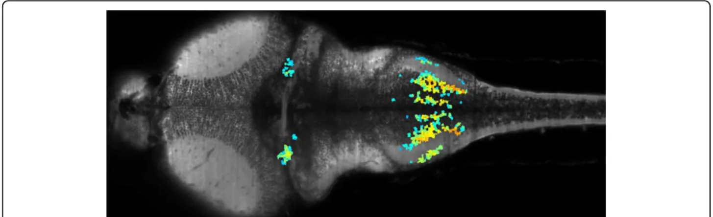
Figure 1Image of the brain of a 6 day-old GCaMP3 zebrafish obtained by SPIM. Colored neurons indicate a set of neurons showing correlated activity.
Candelier et al. BMC Neuroscience 2013, 14(Suppl 1):P97 http://www.biomedcentral.com/1471-2202/14/S1/P97
© 2013 Candelier et al; licensee BioMed Central Ltd. This is an Open Access article distributed under the terms of the Creative Commons Attribution License (http://creativecommons.org/licenses/by/2.0), which permits unrestricted use, distribution, and reproduction in any medium, provided the original work is properly cited.
compromising the signal-to-noise ratio. The extended field of view offered by the SPIM method allowed us to directly identify large scale ensembles of neurons, span-ning several brain regions (see Figure 1), that displayed correlated activity and were thus likely to participate in common neural processes.
Author details
1CNRS / UPMC Univ. Paris 06, FRE 3231, Laboratoire Jean Perrin LJP, F-75005,
Paris, France.2
Ecole Normale Supérieure, Institut de Biologie de l’ENS, IBENS, Paris, F-75005 France.3Inserm, U1024, Paris, F-75005 France.4CNRS, UMR
8197, Paris, F-75005 France.5IBENS, ENS, Paris, France.
Published: 8 July 2013 References
1. Christine Grienberger, Arthur Konnerth: Imaging calcium in neurons. Neuron2012, 73(5):862-885.
2. Michael Weber, Jan Huisken: Light sheet microscopy for real-time developmental biology. Curr Opin Genet Dev 2011, 21(5):566-572. 3. Jerome Mertz: Optical sectioning microscopy with planar or structured
illumination. Nature Methods 2011, 8(10):811-819, October.
4. Raju Tomer, Khaled Khairy, Philipp JKeller: Shedding light on the system: studying embryonic development with light sheet microscopy. Curr Opin Genet Dev2011, 21(5):558-565.
doi:10.1186/1471-2202-14-S1-P97
Cite this article as: Candelier et al.: Fast functional imaging of multiple brain regions in intact zebrafish larvae using Selective Plane Illumination Microscopy. BMC Neuroscience 2013 14(Suppl 1):P97.
Submit your next manuscript to BioMed Central and take full advantage of:
• Convenient online submission
• Thorough peer review
• No space constraints or color figure charges
• Immediate publication on acceptance
• Inclusion in PubMed, CAS, Scopus and Google Scholar
• Research which is freely available for redistribution
Submit your manuscript at www.biomedcentral.com/submit
Candelier et al. BMC Neuroscience 2013, 14(Suppl 1):P97 http://www.biomedcentral.com/1471-2202/14/S1/P97