HAL Id: hal-02457576
https://hal-amu.archives-ouvertes.fr/hal-02457576
Submitted on 28 Jan 2020
HAL is a multi-disciplinary open access
archive for the deposit and dissemination of
sci-entific research documents, whether they are
pub-lished or not. The documents may come from
teaching and research institutions in France or
abroad, or from public or private research centers.
L’archive ouverte pluridisciplinaire HAL, est
destinée au dépôt et à la diffusion de documents
scientifiques de niveau recherche, publiés ou non,
émanant des établissements d’enseignement et de
recherche français ou étrangers, des laboratoires
publics ou privés.
Distributed under a Creative Commons Attribution| 4.0 International License
measurements using ultrashort cantilevers
Claire Valotteau, Fidan Sumbul, Felix Rico
To cite this version:
Claire Valotteau, Fidan Sumbul, Felix Rico. High-speed force spectroscopy: microsecond force
mea-surements using ultrashort cantilevers. Biophysical Reviews, Springer, 2019, 11 (5), pp.689-699.
�10.1007/s12551-019-00585-4�. �hal-02457576�
REVIEW
High-speed force spectroscopy: microsecond force measurements
using ultrashort cantilevers
Claire Valotteau1&Fidan Sumbul1&Felix Rico1
Received: 29 July 2019 / Accepted: 27 August 2019 # The Author(s) 2019
Abstract
Complete understanding of the role of mechanical forces in biological processes requires knowledge of the mechanical properties of individual proteins and living cells. Moreover, the dynamic response of biological systems at the nano- and microscales span over several orders of magnitude in time, from sub-microseconds to several minutes. Thus, access to force measurements over a wide range of length and time scales is required. High-speed atomic force microscopy (HS-AFM) using ultrashort cantilevers has emerged as a tool to study the dynamics of biomolecules and cells at video rates. The adaptation of HS-AFM to perform high-speed force spectroscopy (HS-FS) allows probing protein unfolding and receptor/ligand unbinding up to the velocity of molec-ular dynamics (MD) simulations with sub-microsecond time resolution. Moreover, application of HS-FS on living cells allows probing the viscoelastic response at short time scales providing deep understanding of cytoskeleton dynamics. In this mini-review, we assess the principles and recent developments and applications of HS-FS using ultrashort cantilevers to probe molecular and cellular mechanics.
Keywords Atomic force microscopy . High-speed atomic force microscopy . HS-AFM . High-speed force spectroscopy . HS-FS . Ultrashort cantilevers . Molecular mechanics . Cellular mechanics
Introduction
Mechanical forces play an essential role in many biological processes at different length scales, from nanometers in force bearing adhesion molecular complexes to micrometers in con-tractile muscle cells (Boal and Boal2012). This wide range of length scales combines with a wide range of timescales, from sub-millisecond conformational changes in individual biomol-ecules to minutes or even hours-long processes in living cells. Thus, full understanding of the mechanical properties of bio-logical systems requires a tool capable of accessing these wide ranges of length and time scales. Atomic force microscopy (AFM) is now well established for the visualization of biolog-ical systems in action at near physiologbiolog-ical conditions and with nanometer resolution (Krieg et al.2019). Force spectroscopy (FS) emerged early after AFM invention and was quickly ap-plied to biological systems. In this mode, the AFM cantilever is used as a force sensor to probe the mechanics of individual
biomolecules and living cells with piconewton force resolution (Rief et al.1997; Florin et al.1994; Moy et al.1994; Fritz et al.
1998; Hinterdorfer et al. 1996; Radmacher et al.1992; Hoh et al.1992). While AFM is a widely used technique, the time resolution of force measurements is limited by the response time of the AFM components, mainly the cantilever, common-ly in the millisecond (ms) range. However, many biological processes, such as molecular binding, transmembrane channel gating or protein (un)folding, especially at the single molecule level, occur at shorter times, down to the micro- and nanosec-ond timescales. Recent work using optical tweezers allowed the observation of fast biomolecular processes with a resolu-tion of ~ 10 μs (Neupane et al.2016). The development of high-speed AFM (HS-AFM) using cantilevers with μs-response time allowed an increase in imaging rates by about 1000-fold, providing a new tool to visualize protein and cellu-lar dynamics at video rates (Ando et al.2001; Humphris et al.
2005; Viani et al.1999). The developments by the groups of Hansma and Ando were based on the miniaturization of the main AFM components, such as piezoelectric elements and, especially, adapted optics allowing the use of ultrashort canti-levers (Ando et al. 2001,2008; Walters et al. 1997, 1996; Schaeffer et al.1997). HS-AFM allowed, for example, to the landmark observation of myosin V walking on an actin
* Felix Rico [email protected]
1 Aix-Marseille Univ, INSERM, CNRS, LAI, 13009 Marseille, France
Published online: 7 October 2019 /
filament in real time (Kodera et al.2010). Other dynamic pro-cesses reported using HS-AFM include the glass-like diffusion of membrane proteins, the formation of protein complexes on membranes and the light-induced conformational changes of proton pumps (Ando et al.2008; Casuso et al.2012; Colom et al. 2013; Shibata et al. 2010; Chiaruttini et al. 2015; Munguira et al.2016; Viani et al.2000). HS-AFM is thus an excellent tool to study biological molecules in action thanks to its high spatiotemporal resolution (Ando2018; Ando et al.
2014). The application of ultrashort cantilevers for force spec-troscopy was also considered from the very beginning as they provided enhanced force precision andμs time resolution (Viani et al.1999; Gutsmann et al. 2004). Thus, high-speed force spectroscopy (HS-FS) withμs time resolution emerged as a unique tool to observe rare and short events during bio-molecular processes, such as intermediate states during protein unfolding or bond rupture. Moreover, it provided at long-awaited method to reach the velocities and timescales of all atom molecular dynamics (MD) simulations which are orders of magnitude away from experimental ones but essential to understand the atomic details of biomolecular processes (Izrailev et al.1997; Sotomayor and Schulten2007). In this short review, we discuss the most recent developments and applications in high-speed force spectroscopy (HS-FS), which relies on the use of ultrashort HS-AFM cantilevers for force measurements with microsecond time resolution.
Ultrashort cantilevers and HS-AFM
Ultrashort cantilevers provide advantages that make them op-timal for force measurements with high temporal and force resolution: moderate spring constant (k), low quality factor (Q), reduced viscous drag coefficient (β), and short response time (τ). As shown below, these parameters are interdepen-dent. For a cantilever of rectangular shape, the spring constant is determined by the material properties (Young’s modulus, E) and dimensions (lengthL, width w and thickness t) by k ¼
Ewt3
4L3 (notice the cubic dependence of length and thickness).
Thus, short lengths imply high spring constants and, to achieve moderatek, thin cantilevers are required. The dynam-ic response of a cantilever is often described in terms of the simple harmonic oscillator of effective mass (m) and spring constant (k), whose resonance frequency f0of the first oscilla-tion mode is given by
ω0¼ 2πf0¼ ffiffiffiffi k m r ð1Þ
It is important to notice thatm is an effective mass, which does not coincide with the mass of the cantilever and is affect-ed by the viscosity and density of surrounding maffect-edium
(Morse1983; Timoshenko1937; Leissa1969; Sader1998). This effective mass is proportional to the volume of the cantilever, thus, ω0∝ t/L2 (also dependent on the medium properties). Therefore, light mass and a relatively stiff spring constant is required for high resonance frequencies and, consequently, short response times. The quality factorQ, or the number of oscillations before complete damping out, is proportional to the spring constant and inversely proportional to the viscous drag coefficient and the resonance frequency: Q ¼ ωk
0β ð2Þ
or, using Eq.1,
Q ¼ ω0m
β ð3Þ
The quality factor importantly determines the response time of the cantilever
τ ¼ πfQ
0
ð4Þ
Notice that low quality factors and high resonance frequen-cies are required to achieve short response times. The viscous drag coefficient determines, not only the response time, but, consequently, the maximum velocity that the cantilever can reach. The viscous drag coefficient depends on the geometry and dimensions of the cantilever, mainly on its plan area. Thus, short cantilevers provide lowβ, featuring high k. For example, shortening the length of a rectangular cantilever by two will lead to ~ 2 times lower viscous drag coefficient but ~ 8 times higher spring constant. Importantly, the viscous drag coefficient is inversely proportional to the separation between the cantilever and the substrate (Alcaraz et al.2002; Janovjak et al.2005). Thus, accurate determination of the viscous drag coefficient requires measurement of the viscous drag forces at different distances from the substrate. An elegant approach is to oscillate the cantilever at low amplitude while approaching the tip to the substrate, as it provides an almost continuous value of β as a function of the separation distance (Sunyer et al.2009). Apart from low viscous drag coefficients, ultra-short cantilevers in liquid present low quality factors (Q~1). Accurate experimental determination of the quality factor is difficult when the quality factor itself is low. Moreover, near the substrate, cantilevers present lower resonance frequency and lower Q than in the bulk (Benmouna and Johannsmann
2002). This is also the case for ultrashort cantilevers (Rigato et al.2017). Thus, bothQ and β depend on the topography of the sample and their accurate determination is to be done at the exact point of force measurement. This is important to accu-rately determine the response time. Thus, while the response time of a cantilever may be derived from the equations above
usingf0andQ from the thermal spectrum in bulk, the actual response time at the point of measurement will likely vary from this value, especially when the cantilever is near the surface. An alternative approach to determine the response time involves fitting an exponential decay to the force-time response after a force step at the specific tip-sample separation as shown in Fig.1.
The force resolution (ΔF) is also an essential parameter at short timescales, as it determines the minimum force that can be effectively measured by the cantilever. The force precision of a cantilever at the micrometer scale depends, not on its spring constant, but on its viscous drag coefficient, on the bandwidth
of the measurement (Δf) and on the thermal energy (kBT, with kBthe Boltzmann’s constant and T the absolute temperature) ΔF ¼ ffiffiffiffiffiffiffiffiffiffiffiffiffiffiffiffiffiffiffi4kBTΔfβ
p
ð5Þ
While the force resolution is improved using ultrashort cantilevers, it depends on the bandwidth, i.e. the timescale, of the measurement. To measure forces at short timescales in theμs regime, wide bandwidths in the MHz regime are re-quired, which lowers the force resolution. Given Eqs.4and5, to achieve even shorter response times and improved force precision, it seems reasonable to reduce the Q factor and/or
Fig. 1 High-speed force spectroscopy. a Example of HS-AFM setup for force spectroscopy. The resonance frequency or the bandwidth (BW) of the piezoelectric element, cantilever and photodiode are shown. b Electron micrograph of ultrashort (AC10) compared with conventional
cantilevers (MLCT), revealing the reduced dimensions. c Thermal spectra after removal of 1/f noise and d time response of conventional (MLCT-E and AC40) and ultrashort cantilevers (AC10 and AC7). Cantilever pa-rameters are listed in Table1
the viscous drag coefficient. This strategy led to the applica-tion of ultrashort cantilevers for high-speed force spectrosco-py, as described below.
It is important to note that, in addition of the thermal noise described by Eq.5, the deflection sensitivity of the optical lever system, the spring constant of the cantilever and electronic noise due to the photodiode and associated electronics indirect-ly affect the force precision. Indeed, for cantilevers with high spring constant, the signal to noise ratio is poor compared with softer levers, due to the lower signal (lower deflection) at a same level of force. This is reflected, for example, in the higher background noise of AC7 cantilevers compared with AC10, where same electronics were used (Fig.1). Thus, minimized electronic noise, high deflection sensitivity, and cantilevers with moderate spring constants also improve force resolution.
Current ultrashort cantilevers have typical dimensions of ~ 8μm long by ~ 2 μm wide by 0.1 μm thick, about 10 times smaller than conventional AFM cantilevers. The thermal spec-tra and the time response of various types of cantilevers com-monly used in force spectroscopy are shown in Fig.1. The relevant cantilever parameters described above are reported in Table1for the same cantilevers. The advantage of ultrashort cantilevers (AC10 and AC7 models) is clear, as they feature resonance frequencies of ~ 500 kHz and ~ 1200 kHz, Q-factors ~ 1, and response times ~ 0.2μs and ~ 0.1 μs, respec-tively, in liquid. Moreover, they are the mostly used probes for HS-FS due to the material of the tip (silicon nitride) which allows better functionalization as compared with the carbon of electron beam deposited tips.
Apart from ultrashort cantilevers, HS-AFM and HS-FS re-quire dedicated instrumentation (Ando et al.2008). Advanced electronics for fast force feedback is necessary for HS-AFM
video rate imaging at a precisely controlled applied force (Kodera et al.2006). In addition, high-frequency piezoelectric elements (with resonance frequencies of between 30 and 200 kHz) allow the application of high velocities (up to mm/ s) although within limited travel distances (< 1μm). Finally, the reduced dimensions of ultrashort cantilevers require an optical lever detection system with a small laser spot. Indeed, the main development of AFM systems to allow the use of ultrashort cantilevers was the modification of the optical lever detection system. High power microlenses or microscope objectives are currently used to allow focusing the laser beam onto a spot of diameter of 2–3 μm, comparable with the width of the cantile-ver (Ando et al.2001,2008; Schäffer et al.1996; Fantner et al.
2006). Despite their advantages, due to the requirement of spe-cialized optics, the use of ultrashort cantilevers is limited to a reduced number of commercial AFM systems (RIBM Ando-type, Cypher VRS and FastScan Bruker).
Developments for high-speed force
spectroscopy
To better adapt HS-AFM systems and ultrashort cantilevers for HS-FS measurements, different developments have been implemented. As described above, the temporal and force res-olution of HS-FS measurements depends on the geometry of the cantilevers. Commercially available ultrashort HS-AFM cantilevers (Table1) are not optimal for force measurements. While they provide excellent time resolution (sub-μs), ultra-short cantilevers may present limited force precision perfor-mance and “ringing” in the force response (Edwards et al.
2017). In addition, close proximity to the substrate leads to an important increase in the effective viscous drag, which may limit fast loading velocities. Improved force precision by low-ering the viscous drag coefficient will facilitate detection of small and fast molecular changes and rare events in force spectroscopy measurements. A simple approach to achieve that is reducing the effective separation between the cantilever and the substrate using tilted sample supports (Fig.1a). This strategy was used in our first application of ultrashort cantile-vers for protein unfolding (Rico et al. 2013), reducing the viscous drag coefficient by a factor of two, and allowing the application of high velocities, in the mm/s regime.
Another method of improving the performance of AFM cantilevers is the use of focused ion-beam (FIB) technology to modify the geometry and dimensions of the cantilever (Hodges et al.2001; Bull et al.2014). FIB milling has been employed to improve the force stability and precision while maintaining an excellent temporal resolution. The group of Perkins has modified and tested different milling strategies on commercially available cantilevers to decrease the stiffness and the hydrodynamic drag coefficient (Edwards et al.2017,
2015; Bull et al.2014; Edwards and Perkins2017). Indeed, an
Table 1 Properties of commonly used conventional and ultrashort cantilevers
Cantilever model
MLCT-E AC40 AC10 AC7
Shape V rect rect rect
Length (μm) 140 38 8 6 Width (μm) 18 16 2 2 Thickness (nm) 600 180 130 130 k1 (pN/nm) 112 102 87 592 f0in liquid (kHz)2 7 31 431 1231 Q-factor in liquid2 1.7 1.6 0.8 0.7 β (pN·s/μm) Eq.4 4.59 0.82 0.03 0.05 τ (μs) Eq.4 82 16 0.62 0.18 Experiment 44 17 0.22 0.07
1k was determined from the Sader method in air (Sader et al.
2012) 2f
0andQ were determined from the Lorenztian fit to the thermal spectrum
important assumption in single molecule force spectroscopy (SMFS) analysis methods is the use of overdamped force probes (Q < 0.5) (Bell 1978; Dudko et al. 2006; Evans and Ritchie1997; Merkel et al.1999). Ultrashort HS-AFM canti-levers feature quality factors in liquid ~ 1, which violates the underlying assumption of an overdamped force probe (Edwards et al.2015; Sumbul et al.2018a). Underdamped probes may oscillate at their resonance frequency causing “ringing” effects after a sharp force step. This oscillation may prevent the detection of short-lived, minute changes in the biomolecule structure during single molecule experiments (Faulk et al.2017). Via FIB milling, theQ factor of ultrashort high-speed AFM cantilevers was reduced toQ < 0.5, assuring overdamped response, while improving force stability and maintaining 1-μs-scale temporal resolution.
Another aspect that affects the force stability is the drift in the force arising from the cantilever’s gold coating due to the bimetallic structure. Minimal gold coating reduces the force drift during the measurements. Bull et al. successfully mini-mized drift of commercial cantilevers by preserving a small area of gold coating at the very end of the cantilever covering it with tetraethyl orthosilicate (TEOS). This allowed keeping a reflective surface while reducing the adverse effect of the bimetallic stress and achieving sub-pN force precision over five decades of bandwidth (0.01 Hz–1000 Hz) (Bull et al.
2014). The application of this method to ultrashort cantilevers would also improve the force precision and stability at the shortest timescales.
Apart from modification of the HS-AFM system and canti-levers, the preparation of the sample is an essential bottleneck in FS experiments. In commercially available HS-AFM setups and cantilevers, the type and the size of the sample support as well as the dimensions of the probe are limited. For example, ultrashort cantilevers commonly feature sharp tips, good for imaging, but which lead to low success rate in single molecule HS-FS measurements. Thus, more robust techniques for protein immobilization are important to improve binding efficiency and reproducibility. Moreover, the commonly used gold substrates for thiol grafting of proteins lead to important interference arti-facts (Edwards et al.2015). The recent discovery of mechani-cally ultra-stable receptor/ligand bonds, like those formed in dockerin/cohesin III (Schoeler et al. 2014) and Fgß/SdrG (Milles et al.2018) complexes, now allows protein unfolding experiments by grabbing the molecules from specific sites, with precise knowledge of the pulling direction and with higher efficiency than previous methods based on unspecific attach-ment. Importantly, these strategies are compatible with low re-flective surfaces such as glass or mica, which minimize optical interference artifacts. Therefore, using ultra-stable molecular complexes turns out to be an excellent approach for HS-FS measurements (Ott et al.2016). Finally, working on living cells requires combining the HS-AFM with advanced optical microscopy techniques and modified cantilever tips.
Modification of current setups or coupling the HS-AFM sys-tems with inverted optical microscopes has been already carried out in the recent years, allowing precise positioning of the probe on the cell surface and combination with molecular information using fluorescence markers (Colom et al.2013; Suzuki et al.
2013; Fukuda et al.2013; Shibata et al.2015; Yamashita et al.
2012).
Possible or required developments
for improved force and time resolution
While remarkable achievements have been accomplished thanks to use of ultrashort cantilevers and fast piezoelectric elements, some improvements are still necessary. Calibration of the AFM cantilevers spring constant and of the AFM setup optical lever sensitivity (OLS) is one of the most important steps in force spectroscopy measurements. The available tech-niques used to calibrate AFM cantilevers can be classified in two: contact and noncontact methods. Contact based methods require acquiring force-distance curves on a hard surface to determine the OLS. In order to get a reliable value, the force applied should be high enough to minimize interfacial surface forces. Therefore, there is a risk to damage the cantilever tip and more importantly its coating. After determination of the OLS, the thermal method is commonly used to determine k (Hutter and Bechhoefer 1993; Butt and Jaschke 1995). Conversely, noncontact methods require prior knowledge of spring constant of the cantilever and use the thermal spectrum for OLS determination. Thus, noncontact methods prevent tip damage and have been shown to be more accurate, but require pre-calibrated cantilevers (Schillers et al.2017; Higgins et al.
2006). There are different methods to determine the spring constant of a cantilever. Some of them, like the use of a vibrometer, may be difficult to implement in practice and re-quire expensive instrumentation (Schillers et al. 2017). Another method was developed by Sader et al. (1999,1995) and is easily implemented in almost any AFM system. However, it is valid only for high Q-factor cantilevers,i.e., for ultrashort cantilevers, in air, but not in liquid. Recently, Sader and coworkers initiated a web-based platform for spring constant calibration, the global calibration initiative (GCI) (Sader et al. 2016) in which users are able to upload their own calibrations to the database and/or get the calculated spring constant from the website by only uploading the reso-nance frequency and Q-factor of the cantilever. The accuracy of this method is expected to increase while the pool of cali-brated cantilever data enlarges and may enable a dynamic process of recalibration.
As mentioned before, due to the movement of the sur-rounding fluid relative to the cantilever, a viscous drag force is generated on the cantilever during force spectroscopy mea-surements. This force is proportional to the viscous drag
coefficient and the velocity of the cantilever and causes a deviation in applied force. Therefore, especially at very high velocities, this viscous drag effect should be corrected (Janovjak et al.2005; Rico et al. 2019). Moreover, recent theoretical predictions suggest that the viscous drag of the cantilever may affect the kinetics and measured forces (rup-ture/unfolding) on single molecule force spectroscopy mea-surements (Cossio et al.2016). Thus, cantilevers with even lower viscous drag coefficients may be necessary to further improve our understanding of the mechanics of single mole-cules. The use of milled cantilevers or tilted supports, as ex-posed previously, is a first step in this direction. Substrate tilting will also reduce the interference artifacts produced by laser reflection on the surface (Rico et al. 2013; Yu et al.
2017). Indeed, given the micrometer dimensions of HS-FS cantilevers, part of the laser light reaches the sample surface leading to interference patterns, especially on reflective sur-faces such as gold. To reduce interferences, the optical laser can be substituted by a superluminescent diode (SLED) (Rico et al.2013). While gold substrate were broadly used to easily anchored molecules using thiol-based chemistry, they are nowadays be substituted by non-reflective surfaces such as glass or mica using PEG linkers and site-directed immobiliza-tion strategies (Sumbul et al.2018a; Schoeler et al.2014; Yu et al.2017; Yin et al.2005; Otten et al.2014; Durner et al.
2017; Sumbul et al. 2018b). Nevertheless, interference arti-facts, although minimized, are still present on glass or mica surfaces. Thus, reduction of optical interference remains an important pending improvement for accurate force control. Finally, the force resolution of ultrashort cantilevers is still far from that of optical tweezers, which already achieve time resolution of ~ 10μs at sub-pN force levels (Neupane et al.
2016), and is a limiting factor when probing the mechanics of labile systems. As described above, milled cantilevers offer improved force precision, but further reduction of the viscous drag coefficient seems to be still necessary. As we attain the diffraction limit of the laser spot size, this may require a rad-ically new approach of force sensing.
Force measurements at microsecond
timescales on single molecules and cells
HS-FS or microsecond force measurements have provided important insights on single molecules mechanics, particularly in protein unfolding. It has enabled an accurate description of the protein-unfolding and bond-unbinding pathways on old and well-studied protein systems by revealing closely spaced and/or transiently occupied intermediate states previously un-detected (Florin et al.1994; Merkel et al.1999; Oesterhelt et al.2000).
Bacteriorhodopsin (BR), a model membrane protein formed by seven transmembrane helices and homologous to
biomedically important G protein-coupled receptors (GPCRs, important drug targets), has been extensively studied (Oesterhelt et al.2000). Yu et al. (2017) applied HS-FS to this canonical system with the aim of quantifying the energetics of individual membrane proteins embedded in a native lipid bi-layer. Force extension curves recorded at 300 nm/s (Fig.2a) revealed the three major intermediates, corresponding to the unfolding of helices ED, CB, and A, described in the consen-sus unfolding pathway emerged over the past 15 years (Oesterhelt et al. 2000). However closer inspection of the unfolding traces uncovered “hidden” intermediates and a complex dynamic (un)folding network (Fig.2b). For instance, while only two nonobligate intermediates were described dur-ing unfolddur-ing the ED helix pair over the past 15 years (Fig.2b; left inset), HS-FS enabled to observe 14 intermediates. Changes in contour length between states could be derived from WLC fits to the data, with an estimated accuracy along the polypeptide of ± 1 amino acid. Transitions corresponding to the unwinding of just two amino acids and dwell times as short as 8 ms could be resolved. The higher resolution of HS-FS (~ 100 fold improvement in temporal resolution and ~ 10 fold improvement in force precision in comparison with pre-vious studies on BR) allowed detection of rapid, reversible back-and-forth transitions between two or even three states (Fig. 2b, bottom inset) while stretching. This indicates that the structural elements associated with BR unfolding are smaller than assumed from previous experiments (two or three amino acidsvs. pairs of helices, single helix, or approximately half a helix) but these small changes in the molecular confor-mation are too fast to be detected in previous SMFS experi-ments due to the limited force and time resolution. The wide spreading of refolding events also suggests that the mechani-cal unfolding of BR at standard stretching rates is likely to occur close to equilibrium but usually masked by experimen-tal limitations when using standard force probes.
As reported by Takahashi et al., refolding signatures were also observed while unfolding spectrin, a threeα-helix bundle present in the cell cytoskeleton (Takahashi et al. 2018). Combination of HS-FS up to ~ 15 mm/s and MD simulations down to 5 mm/s revealed the viscoelastic molecular force buffering function of this protein due to a combined mecha-nism of bundle unfolding and α-helix unwinding. Indeed, while at low velocities (≤ 100 μm/s) most traces exhibited a rather flat force plateau of ∼ 25 pN, at high velocity (≥ 100μm/s) they displayed more often individual force peaks corresponding to collapse of the tertiary structure of spectrin repeats. The unfolding peaks were well described by an irre-versible barrier crossing model while the force plateau spec-trum agreed with a near-equilibrium model where unfolding and refolding are possible, disruption and reformation of α-helical hydrogen bonds competing over the stretching path-way. MD simulations supported this interpretation revealing unwinding-rewinding processes even at 5 mm/s stretching
velocities. Thus, HS-FS combined with molecular simulations revealed that spectrin is acting as a soft spring at short exten-sions (70–100 Å) while it is showing a viscous response at larger extensions (100–300 Å, i.e., about 5 times the folded length of spectrin).
HS-FS was also combined with MD simulations to provide detailed and dynamic description of receptor-ligand bonds and their (un)binding pathways (Rico et al.2019). For instance, exploration of the prototypical streptavidin–biotin complex over a large dynamic range of loading rates (11 decades, from 102to 1013pN/s, achieving tip velocities up to 30 mm/s, more than two orders of magnitude faster than conventional AFM) enabled accurate reconstruction of the unbinding free-energy landscape (Fig.2c, left inset). The excellent agreement be-tween experiments and fully atomistic explicit solvent simu-lations at overlapping velocities (Fig.2c) provided the most
direct test of the“computational microscope” and evidenced the unbinding mechanisms at atomistic level. Moreover, tran-sient events far from the binding pocket revealed by MD sim-ulations were supported by the observation of sub-μs binding events using HS-FS. The results stressed out the need of de-veloping theoretical models to consider the dynamic nature of biological bonds. While protein–ligand interactions were commonly described as a unique unbinding pathway, combined HS-FS and MD revealed that biotin-streptavidin unbinding is governed by multiple pathways modulated by transient, timescale-dependent induced fits (Fig.2d). Indeed, during unbinding, biotin crosses multiple energy barriers while streptavidin binding pocket undergoes nonequilibrium conformational changes that depend on the loading rate. This multi-step unbinding process, where several intermediate con-formations have to be visited before the final release, slows
Fig. 2 High-speed force spectroscopy of single molecules. a Unfolding of bacteriorhodopsin using conventional AFM cantilevers revealed three main unfolding peaks, corresponding to helices A, CB and ED. b Force-extension curves of bacteriorhodopsin at 1μs time resolution reveal 14 intermediates when unfolding the ED helix (top right inset), including refolding events (bottom inset), while only two intermediates were report-ed in prior studies (top left inset) (reprintreport-ed from Yu et al.2017) c Dynamic
force spectrum of streptavidin-biotin unbinding. Rupture forces (± SEM) from HS-FS experiments (circles) and MD simulations (triangles). The blue line represents a Brownian dynamics fit to the whole force spectrum describing a free energy landscape with two main barriers (blue line, insert plot). d MD and HS-FS force curves with sub-μs time resolution revealed short binding states out of the binding pocket signature of outer barriers (red line in C, insert plot) (reprinted from Rico et al.2019)
down the unbinding of the two molecules and therefore en-hances rebinding, thus explaining the long lifetime of the complex.
To quantify mechanical properties of cells, HS-FS had to tackle additional challenges. The correction of viscous drag effects is particularly important when probing soft viscoelastic samples such as eukaryotic cells with a Young’s modulus of about 10 kPa or less. Moreover, large scan-range is required to ensure that the tip lifts off the surface in between the force curves. Using short cantilevers, Braunsmann et al. (2014) per-formed force mapping on living mouse embryonic fibroblasts with a resolution of 128 × 128 pixels in less than 1.5 min per map, so 10 to 100 times faster than conventional force map-ping. This allows resolving dynamic processes such as cyto-skeleton reorganization during cell locomotion, growth of individual cytoskeleton fibers, cell blebbing, and formation
of endocytic pits in the cell membrane, while mapping cell elasticity. The force curve rate was increased from 2 to 300 Hz, which facilitated force mapping measurements at high speed, and resulted in an increase of the measured appar-ent Young’s modulus by about 10 times.
Indeed, HS-FS enables to probe living cells at short time scales at which they exhibit different mechanical behavior. By performing microrheology measurements over a large fre-quency range (low amplitude oscillations from 1 Hz to 100 kHz), Rigato et al. (2017) evidenced that cell viscoelas-ticity presented two dynamic regimes. At timescales longer than a millisecond (i.e., at low frequency), the viscoelastic response followed a weak power law dominated by elastic stresses, often interpreted as cytoskeleton deformations at the mesoscale. In contrast, at previously inaccessible shorter timescales (down to 10 μs, or 100 kHz) the viscoelastic
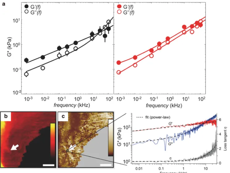
Fig. 3 HS-FS enables to explore the mechanical behaviors of cells over a large range of frequency. a Frequency-dependent complex shear modulus of benign (black) and malignant (red) cancer cells. The response was well described by two power law regimes, one with weak exponent (0.05–0.2) corresponding to cytoskeleton dynamics at the mesoscale and a stronger exponent (0.4–1) interpreted in terms of semiflexible filament dynamics
(reprinted from Rigato et al.2017). Maps of b height and c shear modulus (G0) on a live fibroblast (scale bars 3μm, 64 × 64 pixels). The G0map was built by analyzing the frequency dependant of the storage and loss moduli (right plot, G′, red, and G″, blue) and loss tangent (η, gray) in a frequency range of 5 Hz–30 kHz at each pixel (inset curve) (reprinted from Schachtele et al.2018)
response was better described by semiflexible filaments theo-ries. The possibility of applying semiflexible filament theories allows a mechanistic description of the cytoskeleton state. These findings were corroborated by observations on different cell types under different cytoskeletal conditions using drug treatments. Therefore, the cell mechanical response to high-frequency is rich of information and enables us to characterize the molecular mechanisms, reflecting the morphological and dynamical state of the cytoskeleton. Moreover, malignant can-cer cells exhibit a unique mechanical fingerprint at high fre-quency in comparison with healthy cells (Fig.3a). Thus, prob-ing cells responses over a wide dynamic range may provide a unique characterization of the mechanical phenotype of living cells and give new insights for a better mechanistic under-standing of cell mechanics.
High frequency microrheology of living cells was recently improved. Schächtele et al. (2018) developed a new resonance compensating chirp mode (RCCM) to probe the frequency dependence of the viscoelasticity of living cells (quasi-) con-tinuously over a large frequency range (5 Hz–30 kHz) and with a resolution of 5 Hz. A continuous frequency sweep (chirp) is applied to the z-scanner and is iteratively shaped to compensate for scanner resonances. This allows extending the measurement range far beyond the resonant frequency of the scanner (five times higher). The complex shear modulus of mouse embryonic fibroblast cells exhibited a weak power-law behavior over almost all the frequency range with a stronger frequency dependence at the highest rates (Fig.3c). A short chirp duration (200 ms) and a high approach velocity (320 m/s) allowed mapping live cells and generating spatially resolved images of the power-law parameters (Fig.3b) within minutes (≈ 20 min for 64 × 64 pixels), revealing local visco-elastic variations.
Conclusions and future perspectives
Ultrashort cantilevers and HS-AFM set-ups allow force mea-surement with sub-microsecond time resolution, providing important insights on both cellular and molecular mechanics. By reaching pulling velocities about two orders of magnitude higher than conventional AFM, HS-FS bridges the gap be-tween experimental force measurements and MD simulations, enabling an atomic description of molecular processes sup-ported by experimental data. The improved time resolution allows observation of hidden intermediate states that reveal the complex energy landscapes of biomolecular processes. Thus, HS-FS provides a framework to better understand the-ory, experiments, and simulations. While many applications have revealed the complexity of the mechanics of biomole-cules and living cells, there are still several exciting challenges to come. This may require further improvements and likely radical changes in AFM technology.
The next plausible step seems to be the application of sin-gle molecule HS-FS on living cells to probe receptor-ligand unbinding and/or adhesion molecules unfolding in situ, in the actual physiological environment. The combination of HS-AFM imaging with mechanical intervention of biomolecules starts also to emerge as a promising interactive approach and its application to living cells may arrive soon (Chiaruttini et al.
2015; Ganser and Uchihashi 2019; Owa et al. 2019). Moreover, current efforts point towards the development of even faster force mapping using ultrashort cantilevers. The groups of Schaeffer and Fantner have carried out significant progress in this direction by developing fast force mapping on cells and photothermal off-resonance using ultrashort levers (Schaeffer et al.1997; Nievergelt et al.2018). We expect that the use of ultrashort cantilevers as a force probe with sub-μs response time will grow and continue revealing fast processes in biomolecules and cells.
Funding information This project has received funding from the Agence Nationale de la Recherche (ANR, grant No. BioHSFS ANR-15-CE11-0007), the European Research Council (ERC, grant agreement No 772257), and the Human Frontier Science Program (HFSP, grant No. RGP0056/2018).
Open Access This article is distributed under the terms of the Creative C o m m o n s A t t r i b u t i o n 4 . 0 I n t e r n a t i o n a l L i c e n s e ( h t t p : / / creativecommons.org/licenses/by/4.0/), which permits unrestricted use, distribution, and reproduction in any medium, provided you give appro-priate credit to the original author(s) and the source, provide a link to the Creative Commons license, and indicate if changes were made.
References
Alcaraz J et al (2002) Correction of microrheological measurements of soft samples with atomic force microscopy for the hydrodynamic drag on the cantilever. Langmuir 18:716–721
Ando T (2018) High-speed atomic force microscopy and its future pros-pects. Biophys Rev 10:285–292
Ando T et al (2001) A high-speed atomic force microscope for studying biological macromolecules. Proc Natl Acad Sci 98:12468–12472 Ando T, Uchihashi T, Fukuma T (2008) High-speed atomic force
micros-copy for nano-visualization of dynamic biomolecular processes. Prog Surf Sci 83:337–437
Ando T, Uchihashi T, Scheuring S (2014) Filming biomolecular pro-cesses by high-speed atomic force microscopy. Chem Rev 114: 3120–3188
Bell G (1978) Models for the specific adhesion of cells to cells. Science 200:618–627
Benmouna F, Johannsmann D (2002) Hydrodynamic interaction of AFM cantilevers with solid walls: an investigation based on AFM noise analysis. The European Physical Journal E 9:435–441
Boal D, Boal DH (2012) Mechanics of the cell. Cambridge University Press, Cambridge
Braunsmann C, Seifert J, Rheinlaender J, Schäffer TE (2014) High-speed force mapping on living cells with a small cantilever atomic force microscope. Rev Sci Instrum 85:073703
Bull MS, Sullan RMA, Li H, Perkins TT (2014) Improved single mole-cule force spectroscopy using micromachined cantilevers. ACS Nano 8:4984–4995
Butt HJ, Jaschke M (1995) Calculation of thermal noise in atomic force microscopy. Nanotechnology 6:1–7
Casuso I et al (2012) Characterization of the motion of membrane pro-teins using high-speed atomic force microscopy. Nat Nanotechnol 7: 525–529
Chiaruttini N et al (2015) Relaxation of loaded ESCRT-III Spiral Springs drives membrane deformation. Cell 163:866–879
Colom A, Casuso I, Rico F, Scheuring S (2013) A hybrid high-speed atomic force–optical microscope for visualizing single membrane proteins on eukaryotic cells. Nat Commun 4:2155
Cossio P, Hummer G, Szabo A (2016) Kinetic ductility and force-spike resistance of proteins from single-molecule force spectroscopy. Biophys J 111:832–840
Dudko OK, Hummer G, Szabo A (2006) Intrinsic rates and activation free energies from single-molecule pulling experiments. Phys Rev Lett 96:108101
Durner E, Ott W, Nash MA, Gaub HE (2017) Post-translational Sortase-mediated attachment of high-strength force spectroscopy handles. ACS Omega 2:3064–3069
Edwards DT, Perkins TT (2017) Optimizing force spectroscopy by mod-ifying commercial cantilevers: improved stability, precision, and temporal resolution. J Struct Biol 197:13–25
Edwards DT et al (2015) Optimizing 1-μs-resolution single-molecule force spectroscopy on a commercial atomic force microscope. Nano Lett 15:7091–7098
Edwards DT, Faulk JK, LeBlanc M-A, Perkins TT (2017) Force spec-troscopy with 9-μs resolution and sub-pN stability by tailoring AFM cantilever geometry. Biophys J 113:2595–2600
Evans E, Ritchie K (1997) Dynamic strength of molecular adhesion bonds. Biophys J 72:1541–1555
Fantner GE et al (2006) Components for high speed atomic force micros-copy. Ultramicroscopy 106:881–887
Faulk JK, Edwards DT, Bull MS, Perkins TT (2017) Improved force spectroscopy using focused-ion-beam-modified cantilevers. Methods Enzymol 582:321–351
Florin EL, Moy VT, Gaub HE (1994) Adhesion forces between individ-ual ligand-receptor pairs. Science 264:415–417
Fritz J, Katopodis AG, Kolbinger F, Anselmetti D (1998) Force-mediated kinetics of single P-selectin/ligand complexes observed by atomic force microscopy. Proc Natl Acad Sci U S A 95:12283–12288 Fukuda S et al (2013) High-speed atomic force microscope combined
with single-molecule fluorescence microscope. Rev Sci Instrum 84:073706
Ganser C, Uchihashi T (2019) Microtubule self-healing and defect crea-tion investigated by in-line force measurements during high-speed atomic force microscopy imaging. Nanoscale 11:125–135 Gutsmann T et al (2004) Force spectroscopy of collagen fibers to
inves-tigate their mechanical properties and structural organization. Biophys J 86:3186–3193
Higgins MJ et al (2006) Noninvasive determination of optical lever sen-sitivity in atomic force microscopy. Rev Sci Instrum 77:013701 Hinterdorfer P, Baumgartner W, Gruber HJ, Schilcher K, Schindler H
(1996) Detection and localization of individual antibody-antigen recognition events by atomic force microscopy. Proc Natl Acad Sci U S A 93:3477–3481
Hodges AR, Bussmann KM, Hoh JH (2001) Improved atomic force microscope cantilever performance by ion beam modification. Rev Sci Instrum 72:3880–3883
Hoh JH, Cleveland JP, Prater CB, Revel JP, Hansma PK (1992) Quantized adhesion detected with the atomic force microscope. J Am Chem Soc 114:4917–4918
Humphris ADL, Miles MJ, Hobbs JK (2005) A mechanical microscope: high-speed atomic force microscopy. Appl Phys Lett 86:034106
Hutter JL, Bechhoefer J (1993) Calibration of atomic-force microscope tips. Rev Sci Instrum 64:1868–1873
Izrailev S, Stepaniants S, Balsera M, Oono Y, Schulten K (1997) Molecular dynamics study of unbinding of the avidin-biotin com-plex. Biophys J 72:1568–1581
Janovjak H, Struckmeier J, Müller DJ (2005) Hydrodynamic effects in fast AFM single-molecule force measurements. Eur Biophys J 34: 91–96
Kodera N, Sakashita M, Ando T (2006) Dynamic proportional-integral-differential controller for high-speed atomic force microscopy. Rev Sci Instrum 77:083704
Kodera N, Yamamoto D, Ishikawa R, Ando T (2010) Video imaging of walking myosin V by high-speed atomic force microscopy. Nature 468:72–76
Krieg M et al (2019) Atomic force microscopy-based mechanobiology. Nature Reviews Physics 1:41
Leissa AW (1969) Vibration of plates. Scientific and Technical Information Division, National Aeronautics and Space Administration, Washington, D.C.
Merkel R, Nassoy P, Leung A, Ritchie K, Evans E (1999) Energy land-scapes of receptor-ligand bonds explored with dynamic force spec-troscopy. Nature 397:50–53
Milles LF, Schulten K, Gaub HE, Bernardi RC (2018) Molecular mech-anism of extreme mechanostability in a pathogen adhesin. Science 359:1527–1533
Morse PM (1983) Vibration and sound. (American Institute of Physics reprint)
Moy VT, Florin EL, Gaub HE (1994) Intermolecular forces and energies between ligands and receptors. Science 266:257–259
Munguira I et al (2016) Glasslike membrane protein diffusion in a crowded membrane. ACS Nano 10:2584–2590
Neupane K et al (2016) Direct observation of transition paths during the folding of proteins and nucleic acids. Science 352:239–242 Nievergelt AP, Banterle N, Andany SH, Gönczy P, Fantner GE (2018)
High-speed photothermal off-resonance atomic force microscopy reveals assembly routes of centriolar scaffold protein SAS-6. Nat Nanotechnol 13:696–701
Oesterhelt F et al (2000) unfolding pathways of individual Bacteriorhodopsins. Science 288:143–146
Ott W, Jobst MA, Schoeler C, Gaub HE, Nash MA (2016) Single-mole-cule force spectroscopy on polyproteins and receptor–ligand com-plexes: the current toolbox. J Struct Biol 197:3–12
Otten M et al (2014) From genes to protein mechanics on a chip. Nat Methods 11:1127–1130
Owa M et al (2019) Inner lumen proteins stabilize doublet microtubules in cilia and flagella. Nat Commun 10:1143
Radmacher M, Tillmann RW, Fritz M, Gaub HE (1992) From molecules to cells: imaging soft samples with the atomic force microscope. Science 257:1900–1905
Rico F, Gonzalez L, Casuso I, Puig-Vidal M, Scheuring S (2013) High-speed force spectroscopy unfolds titin at the velocity of molecular dynamics simulations. Science 342:741–743
Rico F, Russek A, González L, Grubmüller H, Scheuring S (2019) Heterogeneous and rate-dependent streptavidin–biotin unbinding re-vealed by high-speed force spectroscopy and atomistic simulations. Proc Natl Acad Sci 116:6594–6601
Rief M, Gautel M, Oesterhelt F, Fernandez JM, Gaub HE (1997) Reversible unfolding of individual titin immunoglobulin domains by AFM. Science 276:1109–1112
Rigato A, Miyagi A, Scheuring S, Rico F (2017) High-frequency microrheology reveals cytoskeleton dynamics in living cells. Nat Phys 13:771–775
Sader JE (1998) Frequency response of cantilever beams immersed in viscous fluids with applications to the atomic force microscope. J Appl Phys 84:64–76
Sader JE, Larson I, Mulvaney P, White LR (1995) Method for the cali-bration of atomic force microscope cantilevers. Rev Sci Instrum 66: 3789–3798
Sader JE, Chon JWM, Mulvaney P (1999) Calibration of rectangu-lar atomic force microscope cantilevers. Rev Sci Instrum 70: 3967–3969
Sader JE et al (2012) Spring constant calibration of atomic force micro-scope cantilevers of arbitrary shape. Rev Sci Instrum 83:103705 Sader JE et al (2016) A virtual instrument to standardise the calibration of
atomic force microscope cantilevers. Rev Sci Instrum 87:093711 Schächtele M, Hänel E, Schäffer TE (2018) Resonance compensating
chirp mode for mapping the rheology of live cells by high-speed atomic force microscopy. Appl Phys Lett 113:093701
Schaeffer TE et al (1997) In: Michalske TA, Wendman MA (eds) Atomic force microscope for small cantilevers, pp 48–52.https://doi.org/10. 1117/12.271228
Schäffer TE, Cleveland JP, Ohnesorge F, Walters DA, Hansma PK (1996) Studies of vibrating atomic force microscope cantilevers in liquid. J Appl Phys 80:3622–3627
Schillers H et al (2017) Standardized nanomechanical atomic force mi-croscopy procedure (SNAP) for measuring soft and biological sam-ples. Sci Rep 7:5117
Schoeler C et al (2014) Ultrastable cellulosome-adhesion complex tightens under load. Nat Commun 5:5635
Shibata M, Yamashita H, Uchihashi T, Kandori H, Ando T (2010) High-speed atomic force microscopy shows dynamic molecular processes in photoactivated bacteriorhodopsin. Nat Nanotechnol 5:208–212 Shibata M, Uchihashi T, Ando T, Yasuda R (2015) Long-tip high-speed
atomic force microscopy for nanometer-scale imaging in live cells. Sci Rep 5:8724
Sotomayor M, Schulten K (2007) Single-molecule experiments in vitro and in silico. Science 316:1144–1148
Sumbul F, Marchesi A, Takahashi H, Scheuring S, Rico F (2018a) High-speed force spectroscopy for single protein unfolding. In: Lyubchenko YL (ed) Nanoscale imaging, vol 1814. Springer, New York, pp 243–264
Sumbul F, Marchesi A, Rico F (2018b) History, rare, and multiple events of mechanical unfolding of repeat proteins. J Chem Phys 148:123335
Sunyer R, Trepat X, Fredberg JJ, Farre R, Navajas D (2009) The temper-ature dependence of cell mechanics measured by atomic force mi-croscopy. Phys Biol 6:025009
Suzuki Y et al (2013) High-speed atomic force microscopy combined with inverted optical microscopy for studying cellular events. Sci Rep 3:2131
Takahashi H, Rico F, Chipot C, Scheuring S (2018)α-Helix unwinding as force buffer in spectrins. ACS Nano 12:2719–2727
Timoshenko S (1937) Vibration problems In engineering. D.Van Nostrand Company Inc., New York
Viani MB et al (1999) Fast imaging and fast force spectroscopy of single biopolymers with a new atomic force microscope designed for small cantilevers. Rev Sci Instrum 70:4300–4303
Viani MB et al (2000) Probing protein–protein interactions in real time. Nat Struct Biol 7:644
Walters DA et al (1996) Short cantilevers for atomic force microscopy. Rev Sci Instrum 67:3583–3590
Walters DA et al (1997) In: Michalske TA, Wendman MA (eds) Atomic force microscopy using small cantilevers, pp 43–47.https://doi.org/ 10.1117/12.271227
Yamashita H et al (2012) Single-molecule imaging on living bacterial cell surface by high-speed AFM. J Mol Biol 422:300–309
Yin J et al (2005) Genetically encoded short peptide tag for versatile protein labeling by Sfp phosphopantetheinyl transferase. Proc Natl Acad Sci U S A 102:15815–15820
Yu H, Siewny MGW, Edwards DT, Sanders AW, Perkins TT (2017) Hidden dynamics in the unfolding of individual bacteriorhodopsin proteins. Science 355:945–950
Publisher’s note Springer Nature remains neutral with regard to jurisdictional claims in published maps and institutional affiliations.